Embed Size (px)
Citation preview
KINESIO TAPING OF THE KNEE FOR CHONDRAMALACIA By Nya Hepburn-Fallah
TOPIC OF DISCUSS FOR IN-SERVICE
FOUNDER OF KINESIO TAPE CHALLENGES FOR USAGE
PRECUTS STYLES PROPERTIES AND BENEFITS
FOUR MAJOR FUNCTIONS OTHER USES
PROPERTIES AND BENEFITS POSITION OF THE KNEE FOR TAPING
HOW TO PERFORM KINESIO TAPING FOR CHONDROMALACIA OF THE KNEE
PREDISPOSED FACTORS OF CHONDROMALACIA
SYMPTOMS, DIAGNOSIS
SPECIAL TEST FOR DIAGNOSING
VARIOUS WAYS OF TREATMENT
MMT FOR PROGRESSION/REGRESSION WHEN TREATMENT UTILIZED
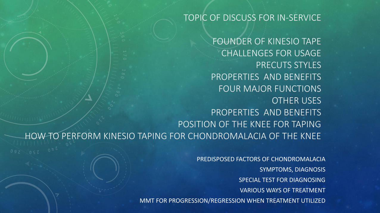
FOUNDER OF KINESIO TAPE • The Kinesio Taping® technique and Kinesio Tex tape was
developed by Dr. Kenzo Kase in Japan In the 1970's.
• In 1973, Dr. Kase's objective was to create a therapeutic tape and taping technique which could support the joints and muscles, without restricting ROM with a added benefit assisting the lymphatic Following two years of research into muscle taping, tape elasticity, adhesiveness and breathability Dr Kase developed Kinesio Tex® tape and the Kinesio Taping method.
• It is based on 3 important concepts: Space, movement and cooling –
• See more at: http://www.physio-pedia.com/Taping#sthash.GCnI3m5T.dpuf
• http://www.kinesiotaping.co.uk/history.jsp
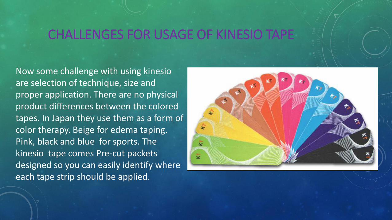
CHALLENGES FOR USAGE OF KINESIO TAPE
Now some challenge with using kinesio are selection of technique, size and proper application. There are no physical product differences between the colored tapes. In Japan they use them as a form of color therapy. Beige for edema taping. Pink, black and blue for sports. The kinesio tape comes Pre-cut packets designed so you can easily identify where each tape strip should be applied.
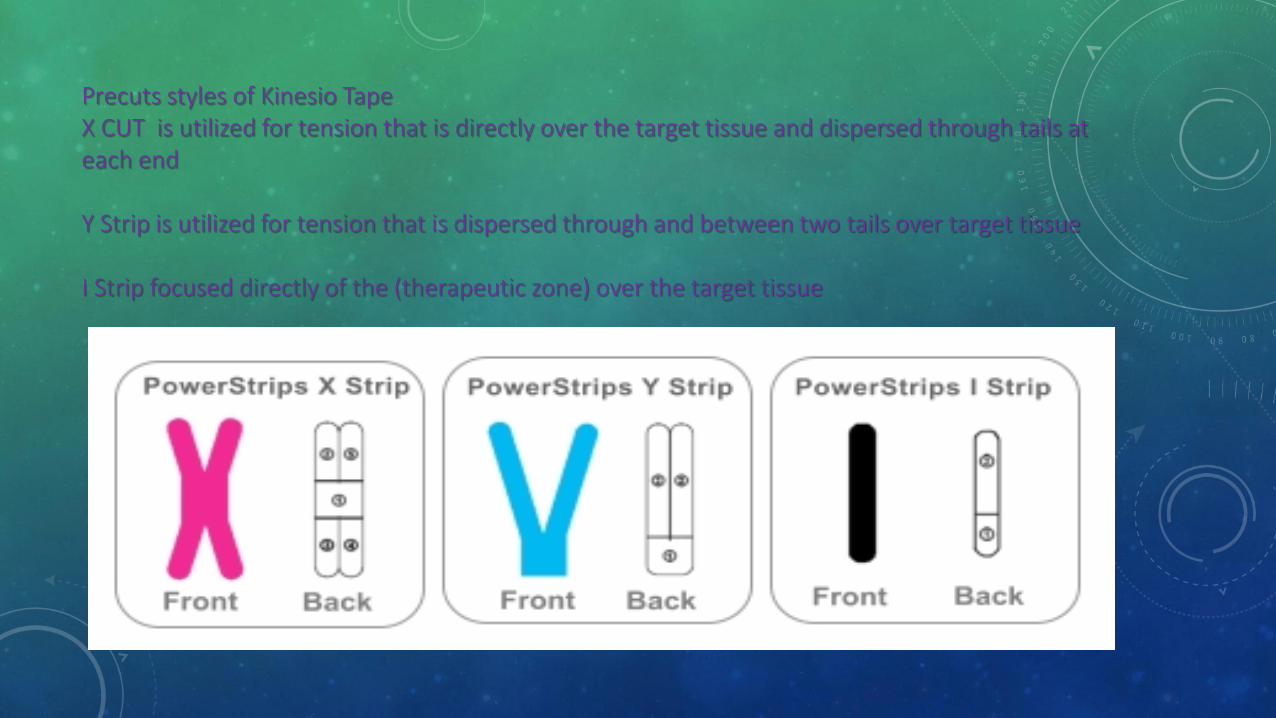
Precuts styles of Kinesio Tape X CUT is utilized for tension that is directly over the target tissue and dispersed through tails at each end Y Strip is utilized for tension that is dispersed through and between two tails over target tissue I Strip focused directly of the (therapeutic zone) over the target tissue
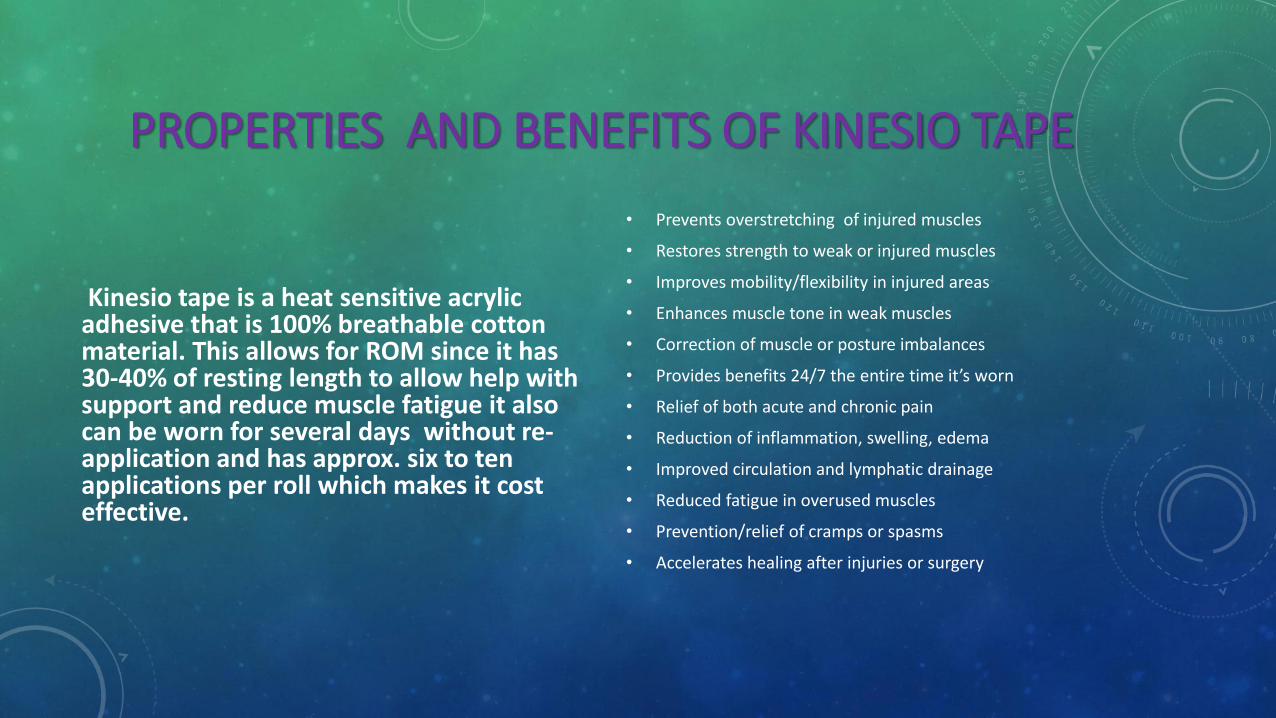
PROPERTIES AND BENEFITS OF KINESIO TAPE
Kinesio tape is a heat sensitive acrylic adhesive that is 100% breathable cotton material. This allows for ROM since it has 30-40% of resting length to allow help with support and reduce muscle fatigue it also can be worn for several days without re-application and has approx. six to ten applications per roll which makes it cost effective.
• Prevents overstretching of injured muscles
• Restores strength to weak or injured muscles
• Improves mobility/flexibility in injured areas
• Enhances muscle tone in weak muscles
• Correction of muscle or posture imbalances
• Provides benefits 24/7 the entire time it’s worn
• Relief of both acute and chronic pain
• Reduction of inflammation, swelling, edema
• Improved circulation and lymphatic drainage
• Reduced fatigue in overused muscles
• Prevention/relief of cramps or spasms
• Accelerates healing after injuries or surgery
”
“ Supporting the muscle -- Proper taping improves the muscle's ability to contract even when it's weakened, reduces a feeling of pain and fatigue, and protects the muscle from cramping, over-extension and over-contraction. Removing congestion to the flow of body fluids -- Kinesiology tape improves blood and lymphatic circulation and reduces inflammation and excess chemical buildup in the tissue. Activating the endogenous analgesic system -- This requirement means that the tape facilitate the body's own healing mechanisms, a central focus in chiropractic medicine. Correcting joint problems -- The goal is improving range of motion and adjusting misalignments that result from tightened muscles.
Dr. Kase lists the following as the four major functions of Kinesio Taping. These functions drive the technology behind the tape:
http://science.howstuffworks.com/kinesiology-tape1.htm
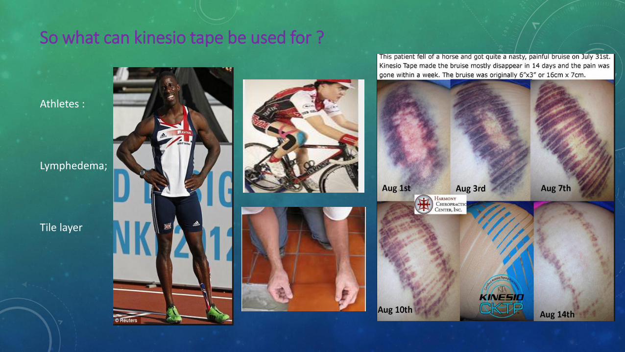
So what can kinesio tape be used for ?
Athletes :
Lymphedema;
Tile layer
FACTS CHONDROMALACIA PATELLA
The patella is designed to glide smoothly over the femur, and the joints in your body are cushioned with articular cartilage this tough, rubbery tissue covers the ends of bones inside a joint. As the joint moves, the cartilage helps to cushion the bones and allows them to glide smoothly against one another. Sometimes, the cartilage behind the kneecap (patella) softens and breaks down causing pain, poor alignment and inflammation these conditions are the causes for the pathological changes indicating chondromalacia which can affect any joint, but the most common location is inside the knee patella which is the most. In severe cases, the damaged cartilage can wear away completely, down to the undersurface of the kneecap. If this happens, the exposed kneecap's bony surface can grind painfully against other knee bones. Also, bits of cartilage can float inside the joint, further irritating the cells that line the joint. In response, these cells produce fluid inside the joint (called a joint effusion).
SOME PREDISPOSED FACTORS OF CHONDROMALACIA
Chondromalacia of the knee affects young adults more than any other age group. It is especially common in runners, joggers, skiers, soccer players, cyclists and other athletes who repeatedly stress their knees. Also, workers who spend a lot of time kneeling – particularly carpet layers, tile setters and floor layers – are more likely to develop this problem. Now weak quadriceps (usually the inner one), or tight quadriceps (usually the outer one) tend to be the problem
• Poor strength in the gluteal muscles resulting in poor knee alignment
• Poor biomechanics with running
• Running too many days in a row without proper recovery
• Excessive Supination or Pronation,
• Worn shoes that result in poor cushioning or instability
• Tightness especially in the IT Bands, hamstrings, quadriceps and gluteal
• Trigger points in the IT Bands, hamstrings, quadriceps and gluteal
• These will be many of the patients that will have a great impact in the orthopedic field of physical therapy
SYMPTOMS, DIAGNOSIS CHONDROMALACIA The most common symptom of Chondromalacia is a dull, aching pain in the front of your knee, behind your kneecap. also can make your knee joint "catch" meaning you suddenly have trouble moving it past a certain point, or "give way" (buckle unexpectedly). In some cases, the painful knee also can appear puffy or swollen can cause a creaky sound or grinding sensation when you move your knee. However, creaking sounds during bending do not always mean that cartilage is damaged.
• Diagnosis
• Your doctor will want to know whether you have ever:
• Fractured your kneecap or any other bone in the knee joint
• Sprained your knee or injured your knee's meniscus (the disk-shaped, shock-absorbing cartilage inside the knee)
• Had knee surgery
• Had bleeding or an infection inside your knee joint
• Been diagnosed with arthritis in your knee
• Your doctor also will ask about the type of work you do and your recreational and sports activities.
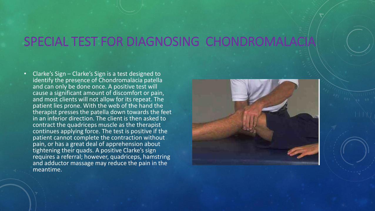
SPECIAL TEST FOR DIAGNOSING CHONDROMALACIA
• Clarke’s Sign – Clarke’s Sign is a test designed to identify the presence of Chondromalacia patella and can only be done once. A positive test will cause a significant amount of discomfort or pain, and most clients will not allow for its repeat. The patient lies prone. With the web of the hand the therapist presses the patella down towards the feet in an inferior direction. The client is then asked to contract the quadriceps muscle as the therapist continues applying force. The test is positive if the patient cannot complete the contraction without pain, or has a great deal of apprehension about tightening their quads. A positive Clarke’s sign requires a referral; however, quadriceps, hamstring and adductor massage may reduce the pain in the meantime.
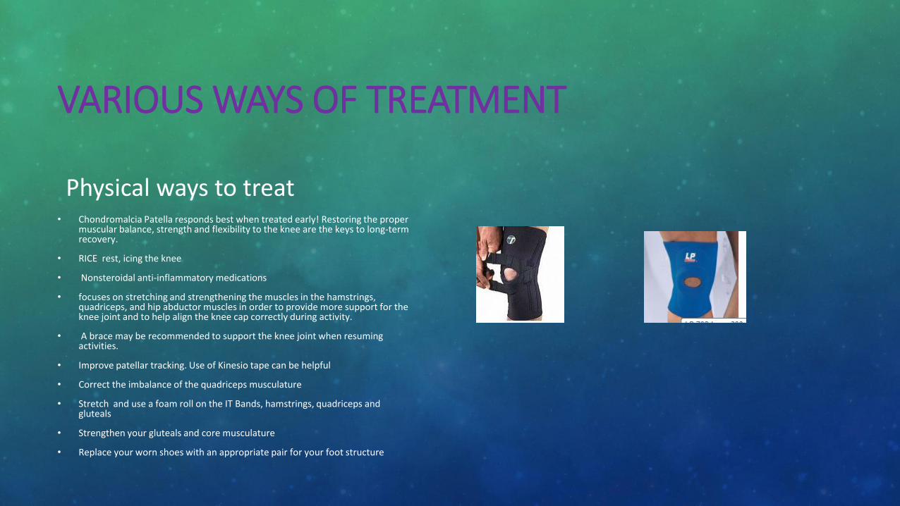
VARIOUS WAYS OF TREATMENT
Physical ways to treat • Chondromalcia Patella responds best when treated early! Restoring the proper
muscular balance, strength and flexibility to the knee are the keys to long-term recovery.
• RICE rest, icing the knee
• Nonsteroidal anti-inflammatory medications
• focuses on stretching and strengthening the muscles in the hamstrings, quadriceps, and hip abductor muscles in order to provide more support for the knee joint and to help align the knee cap correctly during activity.
• A brace may be recommended to support the knee joint when resuming activities.
• Improve patellar tracking. Use of Kinesio tape can be helpful
• Correct the imbalance of the quadriceps musculature
• Stretch and use a foam roll on the IT Bands, hamstrings, quadriceps and gluteals
• Strengthen your gluteals and core musculature
• Replace your worn shoes with an appropriate pair for your foot structure
BEFORE YOU START TAPING !!!!!!
When looking at the knee and patella (knee cap in particular) it is important to remember that the tendon starts above the knee where the individual quad muscles end (there are four quad muscles). This tendon then travels from the end of the femur, over the joint line and inserts onto the tibia tubercle. The knee cap itself sits in the tendon with no direct attachment to those bones. All three bones are lined with cartilage to prevent breakdown and damage to the knee cap. On either side of the knee cap, the tendon is attached to fibrous bands called the retinaculum. These help keep the knee cap from moving too far from side to side. Together, these attachments all help the patella stay in it’s groove as the knee bends and straightens. It is also why this are is such a common spot for injury. In the presence of muscle imbalances or soft tissue restrictions, the knee cap can be pulled out of alignment and inflammation/injury can occur.
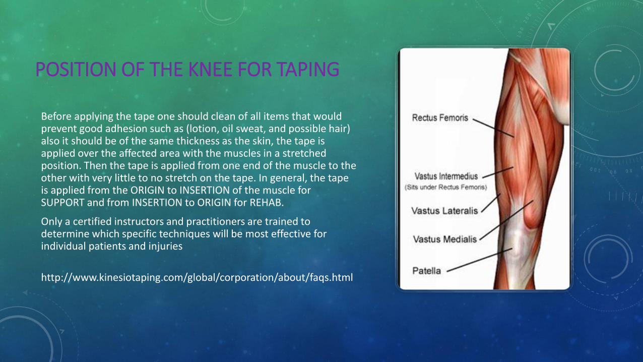
POSITION OF THE KNEE FOR TAPING
Before applying the tape one should clean of all items that would prevent good adhesion such as (lotion, oil sweat, and possible hair) also it should be of the same thickness as the skin, the tape is applied over the affected area with the muscles in a stretched position. Then the tape is applied from one end of the muscle to the other with very little to no stretch on the tape. In general, the tape is applied from the ORIGIN to INSERTION of the muscle for SUPPORT and from INSERTION to ORIGIN for REHAB.
Only a certified instructors and practitioners are trained to determine which specific techniques will be most effective for individual patients and injuries
http://www.kinesiotaping.com/global/corporation/about/faqs.html
HOW TO PERFORM KINESIO TAPING FOR CHONDROMALACIA OF THE KNEE
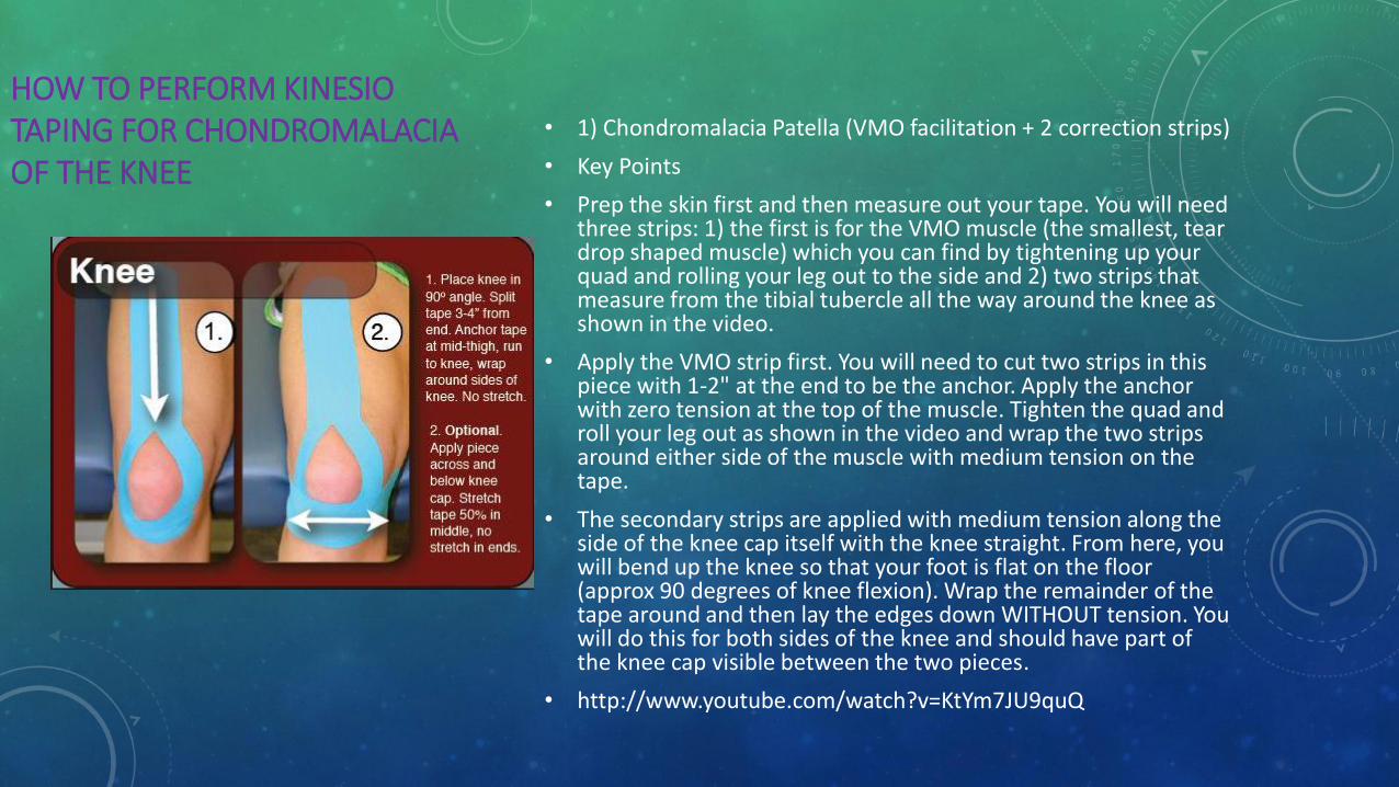
• 1) Chondromalacia Patella (VMO facilitation + 2 correction strips)
• Key Points
• Prep the skin first and then measure out your tape. You will need three strips: 1) the first is for the VMO muscle (the smallest, tear drop shaped muscle) which you can find by tightening up your quad and rolling your leg out to the side and 2) two strips that measure from the tibial tubercle all the way around the knee as shown in the video.
• Apply the VMO strip first. You will need to cut two strips in this piece with 1-2" at the end to be the anchor. Apply the anchor with zero tension at the top of the muscle. Tighten the quad and roll your leg out as shown in the video and wrap the two strips around either side of the muscle with medium tension on the tape.
• The secondary strips are applied with medium tension along the side of the knee cap itself with the knee straight. From here, you will bend up the knee so that your foot is flat on the floor (approx 90 degrees of knee flexion). Wrap the remainder of the tape around and then lay the edges down WITHOUT tension. You will do this for both sides of the knee and should have part of the knee cap visible between the two pieces.
• http://www.youtube.com/watch?v=KtYm7JU9quQ
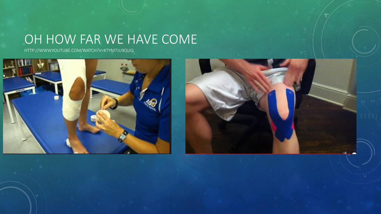
OH HOW FAR WE HAVE COME HTTP://WWW.YOUTUBE.COM/WATCH?V=KTYM7JU9QUQ
•

REFERENCES
http://www.kinesiotaping.co.uk/history.jsp
• http://www.theratape.com/education-center/kinesiology-tape-types/
• http://www.kinesiotaping.co.uk/history.jsp (slide #
• http://www.kinesiotaping.com/global/association/education/certification.html
• http://www.physio-pedia.com/Taping
• http://www.lhup.edu/yingram/jennifer/webpage/knee_motions.htm