Embed Size (px)
Citation preview
ELIANA CAROLINA VESPERO
Caracterização e Epidemiologia Molecular de Cepas de
Klebsiella pneumoniae Produtoras de ESBLs Isoladas de
Pacientes do Hospital Universitário de Londrina, no
período de 2000-2004.
LONDRINA
2007

Livros Grátis
http://www.livrosgratis.com.br
Milhares de livros grátis para download.
ELIANA CAROLINA VESPERO
Caracterização e Epidemiologia Molecular de Cepas de
Klebsiella pneumoniae Produtoras de ESBLs Isoladas de
Pacientes do Hospital Univesitário de Londrina, no
período de 2000-2004. Tese apresentada ao Programa de Pós-Graduação em Microbiologia da Universidade Estadual de Lodrina, como requisito final para obtenção do título de Doutor em Microbiologia. Orientadora: Profa Dra Halha Ostrensky Saridakis Londrina 2007
Este trabalho foi realizado no Laboratório de Bacteriologia do Departamento de Microbiologia, do Centro de Ciências Biológicas, da Universidade Estadual de Londrina e do Laboratório de Biotecnologia do Solo da Embrapa-Soja, sob orientação da Profa
Dra Halha Ostrensky Saridakis
ELIANA CAROLINA VESPERO
Caracterização e Epidemiologia Molecular de Cepas de Klebsiella pneumoniae Produtoras de ESBLs Isoladas de Pacientes do Hospital Univesitário de Londrina, no período de 2000-2004.
COMISSÃO EXAMINADORA
Prof. Dr Afonso Luis Barth Universidade Federal do Rio Grande do Sul Profa Dra. Maria Cristina Tognim Universidade Estadual de Maringá Profa Dra Mariangela Hungria da Cunha
Programa Pós Graduação em Microbiologia Universidade Estadual de Londrina Embrapa-Soja Profa Dra Jacinta Sanchez Pelayo Universidade Estadual de Londrina Profa Dra Halha Ostrensky Saridakis Universidade Estadual de Londrina Londrina, 10 de setembro de 2007

DEDICATÓRIA Aos meus queridos pais, Jacira e Pedro, meus irmãos e sobrinhos pelo que representam em minha vida dedico este trabalho.
AGRADECIMENTOS
A Deus, Meu Caminho, Verdade e Vida. Pela oportunidade que me proporcionou, dando paciência e coragem na hora necessária, sem Ele nada teria conseguido. À Profa Dra Halha Ostresky Saridakis pela orientação, experiência profissional transmitida e amizade no decorrer destes anos. Muito obrigado! À Dra Mariangela Hungria, agradeço pela competência científica, por ter disponibilizado o seu laboratório permitindo a realização dos experimentos e pelo contato proporcionando meu estágio na Argentina. Á amiga que fiz nestes anos, agradeço pelo apoio, carinho, incentivo e agradável convívio. Só tenho à dizer muito obrigado Mariangela! Ao Dr. Fernando Gomes Barcellos, agradeço pelo apoio científico e valiosas discussões. Ao Fernando agradeço pela amizade, apoio, desabafos, compreensão e incentivo. Você é uma das melhores pessoas que já conheci. Muito Obrigado por tudo! Á Ligia Chueire pela amizade, apoio científico e pelo agradável convívio nestes anos. Aos colegas da Cátedra de Microbiologia da Facultad de Farmacia Y Bioquímica de Buenos Aires, em especial a Marcela Radice, Pablo Power e Gabriel Gutkind pela dedicação, colaboração e orientação nos dias em que lá estive. À amiga Glaciela Kaschuk pela sua amizade, carinho, conselhos e momentos de desconcentração. Por ter me proporcionado esta alegria que é ser madrinha do Valentim Agradeço as amigas: doutoras e doutorandas do laboratório de biotecnologia do solo Luciana, Fabiana; à Maria e Adalgisa pela amizade e carinho, e em especial a Jesiane e Pâmela pela amizade, ajuda técnica e discussões cientifícas. Agradeço o carinho e amizade do pessoal da pós-graduação do laboratório de bacteriologia da UEL, aqueles que já se foram: Ariane, Ligiane, Claudia, Raquel, Kathelin, Sérgio, Vanderlei, Marcelo, Tanita e os que ainda estão: Alessandra, Carlos Alfredo, Karen. As docentes, amigas e futuras colegas de trabalho do laboratório de microbiologia clínica do Hospital Univesitário: Márcia Regina Eches Perugini, Marsileni Pelisson, Floristher Elaine Carrara, Regina Mariuza B. Quesada e Vera Lúcia C. Abbondanza, agradeço pelas amostras utilizadas neste estudo, pela amizade e incentivo constante. Aos docentes e à coordenação do programa de pós-graduação em microbiologia, em especial a profa Dra Jacinta Sanchez Pelayo, pela amizade e incentivo constante. Aos estagiários do laboratório de bacteriologia pela amizade, carinho e ajuda, em especial a Camila, Juliana e Rafael KConde pela ajuda contante.
Aos colegas mestrandos e estagiários do laboratório de biotecnologia do solo: Daisi, Leandro, Renan Ribeiro, Rose, Adriana, Susy, Eriana, Nágila, Renan Campos, agradeço pela amizade, em especial ao Alan e Fabinho pela ajuda com os dendrogramas. Aos funcionários do laboratório de biotecnologia do solo: Leny, Dona Rosa, Lílian, Lucas, Rinaldo, Ruth e Dr. Rubens, pela amizade e em especial ao Fabio Mostacio pela ajuda com o Bionumerics. Aos docentes do Departamento de Microbiologia pelo apoio e orientação
Aos amigos do laboratório do Hospital Zona Norte, bioquímicos, técnicos e auxiliares, obrigado pela amizade, colaboração e eternas trocas de plantões.
Ao pessoal do laboratório cepário da Facultad de Farmacia Y Bioquímica de Buenos Aires, em especial a Gisella e Maria, pela ajuda e amizade Aos funcionários do Departamento de Microbiologia, em especial ao Alex e Claci pela amizade e ajuda constante. Aos funcionários, técnicos e estagiários do laboratório de microbiologia clínica do Hospital Universitário pela amizade e colaboração.
Em especial aos meus familiares, pelo eterno incentivo, ajuda e apoio necessários para que realização deste trabalho. A todos que direta e indiretamente contribuíram para a concretização deste trabalho.
Muito obrigado a todos!
Fé é acreditar no que não vemos, e a recompensa dessa fé, é vermos aquilo que acreditamos.
( Santo Agostinho)
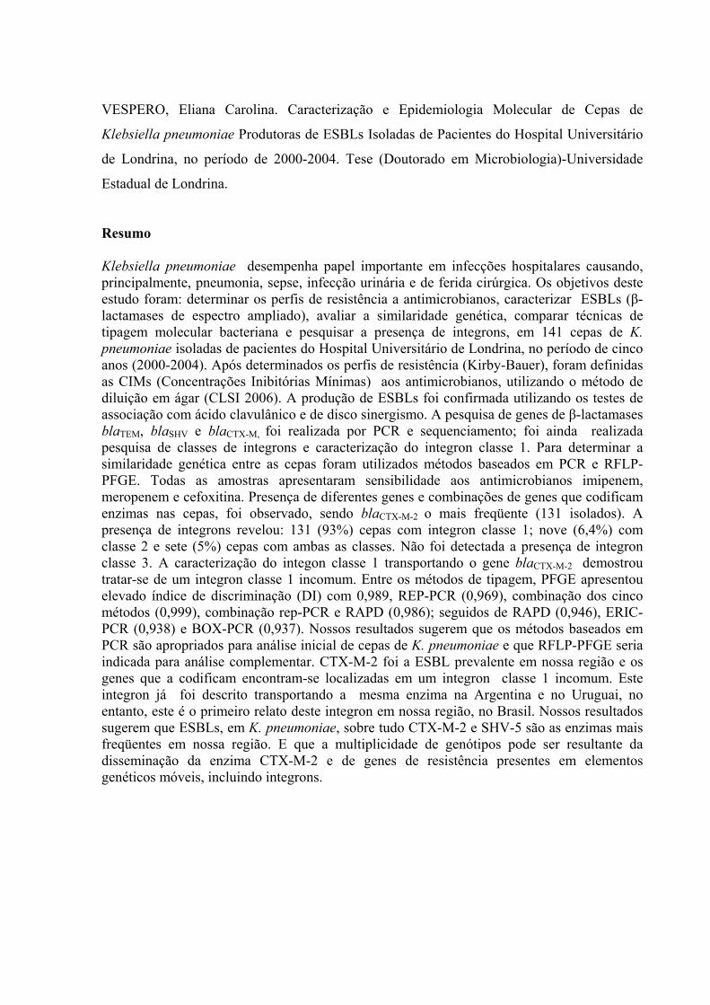
VESPERO, Eliana Carolina. Caracterização e Epidemiologia Molecular de Cepas de
Klebsiella pneumoniae Produtoras de ESBLs Isoladas de Pacientes do Hospital Universitário
de Londrina, no período de 2000-2004. Tese (Doutorado em Microbiologia)-Universidade
Estadual de Londrina.
Resumo Klebsiella pneumoniae desempenha papel importante em infecções hospitalares causando, principalmente, pneumonia, sepse, infecção urinária e de ferida cirúrgica. Os objetivos deste estudo foram: determinar os perfis de resistência a antimicrobianos, caracterizar ESBLs (β-lactamases de espectro ampliado), avaliar a similaridade genética, comparar técnicas de tipagem molecular bacteriana e pesquisar a presença de integrons, em 141 cepas de K. pneumoniae isoladas de pacientes do Hospital Universitário de Londrina, no período de cinco anos (2000-2004). Após determinados os perfis de resistência (Kirby-Bauer), foram definidas as CIMs (Concentrações Inibitórias Mínimas) aos antimicrobianos, utilizando o método de diluição em ágar (CLSI 2006). A produção de ESBLs foi confirmada utilizando os testes de associação com ácido clavulânico e de disco sinergismo. A pesquisa de genes de β-lactamases blaTEM, blaSHV e blaCTX-M, foi realizada por PCR e sequenciamento; foi ainda realizada pesquisa de classes de integrons e caracterização do integron classe 1. Para determinar a similaridade genética entre as cepas foram utilizados métodos baseados em PCR e RFLP-PFGE. Todas as amostras apresentaram sensibilidade aos antimicrobianos imipenem, meropenem e cefoxitina. Presença de diferentes genes e combinações de genes que codificam enzimas nas cepas, foi observado, sendo blaCTX-M-2 o mais freqüente (131 isolados). A presença de integrons revelou: 131 (93%) cepas com integron classe 1; nove (6,4%) com classe 2 e sete (5%) cepas com ambas as classes. Não foi detectada a presença de integron classe 3. A caracterização do integon classe 1 transportando o gene blaCTX-M-2 demostrou tratar-se de um integron classe 1 incomum. Entre os métodos de tipagem, PFGE apresentou elevado índice de discriminação (DI) com 0,989, REP-PCR (0,969), combinação dos cinco métodos (0,999), combinação rep-PCR e RAPD (0,986); seguidos de RAPD (0,946), ERIC-PCR (0,938) e BOX-PCR (0,937). Nossos resultados sugerem que os métodos baseados em PCR são apropriados para análise inicial de cepas de K. pneumoniae e que RFLP-PFGE seria indicada para análise complementar. CTX-M-2 foi a ESBL prevalente em nossa região e os genes que a codificam encontram-se localizadas em um integron classe 1 incomum. Este integron já foi descrito transportando a mesma enzima na Argentina e no Uruguai, no entanto, este é o primeiro relato deste integron em nossa região, no Brasil. Nossos resultados sugerem que ESBLs, em K. pneumoniae, sobre tudo CTX-M-2 e SHV-5 são as enzimas mais freqüentes em nossa região. E que a multiplicidade de genótipos pode ser resultante da disseminação da enzima CTX-M-2 e de genes de resistência presentes em elementos genéticos móveis, incluindo integrons.
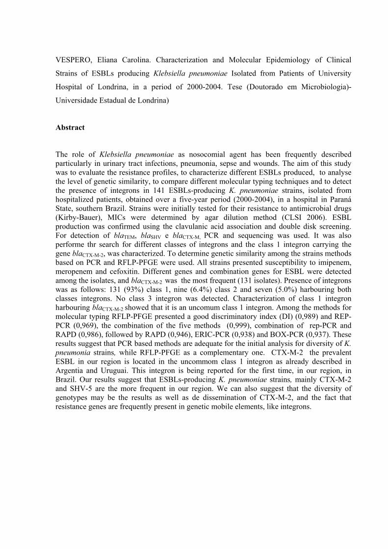
VESPERO, Eliana Carolina. Characterization and Molecular Epidemiology of Clinical
Strains of ESBLs producing Klebsiella pneumoniae Isolated from Patients of University
Hospital of Londrina, in a period of 2000-2004. Tese (Doutorado em Microbiologia)-
Universidade Estadual de Londrina)
Abstract
The role of Klebsiella pneumoniae as nosocomial agent has been frequently described particularly in urinary tract infections, pneumonia, sepse and wounds. The aim of this study was to evaluate the resistance profiles, to characterize different ESBLs produced, to analyse the level of genetic similarity, to compare different molecular typing techniques and to detect the presence of integrons in 141 ESBLs-producing K. pneumoniae strains, isolated from hospitalized patients, obtained over a five-year period (2000-2004), in a hospital in Paraná State, southern Brazil. Strains were initially tested for their resistance to antimicrobial drugs (Kirby-Bauer), MICs were determined by agar dilution method (CLSI 2006). ESBL production was confirmed using the clavulanic acid association and double disk screening. For detection of blaTEM, blaSHV e blaCTX-M, PCR and sequencing was used. It was also performe thr search for different classes of integrons and the class 1 integron carrying the gene blaCTX-M-2, was characterized. To determine genetic similarity among the strains methods based on PCR and RFLP-PFGE were used. All strains presented susceptibility to imipenem, meropenem and cefoxitin. Different genes and combination genes for ESBL were detected among the isolates, and blaCTX-M-2 was the most frequent (131 isolates). Presence of integrons was as follows: 131 (93%) class 1, nine (6.4%) class 2 and seven (5.0%) harbouring both classes integrons. No class 3 integron was detected. Characterization of class 1 integron harbouring blaCTX-M-2 showed that it is an uncomum class 1 integron. Among the methods for molecular typing RFLP-PFGE presented a good discriminatory index (DI) (0,989) and REP-PCR (0,969), the combination of the five methods (0,999), combination of rep-PCR and RAPD (0,986), followed by RAPD (0,946), ERIC-PCR (0,938) and BOX-PCR (0,937). These results suggest that PCR based methods are adequate for the initial analysis for diversity of K. pneumonia strains, while RFLP-PFGE as a complementary one. CTX-M-2 the prevalent ESBL in our region is located in the uncommom class 1 integron as already described in Argentia and Uruguai. This integron is being reported for the first time, in our region, in Brazil. Our results suggest that ESBLs-producing K. pneumoniae strains, mainly CTX-M-2 and SHV-5 are the more frequent in our region. We can also suggest that the diversity of genotypes may be the results as well as de dissemination of CTX-M-2, and the fact that resistance genes are frequently present in genetic mobile elements, like integrons.
SUMÁRIO 1.0 Introdução..…………………………………………………………............. 1
2.0 Objetivos ………………………………………………………………........ 3
2.1 Objetivo geral………………………………………………………....... 3
2.2 Objetivos específicos…………………………………………................ 3
3.0 Revisão Bibliográfica…................................................................................ 4
3.1 Epidemiologia…………………………….............................................. 6
3.2 Resistência a Drogas Antimicrobianas..................................................... 7
3.3 Métodos de Tipagem Molecular............................................................... 14
3.4 Referências Bibliográficas........................................................................ 19
ARTIGOS:
1o: Comparison of molecular typing techniques (rep-PCR, RAPD and RFLP-PFGE) for the epidemiological evaluation of clinical isolates of Klebsiella pneumoniae.
34
2o: Occurrence of Klebsiella pneumoniae Producing Extended-Spesctrum-β-Lactamases (ESBLs) in a Universtiy Hospital, During a Five Years Period, UsingPhenotipic and Molecular Methods.
60
3o: High of incidence of class 1 integron among Klebsiella pneumoniae strains and Characterization of Unusual integron carrying blactxm-2 gene in Isolates from Brazil
76
4.0 Conclusões ………………………………………………………………… 90
1
1. Introdução
Klebsiella pneumoniae está entre as espécies de maior importância em infecção
hospitalar, embora espécies de Klebsiella estejam amplamente distribuídas no ambiente (água,
solo e plantas) e fazem parte da microbiota normal dos tratos respiratório e digestório, em
humanos e outros animais.
K. pneumoniae é a espécie mais importante do gênero e, depois de Escherichia coli,
tem sido considerada o segundo agente gram-negativo nosocomial mais freqüente em
bacteremia. No entanto, infecções por este patógeno podem ocorrer em quase todos os sítios
do corpo humano, causando sepse, pneumonia, infecções do trato urinário e de feridas
cirúrgicas (SCHABERG et al., 1991). Sua capacidade de infecção se deve a vários fatores de
virulência, como a cápsula. Com a aquisição de mecanismos de resistência a novos
antimicrobianos, este microrganismo tem alcançado notoriedade como patógeno hospitalar
responsável por diversos surtos, principalmente em unidades de alto risco, como de terapia
intensiva e neonatal (BEN-HAMOUDA et al., 2004; BAGATTINI et al., 2006).
Espécies pertencentes a este gênero estão presentes em ambientes hospitalares como
pisos, pias, desinfetantes e equipamentos inalatórios (HOBSON et al., 1996), em proporções
que levam à colonização de pacientes a qual é proporcional ao tempo de hospitalização.
Kramer et al. (2006) avaliaram o tempo que um patógeno hospitalar persiste em superfícies
inanimadas e verificaram que Klebsiella spp. foi capaz de sobreviver em ambiente hospitalar
por mais de 30 meses. Cotton et al.(2000), relataram um surto ocorrido em unidade neonatal
por cepa de K. pneumoniae produtora de ESBL (β-lactamase de espectro ampliado), cujo
vetor eram baratas. Segundo alguns autores, a alta taxa de colonização hospitalar por K.
pneumoniae estaria mais associada ao uso inadequado de antimicrobianos do que com os
procedimentos hospitalares (PODSCHUN & ULLMANN, 1998; WALLS, 2000). Em pelo
menos 80% dos pacientes com infecção por cepas de K. pneumoniae produtoras de ESBL a
bactéria tem sido isolada a partir do trato gastrointestinal, sendo este, portanto importante
fonte de transmissão (PENA e tal., 1998).
O principal mecanismo de resistência de bactérias gram-negativas é a produção de β-
lactamases, enzimas que inibem a ação de drogas β-lactâmicos, impossibilitando assim a sua
atividade antimicrobiana (LIVERMORE, 1995). As ESBLs mais freqüentemente
encontradas, em K. pneumoniae, são as pertencentes aos grupos TEM e SHV, sendo
descritos atualmente mais de 160 tipos de TEM e mais de 104 de SHV. Na década de 90,
surgiram ESBLs denominas CTX-M, que hidrolisam preferencialmente cefotaxima e têm
2
sido descritas, com grande freqüência, em cepas de K. pneumoniae, sendo conhecidos
atualmente mais de 69 tipos de CTX-M (MACK & MACK, 2003; CANTON & COQUE,
2006; http: /www.lahey.org/ studies).
ESBLs são codificadas por genes presentes em grandes plasmídios, de 80 a 300 kb,
os quais podem ser transferidos entre espécies bacterianas, facilitando sua disseminação. Em
diversos casos, esses plasmídios podem apresentar outros genes que codificam resistência
antimicrobiana, podendo ocorrer, concomitantemente, a expressão de ESBLs e resistência aos
aminoglicosídeos, sulfonamidas, tetraciclina, cloranfenicol e quinolonas (WINOKUR et al.,
2001; BRADFORD, 2001).
Os métodos de tipagem molecular são de grande importância quando se estuda a
epidemiologia dos microrganismos e no controle de infecções hospitalares. Testes de tipagem
bacteriana têm por objetivo não somente a inclusão ou exclusão de amostras, mas,
prioritariamente, a definição das fontes e rotas de transmissão de microrganismos dentro do
ambiente hospitalar (SINGH et al., 2006).
Os estudos sobre mecanismos de resistência de K. pneumoniae, bem como a
epidemiologia das principais enzimas responsáveis pela resistência às cefalosporinas de
amplo espectro e dos elementos genéticos que facilitam a sua disseminação, podem contribuir
para a compreensão da dinâmica das infecções hospitalares como um todo e a padronização
de ações que visem minimizá-las.
3
2.0 Objetivos
Considerando a importância da bactéria aqui estudada e seus mecanismos de resistência a
antimicrobianos, principalmente em ambiente hospitalar, foram estabelecidos os objetivos
deste estudo.
2.1 Objetivo geral
Caracterização molecular e epidemiológica das enzimas detectadas em cepas de K.
pneumoniae produtoras de ESBLs, isoladas de pacientes do Hospital Universitário de
Londrina, no período de 2000-2004.
2.2 Objetivos específicos
2.2.1. Avaliar o perfil de sensibilidade a antimicrobianos das cepas de K. pneumoniae
produtoras de ESBLs.
2..2.2. Caracterizar as β-lactamases produzidas pelas cepas estudadas, utilizando técnicas de
PCR e de seqüenciamento.
2..2.3. Detectar a similaridade genética das amostras de K. pneumoniae pelas técnicas de
tipagem molecular baseadas em PCR (ERIC, REP, BOX e RAPD) e RFLP-PFGE e
comparar seu poder discriminatório.
2.2.4. Pesquisar a presença de diferentes classes de integrons entre as amostras de K.
pneumoniae e caracterizar integron classe 1.
4
3. Revisão Bibliográfica
As bactérias pertencentes ao gênero Klebsiella (família Enterobacteriaceae) são
bastonetes Gram-negativos, imóveis, geralmente capsulados, anaeróbios facultativos, não
esporulam, fermentam vários carboidratos, inclusive a lactose; algumas espécies produzem
urease e 2,3-butilenoglicol como produto final da fermentação da glicose (teste de Voges
Proskaeur) (FARMER, 1999).
Suas características de patogenicidade lhe conferem a capacidade de sobreviver em
diferentes ambientes na natureza, em hospitais, bem como colonizar e causar infecções em
diferentes sítios do paciente. Dentre essas características a produção de cápsula (WILLIAMS
& TOMAS,1990), a resistência sérica (MONTENEGRO et al., 1985; ALBERTÍ et al., 1993)
e a produção de sideróforos (REISSBRODT & RABSCH, 1988; PODSCHUN &
ULLMANN, 1998), contribuem para sua capacidade invasiva, protegendo-a dos mecanismos
de defesa do hospedeiro (PODSCHUN & ULLMANN, 1998).
As bactérias da espécie K. pneumoniae são caracterizadas como microrganismos
invasores, capacidade esta que lhes é conferida pela presença de cápsula, a qual é essencial
para sua virulência e é composta de uma camada espessa de polissacarídeo responsável pelo
aspecto brilhante e mucóide das colônias em ágar (BRISSE et al., 2004). O polissacarídeo
capsular confere resistência à fagocitose e impede a morte da bactéria pelos fatores de
resistência do soro, através da inibição da ativação do componente C3b do complemento
(WILLIAMS & TOMAS, 1990). Existem 77 sorotipos capsulares sendo K1, K2, K4 e K5
considerados os mais virulentos.
É preciso lembrar que o fenômeno essencial para a instalação de uma bactéria no
hospedeiro é a etapa de adesão, não importa quais sejam as outras propriedades de virulência
(FINLAY & FALKOW, 1997). Em Klebsiella várias adesinas fimbriais (fimbrias tipo 1,
tipo 3 e KPF 28) e afimbriais, são descritas (CF29K e adesina cápsula-like de Klebsiella).
As fímbrias ou pili tipo 1, são hemaglutininas manose sensíveis (HAMS),
comumente encontradas na maioria dos gêneros da família Enterobacteriaceae (CLEGG &
GERLACH, 1987), com variações estruturais entre os diferentes gêneros. São as adesinas
bacterianas mais investigadas, apresentando HAMS com eritrócitos de cobaia. Possuem
duas subunidades importantes: FimA, a maior subunidade que constitui mais de 95% do
cerne fimbrial é estrutural e antigenicamente heterogênea entre as diferentes espécies e a
FimH que reconhece o receptor para fimbria tipo 1 no hospedeiro (SCHEMBRI et al.,
2005; DUNCAN et al., 2005).
5
A fimbria tipo 3 foi originalmente caracterizada pela sua capacidade de aglutinar
eritrócitos tratados com ácido tânico (DUGUID, 1959). Apesar da denominação
“hemaglutinação manose resistente de Klebsiella” (MR/KHA), estudos posteriores
demonstraram que esta fimbria é produzida por outros membros da família
Enterobacteriaceae. Entretanto, fimbrias tipo 3 não são idênticas em todas as enterobactérias,
apresentando diversidade antigênica entre as espécies (CLEGG & GERLACH, 1987).
Possuem duas subunidades importantes a subunidade mrkA codifica o principal componente
fimbrial, a proteína MrkA de 20,5 kDa denominada pilina. A adesina MrkD tem sido descrita
como mediadora da aderência à membrana basal e à superfície basolateral dos epitélios renal
e pulmonar (SCHURTZ et al., 1994), enquanto a adesina MrkA, tem sido descrita como
facilitadora na formação de biofilmes em superfícies abióticas (DI MARTINO et al., 2003;
BODDICKER et al., 2006).
A expressão de cada fator é cuidadosamente coordenada a nível genético. Assim, no
caso de infecção urinária, os mecanismos de adesão são essenciais para sua permanência em
local com fluxo contínuo de líquidos, como é a bexiga. Essas adesinas são também de
grande valor em infecções do trato respiratório, o mesmo ocorrendo nos casos de meningite
por esta espécie, no entanto, a bactéria só atingirá este sítio se estiver adequadamente
protegida por sua cápsula.
As interações mais proeminentes entre diferentes fatores de virulência levam ao
quadro final de infecção. Cápsula e fimbria são componentes estruturais da superfície
bacteriana, essenciais para sobrevivência e virulência de K. pneumoniae, no entanto, tem
ocorrido relatos conflitantes na literatura sobre a expressão simultânea destes fatores.
Tarkkanen et al. (1992) relataram que 29 das 32 cepas capsuladas de K. pneumoniae isoladas
de ITU eram capazes de expressar fimbria tipo 1. Já em 1999 Matatov et al. mostraram, a
partir de cepas isoladas de sepse e ITU, que amostras capsuladas isoladas de sepse não
produziam fimbria tipo 1, enquanto, que a maioria das isoladas de ITU expressavam fimbria
tipo 1 e não eram capsuladas. Os mesmos autores sugerem que cepas capsuladas eram
incapazes de fazer a montagem da fimbria funcional (MATATOV et al.,1999). Sahly et al.
(2000) em estudo realizado com cepas apresentando tipos capsulados e não capsulados,
previamente caracterizadas, relataram que nenhuma produziu fimbria tipo 1. Já Schembri et
al., (2005) relataram que fimbria tipo1 e cápsula possuem importante papel na formação de
biofilme, mas são expressadas em diferentes fases, ou seja, as fimbrias no processo inicial de
adesão e o polissacarídeo capsular durante o desenvolvimento de estruturas de
amadurecimento o biofilme.
6
O mecanismo exato de resistência bactericida ao soro é desconhecido. Várias
proteínas de membrana externa, como a lipoproteína TraT ou porinas (MONTENEGRO et al.,
1985; ALBERTÍ et al., 1993), igualmente a cápsula e antígenos O (LPS) tem sido
implicados (WILLIAMS et al., 1983; TOMÁS et al.,1986). Dos nove sorogrupos de antigeno
O descritos em K. pneumoniae, o O1 é o mais comum (SAHLY et al., 2004).
O crescimento da bactéria no tecido hospedeiro pode se limitado por mecanismos de
defesa deste, bem como pela disponibilidade do ferro. A aerobactina e a yersiniabactina são
sideróforos importantes em K. pneumoniae para a manutenção da infecção no hospedeiro,
contribuindo para um fenótipo mais virulento desta bactéria (PODSCHUN & ULLMANN,
1998; LAWLOR et al., 2007).
Estudos recentes sugerem que a formação de biofilme pode ser um fator de
virulência importante em K. pneumoniae, lembrando que as bactérias encontradas dentro do
biofilme são mais resistentes ao tratamento com antibióticos do que as que crescem
plantonicamente (JAGNOW & CLEGG, 2003). K. pneumoniae é capaz de formar biofilme
em superfícies abióticas, bem como em superfícies revestidas com matriz extracelular humana
e proteínas derivadas do hospedeiro. Biofilmes produzidos por K pneumoniae são detectados
em cateteres e tubos endotraqueais (FUX et al., 2005), sendo, portanto, de grande importância
em ambiente hospitalar interferindo com a terapia antimicrobiana.
3.1. Epidemiologia
A incidência das infecções por K. pneumoniae produtoras de ESBL varia entre países
e entre regiões de um mesmo país (WINOKUR et al., 2001; TURNER, 2005). Segundo
estudo realizado pelo programa MYSTIC (Meropenem Yearly Susceptibility Test Information
Collection) com isolados clínicos de K. pneumoniae produtoras de ESBL, durante os anos
1997-2003, de todo os continentes, verificou-se que nos Estados Unidos 12,3% das cepas
eram produtoras de ESBL. Na Europa, a incidência dessas cepas apresenta variação de
acordo com a região: na Europa oriental, 58,7%, no norte da Europa 16,7% e no sul da
Europa, 24,4%. Na Ásia, 28,2% das amostras de K. pneumoniae eram produtoras de ESBL,
com exceção do Japão onde a incidência foi menor que 5% e na América do Sul, que
apresentou a incidência mais alta, de 51,9% (TURNER, 2005; PATERSON, 2005). De
acordo com Bell et al. (2002) na África do Sul, a incidência das infecções causadas por cepas
de K. pneumoniae produtoras de ESBL foi de 36,1%.

7
Estudo realizado em 2004 na Europa, América Latina e América do Norte, pelo
SENTRY (Antimicrobial Surveillance Program), com a finalidade de avaliar a freqüência e o
padrão de resistência de bactérias isoladas de pacientes pediátricos, revelou que 22,5% das
cepas de Klebsiella spp. eram produtoras de ESBLs (FEDLER et al., 2006).
No Brasil, as taxas das infecções causadas por este microorganismo são maiores do
que as encontradas em diferentes partes do mundo. Em estudo realizado pelo programa
MYSTIC Brasil em 2003, K. pneumoniae foi o 3º bacilo gram negativo mais freqüentemente
isolado em hospitais brasileiros (16,9%), sendo que 51,9% destas amostras eram produtoras
de ESBLs. De acordo com estudos de Kiffer et al. (2005), K. pneumoniae foi responsável por
17,2% das infecções da corrente sangüínea relacionadas a cateter; 12% do trato respiratório;
trato urinário, 18,6% e infecções de pele e tecidos moles 11,5%. Gales (1997) e Marra (2002)
em estudos desenvolvidos no Hospital São Paulo da Universidade Federal de São Paulo
(HSP/UNIFESP) constataram respectivamente, que 39,0% e 52,3% das cepas de K.
pneumoniae isoladas de amostras clínicas eram produtoras de ESBL. Dados fornecidos pela
CCIH do Hospital Universitário-UEL mostram que as freqüências de K. pneumoniae em
isolados de colonização e de infecção hospitalar nos períodos de janeiro a outubro de 2002
foram de 7,8% e de outubro de 2002 a maio de 2003, de 11,6% (comunicado interno). Ainda
no mesmo hospital, a presença de cepas de K. pneumoniae produtoras de ESBLs em 2004 foi
detectada em 28% e em 2005, em 25,1%, do total de isolados desta bactéria (comunicação
pessoal do laboratório de microbiologia).
3.2. Resistência a Drogas Antimicrobianas
O desenvolvimento e a disseminação de diversos mecanismos de resistência a
antimicrobianos resulta no declínio contínuo da eficácia da maioria dos antimicrobianos ao
longo das últimas décadas. A resistência bacteriana a antimicrobianos é, atualmente um
importante problema na maioria dos continentes e uma ameaça à saúde humana. Apesar disso,
permanecem pouco conhecidas informações a respeito de sua disseminação e distribuição
geográfica em países em desenvolvimento (GUZMAN-BLANCO et al., 2000; PATERSON,
2006). A freqüência de resistência bacteriana está intimamente relacionadas às normas de
utilização de agentes antimicrobianos em uma comunidade. O uso indiscriminado dessas
drogas, por vários anos, nos diversos setores da saúde, tem sido um fator importante no
desenvolvimento de resistência, praticamente, em todas as espécies bacterianas. Ainda mais
8
grave é que cepas bacterianas multirresistentes são cada vez mais comuns em ambientes
hospitalares (SAHLY et al., 2004; COLODNER, 2005).
A resistência aos antimicrobianos é o melhor exemplo da rápida adaptação da
bactéria a um novo ecossistema. A habilidade da bactéria em ampliar seu nicho ecológico,
também na presença de determinados antibióticos, pode explicar a aquisição de genes por
transferência de material genético através de conjugação, transformação e transdução e/ou
pelo acumulo de mutações pontuais, levando a modificações de genes existentes
(SUNDSTROM, 1998; CARATOLLI, 2001). Vários mecanismos de resistência estão
envolvidos, entre eles: alteração conformacional e bioquímica do sítio alvo; alteração da
permeabilidade da parede celular da bactéria ao antimicrobiano; efluxo ativo; inativação
enzimática do antimicrobiano (SANDERS & SANDERS, 1992; LIVERMORE, 1998).
Resumidamente pode-se dizer que a resistência ocorre pela pressão seletiva do uso
inadequado de antimicrobianos, pela disseminação clonal ou pela transferência horizontal de
genes de resistência (WITTE, 2004).
A emergência e a disseminação de resistência entre as espécies de
Enterobacteriaceae trazem complicações para o tratamento de infecções hospitalares graves e
podem contribuir para o aparecimento de espécies resistentes a todos antimicrobianos
atualmente disponíveis. Em particular, K. pneumoniae, é um patógeno extremamente
importante em infecções hospitalares uma vez que pode apresentar resistência múltipla a
antimicrobianos de diferentes classes (PATERSON, 2006).
Amostras de K. pneumoniae apresentam resistência intrínseca a ampicilina e
carbenicilina, a qual se deve à presença de genes em seu cromossomo que codificam as ß-
lactamases TEM-1 e SHV-1 (FARMER, 1999). Essas ß-lactamases, foram descritas pela
primeira vez em 1960 em amostras de E. coli e Salmonella paratyphi, logo após a introdução
de ampicilina no tratamento de infeções causadas por esses microrganismos. Com isto, desde
a década de 60 vem ocorrendo a disseminação da ß-lactamase plasmidial TEM-1 a outras
espécies de gram-negativas (PALZKILL, 1998). K. pneumoniae pode ainda produzir a
enzima SHV-1 (“sulphidril variable”) em baixos níveis, o que se traduz por resistência a
ampicilina, amoxicilina, carbenicilina e ticarcilina. Cefalosporinas de primeira geração como
cefalotina e cefalexina também são degradadas por estas enzimas (LIVERMORE, 1995). Com
a introdução do uso clínico de ß-lactâmicos de amplo espectro, em 1978 na Europa e em 1981
nos Estados Unidos, os microrganismos quando submetidos à ação destes antimicrobianos,
sofrem pressão, resultando em resistência às cefalosporinas de amplo espectro. Como
9
conseqüência do uso extensivo de antimicrobianos ß-lactâmicos de amplo espectro, na década
de 80, microrganismos que produziam ß-lactamases mediadas pelos genes blaTEM e blaSHV
desenvolveram ainda resistência a cefalosporinas de amplo espectro e monobactâmicos,
resultando na produção de ESBL (NAUMOVSKI et al., 1992; MEYER et al., 1993).
ESBLs são enzimas capazes de hidrolisar todos os antimicrobianos β-lactâmicos com
exceção das cefamicinas (cefoxitina e cefotetan) e de carbapenems (imipenem e
meropenem). Sua característica fenotípica importante é que geralmente permanecem
sensíveis à ação de inibidores de ß-lactamases como sulbactam e ácido clavulânico,
descritos na década de 70, e tazobactam descrito na década de 80. Esses inibidores formam
um complexo protéico com a ß-lactamase, bloqueando, desta maneira, a atividade
hidrolítica dessas enzimas (WILLIAMS, 1997). Estas características permitem classificar as
ESBLs, de acordo com o esquema proposto para β-lactamases, por Bush-Jacoby-Medeiros,
como pertencentes ao grupo 2be (BUSH et al., 1995). As espécies produtoras de ESBLs
podem sobreviver por longos períodos de tempo em hospitais, com freqüência ocasionando
surtos (THOMSON et al., 1996; BRADFORD, 2001).
Utilizando testes de hibridação ou análise de seqüência de nucleotídeos das ESBLs,
ficou demonstrado que mutações ocorridas nos genes estruturais que codificam as enzimas
TEM-1, TEM-2 e SHV-1, em locais próximos aos seus sítios ativos, resultavam em
alterações na seqüência de aminoácidos, originando as novas ß-lactamases de espectro
ampliado (DU BOIS et al., 1995).
ß-lactamase TEM–1, a enzima mais comumente encontrada em bactérias Gram-
negativas, foi detectada em Atenas, em 1963. O termo TEM deriva de “Temoniera”, nome
da paciente de cujo material clínico, uma hemocultura, foi isolada a primeira cepa de E. coli
produtora desta enzima. TEM-2, a primeira derivada de TEM-1, apresenta um único
aminoácido substituído na molécula da ß-lactamase original. Isto origina a mudança no
ponto isoelétrico de 5,4 para 5,6, mas não altera o substrato. TEM-3, relatada em 1987, foi
a primeira a manifestar o fenótipo ESBL. Desde então, foi observado o aumento rápido no
número e nas variantes de espectro ampliado do tipo TEM. A substituição do aminoácido
na enzima TEM ocorre em número limitado de posições. A combinação na mudança destes
aminoácidos resulta em várias alterações sútis no fenótipo ESBL, como a capacidade de
hidrolisar oximino-cefalosporinas, tais como ceftazidima e cefotaxima (HERITAGE et al.,
1999; BRADFORD, 2001; MACK & MACK, 2003). ß-lactamases do tipo TEM são
10
encontradas em todos os países, sendo predominante na América do Norte (PATERSON &
BONOMO, 2005).
ß-lactamase SHV-1 é a enzima mais comumente encontrada em K. pneumoniae; seu
primeiro relato ocorreu em 1972 e foi assim denominada pela característica química de
apresentar variações na ligação ao seu grupo sulfidrila (MEDEIROS, 1995). Em 1983, três
isolados de K.pneumoniae e um de Serratia marcescens, no oeste da Alemanha,
demonstraram resistência a cefotaxima e às demais novas cefalosporinas de terceira geração.
Esta nova ß-lactamase plasmidial chamada SHV-2, derivou de uma mutação de SHV-1
(KNOTHE, 1983). A mutação, mudança de aminoácido na posição 238 de glicina para
serina, resultou em aumento da afinidade de SHV-1 por oximino-cefalosporinas, com
valores mais elevados dos MICs para cefotaxima e valores menores dos MICs para
ceftazidima. Posteriormente, foi descrita uma variedade de ESBL tipo SHV contendo outras
substituições de aminoácidos, sendo que a mudança de aminoácido nas posições 179, 205 e
240, resultam na produção de altos níveis de enzimas ESBLs (MACK & MACK, 2003).
ESBLs do tipo SHV são as mais freqüentemente produzidas por bactérias isoladas de
materiais clínicos (JACOBY, 1997). Após 15 anos da descoberta de SHV-2, esta enzima foi
detectada em todos os continentes (PATERSON et al., 2003). ESBLs do tipo SHV têm sido
a causa de surtos não só por Enterobacteriaceae, mas também por Pseudomonas aeruginosa
e Acinetobacter spp. (POIREL et al., 2004; HUANG et al., 2004).
A primeira enzima CTX-M foi isolada na Alemanha em 1989, por Bauernfeind et al.
(1990) que relataram o isolamento de uma cepa de E. coli resistente a cefotaxima, cuja ESBL
não era TEM nem SHV, sendo então designada CTX-M-1, devido à capacidade de hidrolisar
cefotaxima. Concomitantemente, grande disseminação de cepas de Salmonella resistentes a
cefotaxima foi relatada na América do Sul (BAUERNFEIND et al., 1992; POWER et al.,
1999). Estas enzimas apresentam ponto isolelétrico superior a 8,0, conferem níveis altos de
resistência à cefotaxima, superiores à ceftazidima, apresentam intensa atividade hidrolítica
contra cefotaxima e sofrem ação inibitória pelo tazobactam, dez vezes superior à do ácido
clavulânico (BUSH et al., 1993; BONNET, 2004).
Embora o primeiro isolado de CTX-M tenha sido relatado em 1989, estas enzimas
tiveram expansão significante somente a partir de 1995. Inicialmente, eram encontradas
predominantemente em três áreas geográficas: América do Sul, Extremo Oriente e Europa
Oriental (POWER et al., 1999; BONNET et al., 2000). Atualmente, vários autores relataram a
detecção desta enzima em regiões como: Estados Unidos, Canadá e Norte da Europa, locais
onde inicialmente, não eram freqüentes (PATERSON & BONOMO, 2005).
11
As enzimas CTX-M são subclassificadas, em 5 grupos, de acordo com a
similaridade nas seqüências de aminoácidos em: CTX-M-1-, CTX-M-2, CTX-M-8, CTX-M-9
e CTX-M-25 (http:/www.lahey.org/studies/webt.stm). As enzimas CTX-M estão fortemente
relacionadas com as β-lactamases de Kluyvera spp e se originaram, provavelmente, da
transferência horizontal de genes e subseqüente mutação dessa enzima em diferentes espécies.
Os genes cromossomais das β-lactamases de Klyuvera ascorbata, KLUA, e de Kluyvera
georgiana, KLUG-1, apresentam, 99% de homologia com os grupos CTX-M-2 e CTX-M-8
(BRADFORD et al., 1998; HUMENIUK et al., 2002). A β-lactamase de Klyuvera
cryocrescens, designada KLUC-1, apresenta 85% a 86% de homologia com as enzimas do
grupo β-lactamases CTX-M-1 (DECOUSSER et al., 2001). Os progenitores dos outros grupos
ainda não são conhecidos. As β-lactamases do tipo CTX-M tem 40% ou menos de
similaridade com as outras ESBLs, TEM e SHV (PATERSON & BONOMO, 2005;
LIVERMORE et al., 2007).
Apesar de serem isoladas em todos os continentes, algumas enzimas CTX-M são
predominantes em algumas regiões. Na América do Sul, a enzima CTX-M-2 são
predominantes, sendo que na Argentina 75% das enzimas CTX-M isoladas em
Enterobacteriaceae são CTX-M-2 (QUINTEROS et al.,1999); no Brasil, CTX-M-2, CTX-M-
8 e CTX-M-16 tem sido relatadas (BONNET et al., 2000; BONNET et al., 2001). No Reino
Unido, onde a primeira CTX-M foi detectada somente no ano de 2000, Livermore et al.
(2007) relataram que 90% dos isolados são produtores de CTX-M-15. Na China, têm sido
descritas CTX-M-3, CTX-M-9, CTX-M-13 e CTX-M-14 em cepas de K. pneumoniae e
Enterobacter cloacae (XIONG et al., 2002; WANG et al., 2003). Em Taiwan, estudos
demonstraram disseminação clonal de K. pneumoniae produzindo CTX-M-3 e CTX-M-14 e
na Coréia do Sul, CTX-M-14 também é predominante (YU et al., 2002;WANG et al., 2003).
Na África, Kenya, clones bacterianos produtores de CTX-M-12 foram isolados (KARIUK et
al., 2001).
Os tipos de enzimas CTX-M mais disseminadas pelo mundo são CTX-M-2, CTX-M-
3 e CTX-M-14 (BRINAS et al.; 2003; BONNET, 2004) e tem sido detectados em isolados de
humanos e de animais saudáveis, podendo, estes, serem reservatórios de bactérias produtoras
dessas enzimas (WANG et al., 2003).Estas enzimas estão envolvidas em surtos de ESBLs em
hospitais, disseminadas entre estados, países ou até mesmo, ente os continentes. O
aparecimento e a disseminação destas enzimas é decorrente da transmissão de plasmídios ou
de amostras epidêmicas, ou ainda através de elementos móveis como seqüências de inserção,
transposons e integrons (COQUE et al., 2002; BONNET et al, 2004).
12
Genes que codificam ESBLs blaTEM ou blaSHV são usualmente localizadas em
plasmídios conjugativos, podendo se localizar em elementos genéticos, como integrons, que
facilitam sua disseminação, como ocorre com blaCTX-M.. (BRADFORD, 2001; CANTÓN et
al., 2003; BONNET, 2004) Os integrons são sistemas de recombinação e expressão eficientes
e naturais, capazes de capturar genes como parte de elementos conhecidos, como cassetes
gênicos (SABATÉ et al., 2000). São conhecidas cinco classes de integrons que carream genes
de resistência aos antimicrobianos, classificados de acordo com a homologia dos genes de sua
enzima integrase. Os integrons da classe 1 são os mais freqüentes em ambiente hospitalar e na
comunidade, seguidos pelos da classe 2, e sendo as demais classes menos relatadas
(GOLDSTEIN et al., 2001; SCHMITZ et al., 2001). Nos últimos anos, tem-se observado em
várias instituições hospitalares, o aumento no número de isolados clínicos contendo integrons
com genes de resistência aos antimicrobianos, com variações nas regiões cassetes,
principalmente nos integrons da classe 1 (SCHMITZ et al., 2001;YU et al., 2003).
Apesar de ESBLs não hidrolisarem as cefamicinas (cefoxitina), cepas clínicas
podem se tornar resistentes a estes agentes. A resistência a cefoxitina e cefotetan, durante
tratamento, tem sido relatada em pacientes com infecção por cepas de K. pneumoniae
produtoras de ESBL, resistência essa decorrente da produção concomitante de ß-lactamase
AMP-C e/ou da perda de porinas, com conseqüente diminuição de permeabilidade da
membrana externa, da bactéria, a essas drogas (MARTINEZ-MARTINEZ et al., 1996;
ARDANUY et al., 1998).
A resistência de bactérias gram negativas às quinolonas ocorre, principalmente, por
mutações cromossômicas nos genes gyrA e gyrB, parC e parE das topoisomerases
(EVERETT et al., 1996). Martinez-Martinez et al. (1998), descreveram o primeiro plasmídio
conjugativo que transportava os genes da proteína Qnr, de um isolado de K. pneumoniae a
partir de urina, em Birmingham, USA (posteriormente denominada QnrA) responsável pela
resistência às quinolonas. Resistência às quinolonas tem sido encontrada em amostras de
bactérias produtoras de ESBLs com freqüência de 18% a 56% (PATERSON et al., 2000;
WANG et al., 2004). Estudo realizado pelo programa MYSTIC brasileiro durante ano de
2003, demonstrou que 64,1% das amostras hospitalares de K. pneumoniae permaneciam
sensíveis a ciprofloxacina (KIFFER et al., 2005).
Na década de 70, tornou-se freqüente o isolamento de cepas clínicas de K.
pneumoniae resistentes à gentamicina (PENA et al., 1998). A resistência aos
aminoglicosideos em bactérias gram negativas é freqüentemente mediada por uma série de
enzimas modificadoras das moléculas destes fármacos. Os determinantes genéticos destas
13
enzimas estão localizados em elementos móveis, facilitando a rápida disseminação dos genes
em várias populações bacterianas (OVER et al., 2001). Colodner et al. (2004), em estudo
realizado em um hospital no norte de Israel, relataram que amicacina continuava como droga
de escolha em 88% de cepas de bactérias produtoras de ESBLs susceptíveis a esse
antimicrobiano. Estudo realizado pelo programa MYSTIC (2003) com bactérias gram
negativas, demonstrou que 52,3% das cepas de K. pneumoniae dos hospitais brasileiros eram
susceptíveis à gentamicina e 81,7% à amicacina (KIFFER et al., 2005). Itokazu et al.(1996)
demonstraram elevadas taxas de resistência frente aos aminoglicosídeos (73%) e às
quinolonas (52%), em uma população resistente à ceftazidima, enquanto nas amostras
sensíveis às cefalosporinas de terceira geração, a resistência a este grupo não passou de 5%.
Os carbapenems (meropenem, imipenem e ertapenem), por serem estáveis frente à
maioria das ß-lactamases com base em serina, inclusive aquelas que hidrolisam as
cefalosporinas de terceira geração, apresentam excelente atividade in vitro contra a maioria de
cepas K. pneumoniae produtoras de ESBLs (MENDES & TURNER, 2001). Entretanto, nos
últimos cinco anos, ocorreram vários relatos em diferentes países, inclusive no Brasil, de
amostras de K. pneumoniae resistentes aos carbapenems (LINCOPAN et al., 2005). Martinez-
Martinez et al. (1999) demonstraram, em isolados clínicos de K. pneumoniae produtoras de
ESBLs, que a resistência aos carbapenems pode ocorrer devido à combinação de perda de
porinas e de produção de metalo-betalactamases.
Outro mecanismo de resistência aos carbapenems, mediado por plasmídio, é a
produção de enzimas designadas carbapenemases. Dentre essas, um grupo foi denominado
metalo-betactamases, carbapenemases pertencentes à classe B na classificação de Ambler, e
grupo 3 na classificação de Bush-Jacoby-Medeiros. Estas, degradam ß-lactâmicos, com
exceção dos monobactâmicos e compreendem um grupo de quatro enzimas principais
nomeadas: IMP, VIM, SPM e GIM (BUSH et al., 1995; SENDA et al., 1996; LAURETTI et
al., 1999; PATERSON & BONOMO, 2005). No Japão em 1994, foi descrito o primeiro
isolado de K. pneumoniae produtor de uma metalo-betactamase uma IMP-1. Lincopan et al.
(2005) descreveram o primeiro caso de metalo-betactamases na América Latina, no Brasil, de
um isolado de K. pneumoniae produzindo a enzima IMP-1, da amostra de um paciente de 75
anos, com pneumonia hospitalar, internado no Hospital São Paulo. Essas enzimas tem sido
encontradas em várias cepas de bactérias gram negativas de origem clínica, principalmente no
Extremo Oriente, Japão, e região do Mediterrâneo, na Grécia (SENDA et al., 1996;
NORDMANN et al., 2002).
14
A segunda carbapenemase detectada em K. pneumoniae foi uma nova ß-lactamase
que pertence ao grupo 2f da classificação de Bush-Jacoby-Medeiros, denominada KPC
(BUSH et al., 1995). Esta enzima é endêmica em muitas cidades da Costa Leste dos EUA,
onde foi detectada pela primeira vez, na cepa K. pneumoniae 1534, tratando-se de uma KPC-1
ß-lactamase resistente a carbapenems, isolada de paciente em um hospital da Carolina do
Norte (YIGIT et al., 2001). Relatos de produção de carbapenemases, KPC-2 e KPC-3 têm
sido freqüentes, principalmente na cidade de Nova York (BRADFORD et al., 2004;
WOODFORD et al., 2004). Estudos realizados por Bratu et al. (2005) demonstraram que 24%
das cepas de K. pneumoniae isoladas em Nova York eram produtoras de KPCs.
Como carbapenems são ainda as drogas de escolha em amostras clínicas de K.
pneumoniae produtoras de ESBLs, somente tigeciclina ou polimixinas poderiam ser
consideradas opções terapêuticas, no caso de isolamento de Klebisella resistente aos
carbapenems (PATERSON & BONOMO, 2005).
3.3 Métodos de Tipagem Molecular
Diferentes técnicas de tipagem para a espécie K. pneumoniae têm sido relatadas
com a finalidade de determinar se os isolados são geneticamente relacionados. Os métodos
utilizados na diferenciação de amostras de K. pneumoniae podem ser divididos em métodos
fenotípicos e genotípicos. Dentre as características fenotípicas estão a biotipagem, a tipagem
de bacteriocinas, a sorotipagem e os perfis de sensibilidade a antimicrobianos (HOLMBERG
et al., 1984; BARENFANGER et al., 1999). Dentre os métodos moleculares genotípicos
utilizados na diferenciação de amostras de K. pneumoniae destacam-se: análise do perfil
plasmidial, técnica de rep-PCR (CAO et al., 2002; GALANI et al., 2002; CARTELLE et al.,
2004), RAPD (randomly amplified polymorphic DNA) (VOGEL et al., 1999), ribotipagem
(CHANG et al., 2001; YU et al., 2002; DIANCOURT et al., 2005), PFGE (pulsed field gel
electrophoresis) (TENOVER et al, 1997; 2005; DIPERSIO et al., 2005), AFLP (amplified
fragment length polymorphism) (VAN DER ZEE et al., 2003; AL NAIEMI et al., 2006),
MLST (multilocus sequence typing) (DIANCOURT et al., 2005) e SLST (single-locus
sequence typing) (FEY & RUPP, 2003; SINGH et al., 2006).
A análise plasmidial, o primeiro método molecular usado na epidemiologia das
infecções causadas por bactérias (MEYER et al., 1976; TENOVER, 1985) tem sido
15
amplamente utilizada em estudos de K. pneumoniae (FRENCH et al., 1996, SILVA et al.,
2001; COQUE et al., 2002; ANDREI & ZERVOS, 2006), porém, é uma técnica limitada em
termos da epidemiologia e deve ser utilizada em associação com outras técnicas moleculares.
A análise dos perfis plasmidiais, após a clivagem com diferentes enzimas de restrição,
aumenta o poder discriminatório desta técnica e tem sido utilizada em situações clínicas para
determinar a evolução ou disseminação de resistência aos antimicrobianos, entre isolados com
diferentes perfis de PFGE, ou entre diferentes espécies de microrganismos dentro do hospital
(SINGH et al., 2006).
A técnica de rep-PCR faz uso de oligonucleotídeos iniciadores complementares de
sequências de DNA repetitivas e altamente conservadas, presentes em múltiplas cópias no
genoma da maioria das bactérias gram-negativas e várias gram-positivas. Três famílias de
seqüências repetitivas foram identificadas, REP (“Repetitive Extragenic Palindromic”) de 35-
40 pares de bases (pb), sequência ERIC (“Enterobacterial Repetitive Intergenic Consensus”)
de 124-127 pb (DE BRUIJIN, 1992) e o elemento BOX com 154 pb (VERSALOVIC et al.,
1994). Estas sequências parecem estar localizadas em posições intergênicas distintas no
interior do genoma. Os elementos repetitivos podem estar presentes em ambas as orientações,
e os oligonucleotídeos iniciadores foram desenhados de modo a promover a síntese de DNA a
partir da repetição invertida nas seqüências REP e ERIC, e da subunidade boxA nas
sequências BOX, amplificando, assim, regiões genômicas distintas, localizadas entre os
elementos repetitivos. Os métodos correspondentes são designados REP-PCR, ERIC-PCR,
BOX-PCR, e rep-PCR em geral para os três métodos, além de outros oligonucleotídeos
repetitivos (LOUWS et al., 1999). Técnicas baseadas em rep-PCR, principalmente ERIC e
REP, têm sido freqüentemente utilizadas nos estudos epidemiológicos hospitalares de
isolados de K. pneumoniae, por serem técnica rápidas, baratas e de fácil execução (GORI et
al., 1996; SILVA et al., 2001; CARTELLE, et al., 2004). A metodologia de BOX-PCR tem
sido pouco utilizada na epidemiologia de amostras de K. pneumoniae, porém com outras
espécies de Enterobacteriaceae, como E. coli (YANG et al, 2004; SIGLER & PASUTTI,
2006) e Salmonella spp. (WEIGEL et al., 2004; WOO & LEE, 2006), foram relatados, bons
resultados com esta técnica. A utilização simultânea destas técnicas aumenta o poder
discriminatório da tipagem. Além disso, os resultados apresentam uma boa correlação com
os resultados de RFLP-PFGE, tendo, no entanto, menor poder discriminatório (LOUWS et
al., 1999).
16
RAPD, outra técnica de tipagem molecular baseada em PCR, é amplamente
utilizada em isolados de K. pneumoniae e outras espécies de Enterobacteriaceae (GORI et al.,
1996;.VOGEL et al., 1999; SILVA et al., 2001; CARTELLE, et al., 2004). A técnica se
baseia, como o nome indica, na análise do polimorfismo dos fragmentos de DNA amplificado
aleatoriamente. Resulta da utilização de uma sequência iniciadora arbitrária (com cerca de 9 a
10 pb), combinada com dois ciclos de PCR de baixa restringência e muitos ciclos de alta
restringência, o que gera um conjunto de produtos de amplificação, com diferentes tamanhos
e reprodutibilidades, característico de um genoma particular (LOUWS et al., 1999).
Também a ribotipagem está entre os métodos moleculares de tipagem utilizados em
K. pneumoniae (CHANG et al., 2001; DIANCOURT et al., 2005). A versão mais utilizada da
técnica se baseia na combinação da análise de fragmentos de restrição com hibridação, usando
sondas específicas de modo a reduzir o número de bandas a analisar, é a versão mais utilizada
desta técnica. Como os genes de rDNA estão presentes em várias cópias no genoma, a
utilização dos rDNA’s como sondas permite detectar, após a hibridação, vários fragmentos de
restrição (GRIMONT & GRIMONT et al., 1986; WEISBURG et al., 1991). Algumas
seqüências desta região genômica são altamente conservadas, tanto que mesmo sondas
heterólogas hibridizam fortemente com elas, enquanto outras regiões genômicas variam
consideravelmente, mesmo entre níveis taxonômicos próximos (WOESE, 1987).
Caracteristicamente, tem sido demonstrado que o poder discriminatório da ribotipagem é
inferior ao do PFGE e alguns métodos baseados em PCR, contudo, várias espécies têm sido
estudadas utilizando esta técnica. Um ponto favorável desta metodologia é que pode ser
realizada de maneira totalmente automatizada, diminuindo a variabililidade técnica (BAILEY
et al., 2002). Um dos sistemas utlizados para caracterização microbiana é o RiboPrinter
(Qualicon, Inc.Wilmington, DE) (SINGH et al., 2006).
A eletroforese de campo alternado (RFLP-PFGE) têm sido empregado como
método de tipagem molecular, em pelo menos 40 grupos de patógenos, inclusive espécies de
Candida (SINGH et al., 2006) e, em muitos microrganismos, como K. pneumoniae, tem sido
utilizada como método de referência (CARTELLE et al., 2004). O método se baseia na
análise do DNA cromossômico clivado com enzimas de restrição, resultando em uma série de
fragmentos (geralmente de 5 a 20) de diferentes tamanhos (10 a 800 kb) que formam padrões
diferentes quando analisados por eletroforese em gel de agarose (FINNEY, 1993). Com a
alternância periódica do campo elétrico, à qual o DNA digerido com enzima de restrição é
submetido, as moléculas são permanentemente forçadas a modificar a orientação com que se
17
movem (SAMBROOK et al., 1989). Quanto mais longa for a molécula, maior o tempo que
necessita para encontrar uma orientação que favoreça o movimento ao longo do gel. Estes são
os princípios que determinaram a criação de configurações que permitissem a aplicação de
dois campos elétricos, com diferente orientação, em um gel de agarose. Foram desenvolvidos
aparelhos com diversos tipos de configurações de eletrodos (CARLE et al., 1986; FINNEY,
1993). O sistema de campo elétrico homogêneo, (CHEF, "contour-clamped homogeneous
electrical field") é, atualmente, o mais usado e apresenta uma série de eletrodos dispostos nos
lados de um hexágono e com os dois campos elétricos formando um ângulo de 120º. Ao longo
de cada um dos lados do hexágono forma-se uma distribuição gradual de potenciais,
resultando em um campo elétrico homogêneo em todo o gel (FINNEY, 1993; SINGH et al.,
2006). Para interpretar os padrões de fragmentos de DNA gerados por PFGE, podem ser
usadas as normas propostas por Tenover et al. (1995), ou a analise em softwares como o
Bionumerics (Applied Mathematics, Kortrijk, Belgium). Para a maioria das bactérias é o
método de tipagem molecular com maior reprodutibilidade e melhor poder discriminatório,
porém, necessita de aparelho próprio, é trabalhoso e de custo elevado ao laboratório (GORI et
al., 1996; CARTELLE et al., 2004).
Os recentes avanços na epidemiologia molecular incluem as análises baseadas nas
seqüências nucleotídicas. As técnicas utilizadas mais recentemente são a SLST, relatada em
trabalhos de tipagem com S. aureus, e a MLST utilizada em trabalho por Diancourt et al.
(2005), com K. pneumoniae. Entretanto, de acordo com Singh et al. (2006) essas técnicas
ainda necessitam melhor avaliação, como métodos de tipagem molecular e precisam ser
comparadas com métodos de referência, como o PFGE.
Existem vários coeficientes que podem ser utilizados para calcular a similaridade
entre amostras e sua escolha pode interferir no resultado final de uma análise. Os coeficientes
de Cosine e Pearson são baseados na curva usando a presença ou ausência da banda de DNA
e a intensidade do pico de cada banda como variável. Os coeficientes de Jaccard, Dice,
Jeffrey e Ochiai se baseiam somente na presença ou ausência banda de DNA. Atualmente,
não há consenso sobre o método mais exato a ser utilizado em uma análise (RADEMAKER
& DE BRUIJIN, 1997). O coeficente de Pearson é utilizado para calcular similaridade entre
REP-PCR e ribotipos, o de Cosine para calcular similaridade entre rep-PCR e PFGE
(CARSON et al., 2003). Os coeficientes de Jaccard e Dice foram, inicialmente, utilizados para
calcular similaridades entre os métodos baseados em PCR e, posteriormente, para ribotipos,
PFGE e, também, para a análise gerada pela amplificação da região intergênica 16S-23S
(HARTEL et al., 2003; HASSAN et al., 2005).

18
Na escolha dos métodos de tipagem a serem empregados em um estudo
epidemiológico, vários aspectos devem ser analisados, como, tipabilidade, reprodutibilidade,
poder discriminatório, além de oferecer facilidades técnicas e de interpretação e custos
acessíveis. A função da tipagem molecular de microrganismos é determinar se isolados
epidemiologicamente relacionados também o são geneticamente. O conhecimento da
identidade e da distribuição de um microrganismo é essencial para determinar a
epidemiologia das infecções hospitalares e contribui para utilização de métodos racionais para
seu controle.(STRUELENS, 1998; TRINDADE et al., 2003; SINGH et al., 2006).
19
3.4 Referências Bibliográficas
ALBERTI, S.; MARQUES, G.; CAMPRUBI, S.; MERINO, S.; TOMAS, J. M.; VIVANCO, F.; BENEDI V. J. C1q binding and activation of the complement classical pathway by Klebsiella pneumoniae outer membrane proteins. Infect. Immun., v.61, p.852-860, 1993.
AL NAIEMI N.; HEDDEMA, E. R.; BART, A.; DE JONGE, E.; VANDENBROUCKE-GRAULS, C. M.; SAVELKOUL, P. H.; DUIM, B. Emergence of multidrug-resistant Gram-negative bacteria during selective decontamination of the digestive tract on an intensive care unit. J. Antimicrob. Chemother., v.58, p.853-856, 2006.
ANDREI, A.; ZERVOS, M. J. The application of molecular techniques to the study of hospital infection. Arch. Pathol. Lab. Med., v.130, p.662-667, 2006.
ARDANUY, C.; LINHARES, J.; DOMINGUEZ, M. A.; HERNANDEZ-ALLES, S.; BEVEDI, V. J.; MARTINEZ-MARTINEZ, L. Outer membrane profiles of clonally related Klebsiella pneumoniae isolates from clinical samples and activities of cephalosporins and carbapenems. Antimicrob. Agents Chemother., v.42, p.1636-1640, 1998.
BAGATTINI, M.; CRIVARO, V.; DI POPOPLO, A.; GENTILE, F.; SCARCELLA, A.; TRIASSI, M.; VILLARI, P.; ZARRILLI, R. Molecular epidemiology of extended-spectrum beta-lactamase-producing Klebsiella pneumoniae in a neonatal intensive care unit. J Antimicrob Chemother., v.57, p. 979-982, 2006.
BAILEY, J. S.; FEDORKA-CRAY, P. J.; STERN, N. J.; CRAVEN, S. E.; COX, N. A.; COSBY, D. E. Serotyping and ribotyping of Salmonella using restriction enzyme PvuII. J. Food Prot.v.65, p.1005–1007, 2002.
BARENFAGER, J.; DRAKE, C.; KACICH, G. Clinical and financial benefits of rapid bacterial identification and antimicrobial susceptibility testing. J. Clin. Microbiol. v.37, p.1415–1418,1999.
BAUERNFEIND, A.; GRIMM, H.; SCHWEIGHART, S. A new plasmidic cefotaximase in a clinical isolate of Escherichia coli. Infection, v.18, p. 294-298, 1990.
BAUERNFEIND, A.; CASELLA, J. M.; GOLDBERG, M.; HOLLEY, M.; JUNGWIRTH, R.; MANGOLD, P.; ROHNISHC, T.; SCHWEIGHART, S.; WILHELM, R. A new plasmidic cefotaximase from patients infected with Salmonella typhimurium. Infection, v.20, p.158–163, 1992.
20
BELL, J. M.; TURNIDGE, J. D.; GALES, A. C.; PFALLER, M. A.; JONES, R. N. Sentry APAC Study Group. Prevalence of extended spectrum beta-lactamase (ESBL)-producing clinical isolates in the Asia-Pacific region and South Africa: regional results from SENTRY Antimicrobial Surveillance Program (1998-99). Diagn. Microbiol. Infect. Dis.., v.42, p193-198, 2002.
BEN-HAMOUDA, T.; FOULON. T.; BEN-MAHREZ, K. Involvement of SHV-12 and SHV-2a encoding plasmids in outbreaks of extended-spectrum beta-lactamase-producing Klebsiella pneumoniae in a Tunisian neonatal ward. Microb. Drug Resist., v.10, p.132-138, 2004.
BODDICKER, J. D.; ANDERSON, R. A.; JAGNOW, J.; CLEGG, S. Signature-tagged mutagenesis of Klebsiella pneumoniae to identify genes that influence biofilm formation on extracellular matrix material. Infect. Immun., v. 74, p.4590-4597, 2006.
BONNET, R.; SAMPAIO, J. L. M.; LABIA, R.; DE CHAMPS, C.; SIROT, D.; CHANAL, C.; SIROT, J. A novel CTX-M beta-lactamase (CTX-M-8) in cefotaxima resistant Enterobacteriaceae isolated in Brazil. Antimicrob. Agents Chemother., v.44, p.1936–1942, 2000.
BONNET, R.; DUTOUR, C.; SAMPAIO, J. L. M.; CHANAL, C.; SIROT, D.; LABIA, R.; DE CHAMPS, C. Novel cefotaximase (CTX-M-16) with increased catalytic efficiency due to substitution Asp240Gly. Antimicrob. Agents Chemother., v. 45, p.2269–2275, 2001.
BONNET, R. Growing group of extended-spectrum beta-lactamases: the CTX-M enzymes. Antimicrob. Agents Chemother., v.48, p.1-14, 2004.
BRADFORD, P. A.; YANG, Y.; SAHM, D.; GROPE, I.; GARDOVSKA, D.; STORCH, G. CTX-M-5, a novel cefotaxime-hydrolyzing β-lactamase from an outbreak of Salmonella typhimurium in Latvia. Antimicrob. Agents Chemother., v.42, p.1980–1984, 1998.
BRADFORD, A. P. Extended-spectrum ß-lactamases in the 21st century: characterization, epidemiology, and detection of this important resistance threat. Clin. Microbiol. Rev., v.14, p.933-951, 2001.
BRADFORD, P. A.; BRATU, S.; URBAN, C.; VISALLI, M.; MARANAO, N.; LANDMAN, D.; RAHAL, J. J.; BROOKS, S.; CEBULAR, S.; QUALE, J. Emergence of carbapenem-resistant Klebsiella species possessing the class A carbapenemhydrolyzing KPC-2 and inhibitor-resistant TEM-30 beta-lactamases in New York City. Clin. Infect. Dis., v.39, p.55–60, 2004.
BRATU, S.; MOOTY, M.; NICHANI, S.; LANDMAN, D.; GULLANS, C.; PETTINATO, B.; KARUMUDI, U.; TOLANEY, P.; QUALE, J. Emergence of KPC-possessing Klebsiella
21
pneumoniae in Brooklyn, New York: epidemiology and recommendations for detection. Antimicrob. Agents Chemother. v.49, p.3018–3020, 2005.
BRINAS, L.; MORENO, M. A.; ZARAZAGA, M.; PORRERO, C.; SAENZ, Y.; GARCIA, M.; DOMINGUEZ, L.; TORRES, C. Detection of CMY-2, CTX-M-14, and SHV-12 beta-lactamases in Escherichia coli fecal-sample isolates from healthy chickens. Antimicrob. Agents Chemother., v.47, p.2056–2058, 2003.
BRISSE, S.; JEANJEAN, S. I.; GRIMONT, P. A. D. Molecular serotyping of Klebsiella species isolates by restriction of the amplified capsular antigen gene cluster. J. Clin. Microbiol., v.42, p.3388-3398, 2004.
BUSH, K.; MACALINTAL, C.; RASMUSSEN, B. A.; LEE, V. J.; YANG, Y. Kinetic interactions of tazobactam with beta-lactamases from all major structural classes. Antimicrob. Agents Chemother., v.37, p.851-858, 1993.
BUSH, K.; JACOBY, G. A.; MEDEIROS, A. A. A functional classification scheme for beta-lactamases and its correlation with molecular structure. Antimicrob. Agents Chemother., v.39, p.1211–1233, 1995.
CAO, V.; LAMBERT, T.; NHU, D. Q.; LOAN, H. K.; HOANG, N. K.; ARLET, G.; COURVALIN, P. Distribution of extended-spectrum beta-lactamases in clinical isolates of Enterobacteriaceae in Vietnam. Antimicrob. Agents Chemother., v 46, p.3739–3743, 2002.
CANTÓN, R.; COQUE, T. M .; BAQUERO, F. Multi-resistant Gram-negative bacilli: from epidemics to endemics. Curr. Opin. Infect. Dis., v.16, p.315–325, 2003.
CANTÓN, R.; COQUE, T. M. The CTX-M b-lactamase pandemic. Curr. Opin. Microbiol., v.9, 466–475, 2006.
CARATOLLI, A. Importance of integrons in the diffusion of resistance. Vet. Res., v.32, 243-259, 2001
CARLE, G. F.; FRANK, M.; OLSON, M. V. Electrophoretic separations of large DNA molecules by periodic inversion of the electric field. Science, v.232, p. 65–68, 1986.
CARSON, C. A.; SHEAR, B. L.; ELLERSIECK, M. R.; SCHNELL, J. D. Comparison of ribotyping and repetitive extragenic palindromic-PCR for identification of fecal Escherichia coli from humans and animals. Appl. Environ. Microbiol., v.69,1836–1839, 2003.
22
CARTELLE, M.; TOMAS, M. M.; PERTEGA, S.; BECEIRO, A.; DOMINGUEZ, M.A.; VELASCO, D.; MOLINA, F.; VILLANUEVA, R.; BOU, G. Risk factors for colonization and infection in a hospital outbreak caused by a strain of Klebsiella pneumoniae with reduced susceptibility to expanded-spectrum cephalosporins. J. Clin. Microbiol. v.42, p.4242-4249, 2004.
CHANG, F.Y.; SIU, L. K.; FUNG, C. P.; HUANG, M. H.; HO, M. Diversity of SHV and TEM beta-lactamases in Klebsiella pneumoniae: gene evolution in Northern Taiwan and two novel beta-lactamases, SHV-25and SHV-26. Antimicrob. Agents Chemother., v.45, p. 2407-2413, 2001.
CLEGG, S.; GERLACH, G. F. Enterobacterial fimbriae. J. Bacteriol., v.16, p.934-938, 1987.
COLODNER R.; RAZ, R.; CHAZAN, B.; SAKRAN, W. Susceptibility pattern of extended-spectrum beta-lactamase producing bacteria isolated from inpatients to five antimicrobial drugs in a community hospital in Northern Israel. Int. J. Antimicrob. Agents., v.24, p.409-410, 2004.
COQUE, T. M.; OLIVER, A.; PEREZ-DIAZ, J. C.; BAQUERO, F.; CANTON R. Genes encoding TEM-4, SHV-2, and CTX-M-10 extended-spectrum beta-lactamases are carried by multiple Klebsiella pneumoniae clones in a single hospital (Madrid, 1989 to 2000). Antimicrob. Agents Chemother., v.46, p.500-510, 2002.
COTTON, M. F.; WASSERMAN, E.; PIEPER, C. H.; THERON, D. C.; VAN TUBBERGH, D.; CAMPBELL, G.; FANG, F. C.; BARNES, J. Invasive disease due to extended spectrum beta-lactamase-producing Klebsiella pneumoniae in neonatal unit: the possible role of cockroaches. J. Hosp. Infect., v.44, p.13–17, 2000.
DECOUSSER, J. W.; POIREL, L.; NORDMANN, P. Characterization of a chromosomally encoded extended-spectrum class A beta-lactamase from Kluyvera cryocrescens. Antimicrob. Agents Chemother., v.45, p.3595–3598, 2001.
DE BRUIJIN, F. Use of repetitive (repetitive extragenic palindromic and enterobacterial repetitive intergenic consensus) sequences and the polymerase chain reaction to fingerprinting the genomes of Rhizobiun meliloti isolates and the others soils bacteria. Appl. Environ. Microbiol., v.58, p.2180-2187, 1992.
DIANCOURT L.; PASSET, V.; VERHOEF, J.; GRIMONT, P.A.; BRISSE, S. Multilocus sequence typing of Klebsiella pneumoniae nosocomial isolates. J. Clin. Microbiol., v.43, p.4178-4182, 2005.
23
DI MARTINO, P.; CAFFERINI, N.; JOLY, B.; DARFEUILLE-MICHAUD, A. Klebsiella pneumoniae type 3 pili facilitate adherence and biofilm formation on abiotic surfaces. Res. Microbiol., v. 154, p-9-16, 2003.
DI PERSIO, J. R.; DESHPANDE, L. M.; BIEDENBACH, D. J.; TOLEMAN, M. A.; WALSH, T. R.; JONES, R. N. Evolution and dissemination of extended spectrum β-lactamase-producing Klebsiella pneumoniae: epidemiology and molecular report from the SENTRY Antimicrobial Surveillance Program (1997–2003). Diagn. Microbiol. Infect. Dis. v.51,1–7, 2005.
DU BOIS, S. K., MARRIOT, M. S., AMYES, S.G. TEM- and SHV-derived extended-spectrum beta-lactamases: relationship between selection, structure and function. J. Antimicrob Chemother., v. 35, p.7-22, 1995.
DUGUID, J. P. Fimbriae and adhesive properties in Klebsiella strains. J. Gen. Microbiol. v. 21, p. 271-286, 1959.
DUNCAN, M. J.; MANN, E. L.; COHEN, M. S.; OFEK, I.; SHARON, N.; ABRAHAM, S. N. The distinct binding specificities exhibited by Enterobacterial type 1 fimbriae are determined by their fimbrial shafts. J. Biol. Chem. ,v.280, p.37707-37716, 2005.
EVERETT, M. J.; JIN, Y. F.; RICCI, V.; PIDDOCK, L. J. Contributions of individual mechanisms to fluoroquinolone resistance in 36 Escherichia coli strains isolated from humans and animals. Antimicrob. Agents Chemother., v.40, p.2380-2386, 1996.
FARMER, J. J. Enterobacteriaceae: Introduction and Identification In: MURRAY, R. P. Manual of Clinical Microbiology. 7 ed. Washington, American Society for Microbiology. 1999, p.442-458.
FEDLER, K. A.; BIEDENBACH, D. J.; JONES, R. N. Assessment of pathogen frequency and resistance patterns among pediatric patient isolates: report from the 2004 SENTRY Antimicrobial Surveillance Program on 3 continents. Diagn. Microbiol. Infect. Dis., v.56, p.427-436, 2006.
FEY, P.D.; RUPP, M. E. Molecular epidemiology in the public health and hospital environments. Clin. Lab. Med., v.23, p.885-901, 2003.
FINLAY, B. B.; FALKOW, S. Common themes in microbial pathogenicity revisited microbiol .Mol. Biol. Rev., v. 61, p.136-169, 1997.
24
FINNEY, M. Pulsed-field gel electrophoresis. In: AUSUBEL, F. M.; BRENT, R.; KINGSTON R. E.; MOORE, D. D.; SEIDMAN, J. G.; SMITH, J. A.; STRUHL K. Current protocols in molecular biology. Greene-Wiley, New York, N.Y. 1993, v.1 p. 2.5.9–2.5.17
FRENCH, G.L., SHANNON, K.P., SIMMONS, N. Hospital outbreak of Klebsiella pneumoniae resistant to broad-spectrum cephalosporins and beta-lactam-beta-lactamase inhibitor combinations by hyperproduction of SHV-5 beta-lactamase. J. Clin. Microbiol., v.34, p.358-363, 1996.
FUX, C. A.; COSTERTON, J. W.; STEWART, P. S.; STOODLEY, P. Survival strategies of infectious biofilms. Trends Microbiol., v.13, p.34–40, 2005.
GALANI, I.; XIROUCHAKI, E.; KANELLAKOPOULOU, K.; PETRIKKOS, G.; GIAMARELLOU, H. Transferable plasmid mediating resistance to multiple antimicrobial agents in Klebsiella pneumoniae isolates in Greece. Clin. Microbiol. Infect., v.8, p.579–588, 2002.
GALES, A. C. Prevalência, sensibilidade a antimicrobianos e tipagem molecular de amostras de K. pneumoniae produtoras de beta-lactamase de espectro ampliado. 1997. Mestrado em doenças infecciosas e parasitárias. Universidade Federal de São Paulo, São Paulo.
GOLDSTEIN, C.; LEE, M. S.; SANCHES, S.; HUDSON, C.; PHILLIPS, B.; REGISTER, B.; GRADY, M.; LIEBERT, C.; SUMMERS, A. O.; WHITE; D. G.; MAURER, J. J. Incidence of class 1 and 2 integrases in clinical and commensal bacteria from livestock, companion animals, and exotics. Antimicrob. Agents Chemother., v.45, p.723–726, 2001.
GORI, A.; ESPINASSE, F.; DEPLANO, A.; NONHOFF, C.; NICOLAS, M. H.; STRUELENS, M. J. Comparison of pulsed-field gel electrophoresis and randomly amplified DNA polymorphism analysis for typing extended-spectrum-beta-lactamase-producing Klebsiella pneumoniae. J. Clin. Microbiol., v.34, p. 2448-2453, 1996.
GRIMONT, F.; GRIMONT, P. A. D. Ribosomal ribonucleic acid gene restriction patterns as potential taxonomic tools. Ann. Inst. Pasteur. Microbiol., v.137B, p.165–175, 1986.
GUZMAN-BLANCO, M.; CASELLAS, J. M.; SADER, H. S. Bacterial resistance to antimicrobial agents in Latin America. The giant is awakening. Infect. Dis. Clin. Norh Am., v.14, p.67-81, 2000.
HARTEL, P.; SUMMER, G., J. D.; SEGARS, W. I. Deer diet affects ribotype diversity of Escherichia coli for bacterial source tracking. Water Res., v.37, p.3262–3268, 2003.
25
HASSAN, W. M.; WANG, S. Y.; ELLENDER, R. D. Methods to increase fidelity of repetitive extragenic palindromic PCR fingerprint-based bacterial source tracking efforts. Appl. Envir. Microbiol., v.71, p.512-518, 2005.
HERITAGE, J.; M’ZALI, F. H.; GASCOYNE-BINZI, D.; HAWKEY, P. M. Evolution and spread of SVH extended-spectrum beta-lactamases in Gram negative bacteria. J. Antimicrob. Chemother., v.44, p.309-318,1999.
HOBSON, K.; BONTEN, M. J. M.; JARVIS, W. R. Nosocomial pneumoniae in mechanically ventilated patients receiving anti-acidi, ranitidine, or sucrafate as prophylaxis for stress ulcer: a randomized controlled trial. Ann. Intern. Med., v.120, p.653-662, 1996.
HOLMBERG, S.; WACHSMUTH, D. I. K.; HICKMAN-BRENNER, F. W.; COHEN, M. L. Comparison of plasmid profile analysis, phage typing, and antimicrobial susceptibility testing in characterizing Salmonella typhimurium isolates from outbreaks. J. Clin. Microbiol., v.19, p.100–104, 1984.
HUANG, Z. M.; MAO, P. H.; CHEN, Y.; WU, L.; WU, J. Study on the molecular epidemiology of SHV type beta-lactamase-encoding genes of multiple-drug-resistant Acinetobacter baumannii. Zhonghua Liu Xing Bing Xue Za Zhi., v.2, p.5425–427, 2004.
HUMENIUK, C.; ARLET, G.; GAUTIER, V.; GRIMONT, P.; LABIA, R.; PHILIPPON, A. Beta-lactamases of Kluyvera ascorbata, probable progenitors of some plasmid-encoded CTX-M types. Antimicrob. Agents Chemother., v.46, p.3045–3049, 2002.
ITOKAZU, G. S.; QUINN, J. P.; BELL-DIXON, C.; KAHAN, F. M.; WEINSTEIN, R. A. Antimicrobial resistance rates among aerobic gram-negative bacilli recovered from patients in intensive care units: evaluation of a national postmarketing surveillance program. Clin. Infect. Dis., v.23, p-779-784, 1996.
JACOBY, G. A. Extended-spectrum beta-lactamases and other enzymes providing resistance to oxyimino-beta-lactams. Infect. Dis. Clin. N. Am., v.11, p.875–887, 1997.
JAGNOW, J.; CLEGG, S. Klebsiella pneumoniae MrkD-mediated biofilm formation on extracellular matrix- and collagen-coated surfaces. Microbiol., v.149, p.2397–2405, 2003.
KARIUKI, S.; CORKILL, J. E.; REVATHI, G.; MUSOKE, R.; HART, C. A. Molecular characterization of a novel plasmid-encoded cefotaximase (CTX-M-12) found in clinical Klebsiella pneumoniae isolates from Kenya. Antimicrob. Agents Chemothe., v.45, p.2141–2146, 2001.
26
KIFFER, C.; HSUING, A.; OPLUSTIL, C.; SAMPAIO, J.; SAKAGAMI, E.; TURNER, P.; MENDES, C. and the MYSTIC Brazil Group. Antimicrobial susceptibility of gram-negative bacteria in brazilian hospitals: The MYSTIC Program Brazil 2003. Braz. J. Infect. Dis., v.9, p.216-224, 2005.
KNOTHE, H. In-vitro activity of cefotaxima. Wien Klin Wochenschr Suppl., v.142, p.4-7, 1983.
KRAMER, A.; SCHWEBKE, I.; KAMP, G. How long do nosocomial pathogens persist on inanimate surfaces? A systematic review. BMC Infect. Dis.,v.6, p.130, 2006.
LAURETTI, L.; RICCIO, M. L.; MAZZARIOL, A.; CORNAGLIA, G.; AMICOSANTE, G.; FONTANA, R.; ROSSOLINI, G. M. Cloning and characterization of blaVIM a new integron-borne metallo-lactamase gene from a Pseudomonas aeruginosa clinical isolate. Antimicrob. Agents Chemother., v.43, p.1584–1590, 1999.
LAWLOR, M. S.; O'CONNOR, C.; MILLER, V. L. Yersiniabactin is a virulence factor for Klebsiella pneumoniae during pulmonary infection. Infect. Immun., v.75, p. 1463-1472, 2007.
LINCOPAN, N.; MCCULLOCH, J.A.; REINERT, C.; CASSETTARI, V. C.; GALES, A. C.; MAMIZUKA, E. M. First isolation of metallo-beta-lactamase-producing multiresistant Klebsiella pneumoniae from a patient in Brazil. J. Clin. Microbiol., v.43, p.516-519, 2005.
LIVERMORE, D. M. ß-lactamases in laboratory and clinical resistance. Clin. Microbiol. Rev., v. 8, p.557-584, 1995.
LIVERMORE, D. M. β-lactamase-mediated resistance and opportunities for its control. J. Antimicrob. Rev., v.8, p.557-584, 1998.
LIVERMORE, D. M.; CANTON, R.; GNIADKOWSKI, M.; NORDMANN, P.; ROSSOLINI, G. M.; ARLET, G.; AYALA, J.; COQUE, T. M.; KERN-ZDANOWICZ, I.; LUZZARO, F.; POIREL, L.; WOODFORD, N. CTX-M: changing the face of ESBLs in Europe. J. Antimicrob. Chemother., v.59, p.165-174, 2007.
LOUWS, F. J.; RADEMAKER, J. L. W.; BRUIJIN, F. J. The three Ds of PCR-based genomic analysis of Phytobacteria: Diversity, Detection and Disease Diagnosis. Annu. Rev. Phytopathol., v.37, p.81-125, 1999.
MACK, E.; MACK, D. Extended-spectrum-lactamases: implications for the clinical microbiology laboratory, therapy, and infection control. J. Infect., v.47, p. 273-295, 2003.
27
MARTINEZ-MARTINEZ, L.; HERNANDEZ-ALLES, S.; ALBERTI, S.; TOMAS, J. M.; BENEDI, V.J.; JACOBY, G. A. In vivo selection of porin-deficient mutants of Klebsiella pneumoniae with increased resistance to cefoxitin and expanded-spectrum cephalosporins. Antimicrob.Agents Chemother., v. 40, p. 342-348, 1996.
MARTÍNEZ-MARTÍNEZ, L.; PASCUAL, A.; JACOBY, G. A. Quinolone resistance from a transferable plasmid. Lancet, v.351, p.797–799, 1998.
MARTÍNEZ-MARTÍNEZ, L.; PASCUAL, A.; HERNANDEZ-ALLES, S.; ALVAREZ-DIAZ, D.; SUAREZ, A. I.; TRAN, J.; BENEDI, V. J.; JACOBY, G. A. Roles of beta-lactamases and porins in activities of carbapenems and cephalosporins against Klebsiella pneumoniae. Antimicrob. Agents Chemother., v.43, p.1669-1673, 1999.
MARRA, A.R. Análise dos fatores de risco relacionados à letalidade das infecções da corrente sanguinea hospitalar por Klebsiella pneumoniae. 2002. Mestrado em doenças infecciosas e parasitárias. Universidade Federal de São Paulo, São Paulo.
MATATOV, R.; GOLDHAR, J.; SKUTELSKY, E.; SECHTER, I.; PERRY, R.; PODSCHUN, R.; SAHLY, H.; THANKAVEL, K.; ABRAHAM, S. N.; OFEK, I. Inability of encapsulated Klebsiella pneumoniae to assemble functional type 1 fimbriae on their surface. FEMS Microbiol. Lett., v.179, p.123–130, 1999.
MEDEIROS, A. A. Evolution and dissemination of beta-lactamases accelerated by generations of beta-lactam antibiotics. Clin. Infect. Dis., v. 24 (suppl 1), p. S19-S45, 1995.
MENDES, C.; TURNER, J. P. Diferenças na ocorrência de patógenos entre as unidades do programa MYSTIC europeu (1997-2000). Diag. Microbiol. Infect.Dis., v.41, p-191-196, 2001.
MEYER, J.A.; SANCHEZ, D.; ELWELL, L.P.; FALKOW, S. Simple agarose gel electrophoretic method for the identification and characterization of plasmid deoxyribonucleic acid. J. Bacteriol., v.127, p.1529–1537, 1976.
MEYER, K.S.; URBAN, C.; EAGAN, J.; BERGER, B. J.; RAHAL, J.J. Nosocomial outbreak of Klebsiella pneumoniae infection resistant to late-generation cephalosporins. Ann. Intern. Med., v.119, p. 353-358, 1993.
MONTENEGRO, M. A.; BITTER-SUERMANN, D.; TIMMIS, J. K.; AGUERO, M. E.; CABELLO, F. C.; SANYAL, S. C.; TIMMIS, K. N. TraT gene sequences, serum resistance and pathogenicity-related factors in clinical isolates of Escherichia coli and other gram-negative bacteria. J. Gen. Microbiol., v.131, p.1511-1521, 1985.
28
NAUMOVSKI, L.; QUINN, J. P.; MIYASHIRO, D.; PATEL, M., BUSH, K.; SINGER, S.; GRAVES, D.; PALZKILL, T.; ARVIN, A. M. Outbreak of ceftazidime resistance due to novel extended-spectrum β-lactamase in isolates from cancer patients. Antimicrob. Agents Chemother., v.36, p. 1991-1996, 1992.
NORDMANN, P.; POIREL, L. Emerging carbapenemases in Gram negative aerobes. Clin. Microbiol. Infect., v. 8, p.321–331, 2002.
ÖVER, U.; GÜR, D.; ÜNAL, S.; MILLER, G. H. The changing nature of aminoglycoside resistance mechanisms and prevalence of newly recognized resistance mechanisms in Turkey. Clin. Microbiol. Infect., v.7, p.470-478, 2001.
PALZKILL, T. β-Lactamases are changing their activity spectrums. ASM News, v.64, p. 90-95, 1998.
PATERSON, D. L.; MULAZIMOGLU, J.; CASELLAS, M. ; KO, W. C.; GOOSSENS, H.; VON GOTTBERG, A.; MOHAPATRA, S.; TRENHOLME, G. M.; KLUGMAN, K. P.; MC CORMACK, J. G.; YU, V. L. Epidemiology of ciprofloxacin resistance and its relationship to extended-spectrum lactamase production in Klebsiella pneumoniae isolates causing bacteremia. Clin. Infect. Dis., v.30, p.473–478, 2000.
PATERSON, D. L.; HUJER, K. M.; HUJER, A. M.; YEISER, B.; BONOMO, M. D. ; RICE, L. B.; BONOMO, R. A. Extended-spectrum beta-lactamases in Klebsiella pneumoniae bloodstream isolates from seven countries: Dominance and widespread prevalence of SHV- and CTX-M-type beta-lactamases. Antimicrob. Agents Chemother., v. 47, p.3554–3560, 2003.
PATERSON, D. L; BONOMO, R. A. Extended-spectrum β-lactamases: a clinical update. Clin. Microbiol. Rev., v. 18, p.657-686, 2005.
PATERSON, D. L. Resistance in gram-negative bacteria: Enterobacteriaceae. Am. J. Med. v.119, p.S20-S28, 2006.
PENA, C.; PUJOL, M.; ARDANUY, C.; RICART, A.; PALLARES, R.; LINARES, J.; ARIZA, J.; GUDIOL, F. Epidemiology and successful control of a large outbreak due to Klebsiella pneumoniae producing extended-spectrum betalactamases. Antimicrob. Agents Chemother., v. 42, p.53–58, 1998..
PODSCHUN, R.; ULLMANN, U. Klebsiella spp. as nosocomial pathogens: epidemiology, taxonomy, typing methods, and pathogenicity factors. Clin. Microbiol. Rev., v.11, p.589-603, 1998.
29
POIREL, L.; LEBESSI, E.; CASTR, M.; FEVRE, C.; FOUSTOUKOU, M.; NORDMANN, P. Nosocomial outbreak of extended-spectrum beta-lactamase SHV-5-producing isolates of Pseudomonas aeruginosa in Athens, Greece. Antimicrob. Agents Chemother., v.48, p.2277–2279, 2004.
POWER, P.; RADICE, M.; BARBERIS, C.; DE MIER, C.; MOLLERACH, M.; MALTAGLIATTI, M.; VAY, C.; FAMIGLIETTI, A.; GUTKIND, G. Cefotaxime-hydrolysing beta-lactamases in Morganella morganii. Eur. J. Clin. Microbiol. Infect. Dis., v 18, p.743–747, 1999.
QUINTEROS, M.; MOLLERACH, M.; RADICE, M. Abstr. 39th Intersci. Conf. Antimicrob. Agents Chemother .,abstr. 893, 1999.
RADEMAKER, J.; DEBRUIJIN, F. Characterization and classification of microbes by REP-PCR genomic fingerprinting and computer assisted pattern analysis. In: DNA markers: protocols, applications, and overviews. 1997. Wiley-Liss, Inc., New York, N.Y..
REISSBRODT, R.; RABSCH, W. Further differentiation of Enterobacteriaceae by means of siderophore-pattern analysis. Zentbl. Bakteriol. Mikrobiol. Hyg. Ser. A, v. 268, p.306-317, 1988.
SABATÉ, M.; TARRAGÓ, R.; NAVARRO, F.; MIRÓ, E.; VERGÉS, C.; BARBÉ, J.; PRATS, G. Cloning and sequence of the gene encoding a novel cefotaxima hydrolyzing lactamase (CTX -M-9) from Escherichia coli in Spain. Antimicrob. Agents Chemother., v.44, p.1970–1973, 2000.
SAHLY, H.; PODSCHUN, R.; OELSCHLAEGER,T. A.; GREIWE, M.; PAROLIS, H.; HASTY, D.; KEKOW, J.; ULLMANN,U.; OFEK, I.; SELA, S. Capsule impedes adhesion to and invasion of epithelial cells by Klebsiella pneumoniae. Infect. Immun., v.68, p.6744–6749, 2000.
SAHLY, H.; AUCKEN, H.; BENEDÍ, V. J.; FORESTIER, C.; FUSSING, V.; HANSEN, D. S.; OFEK, I.; PODSCHUN, R.; SIROT, D.; TOMÁS, J. M.; SANDVANG, D.; ULLMANN, U. Increased serum resistance in Klebsiella pneumoniae strains producing extended-spectrum ß-lactamases. Antimicrob. Agents Chemother., v. 48, p.3477-3482, 2004.
SAMBROOK, J.; FRITSCH, E. F., MANIATIS. T. 1989. Molecular cloning: a laboratory manual, 2nd ed., Cold Spring Harbor Laboratory Press, Cold Spring Harbor, N.Y.
SANDERS, C. C.; SANDERS, W.E. JR. β-lactam resistance in gram-negative bacteria: global trends and clinical impact. Clin. Infect. Dis., v.15, p.824-839, 1992.
30
SCHABERG, D. R.; CLUVER, D. H.; GAYNES, R. P. Major trends in the microbial etiology of nosocomial infection. Am. J. Med., v.3, p.72-75, 1991.
SCHEMBRI, M. A.; BLOM, J.; KROFELT, K. A.; KLEMM, P. Capsule and fimbria interaction in Klebsiella pneumoniae. Infect Immun., v.73, p.4626-4633, 2005.
SCHMITZ, F. J.; HAFNER, D.; GEISEL, R.; FOLLMANN, P.; KIRCHKE, K.; KÖHRER, K.; VERHOEF, J.; FLUIT, A. C. Increased prevalence of class 1 integrons in Escherichia coli, Klebsiella species, and Enterobacter species isolates over a 7-year period in a German university hospital. J. Clin. Microbiol., v.39, p.3724–3726, 2001.
SCHURTZ, T. A.; HORNICK, D. B.; KORHONEN, T. K.; CLEGG, S. The type 3 fimbrial adhesin gene (mrkD) of Klebsiella species is not conserved among all fimbriate strains. Infect. Immun., v.62, p.4186-4191, 1994.
SENDA, K.; ARAKAWA, Y.; ICHIYAMA, S.; NAKASHIMA, K.; ITO, H.;OHSUKA, S.; SHIMOKATA, K.; KATO, N.; OHTA, M. 1 PCR detection of metallo-lactamase gene (blaIMP) in gram-negative rods resistant to broad-spectrum-lactams. J. Clin. Microbiol., v. 34, p.2909–2913, 1996.
SIGLER, V.; PASUTTI, L Evaluation of denaturing gradient gel electrophoresis to differentiate Escherichia coli populations in secondary environments. Environ. Microbiol., v.8, p.1703-1711, 2006.
SINGH, A.; GOERING, R. V.; SIMJEE, S.; FOLEY, S. L.; ZERVOS, M. Appliccation of molecular techniques to the study of hospital infection. Clin. Microbiol. Rev., v.19, n.3, p-512-530, 2006.
SILVA J,; GATICA R.; AGUILAR, C.; BECERRA, Z., GARZA-RAMOS, U.; VELAZQUEZ, M., MIRANDA, G.; LEANOS, B.; SOLORZANO, F.; ECHANIZ, G. Outbreak of infection with extended-spectrum beta-lactamase-producing Klebsiella pneumoniae in a Mexican hospital. J Clin. Microbiol., v.39, p.3193-3196, 2001.
STRUELENS, M. J. Molecular epidemiologic typing systems of bacterial pathogens: current issues and perspective. Mem. Inst. Oswaldo Cruz, v. 93, p.581-585, 1998.
SUNDSTROM, L. The potential of integrons and connected programmed rearrangements for mediating horizontal gene transfer. APMIS,v.84, p.37-42, 1998.
31
TARKKANEN, A. M.; ALLEN,B. L.; WILLIAMS, P. H.; KAUPPI, M.; HAAHTELA, K; SIITONEN, A.; ORSKOV, I.; ORSKOV, F.; CLEGG, S.; KORHONEN, T. K. Fimbriation, capsulation, and iron-scavenging systems of Klebsiella strains associated with human urinary tract infection. Infect. Immun. v.60, p.1187–1192, 1992.
TENOVER, F. C. Plasmid fingerprinting: a tool for bacterial strain identification and surveillance of nosocomial and community acquired infections. Clin. Lab. Med.,v. 5, p.413–436, 1985.
TENOVER, F. C.; ARBEIT, R. D.; GOERING R V. How to select and interpret molecular strain typing methods for epidemiological studies of bacterial infections: a review for healthcare epidemiologists. Infect. Control. Hosp. Epidemiol., v.18, p.426-439, 1997.
TOMAS J. M.; BENEDI, V. J.; CIURANA, B.; JOFRE, J. Role of capsule and O antigen in resistance of Klebsiella pneumoniae to serum bactericidal activity. Infect. Immun., v.54, p.85-89, 1986.
THOMSON, K. S.; PREVAN, A. M.; SANDERS, C. C. Novel plasmid-mediated ß-lactamases in Enterobacteriaceae: emerging problems for new ß-lactam antibiotics. Curr. Clin. Top. Infect. Dis. v.16, p.151-163, 1996.
TURNER, J. P. Extended-spectrum β-lactamases. Clin. Infect. Dis., v.41, p.S273-S275, 2005
TRINDADE, P. A., MCCULLOCH, J. A., OLIVEIRA, G. A., MAMIZUKA, E. M. Molecular techniques for MRSA typing: current issues and perspectives. Braz. J. Infect. Dis., v.7, p.32-43, 2003 .
VAN DERN ZEE, A.; STEER, N., THIJSSEN, E.; NELSON, J.; VAN'T VEEN, A.; BUITING, A. Use of multienzyme multiplex PCR amplified fragment length polymorphism typing in analysis of outbreaks of multiresistant Klebsiella pneumoniae in an intensive care unit. J. Clin. Microbiol., v.4, p.1798-802, 2003.
VOGEL, L.; JONES, G.; TRIEP, S.; KOEK, A.; DIJKSHOOM, L. RAPD typing of Klebsiella pneumoniae, Klebsiella oxytoca, Serratia marcescens, and Pseudomonas aeruginosa isolates using standardized reagents. Clin. Microbiol. Infect., v.5, p. 270–276, 1999.
XIONG, Z.; ZHU, D.; WANG, F.; ZHANG, Y.; OKAMOTO, R.; INOUE, M. Investigation of extended-spectrum beta-lactamase in Klebsiellae pneumoniae and Escherichia coli from China. Diagn. Microbiol. Infect. Dis., v.44, p.195–200, 2002.
32
YANG, H. H.; VINOPAL, R. T.; GRASSO, D.; SMETS, B. F. High diversity among environmental Escherichia coli isolates from a bovine feedlot. Appl. Environ. Microbiol., v.70, p.1528-1536, 2004.
YIGIT, H.; QUEENAN, A. M.; ANDERSON, G. J.; DOMENECH-SANCHEZ, A.; BIDDLE, J. W.; STEWARD, C. D.; ALBERTI, S.; BUSH, K.; TENOVER, F. C. Novel carbapenem-hydrolyzing beta-lactamase, KPC-1, from a carbapenem-resistant strain of Klebsiella pneumoniae. Antimicrob. Agents Chemother., v. 45, p.1151–1161, 2001.
YU, W. L.; WINOKUR, P. L.; VON STEIN, D. L.; PFALLER, M. A.; WANG, J. H.; JONES, R. N. First description of Klebsiella pneumoniae harboring CTX-M beta-lactamases (CTX-M-14 and CTX-M-3) in Taiwan. Antimicrob. Agents Chemother., v. 46, p.1098–1100, 2002.
YU, H. S.; LEE, J. C.; KANG, H. Y.; RO, D. W.; CHUNG, J. Y.; JEONG, Y. S.; TAE, S. H.; CHOI,C. H.; LEE, E. Y.; SEOL, S. Y.; LEE, Y. C.; CHO, D. T. Changes in the gene cassettes of class 1 integrons among Escherichia coli isolates from urine specimens collected in Korea during the last two decades. J. Clin. Microbiol., v.41, p.5429–5433, 2003.
WALLS, C. Risk for nosocomial infection in a high-risk nursery. Infec. Control. Hosp. Epidemiol., v.21, p. 250-254, 2000.
WANG, H.; KELKAR, S.; WU, W.; CHEN, M.; QUINN, J. P. Clinical isolates of Enterobacteriaceae producing extended-spectrum beta-lactamases: prevalence of CTX-M-3 at a hospital in China. Antimicrob. Agents Chemother., v. 47, p.790–793, 2003..
WANG, M.; SAHM, D. F.; JACOBY, G. A.; HOOPER, D. C. Emerging plasmid-mediated quinolone resistance associated with the qnr gene in Klebsiella pneumoniae clinical isolates in the United States. Antimicrob. Agents Chemother., v. 48, p. 1295–1299, 2004.
WEIGEL, R. M.; QIAO, B.; TEFEREDEGNE, B.; SUH, D. K.; BARBER, D. A.; ISAACSON, R. E.; WHITE, B. A. Comparison of pulsed field gel electrophoresis and repetitive sequence polymerase chain reaction as genotyping methods for detection of genetic diversity and inferring transmission of Salmonella. Vet. Microbiol., v.100, p.205-217, 2004.
WEISBURG, W. G.; BARNS, S. M.; PELLETIER, D. A.; LANE, D. J. 16S ribosomal DNA amplification for phylogenetic study. J. Bacteriol., v.173, p.697-703, 1991.
VERSALOVIC, J.; SCNEIDER, M.; DE BRUJIN, F.J.; LUPSKI, J. R. Genomic fingerprinting of bacteria using repetitive sequence-based polymerase chain reaction. Methods Mol. Cell. Biol., v.4, p.25-40, 1994.
33
WILLIAMS, P.; LAMBERT, P. A.; BROWN, M. R.; JONE, R. J. The role of the O and K antigens in determinig the resistance of Klebsiella aerogenes to serum killing and phagocytosis. J. Gen. Microbiol., v.129, p.2181-2191, 1983.
WILLIAMS, P.; TOMAS, J. M. The pathogenicity of Klebsiella pneumoniae. Rev. Med. Microbiol. v.1, p.196-204, 1990.
WILLIAMS, J. D. ß-lactamase inhibition and in vitro activity of sulbactam and sulbactam/cefoperazone. Clin. Infect. Dis., v. 24, p.494-497, 1997.
WINOKUR, P. L.; CANTON, R.; CASELLAS, J. M.; LEGAKIS, N. Variações na prevalência de cepas que expressam um fenótipo da β-lactamase de espectro estendido e caracterização de isolados provenientes da Europa, das Américas e da região do Pacífico Oeste. Clin. Infec. Dis., v.32, supl.2, S94-S103, 2001.
WITTE, W. International dissemination of antibiotic resistant strains of bacterial pathogens. Infect. Genet. Evol, v.4, p.187-191, 2004.
WOESE, C. R. Bacterial evolution. Microbiol.Rev.,v.51, p.221-271, 1987.
WOODFORD, N.; TIERNO, P. M. J. R.; YOUNG, K.; TYSALL, L.; PALEPOU, M. F.; WARD, E.; PAINTER, R. E.; SUBER, D. F.; SSHUNGU, D.; SILVER, L.; INGLIMA, K.; KORNBUM, J.; LIVERMORE, D. M. Outbreak of Klebsiella pneumoniae producing a new carbapenem-hydrolyzing class A beta-lactamase, KPC-3, in a New York Medical Center. Antimicrob. Agents Chemother., v. 48, p.4793–4799, 2004.
WOO, Y. K.; LEE, S. H. Genetic diversity of multi-resistant Salmonella enterica serotype Typhimurium isolates from animals and humans. J. Microbiol., v. 44, p.10.
34
Comparison of molecular typing techniques (rep-PCR, RAPD and RFLP-
PFGE) for the epidemiological evaluation of clinical isolates of Klebsiella
pneumoniae
Eliana Carolina Vespero1, Fernando Gomes Barcellos2, Ligia Maria Chueire2, Halha
Ostrensky Saridakis1, Mariangela Hungria2
1-Universidade Estadual de Londrina, Departamento de Microbiologia, Campus
Universitário, Cx. Postal 6001, 86051-990 Londrina, PR, Brazil.
2-Embrapa Soja, Cx. Postal 231, 86001-970 Londrina, PR, Brazil.
Abstract
Molecular techniques have greatly contributed to a better understanding of the epidemiological
characteristics of nosocomial infections. Klebsiella pneumoniae is an important pathogen
associated with hospital-acquired infections and several genotypic molecular methods have been
used for the differentiation of isolates. In this study, 141 antibiotic-resistant K. pneumoniae
isolates with extended spectrum β-lactamase (ESBL) obtained in a five-year period from patients
at a hospital in Paraná State in southern Brazil, were analyzed by rep-PCR (with ERIC, REP and
BOX primers), RAPD and RFLP- PFGE techniques aimed at fingerprinting the isolates and
trying to understand the epidemiology of the bacterial species. The discriminatory power of each
method was evaluated by both Simpson’s index (DI) and the genetic similarity of cluster
analysis. A high level of genetic diversity was observed among the isolates, with final
similarities in the cluster analyses lower than 46%. The highest DI value was observed with the
35
RFLP-PFGE (0.989), but the PCR-based methods were very effective, noted as follows: REP-
PCR (0.969), RAPD (0.946), ERIC-PCR (0.938) and BOX-PCR (0.937). The spread of ESBL-
producing K. pneumoniae was not primarily due to clonal dissemination, but involved horizontal
transfer as well. Considering the costs, equipment and technical labor involved in each analysis,
the results suggest that PCR-based methods are appropriate for large-scale assessment of K.
pneumoniae diversity, while PFGE is recommended as a complementary analysis in
epidemiological studies.
1. Introduction
Klebsiella pneumoniae is an important human pathogen associated with hospital-acquired
infections such as pneumonia, urinary tract infection or bacteremia (41). Antibiotic-resistant K.
pneumoniae with extended-spectrum β-lactamase (ESBL) is an important cause of nosocomial
infections among critically-ill hospitalized patients (9,38).
ESBL genes from K. pneumoniae are usually located on conjugative plasmids and encode
for enzymes derived from the classical TEM- and SHV-type β-lactamases. However, these genes
have recently been described as occurring within integron structures such as the CTX-M family
β-lactamases which confer different levels of resistance to cefotaxime, ceftazidime, and other
broad-spectrum cefhalosporins and monobactams (39,44). Resistance determinants to other
antibiotics such as aminoglycosides, tetracyclines, chloramphenicol, trimethoprim, sulfonamides
and quinolones are also usually located on transferable elements such as plasmids, transposons,
integrons and other genetic elements (9,16,33,39).
Intra-hospital dissemination of K. pneumoniae has usually been associated with the
continuous selection of new types of ESBLs, as well as with horizontal transfer of bla-genes,
mainly among Enterobacteriaceae species (62). The epidemiology of ESBLs-producing K.
pneumoniae is therefore complex and varies among institutions, and although several surveys
36
have reported both identical genes encoding drug resistance and plasmid dissemination among
different isolates (4,12,37), the spread of an epidemic strain remains the most common
mechanism of ESBL dissemination (31,39). Outbreaks of K. pneumoniae producing ESBLs are
usually limited to high-risk areas, such as intensive care or neonatal units, where a fast selection
of ESBL-producing clones and the horizontal transfer of genes have been observed (2, 3).
Typing methods involving phenotypic or genotypic traits of bacterial pathogens have been
used in clinical settings as a common surveillance tool or in the investigation of infection
outbreaks (40,45,57). Phenotypic methods developed for typing Klebsiella include phage-,
bacteriocin-, serological-, and biochemical-typing, as well as the antibiotic resistance phenotype.
However, an effective discrimination is only achieved when several of those methods are
combined, even though their application in epidemiological studies is very limited (49).
Genotypic methods such as pulse field gel electrophoresis analysis of genome
macrorestriction fragments (RFLP-PFGE) (6,15), amplified fragment length polymorphism
(AFLP) (57), ribotyping (15), multilocus sequence typing (MLST) (14), single-locus sequence
typing (SLST) (16) and several polymerase chain reaction (PCR) approaches have gained
popularity due to their increased discriminatory power and sensitivity (7,9, 60).
RFLP-PFGE has proved to be highly discriminatory for typing different bacterial species,
including K. pneumoniae, but it is technically demanding, time consuming and requires
sophisticated equipment (9,18). The repetitive-element polymerase chain reaction (rep-PCR)
uses primers complementary to naturally-occurring highly-conserved and repetitive DNA
sequences, present in multiple copies in the genomes of most Gram-negative and several Gram-
positive bacteria (13,58). The three main sequences used in rep-PCR correspond to repetitive
extragenic palindromic (REP), enterobacterial repetitive intergenic consensus (ERIC), and BOX
element (BOX) sequences (13,58), and their application in typing bacteria has been very
37
successful and broadly adopted by several laboratories, due to the low cost, ease of performing,
and high speed of fingerprinting (9, 18, 60).
PCR-based methods (rep-PCR and random amplified polymorphic DNA - RAPD) and
RFLP-PFGE fingerprinting have been previously described for typing K. pneumoniae strains (6,
9, 45, 60). Cartelle et al. (9) were the first to describe a comparative study using four techniques
to evaluate typing methods for K. pneumoniae, but used 21 isolates from an outbreak in a
neonatal unit, and concluded that PCR-based methods were useful and expeditious in typing
strains of K. pneumoniae. Nonetheless, there are very few reports of comparative studies of
PCR-based methods used in epidemiological studies (45). In Brazil, there are no reports of
studies comparing the methods using a large sample of isolates collected over many years.
Therefore, 141 ESBL-producing K. pneumoniae isolates from hospitalized patients, obtained
over a five-year period in a hospital in Paraná, a southern Brazilian state, were analyzed by rep-
PCR, RAPD and RFLP-PFGE, aimed at both comparing the methods for the fingerprinting of
clinical isolates and getting a better understanding of the epidemiology of Klebsiella species.
2. Materials and methods
2.1 Clinical isolates
One-hundred forty-one clinical isolates of ESBL-producing K. pneumoniae were obtained
over a five-year period, from January of 2000 to December of 2004; the sources included urine,
blood, surgical wound, tracheal aspirate, central venous catheter, pleural liquid and cerebrospinal
fluid. Isolates were prospectively and randomly collected from clinical specimens, one isolate
per hospitalized patient, at the State University of Londrina Hospital, Paraná State, Brazil. The
isolates were initially identified using MicroScan Walkaway system (Dade Behring, Sacramento,
CA, USA) followed by confirmation with API 20E (Bio-Merieux, Marcy I’Etoile, France), and
stored in brain heart infusion (BHI) broth +20% glycerol, at –200C.
38
2.2 References strains
K. pneumoniae strains ATCC 13883, ATCC 10031 and Escherichia coli strain ATCC
25922 provided by the Bacteriology Laboratory of the State University of Londrina and ATCC
700603 from the Federal University of São Paulo (UNIFESP), were used as reference strains.
2.3 Screening for ESBL production
Isolates showing resistance profiles to cefpodoxime (MIC≥8 μg mL-1) and/or to
cefotaxime/ceftazidime/aztreonam (MIC≥2 μg mL-1), determined by automated methods, were
tested for ESBL production according to the Clinical and Laboratory Standards Institute
guidelines (11). ESBL production was confirmed by the double-disk screening (DDS) and
combination disk (CD) methods using Oxoid disks (Basingstoke, Hampshire, England), as
described previously (21,24). K. penumoniae ATCC 700603 and E. coli 25922 were used as
positive and negative controls, respectively.
2.4 Amplification with specific (ERIC-, REP- and BOX-PCR) and arbitrary (RAPD)
primers
DNA was extracted from all strains as described before (25). Total DNA was extracted
from each isolate or strain, and 50 ng were used in each amplification by rep-PCR with ERIC1R,
ERIC2, REP1R and REP2I primers (13), as described before (47). rep-PCR was also performed
with BOXA1R primer (58), as described before (25).
For the RAPD analysis, the primer was chosen as described by Lopes et al. (28), and 14
primers were evaluated based on preliminary assays with K. pneumoniae strains. The primer 793
(5’-GACCGACCCA-3’) was selected based on the accuracy and reproducibility of the
amplification profiles, and amplification was performed as described by Lopes et al. (28).
39
Amplifications were performed in an MJ Research Inc. PT 100 thermocycler. Amplified
fragments were separated by horizontal electrophoresis on 1.5% agarose (low EEO, type I-A,
Gibco BRL) gels (20 x 25 cm) at 120V for 6 h. The 1-kb Plus DNA Ladder (InvitrogenTM) was
used as a molecular size marker on the right, left and central lanes of each gel. Products were
detected after ethidium bromide staining and photographed with a Kodak Digital Science 120
apparatus.
2.5 Genome macrorestriction analysis by RFLP-PFGE
Analysis of chromosomal DNA macrorestriction was carried out by RFLP-PFGE as
described by Chang & Chiu (10). Chromosomal DNA was digested with XbaI (InvitrogenTM)
and the restricted DNA fragments were separated using the CHEF-DRIII system (Biorad, USA),
with pulses ranging from 5 to 50 s, with a voltage of 6 V cm-1, at 14oC for 20 h. The λ ladder
(Bio Rad) was used as molecular marker in each gel. Products were visualized as described in
Section 2.3.
2.6 Cluster analysis
Cluster analyses were performed with the ERIC-, REP-, and BOX-PCR, RAPD and
RFLP-PFGE products. The sizes of the fragments in each analysis were first normalized
according to the MW of the DNA markers and the fingerprintings were analyzed using
BioNumerics software (Applied Mathematics, Kortrijk, Belgium, version 4.6), setting up a
position tolerance of 3%. DNA fragments greater than 12,000 bp or smaller than 200 bp were
excluded. PCR products were submitted to similarity analyses using the UPGMA algorithm
(unweighted pair-group method, with arithmetic mean) (50) with the coefficient of Jaccard (23).
First, the profiles obtained with each method were analyzed, and isolates showing similarity
40
≥85% were grouped into the same cluster (19, 46). Polyphasic analyses combining profiles
obtained with the different methods were also performed.
2.7 Estimates of discriminatory indices
The discriminatory index (DI), aimed at estimating the probability of two unrelated strains
sampled from the test population being placed into different typing groups, was determined by
the application of Simpson's index of discrimination (SID) (20).
3. Results
3.1 Genotyping with PCR-based methods (rep-PCR and RAPD)
According to Versalovic et al. (58), the optimal number of bands in the rep-PCR analyses
should range from eight to 15. Using ERIC primer, six to 21 bands were obtained, and allowed
the discrimination of 76 genotypes (Table 1). Those isolates showing 85% similarity (19,46) in
the clustering analysis with the UPGMA algorithm and the Jaccard coefficient were considered
similar. As a result, 83 isolates were grouped into 18 clusters, with a maximum of 14 isolates per
cluster, and the other 58 isolates had unique profiles. Isolates obtained in every one of the five
years were spread all over the dendrogram (data not shown), and all isolates were joined at a
very low level of similarity of only 28.1%. SID of the ERIC-PCR analysis was estimated at
0.938 (Table 1).
In the analysis with the REP-primer, five to 23 bands were obtained, which allowed the
identification of 92 genotypes. A high level of genetic diversity was detected; 17 clusters were
observed, including 66 isolates, while 53% of the profiles were unique. The final level of
similarity in the clustering analysis was only 22.7%, and SID was estimated at 0.969 (Table 1).
Eight to 18 products were obtained in the BOX-PCR analysis, and a common band of ≈550
bp was present in all isolates. The lowest number of genotypes (51) was obtained in this analysis,
41
and 14 clusters were observed, including a major one with 55 strains. Finally, 37 isolates showed
unique profiles, and all isolates were joined at a final level of similarity of 46.7%, with SID
estimated at 0.937 (Table 1).
In the RAPD analysis, a common band of ≈650 bp was present in all strains, and seven to
18 bands allowed the discrimination of 76 genotypes. Seventy-five isolates fit into ten clusters,
grouping up to 17 isolates per cluster, and 66 isolates showed unique profiles. All isolates were
joined at a final level of similarity of 32.2%. A very high SID of 0.946 was also estimated for
RAPD (Table 1).
A polyphasic analysis was performed using the results from all four PCR-based methods,
and the dendrogram constructed allowed the discrimination of 126 genotypes, with 12 main
clusters including 27 isolates, in addition to 114 isolates showing unique combinations of
profiles (Fig. 1). None of the clusters was related to a specific year, and all isolates were joined
at a final level of similarity of 18.7% (Fig. 1). The analysis of congruence between the different
PCR-based methods resulted in values ranging from 7.9 (RAPD and ERIC-PCR) to 18.3%
(ERIC and REP-PCR) (Fig. 2), indicating that although all methods were very effective in the
identification of different genotypes, the hierarchy in the clustering analysis of the isolates varied
within each analysis.
3.2 Macrorestriction analysis of total genomic DNA (RFLP-PFGE)
Macrorestriction patterns generated by XbaI consisted of seven to 15 bands ranging from
48.5 to 630.5 kb, and allowed the discrimination of the highest number of genetic types, 94
(Table 1). Seventy-two isolates showed unique profiles (Table 1), and the clustering analysis
joined all isolates at a final level of similarity of 36.6% (Table 1, Fig. 3). The highest SID was
achieved with this method, and estimated at 0.989 (Table 1).
42
The congruence between RFLP-PFGE and the PCR-based methods was very low,
ranging from 0% with BOX-PCR to a maximum of 5.9% with ERIC-PCR (Fig. 2). A combined
clustering analysis joining the profiles obtained in all five analyses (ERIC-, REP- and BOX-
PCR, RAPD and RFLP-PFGE) was also performed, and was highly effective in identifying
genotypes, resulting in 133 different genetic types (Table 1), joined at a final level of similarity
of 43.2%. The congruence between each method and the result that combined all of them ranged
from 59.8% with the REP-PCR to 35.5% with the RFLP-PFGE (Fig. 2). Simpson's index of
discrimination obtained in the polyphasic analysis was 0.999 (Table 1).
4. DISCUSSION
A variety of criteria have been developed to aid in the selection and interpretation of
molecular typing methods applied to epidemiological studies (43, 54). Although a particular
typing method may have a high discriminatory power and good reproducibility, its complexity or
high cost could be beyond the capabilities of many laboratories (48), especially in countries such
as Brazil, where molecular methods are not routinely used to evaluate the epidemiology of
microorganisms in hospitalized patients. Therefore, in this study we report the results of a
comparative study performed to evaluate the efficiencies of five molecular typing methods to
differentiate clinical isolates of K. pneumoniae obtained in a hospital in southern Brazil during a
five-year period.
The knowledge of the epidemiology of the ESBL-producing K.pneumoniae requires the
use of accurate markers capable of differentiating between the spread of resistance plasmids and
strain dissemination. Initially, K. pneumoniae strains were characterized by capsular serotyping
and plasmid analysis (1,36,55), but later, PFGE was proved to be highly discriminatory, and thus
became extensively used (9,43,4).
43
The RFLP-PFGE methodology allows the clear separation of a large range of molecular
weight DNA fragments (10 to 800 kb) (35). Additionally, the criteria to analyze the results are
well established, and the process generates a limited number of bands (from 5 to 20), simplifying
the analysis (42). The data obtained in our study also showed that RFLP-PFGE has an excellent
discriminatory power for typing K. pneumoniae. RFLP-PFGE has also proved to be very
effective in the genotyping of K. pneumoniae (9,18), and in our study allowed the identification
of 95 different genotypes in a population of 141 isolates. In the estimates of SID (20), a DI of 1.0
indicates that a typing method was able to distinguish each member of a strain population from
all other members of that population, while a null value would indicate that all members of a
strain population were of one identical type. The estimates using the Brazilian isolates analyzed
by RFLP-PFGE resulted in a very high SID of 0.989.
PCR-based methods (rep-PCR and RAPD) have been previously described for typing K.
pneumoniae strains (45, 60). In this study, the dendrograms generated by PCR-based typing
methods confirmed high genetic diversity among the K. pneumoniae isolates obtained from the
hospital environment between 2000-2004. In addition, comparable indices of SID were obtained
with all four PCR-based methods, a strong indication of similar performances for the
discrimination of genotypes.
ERIC-PCR and REP-PCR have been more extensively used for typing important human
pathogens, and since 1996, BOX-PCR has also been used for typing microorganisms such as
Streptococcus pneumoniae (56), E. coli (64), Salmonella spp. (63), Aeromonas spp. (53),
Pseudomonas aeruginosa (52), and Yersinia pseudotuberculosis (26), among others. Mantilla et
al. (31) were the first to report the use BOX-PCR in K. pneumoniae isolates, and the results
obtained in their study were similar to ours, confirming that the discriminatory power of the
BOX elements is as good as that obtained with REP and ERIC primers. Furthermore, considering
the coefficient of similarity of the cluster analysis, the categorization efficiency of the REP-PCR
44
analysis would be higher than that of the RFLP-PFGE. In the characterization of a nosocomial
outbreak caused by Acinetobacter baumannii, Bou et al. (5) found REP-PCR to have a higher
discriminatory power than RAPD, with RFLP-PFGE as the reference technique. The latter
reports corroborate our results, but we demonstrated that the discrimination of K. pneumoniae
with RAPD was also very good, considering both DI and the genetic similarity in the cluster
analysis.
The highest number of genotypes (92) was obtained with the REP-PCR, while the lowest
(51) was observed with the BOX-PCR, but high SID indices (≥0.937) were obtained with all four
PCR-based methods, indicating a similar performance in the discriminatory analysis. Relatively
low congruence was demonstrated among the methods in the clustering analysis, but a careful
examination indicates that a single genotype almost always showed unique profiles in all
analyses. Differences were then attributed to the occupation of different branch positions in each
clustering analysis, which could be attributed to the analysis of different regions of the genome.
According to Singh et al. (49), RFLP-PFGE analysis provides relatively global chromosomal
overview, scanning more than 90% of the chromosome, but it has only moderate sensitivity,
since minor genetic changes may go undetected. Conversely, PCR-based methods generally
survey relatively limited regions, representing less than 10% of the genome. Since PCR products
are usually relatively small (≤5 kb), electrophoretic analysis can detect even smaller genetic
changes affecting their size; however, if the change occurs outside of the amplified region, it will
not be detected. Gori et al. (18) suggested that discrepancies between RAPD and RFLP-PFGE
typing results may be explained by the fact that these methods explore different levels of DNA
polymorphism. RFLP-PFGE is based on restriction fragment length polymorphism and can
resolve the whole chromosomal DNA. In contrast, RAPD examines local sequence
polymorphisms associated with repeat motifs occurring within genomic DNA and also detects
short-range length polymorphism of the amplified segments.
45
The polyphasic approach, joining the profiles obtained in all five analyses resulted in a
highly discriminatory index (0.999), very similar to the one obtained exclusively with RFLP-
PFGE. The effectiveness of RFLP-PFGE has been widely demonstrated, with reports of similar
DI values, such as 0.98 in P. aeruginosa (48), or lower, such as 0.87 in Streptococcus suis (30).
Therefore, RFLP-PFGE together with MLST are currently the preferential methods used by
European microbiology laboratories involved in epidemiological typing (32). However, RFLP-
PFGE requires specific sophisticated instrumentation and high-cost reagents, and is rarely used
in South America, except in the case of a few investigational studies (48)
The PCR-based methods examined in this study were also very reliable, with good
reproducibility, lower costs and a high power of discrimination, being very effective in
categorizing ESBL-producing K. pneumoniae. The DI obtained in those analyses decreased in
the following order: REP-PCR>RAPD>BOX-PCR>ERIC-PCR, but all were higher than 0.900,
the standard value (20). High DI values for PCR-based methods were also reported by other
authors, e.g., Woo and Lee (63) who found that REP-PCR was the most discriminatory method
for Salmonella enterica, with a DI value of 0.954, very similar to the estimate in our study. Also,
in the Acinetobacter calcoaceticus-A. baumannii complex, a DI of 0.99 was reported for REP-
PCR and of 0.94 for ERIC-PCR (59), similar to the results obtained in A. baumanii (51).
However, there are reports of lower DI values and better performance for other PCR-based
methods, e.g., in a study performed by Cartelle et al. (9), with the following values: ERIC-PCR
(0.828)>RAPD (0.826)>REP-PCR (0.773).
In our study, as also described for other pathogens (34), the reproducibility of band
number, size, position and intensity in PCR-based methods was excellent. Furthermore, in our
study the DI detected with RFLP-PFGE was very similar to that observed with all PCR-based
methods, particularly REP-PCR. Northey et al. (34) also reported an excellent agreement and
discrimination capacity between RFLP-PFGE and REP-PCR profiles of 200 clinical isolates of
46
Clostridium difficile. Therefore, all these results, including ours, may encourage the adoption of
PCR-based methods in hospitals, facilitating surveys and helping to prevent outbreaks in
countries where molecular analyses are still not routinely used, as in Brazil.
There are several reports attributing outbreaks or an increase in the frequency of ESBL
infections mainly to clonal dissemination, e.g., for K. pneumoniae (9), and for E. coli (45).
However, for various Enterobacteriaceae species, ESBLs have been attributed to plasmid
dissemination (8,29,37). In our study, 51% of the 141 isolates had unique RFLP-PFGE profiles,
or 91% if the polyphasic approach is considered. Therefore, the main spreading mechanism in
the large sample of ESBL-producing K. pneumoniae collected in a five-year period in this
hospital in southern Brazil was not attributed to only clonal dissemination, but to a horizontal
transfer mechanism as well (data not shown). As suggested by Romero et al. (45), further studies
are necessary and are now underway to determine if the variety of K. pneumoniae profiles
obtained in our study is related to plasmid dissemination, or transposable elements, or even to the
evolutionary success of a particular enzyme; the identification of the main mechanism is
currently in progress in our laboratory. Additionally, it is also necessary to investigate the impact
of antibiotic selection and the dynamic flow of organisms and genes between the hospital and the
community. Therefore, the feasibility of using easy, inexpensive and fast PCR-based techniques
in routine typing can be extremely valuable in epidemiology studies.
5. REFERENCES
1. Arlet, G., M. J. Sanson-le-Pors, M. Rouveau, G. Fournier, O. Marie, B. Schlemmer, and
A. Philippon. 1990. Outbreak of nosocomial infections due to Klebsiella pneumoniae
producing SHV-4 β-lactamase. Eur. J. Clin. Microbiol. Infect. Dis. 9:797–803.
2. Bagattini, M., V. Crivaro, A. Di Popolo, F. Gentile, A. Scarcella, M. Triassi, P. Villari,
and R. Zarrilli. 2006. Molecular epidemiology of extended-spectrum β-lactamase-producing
47
Klebsiella pneumoniae in a neonatal intensive care unit. J. Antimicrob. Chemother. 57: 979-
982.
3. Ben-Hamouda, T., T. Foulon, and K. Ben-Mahrez. 2004. Involvement of SHV-12 and
SHV-2a encoding plasmids in outbreaks of extended-spectrum β-lactamase-producing
Klebsiella pneumoniae in a Tunisian neonatal ward. Microb. Drug. Resist. 10:132-8.
4. Bonnet, R., C. Dutour, J. L. Sampaio, C. Chanal, D. Sirot, R. Labia, C. De Champs, and
J. Sirot. 2001. Novel cefotaximase (CTX-M-16) with increased catalytic efficiency due to
substitution Asp-240-Gly. Antimicrob. Agents Chemother. 45:2269–2275.
5. Bou, G., G. Cerveró, M. A. Domínguez, C. Quereda, and J. Martinez-Beltrán. 2000.
PCR-based DNA fingerprinting (REP-PCR, AP-PCR) and pulsed-field gel electrophoresis
characterization of a nosocomial outbreak caused by imipenem- and meropenem-resistant
Acinetobacter baumannii. Clin. Microbiol. Infect. 6:635-643.
6. Bouallegue-Godet, O., F. Grimont, Y. Ben Salem, M. Saidani, R. Mzoughi, H. Sboui, and
P. A. Grimont. 2005. Investigation of the clonal dissemination of Klebsiella pneumoniae
isolates producing extended-spectrum beta-lactamases in a neonatal ward, Sousse, Tunisia.
Pathol. Biol. 53:75-80.
7. Cao, V., T. Lambert, D. Q. Nhu, H. K. Loan, N. K. Hoang, G. Arlet, and P. Courvalin.
2002. Distribution of extended-spectrum β-lactamases in clinical isolates of
Enterobacteriaceae in Vietnam. Antimicrob. Agents Chemother. 46:3739–3743.
8. Caccamo, M., M. Perilli, G. Celenza, G. Bonfiglio, G. Tempera, and G. Amicosante.
2006. Occurrence of extended spectrum beta-lactamases among isolates of Enterobacteriaceae
from urinary tract infections in southern Italy. Microb. Drug. Resist. 12:257-64.
9. Cartelle, M., M. M. Tomas, S. Pertega, A. Beceiro, M. A. Dominguez, D. Velasco, F.
Molina, R. Villanueva, and G. Bou. 2004. Risk factors for colonization and infection in a
hospital outbreak caused by a strain of Klebsiella pneumoniae with reduced susceptibility to
expanded-spectrum cephalosporins. J. Clin. Microbiol. 42:4242-4249.
48
10. Chang, N., and L. Chui. 1998. A standardized protocol for the rapid preparation of bacterial
DNA for pulsed-field gel electrophoresis. Diagn. Microbiol. Infect.Dis. 31:275-279.
11. Clinical and Laboratory Standards Institute (2005). Performance standards for
antimicrobial disk susceptibility tests. 8th ed. (documented M2-A8, v.23, n.1)
12. Coque, T. M., A. Oliver, J. C. Pérez-Diaz, F. Baquero, and R. Cantón. 2002. Genes
encoding TEM-4, SHV-2, and CTX-M-10 extended-spectrum beta-lactamases are carried by
multiple Klebsiella pneumoniae clones in a single hospital (Madrid, 1989 to 2000).
Antimicrob. Agents Chemother. 46:500-510.
13. de Bruijin, F. 1992. Use of repetitive (repetitive extragenic palindromic and enterobacterial
repetitive intergenic consensus) sequences and the polymerase chain reaction to fingerprinting
the genomes of Rhizobiun meliloti isolates and the others soils bacteria. App. Env. Microbiol.
58:2180-2187.
14. Diancourt, L., V. Passet, J. Verhoef, P.A. Grimont, and S. Brisse. 2005. Multilocus
sequence typing of Klebsiella pneumoniae nosocomial isolates. J. Clin. Microbiol. 43:4178-
4182
15. DiPersio, J. R., L. M. Deshpande, D. J. Biedenbach, M. A. Toleman, T. R. Walsh, and
R. N. Jones. 2005. Evolution and dissemination of extended-spectrum β-lactamase-producing
Klebsiella pneumoniae: epidemiology and molecular report from the SENTRY Antimicrobial
Surveillance Program (1997-2003). Diagn. Microbiol. Infect. Dis. 51:1-7.
16. Fey, P.D., and M.E. Rupp. 2003. Molecular epidemiology in the public health and hospital
environments. Clin. Lab. Med. 23:885-901.
17. Garaizar, J., N. Lopez-Molina, I. Laconcha, D. L. Baggesen, A. Rementeria, A.
Vivanco, A. Audicana, and I. Perales. 2000. Suitability of PCR fingerprinting, infrequent-
restriction-site PCR, and pulsed-field gel electrophoresis, combined with computerized gel
analysis, in library typing of Salmonella enterica serovar enteritidis. Appl. Env. Microbiol.
66:5273-5281.
49
18. Gori, A., F. Espinasse, A. Deplano, C. Nonhoff, M. H. Nicolas, and M. J. Struelens.
1996. Comparison of pulsed-field gel electrophoresis and randomly amplified DNA
polymorphism analysis for typing extended-spectrum-beta-lactamase-producing Klebsiella
pneumoniae. J. Clin. Microbiol. 10:2448-53.
19. Grundmann, H., C. Schneider, D. Hartung, F. D. Daschner, and T. L. Pitt. 1995. Discriminatory
power of three DNA-based typing techniques for Pseudomonas aeruginosa. J. Clin. Microbiol.
33:528-534.
20. Hunter, P. R., and M. A. Gaston. 1988. Numerical index of the discriminatory ability of
typing systems: an application of Simpson’s index of diversity. J. Clin. Microbiol. 26:2465–
2466.
21. Jacoby, G. A., and P. Han. 1996. Detection of extended-spectrum Beta-lactamases in
clinical isolates of Klebsiella pneumoniae and Escherichia coli. J. Clin. Microbiol. 34:908-911.
22. Jacoby, G. A., and L. Sutton. 1991. Properties of plasmids responsible for production of
extended spectrum ß-lactamases. Antimicrob. Agents Chemother. 35:164–169.
23. Jaccard, P. 1912. The distribution of flora in the alpine zone. New Phytol. 11:37–50.
24. Jarlier, V., M. H. Nicolas, G. Fournier, and A. Philippon. 1988. Extended broad-spectrum
beta-lactamases conferring transferable resistance to newer beta-lactam agents in
Enterobacteriaceae: hospital prevalence and susceptibility patterns. Rev. Infect. Dis. 10:867-
878.
25. Kaschuk, G., M. Hungria, D. S. Andrade, R. J. Campo. 2006. Genetic diversity of
rhizobia associated with common bean (Phaseolus vulgaris L.) grown under no-tillage and
conventional systems in Southern Brazil. App. Soil Ecology 32:210-220.
26. Kim, W., M. O. Song, W. Song, K. J. Kim, S. I. Chung, C. S. Choi, and Y. H. Park.
2003. Comparison of 16S rDNA analysis and rep-PCR genomic fingerprinting for molecular
identification of Yersinia pseudotuberculosis. Antonie Van Leeuwenhoek 83:125-133.
50
27. Lim, Y.C., K. J. Kim, M. O. Song, K. J. Kim, S. I. Chung, C. S. Choi and Y. H. Park.
2002. Evaluation of rep-PCR genomic fingerprinting for the molecular systematic study of
Yersinia species. J. Bact.Virol. 32:367-380.
28. Lopes, A. C. S., J. F. Rodrigues, and M. A. Morais-Junior. 2005. Molecular typing of
Klebsiella pneumoniae isolates from public hospitals in Recife, Brazil. Microbiol. Res. 160:
37-46.
29. Lucet, J. C., S. Chevret, and D. Decré. 1996. Outbreak of multiply resistance
enterobacteriaceae in an intensive care unit: epidemiology and risk factors for acquisition. Clin.
Infect. Dis. 22:430-436.
30. Luey, C. J. Y., Y. W. Chu, T. K. M. Cheung, C. C. P La, M. Y. Chu, , D. T. L. Cheung,
and K. M. Kam. 2007. Rapid pulsed-field gel electrophoresis protocol for subtyping of
Streptococcus suis serotype 2. J. Microbiol. Methods. 68:648-650.
31. Mantilla, J. R., M. T. Reguero, E. B. González, I. A. García, A. L. Leal, P. A. Espinal,
C. Alpuche, I. A. Valderrama, M. I. Garzón, and N. M. Olarte. 2006. Caracterización
molecular de un brote por Klebsiella pneumoniae productora de CTX-M-12 en la unidad de
cuidado intensivo neonatal de un hospital colombiano. Biomédica 26:406-414.
32. Maquelin, K., B. Cookson, P. Tassios, and A. van Belkum. 2007. Current trends in the
epidemiological typing of clinically relevant microbes in Europe. J. Microbiol Methods
69:371-375.
33. Nordmann, P. 1998. Trends in ß-lactam resistance among Enterobacteriaceae. Clin. Infect.
Dis. 27(Suppl. 1):S1000–S1006
34. Northey, G., M. Gal, A. Rahmati, and J. S. Brazier. 2005. Subtyping of Clostridium
difficile PCR ribotype 001 by REP-PCR and PFGE. J. Med. Microbiol. 54:543–547
35. Olive, M. D., and P. Bean. 1999. Principles and applications of methods for DNA based
typing of microbial organisms. J. Clin. Microbiol. 37:1661-1669
51
36. Orskov, I., and F. Orskov. 1984.Serotyping of Klebsiella.Methods Microbiol.14:143–164.
37. Palucha, A., B. Mikiewicz, W. Hryniewicz, and M. Gniadkowski. 1999. Concurrent
outbreaks of extended spectrum ß-lactamase-producing organism of the family
Enterobacteriaceae in a Warsaw hospital. J. Antimicrob. Chemother.44:489–499.
38. Paniara, E., H. Platsouka, E. Dimopoulou, V. Tzelepi, Miriagou, and L. S.Tzouvelekis.
2000. Diversity of beta-lactam resistance levels among related Klebsiella pneumoniae strains
isolated in an intensive care unit. J. Chemother.12:204–207.
39. Paterson, D. L., and R.A. Bonomo. 2005. Extended-Spectrum β-lactamases: a Clinical
Update. Clin. Microbiol. Rev. 18:657-686.
40. Patton, T. G., S. Katz, R. J. Sobieski, and S. S. Crupper. 2001. Genotyping of clinical
Serratia marcescens isolates: a comparison of PCR-based methods. FEMS Microbiol. Lett.
194:19-25.
41. Podschun, R., and U. Ullmann. 1998 Klebsiella spp. as nosocomial pathogens:
epidemiology, taxonomy, typing methods, and pathogenicity factors. Clin. Microbiol. Rev.
11:589-603.
42. Pfaller, M.A., R. J. Hollis, H. S. Sader. 1992 Molecular biology: PFGE analysis of
chromosomal restriction fragments. In: Isenberg HD, ed. Clinical Microbiology Procedures
Handbook. Washington, DC: ASM Press; 1992:10.5.c.1-10.5.c.11
43. Pfaller, M.A., J. Acar, R. N. Jones, J. Verhoef, J. Turnidge, and H. S. Sader. 2001.
Integration of molecular characterization of microorganisms in a global antimicrobial
resistance surveillance program. Clin. Infect. Dis. 32:S104-S113.
44. Philippon, A., G. Arlet, and P. H. Lagrange. 1994. Origin and impact of plasmid-mediated
β-lactamases. Eur. J. Clin. Microbiol. Infect. Dis. 13 (Supp.1):S17-S29.
52
45. Romero, L., L. López, J. Rodríguez-Baño, J. Ramón Hernández, L. Martínez-Martínez,
and A. Pascual. 2005. Long-term study of the frequency of Escherichia coli and Klebsiella
pneumoniae isolates producing extended-spectrum ß-lactamases. Clin. Microbiol.
Infect.11:625-631.
46. Ross, T. L., W. G. Merz, M. Farkosh, and K. C. Carroll. 2005. Comparison of an
automated repetitive sequence-Based PCR microbial typing system to pulsed-field gel
electrophoresis for analysis of outbreak of methicillin-resistant Staphylococcus aureus. J. Clin.
Microbiol. 43:5642-5647.
47. Santos, M. A., M. A. T Vargas, and M. Hungria. 1999. Characterization of soybean
Bradyrhizobium strains adapted to the Brazilian savannas. FEMS Microbiol. Ecol. 30:261-272.
48. Silbert, S., M. A. Pfaller, R. J. Hollis, A. L. Barth, and H. Sader. 2004. Evaluation of
three molecular typing techniques for nonfermentative gram-negative bacilli. Infec. Control
Hosp. Epidemiol. 25:847-851.
49. Singh, A., R. V Goering, S. Simjee, S. L. Foley, and M. Zervos. 2006. Application of
molecular techniques to the study of hospital infection. Clin. Microbiol. 19:512-530.
50. Sneath, P. B. A., and R. R. Sokal. 1973. Numerical taxonomy. W. H. Freeman and Co., San
Francisco, CA.
51. Snelling, A. M., P. Gerner-Smidt, P. M. Hawkey, J. Heritage, P. Parnell, C. Porter, A.
R. Bodenham, and T. Inglis. 1996. Validation of use of whole-cell repetitive extragenic
palindromic sequence-based PCR (REP-PCR) for typing strains belonging to the Acinetobacter
calcoaceticus-Acinetobacter baumannii complex and application of the method to the
investigation of a hospital outbreak. J. Clin. Microbiol. 34:1193–1202.
52. Syrmis, M.W., M.R. O'Carroll, T. P. Sloots, C. Coulter, C. E. Wainwright, S. C. Bell,
and M.D. Nissen. 2004. Rapid genotyping of Pseudomonas aeruginosa isolates harboured by
adult and pediatric patients with cystic fibrosis using repetitive-element-based PCR assays. J.
Med. Microbiol. 53:1089-1096.
53
53. Tacão, M, A. Alves, M. J. Saavedra, and A. Correia. 2005. BOX-PCR is an adequate tool
for typing Aeromonas spp. Antonie Van Leeuwenhoek. 88:173-179.
54. Tenover, F. C., R. D. Arbeit, and R. V. Goering. 1997. How to select and interpret
molecular strain typing methods for epidemiological studies of bacterial infections: a review
for healthcare epidemiologists. Infect. Control Hosp. Epidemiol. 18:426-439
55. Thompson, W., L. Romance, H. Bialkowska-Hobrazanska, R. P. Rennie, F. Ashton, and
L. E. Nicolle. 1993. Klebsiella pneumoniae infection on a rehabilitation unit: comparison of
epidemiologic typing methods. Infect. Control Hosp. Epidemiol. 14:203–210.
56. van Belkum A, M. Sluijuter, R. de Groot, H. Verbrugh, and P. W. Hermans. 1996.
Novel BOX repeat PCR assay for high-resolution typing of Streptococcus pneumoniae strains.
J. Clin. Microbiol. 34:1176-1179.
57. van der Zee, A., N. Steer, E. Thijssen, A. V. Nelson, and A. Buiting. 2003. Use of
multienzyme multiplex PCR amplified fragment length polymorphism typing in analysis of
outbreaks of multiresistant Klebsiella pneumoniae in an intensive care unit. J. Clin. Microbiol.
41:798-802.
58. Versalovic, J., M. Scneider, F. J. de Bruijn, and J. R. Lupski. 1994. Genomic
fingerprintinfg of bacteria using repetitive sequence-based polymerase chain reaction. Methods
Mol.Cell.Biol.4:25-40.
59. Vila, J., M. A. Marcos, and M.T. Jimenez de Anta. 1996. A comparative study of
different PCR-based DNA fingerprinting techniques for typing of the Acinetobacter
calcoaceticus-A. baumannii complex. J. Med. Microbiol. 44:482-489
60. Vogel, L., G. Jones, S. Triep, A. Koek, and L. Dijkshoorn. 1999. RAPD typing of
Klebsiella pneumoniae, Klebsiella oxytoca, Serratia marcescens, and Pseudomonas
aeruginosa isolates using standardized reagents. Clin. Microbiol. Infect. 5:270–276.
54
61. Voss, A., M. A. Pfaller, R. J. Hollis, W. J. Melchers, and J. F. Meis. 1998. Evaluation of
the discriminatory power of pulsed-field gel electrophoresis and PCR fingerprinting for
epidemiologic typing of Candida species. Clin. Microbiol. Infect. 4:82-87.
62. Witte, W. 2004. International dissemination of antibiotic resistant strains of bacterial
pathogens. Infect. Genet. Evol. 4:187–191.
63. Woo, Y. K., and S. H. Lee. 2006. Genetic diversity of multi-resistant Salmonella enterica
serotype typhimurium isolates from animals and humans. J. Microbiol. 44:106-112.
64. Yang, H. H., R. T Vinopal, D. Grasso, and B. F. Smets. 2004. High diversity among
environmental Escherichia coli isolates from a bovine feedlot. Appl Environ Microbiol.
70:1528-1536.
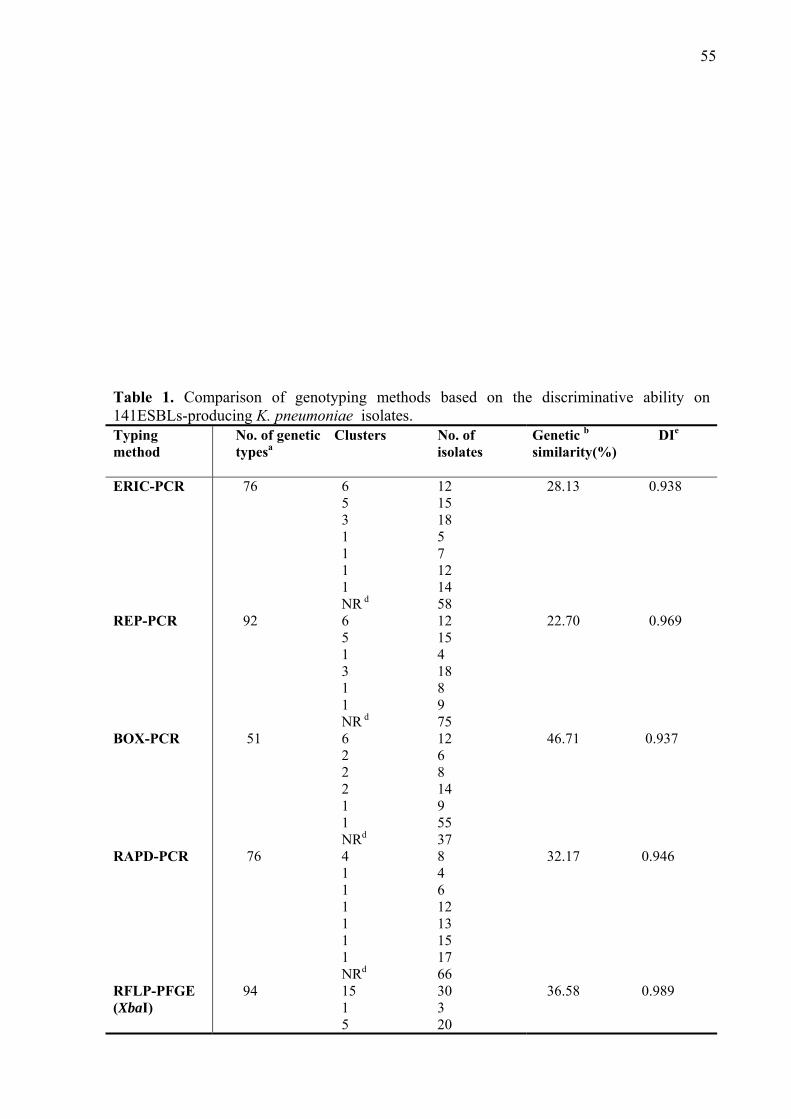
55
Table 1. Comparison of genotyping methods based on the discriminative ability on 141ESBLs-producing K. pneumoniae isolates. Typing method
No. of genetic typesa
Clusters No. of isolates
Genetic b
similarity(%)
DIe
ERIC-PCR REP-PCR BOX-PCR RAPD-PCR RFLP-PFGE (XbaI)
76 92 51 76 94
6 5
3 1 1 1 1 NR d
6 5 1 3 1 1 NR d
6 2 2 2 1 1 NRd
4 1 1 1 1 1 1 NRd
15 1 5
12 15 18 5 7 12 14 58 12 15 4 18 8 9 75 12 6 8 14 9 55 37 8 4 6 12 13 15 17 66 30 3 20
28.13 22.70 46.71 32.17 36.58
0.938 0.969 0.937 0.946 0.989
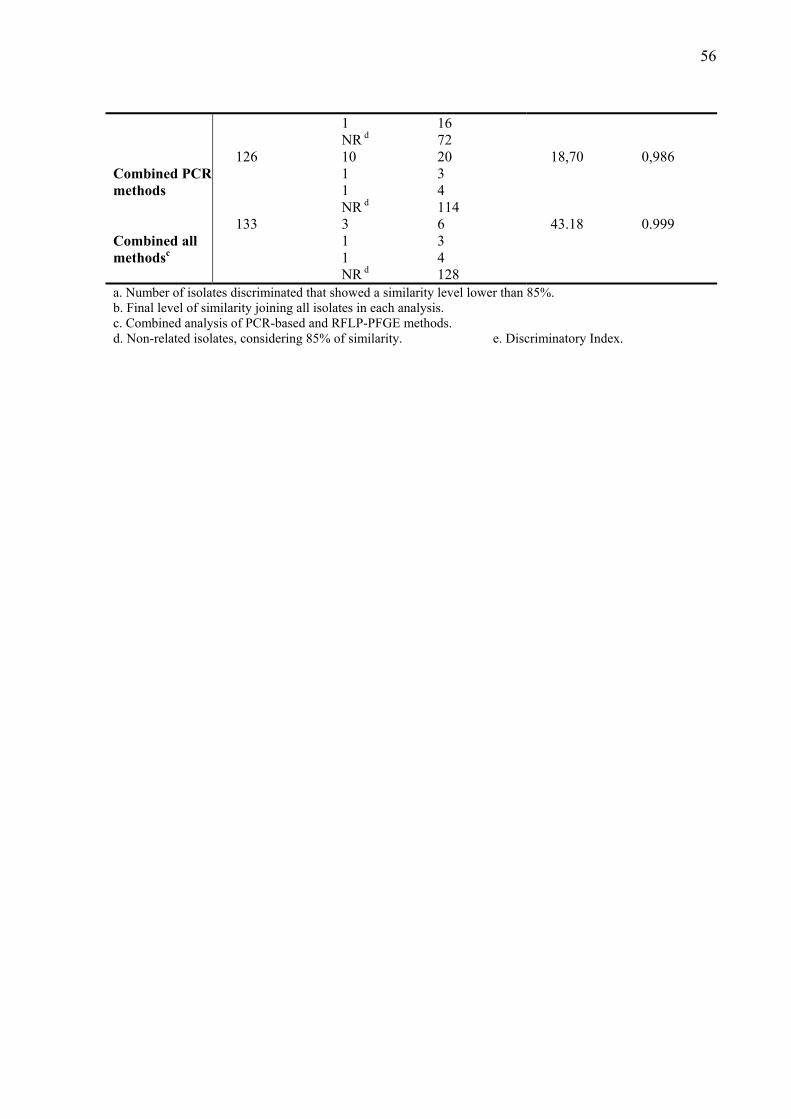
56
Combined PCR methods Combined all methodsc
126 133
1 NR d
10 1 1 NR d
3 1 1 NR d
16 72 20 3 4 114 6 3 4 128
18,70
43.18
0,986 0.999
a. Number of isolates discriminated that showed a similarity level lower than 85%. b. Final level of similarity joining all isolates in each analysis. c. Combined analysis of PCR-based and RFLP-PFGE methods. d. Non-related isolates, considering 85% of similarity. e. Discriminatory Index.
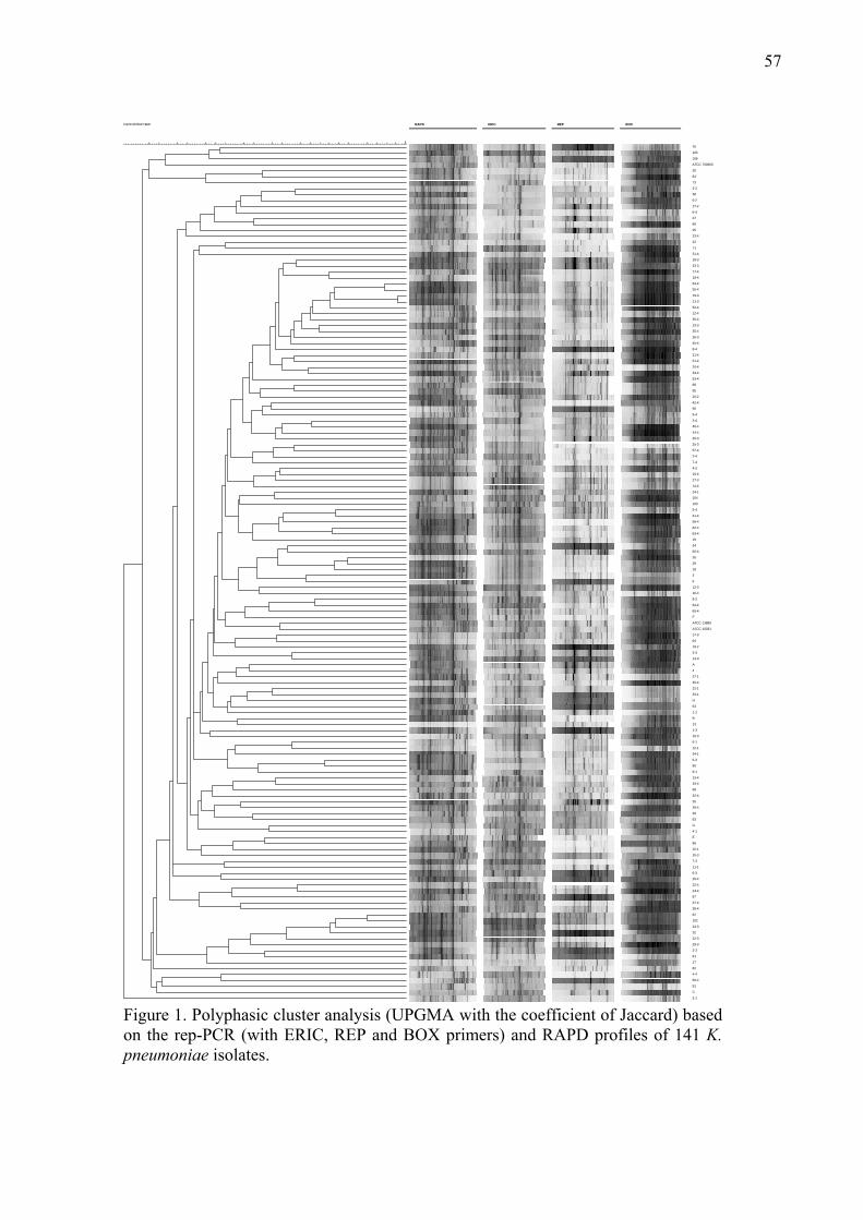
57
rep+eric+box+rapd
100
9896949290888684828078767472706866646260585654525048464442
RAPD ERIC REP BOX
76
105
106
ATCC 700603
30
84
73
3-2
38
6-2
17-2
6-4
27
68
25
23-4
22
71
31-4
18-3
23-3
17-4
18-4
54-4
55-4
19-3
21-3
52-4
12-4
36-4
13-3
35-4
26-3
45-4
8-4
21-4
51-4
20-4
33-4
53-4
89
95
20-2
42-4
96
9-4
3-4
46-4
44-4
20-3
25-3
57-4
2-4
7-4
4-2
15-4
27-3
14-4
24-1
104
103
5-4
41-4
56-4
62-4
63-4
19
34
60-4
26
29
18
3
6
12-3
16-4
8-3
64-4
65-4
F
ATCC 13889
ATCC 10031
17-3
64
19-2
3-3
14-3
A
4
27-1
30-4
22-1
33-1
H
63
1-1
B
13
1-3
10-3
6-1
12-1
34-1
5-3
80
9-1
13-4
19-4
98
32-4
35
39-4
39
93
G
4-1
E
88
10-1
15-3
7-3
11-1
6-3
29-4
22-4
24-4
87
37-4
38-4
62
102
24-3
32
22-3
29-3
2-3
61
17
82
4-4
59-4
91
C
3-1 Figure 1. Polyphasic cluster analysis (UPGMA with the coefficient of Jaccard) based on the rep-PCR (with ERIC, REP and BOX primers) and RAPD profiles of 141 K. pneumoniae isolates.
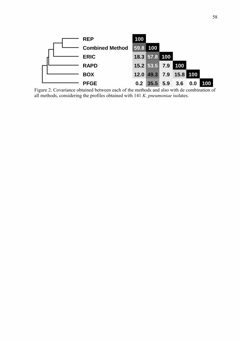
58
REPCombined MethodERICRAPDBOXPFGE
10059.818.315.212.00.2
10057.853.549.335.5
1007.97.95.9
10015.83.6
1000.0 100
Figure 2. Covariance obtained between each of the methods and also with de combination of all methods, considering the profiles obtained with 141 K. pneumoniae isolates.
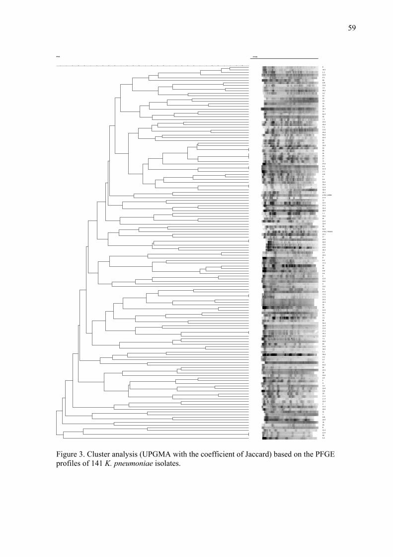
59
PFGE
100
98969492908886848280787674727068666462605856545250484644424038
PFGE
E
42-4
1-3
15-3
6-1
89
103
18-4
4-4
35-4
4-2
19
93
3-4
76
26
24-4
73
62-4
95
7-4
23-3
45-4
7-3
13-3
25-3
55-4
63-4
62
18
32-4
63
88
61
25
27
71
16-4
8-3
51-4
2-3
104
3
8-4
65-4
33-4
12-4
46-4
24-1
ATCC 13889
17-3
22
53-4
37-4
31-4
39-4
1-1
33-1
87
19-4
30-4
35
52-4
ATCC 700603
22-1
17
20-4
59-4
19-3
10-3
44-4
2-4
26-3
H
6-3
27-3
82
96
102
9-4
6
12-3
20-2
A
19-2
9-1
10-1
21-3
57-4
22-3
24-3
32
39
14-4
12-1
30
3-1
84
64-4
21-4
15-4
27-1
34-1
54-4
4-1
56-4
80
14-3
38-4
5-4
36-4
3-3
6-2
13
60-4
64
20-3
38
18-3
6-4
F
G
3-2
23-4
106
29
17-2
17-4
22-4
C
11-1
29-3
91
4
105
29-4
34
68
B
41-4
13-4
98
5-3
Figure 3. Cluster analysis (UPGMA with the coefficient of Jaccard) based on the PFGE profiles of 141 K. pneumoniae isolates.
60
Occurrence of Klebsiella pneumoniae Producing Extended-Spesctrum-β-
Lactamases (ESBLs) in a Universtiy Hospital, During a Five Years Period,
Using Phenotipic and Molecular Methods
1.Introduction
Klebsiella pneumoniae has emerged as a common cause of serious epidemic and
nosocomial infections in hospitals, resulting in high morbidity and mortality (DiPersio et al;.
2005). K. pneumoniae infections occur in almost all age groups, with urinary and respiratory
tract infections being most commonly described. K. pneumoniae accounts for a significant
proportion of hospital-acquired pneumonia, bacteremia, meningitis, septicemia, and soft tissue
infections. Neonatal infections by this bacterium are becoming a major concern of
pediatricians, as septicemia and meningitis in newborns are now often caused by multidrug-
resistant strains (Kurupati et al., 2007). Community-acquired-K. pneumoniae infections have
been also reported (Tumbarello et al., 2006).
Since extended-spectrum-lactamases (ESBLs) were initially reported in K.
pneumoniae in Germany in 1983 (Knothe et al., 1983), they have been increasingly described
worldwide. ESBL production is generally the result of point mutations in the blaTEM and
blaSHV genes which alter the primary amino sequences of the respective β-lactamase
enzyme. ESBL are also capable of hydrolyzing the oxyiminocephalosporins and
monobactams (Philippon et al., 1994). In 1989, a novel type of ESBL that conferred a high
level of resistance to cefotaxime and a low level of activity against ceftazidime, was nearly
simultaneously identified in an Escherichia coli strain isolated in Germany and in a
Salmonella enterica serovar Typhimurium isolate recovered in Argentina (Tzouvelekis et al.,
2000). This new family of plasmid-mediated ESBLs of Ambler class A were named
cefotaximase (CTX-M), and has been reported with increasing frequency throughout the word
(Edelstein et al., 2003; Lovallay et al., 2006).
The new epidemiology scenario of ESBL includes the increase in the number of
different CTX-M enzymes and the recognition of multiple clones and genetic elements that
carry blaCTX-M genes. The way by which CTX-M enzymes spread might follow an allodemic,
rather than an epidemic pattern; this term reflecting that the increase of CTX-M enzymes has
not been the result of the dissemination of particular clones, but of the spread of both
multiple specific clones and/or mobile genetic elements (Cantón et al., 2003). Some of the
CTX-M enzymes are widely present in specific countries, such as CTX-M-9 and CTX-M-14
61
in Spain (Hernandez et al., 2005; Novais et al., 2006), CTX-M-1 in Italy (Brigante et al.,
2005) and CTX-M-2 in most South American countries, Japan and Israel (Bonnet, 2004; Ben-
Ami et al., 2006), whereas others such as CTX-M-15 have been detected worldwide
(Brigante et al., 2005; Lavollay et al., 2006)
In Europe, United States and Latin America, the number of infections caused by
ESBL-producing strains of the family Enterobacteriaceae is increasing, and this trend has a
significant impact on mortality rates and hospitals costs (Bisson et al., 2002; Cartelle et al.,
2004). Although ESBLs have been detected in many gram-negative species, K. pneumoniae is
still the most frequently reported producer of these enzymes. Since the ESBLs genes are
usually found in large plasmids, that also contain other antimicrobial resistance genes, ESBL-
producing organisms may also be resistant to aminoglycosides, tetracyclines,
chloramphenicol, and/or sulfonamides (Podschun and Ullmann, 1998; Bradford, 2001).
ESBL-producing K. pneumoniae strains are more likely to be resistant to fluoroquinolones
than their non-ESBL-producing counterparts (Paterson, 2000). ESBL production has an
important clinical impact even when cephalosporin MICs are in the susceptible range (Kang
et al., 2004). Carbapenems are the mainstay of therapy for infections caused by multidrug-
resistant ESBL-producing organisms, and recent reports of acquired carbapenem resistance
among these organisms are thus another serious concern (Woodford et al., 2004).
This work aimed at detecting and characterizing of ESBLs produced by K.
pneumoniae, using molecular methods, 141 isolates from hospitalized patients, collected
during a five year period (2000-2004) in a hospital of Londrina, Parana State, Southern Brazil.
To our knowledge this is the first study of this kind in our state with such a large sample of
isolates. 2. Material and methods
2.1 Clinical isolates
One-hundred and forty- one isolates of ESBLs producing K. pneumoniae were studied, which
were collected during the period of January 2000 to December 2004, from different sources,
including urine (107), blood (19), surgical wound (6), tracheal aspirate (4), central venous
catheter (2), pleural liquid (2) and cerebrospinal fluid (1). Strains were select from clinical
specimens, one isolate per patient, at the University Hospital, Londrina, Parana State, southern
Brazil. The isolates were initially identified using Microscan Walkaway (Dade Behring,
62
Sacramento, CA, USA) confirmed with API 20E (Bio-Merieux, Marcy I’Etoile, France), and
stored in brain heart infusion (BHI) broth +20% glicerol at –200C.
2.2 Screening for ESBL producers
The isolates presenting a cefpodoxime MIC≥8 μgmL-1 and/or a cefotaxime/
ceftazidime/aztreonam MIC≥2 μgmL-1, by automated method were tested for ESBL
production according to CLSI (Clinical and Laboratory Standards Institute) 2005 guidelines.
ESBLs production was confirmed by the double disk screening (DDS) and combination disk
(CD) methods using Oxoid disks (Basingstoke, Hampshire, England) as described previously
(Jarlier et al., 1988; Jacoby and Han, 1996). K. penumoniae ATCC 700603 and E. coli ATCC
25922 were used as positive and negative controls.
2.3 Antimicrobial Susceptibility Testing MICs were determined by agar dilution method according to the CLSI (2005). The following
antimicrobial agents were tested, obtained from their respective manufacturers: aztreonam
(Bristol-Myers-Squibb, Brasil), cefepime (Bristol-Myers-Squibb, Brasil), ceftazidime (Glaxo-
Smith-Kline-Brasil), ceftriaxone (Roche, Brasil), cefotaxime (Aventis Pharma, Brasil),
cefazoline (Eli Lilly, Brasil), cefoxitin (Merck, Sharp & Dohme, Brasil), imipenem (Merck,
Sharp & Dohme, Brasil), meropenem (AztraZeneca, Brasil), ampicillin (Eurofarma, Brasil),
amoxicillin/clavulanate (Glaxo-Smith-Kline-Brasil), ampicillin/sulbactam (Pfizer,Brasil),
piperacillin/tazobactam (Wyeth-Whitehall, Brasil), amikacin (Bristol-Meyers-Squibb, Brasil)
gentamicin (Schering -Ploug, Brasil), ciproflixacin (Bayer, Brasil) levofloxacin (Aventis
Pharma, Brasil) K. penumoniae ATCC 700603 and ATCC E. coli 25922 were used as controls.
2.4 β-Lactamase gene characterization The blaSHV, blaTEM, blaCTX-M ESBL-encoding genes were characterized by PCR as described
previously (Arlet and Philippon, 1991; Bonnet et al., 2000; Bedenic et al., 2001). PCR
products obtained were digested with the following restriction endonucleases: for blaSHV,
DdeI, HhaI, HaeIII and NheI were used; for blaTEM, was used MseI, HhaI and HpaII; and for
blaCTX-M, HhaI, HinfI, DdeI and PstI. The DNA fragments were analyzed by 2% agarose gel
63
electrophoresis. The ESBL-encoding genes that showed differences in the RFLP analysis
were subsequently cloned into the TOPO TA vector (InvitrogenTM), following the procedure
described by the manufacturer, and sequenced. The PCR products were purified as described
before (Menna et al., 2006). The sequencing was performed with the use of the DYEnamic
ET terminator reagent (GE Healthcare) and analyzed in a MegaBace 1000 DNA Analysis
System (GE Healthcare), according to the parameters previously described (Menna et al.,
2006). The high-quality sequences obtained in both 3’ and 5’ directions were assembled using
the programs phred (Ewing & Green, 1998), phrap (http://www. phrap.org), and Consed
(Gordon et al., 1998) and were submitted to the GenBank database
(http://www.ncbi.nlm.nih.gov/BLAST) to seek for significant alignments.
2.5 Molecular typing
2.5.1 ERIC-PCR: Enterobacterial repetitive intergenic consensus polymerase chain reaction
(ERIC-PCR) was performed with primers ERIC1R and ERIC2 (de Bruijin, 1992), and the
PCR conditions as described by Santos et al. (1999). Amplification was performed in an MJ
Research Inc.PT 100 thermocycler. Amplified fragments were separated by horizontal
electrophoresis on a 1,5% agarose (low EEO, type I-A, GibcoBRL) gel (20x25 cm) at 120V
for 6h. Interpretation of patterns generated by ERIC-PCR was performed using Bionumerics
software (Applied Mathematics, Kortrijk, Belgium, version 4.6). The sizes of the fragments in
each analysis were normalized according to the molecular weight of the 1-kb molecular size
marker (Invitrogen, CA, USA). Clusters analisys was performed using the UPGMA
algorithm (unweighted pair-group method, with arithmetic mean, Sneath & Sokal, 1973) and
the coefficient of Jaccard (Jaccard, 1912). Isolates were considered similar if the similarity
coefficient between their patterns was of 0.85 or greater (Grundmann et al., 1995).
2.5.2 RFLP-PFGE: pulsed field gel electrophoresis (PFGE) analysis of genome
macrorestriction fragments (RFLP-PFGE) was performed as described by Chang & Chiu,
(1998). Total DNA was digested with XbaI (InVitrogenTM) and restricted DNA fragments were
separated using the CHEF-DRIII system (Biorad, USA), with pulses ranging from 5 to 50 s, at a
voltage of 6 V cm-1, at 14oC for 20 h and the λ ladder (Bio Rad) was used as molecular weight.
Products were detected after ethidium bromide staining and photographed with a Kodak Digital
64
Science 120 apparatus. The results were analyzed with the Bionumerics software (Applied
Mathematics, Kortrijk, Belgium, version 4.6) to determine similarities and differences among
the genotypes of different bacterial isolates.
3.RESULTS
3.1 Antimicrobial Susceptibility of ESBL-producing Klebsiella pneumoniae
All isolates were uniformly resistant to ampicillin and cefazolin (MIC≥ 128 μg mL-1).
Potential ESBL phenotypes (MIC≥2 mg L-1 for aztreonam, or ceftriaxone, or ceftazidime)
were observed in almost strains. Among the susceptible strains only 5.0% were susceptible
to cefotaxime, 5.7% to ceftriaxone, 6.4% to ceftazidime, 5.7% to aztreonam and 10.6% to
cefepime. Among the antimicrobial agents tested, only imipenem and meropenem
demonstrated 100% activity against K. pneumoniae isolates. The penicillin-β-lactamase
inhibitor combination of piperacillin-tazobactam was active against 98.6% of the isolates,
whereas only 49.6% were susceptible to ampicillin-sulbactam and 67% for amoxicillin-
clavulanate. Susceptibility to the aminoglycosides, 23.5% were susceptible to amikacin and
20.% to gentamicin. Among fluorquinolones, levofloxacin was active against 54.5% of the
isolates, whereas only 20.6% of isolates were susceptible to ciprofloxacin. The distribution of
the MICs values are shown in Table 1.
3.2 Screening for ESBL producers
Strains presenting MIC suggestive of ESBL production as cefpodoxime MIC≥8μg
mL-1 and/or a cefotaxime/ceftazidime/aztreonam MIC≥2μg mL-1, in the automated method
were confirmed using DDS and CD methods.
3.3 Genetic characterization of β-lactamases: DNA amplification and sequencing
When the TEM, SHV, and CTX-M β-lactamases from K. pneumoniae were amplified a
858-bp product characteristic of TEM was obtained in six (4.3%) isolates (two from 2000 and
four in 2003) and with the different restriction endonucleases two patterns were obtained.
65
However, we did not succeed in obtaining sequences with TEM-type β-lactamases, therefore
not it was possible to characterize.
For the SHV-type β-lactamases a 950-bp product was obtained in 135 (96%) isolates,
of which 92 (68%) were SHV-types ESBLs. The analysis of the restriction endonucleases
yielded four different patterns corresponding to the following SHV-types ESBLs observed after
sequencing: SHV-2 (n=23; 17.0%), SHV-5 (n=40; 29.7%), SHV-11 (n=29; 21.5%); and 43
(31.8%) isolates presented only the SHV-1-type β-lactamases. These β-lactamases were also
associated with other enzymes like TEM, CTX-M-2 and CTX-M-54 in the same the isolates
(Table 2). Four (3.0%) strains produced only SHV-types ESBLs, being three SHV-11 and one
SHV-5.
For the β-lactamases type CTX-M, a 550-bp product was obtained with 133 (94,3%)
isolates. The restriction analysis with endonucleases yielded two different patterns and after
sequencing the follow CTX-M-type ESBL were observed: CTX-M-2 in 131 (92.9%) and 2
strains CTX-M-54 (1.4%), these last ones during 2003. Different combination of these enzymes
were observed among the strains, as shown in Table 2.
The results obtained, showed 88 (62%) strains containing genes for two ESBLs in the
same isolate. Several combinations were described as SHV-2/CTXM-2 (n=23; 26.2%), SHV-
5/CTX-M-2 (n=38; 43.2%) and SHV-11/CTX-M-2 (n=23; 26.2%), SHV-5/TEM (1; 1.1%) ,
TEM/CTX-M-2( n=1; 1.1%) and SHV-11/TEM (2; 2.2%).
3.4 Molecular typing
Cluster analysis of the restriction profiles produced by RFLP-PFGE and bands
generated by ERIC-PCR typing revealed low relatedness among the isolates. When the
dendrogram was generated, ERIC-PCR analysis resulted in 76 different genotypes, six with
two strains each, five with three stains each, three with six strains and single genotype with:
five, seven, twelve and fourteen strains each (Fig. 1). The other isolates showed genetic
similarity lower than 85% (between 28,13% and 85%). The dendrogram generated by RFLP-
PFGE, resulted in 94 different genotypes, fifteen different clusters containing two strains each,
five with four strains each, one with three and a cluster with sixteen strains (isolated in
different years: one in 2000, five in 2002, two in 2003 and seven in 2004).The other isolates
presented genetic similarity between 36.58% a 85.0% (Table 2).
66
4. Discussion
ESBLs are an increasing problem in human medicine, inducing resistance to third-
generation cephalosporins among Enterobacteriaceae, especially K. pneumoniae. In addition,
bacteria producing these enzymes are frequently resistant to many classes of non-β-lactam
antibiotics, resulting in difficult-to-treat infections (Mac Kenzie et al., 2002; Mack & Mack,
2003). Due to their importance, these enzymes have been extensively investigated. The TEM-,
SHV-,0XA-, and more recently, CTX-M-type enzymes emerged in many countries of the
world, including Brazil (Bonnet et al, 2000; Hernández et al., 2005; DiPersio et al., 2005;
Livermore et al., 2007)
Although antibiotic resistance is becoming a major threat to human health
worldwide, information concerning the dissemination and geographical distribution of
antibiotic-resistant bacteria remains scarce (Villa et al., 2000). The resistance patterns of
ESBL-producing K. pneumoniae are remarkable for the high rate of co-resistance to other
classes of antibiotics. The 92.7% of CTX-M producing isolates, cefotaxime, ceftriaxone and
cefepime MICs presented high ( ≥128µgmL-1) for MIC50 and MIC90, while for ceftazidime
they were lower (MIC50=16 µg mL-1 and MIC90 64 µgmL-1). Considering that 62% of the
strains were SHV-type ESBL produces, this could explain the lower values for ceftazidime,
since this group of enzymes is able to hydrolise better ceftzidime. The β-Lactam-β-
Lactamase inhibitor combinations, as well as carbapenems, aminoglycosides and
fluorquinolones are considered to be potentially active drugs against ESBLs-producing K.
pneumoniae (Villegas et al., 2004). In a study performed with CTX-M producing K.
pneumoniae in Russian hospitals, Edelstein et al. (2003) found results similar to ours about
piperacillin-tazobactam and concluded that the increased activity of this drug association
could be explained by the fact that CTX-M enzymes are better inhibited by tazobactam than
by clavulanate. In relation to the aminoglicosydes, these authors obtained similar results to
ours, that is, 85.1% of the K. pneumoniae isolates were resistant to gentamicin. Liao et al.
(2006) studying ESBL-K. pneumoniae in two regional hospitals in Taiwan reported that only
37% of the isolates were susceptible to ciprofloxacin, in agreement with our results of 20.6%.
Hernández et al. (2005) reported that the concurrence of ciprofloxacin resistance with ESBL
production, particularly in isolates of K. pneumoniae, was also observed in a nationwide study
in Spain; according to the authors the actual causes of this association are not well known but
67
may be related not only to target mutations in DNA gyrase or toposisomerase IV, but also to
other mechanisms, including porin loss, active efflux, and target protection.
The SHV-type ESBL is often produced by K. pneumoniae, eventhough it is also
found in other species of Enterobacteriaceae and in P.aeruginosa (Neonakis et al., 2003). The
blaSHV-1 is detected in more than 90% of clinical isolates of K. pneumoniae, carrying a
chromosomal copy. Its prevalence provides an explanation for the number of variants to
which is directly related. In South America, the first reports on SHV-type ESBL occurred in
1988 and 1989, of K. pneumoniae isolates ( in Chile and Argentina) harboring SHV-2 and
SHV-5. In Brazil, SHV was first described by Corkill et al. (2001), a K. pneumoniae strain
producing SHV-27, isolated in a blood culture in Aracaju State of Sergipe, Northeast Region.
In this study we found different types of SHV produced by 135 (96%) of the strains: SHV-1
(n=43;31.8%), SHV-2 (n=23; 17.0%), SHV-5 (n=40; 29.7%) and SHV-11 (n=29; 21.5%).
Considering that SHV-1 is not an ESBL, we could say that SHV-5 was the predominant
ESBL type in this study, in agreement with the results obtained in Greece (88%) (Legakis et
al., 1995). The first report on SHV-11 producer K. pneumoniae occurred in 1997 in
Switzerland (Nüesch-Inderbinen et al., 1997) and in 1999 it was detected in several
Enterobacteriaceae species, including Shigella dysenteriae (Ahamed & Kundu, 1999) and
thereafter was spread worldwide. More recently it was observed that, K .pneumoniae strains
carrying the chromosomal SHV-11 β-lactamase gene produce the plasmid-mediated SHV-12
ESBL more frequently than those carrying the chromosomal gene (Lee et al., 2006).
The TEM-type ESBLs are derived from TEM-1 and TEM-2. The first described
TEM-ESBL was a strain of K. oxytoca harboring a plasmid carrying a gene encoding
ceftazidime resistance, in Liverpool, England, in 1982. Well over 161 TEM-type β-lactamases
have been described and the amino acid changes in comparison with TEM-1 and TEM-2 are
documented at http://www.lahey.org/studies/ttemtabele.htm. Curiously, TEM-type ESBLs
have been very rarely reported in South America, and the enzymes seem to be prevalent in
North America and Canada. In this study a low number of strains (six) harboring blaTEM was
detected, and in five of them were associated with the presence of blaSHV and in another with
CTX-M-2; in addition the association of blaTEM and blaSHV was also observed in 4.3% of
isolates. More studies are necessary to analyze if the low prevalence of blaTEM in our study
could be related to the intensive spread of CTX-M.
In South America, CTX-M-type-ESBL occurred in 1989 during an outbreak of
multiresistant Salmonella enterica serovar Typhimurium infections in Argentina. From these
isolates a new non-SHV, non-TEM ESBL named CTX-M-2 was identified and this enzyme
68
has since then been described in many Enterobacteriaceae species spread throughout different
parts of South America continent. In Brazil, besides CTX-M-2 other CTX-M enzymes (CTX-
M-8, 9 and 16), have been described. Radice et al (2002) detected CTX-M-2 in 75% of the
ESBL-producing enterobacteria in Buenos Aires and recently the first time was in Colombia
(Silva et al., 2006). Our study, reported and confirm the dissemination of CTX-M-2 in South
America. Furthermore, in our study strains carrying blaCTX-M-2 were found in 92.9% and CTX-
54 in 2 isolates (1.4%). The way by which CTX-M-2 enzymes spread in South America
countries may be not a result of the dissemination of particular clones, but of the spread of
both multiple specific clones and/or mobile genetic elements as has already been described in
integrons (Power et al., 2005; Vignoli et al., 2006). The CTX-M-54 enzyme was first reported
in Korea 2006 in isolates of K. pneumoniae (Bae et al., 2006), however in our study two
strains producing this enzyme were isolated in 2003.
Our molecular analyses indicate that dissemination of ESBL-producing K.
pneumoniae strains in our hospital probably involved horizontal transfer of the resistance
determinants, although simultaneous spread of several clones could also have played a role.
This possibility is supported by the finding, in the analysis of ERIC-PCR, of several clones
(from two to sixteen) with the same gene or the association of genes (e.g. only CTX-M-2 or
SHV-2/CTX-M-2, SHV-5/CTX-M-2, SHV-11/CTX-M-2). In the RFLP-PFGE analysis, one
large clone was obtained with sixteen strains harboring CTX-M-2 or CTXM-2 associated with
SHV, isolated in different years and several small clones (from two to four). The presence of
endemic CTX-M-2 enzyme distribution of strains was observed. The abundance of different
genotypes in relation to different enzymes shows the complexity and diversity of the
epidemiology of ESBLs in the University Hospital of Londrina. It was not possible to
correlate each clone with a specific enzyme, since each clone had different isolates with
CTXM-2 alone or associated to different SHVs. If the clustered isolates are genotypically
different, then the clustering may be due to chance alone, or to excessive drug pressure,
resulting in the selection of a resistant phenotype within a group of unrelated isolates.
K. pneumoniae producing two or more ESBLs have been reported scarcely, but
when detected in most cases they include TEM-and/or SHV-derived enzymes (Melano et al.,
2003). Here we report the identification of 62% multiresistant K. pneumoniae isolates which
produced several combinations of two ESBLs in the same isolate. The more frequent
combination detected was of SHV-5/CTX-M-2, which was found in 38 (27%) isolates. The
emergence of bacteria with multiple β-lactamases carries several implications as likelihood
69
that β-lactamase inhibitors will be overwhelmed and difficulties in the interpretation of
antibiograms using manually or automated systems (Essack et al., 2004).
In summary, this study shows the presence of multiresistant and ESBL producing
strains of K. pneumoniae in a large sample of isolates (141) collected during the period of
January 2000 to December 2004. We also detected the dominant presence of CTX-M-2 and
just few (six) strains of TEM producers. Four types SHV were detected, being SHV-5 the
most frequent 29.7%. Our results show the prevalence of CTX-M-2 in our region. These data
could be used to better understand the epidemiology and to establish policies for the use of
new antimicrobial drugs, in addition to improving hospital hygiene, a great challenge for
hospital epidemiology
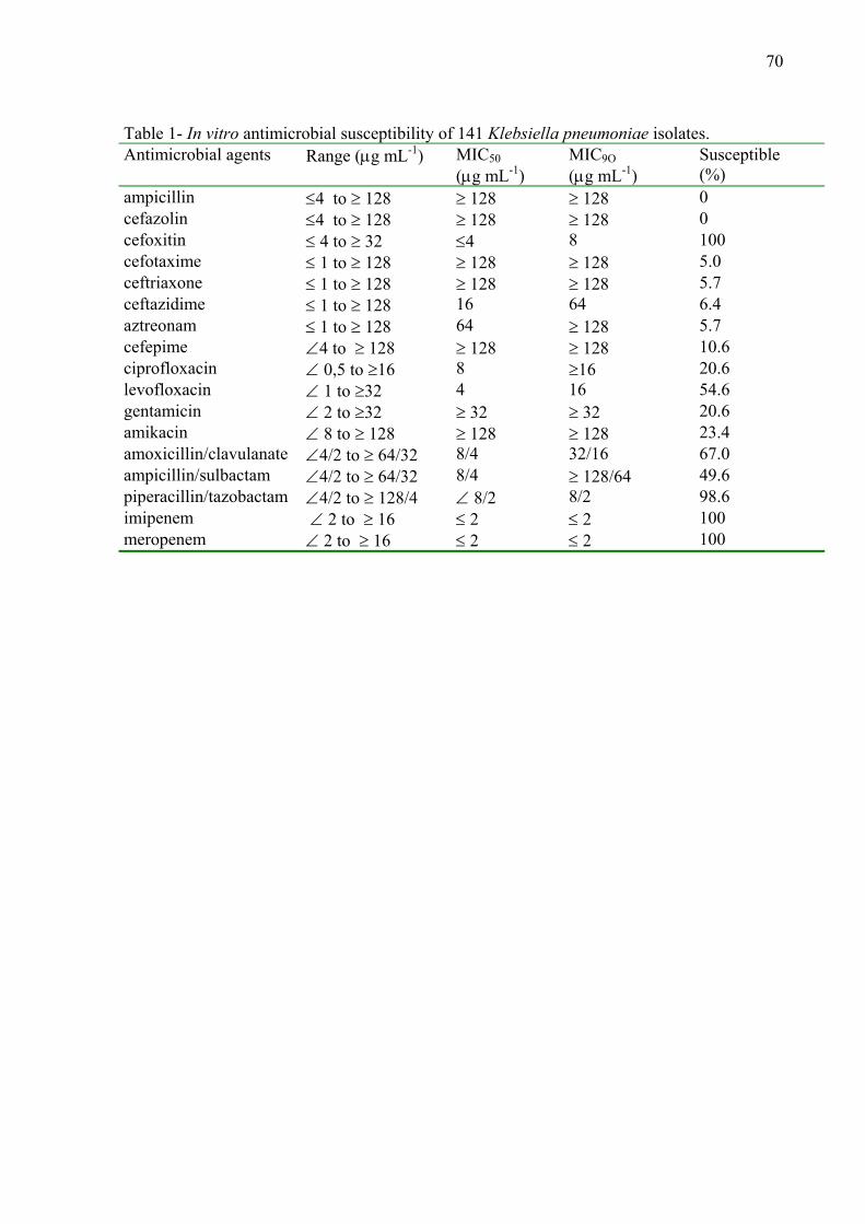
70
Table 1- In vitro antimicrobial susceptibility of 141 Klebsiella pneumoniae isolates. Antimicrobial agents Range (μg mL-1) MIC50
(μg mL-1) MIC9O
(μg mL-1) Susceptible (%)
ampicillin ≤4 to ≥ 128 ≥ 128 ≥ 128 0 cefazolin ≤4 to ≥ 128 ≥ 128 ≥ 128 0 cefoxitin ≤ 4 to ≥ 32 ≤4 8 100 cefotaxime ≤ 1 to ≥ 128 ≥ 128 ≥ 128 5.0 ceftriaxone ≤ 1 to ≥ 128 ≥ 128 ≥ 128 5.7 ceftazidime ≤ 1 to ≥ 128 16 64 6.4 aztreonam ≤ 1 to ≥ 128 64 ≥ 128 5.7 cefepime ∠4 to ≥ 128 ≥ 128 ≥ 128 10.6 ciprofloxacin ∠ 0,5 to ≥16 8 ≥16 20.6 levofloxacin ∠ 1 to ≥32 4 16 54.6 gentamicin ∠ 2 to ≥32 ≥ 32 ≥ 32 20.6 amikacin ∠ 8 to ≥ 128 ≥ 128 ≥ 128 23.4 amoxicillin/clavulanate ∠4/2 to ≥ 64/32 8/4 32/16 67.0 ampicillin/sulbactam ∠4/2 to ≥ 64/32 8/4 ≥ 128/64 49.6 piperacillin/tazobactam ∠4/2 to ≥ 128/4 ∠ 8/2 8/2 98.6 imipenem ∠ 2 to ≥ 16 ≤ 2 ≤ 2 100 meropenem ∠ 2 to ≥ 16 ≤ 2 ≤ 2 100
71
5. References
Ahamed, J., Kundu, M., 1999. Molecular characterization of the SHV-11 beta-lactamase of Shigella dysenteriae. Antimicrob. Agents Chemother. 8:2081-2083.
Arlet, G., Philippon, A. Construction of polymerase chain reaction and use of intragenic Dna probes for thre main types of transferable Beta-lactamases (TEM,SHV,CARB). Fems Microbiol. Lett. 66:19-25, 1991.
Bae, I .K., Lee, B. H., Hwang, H. Y., Jeong, S. H., Hong, S. H., Chang, C. L., Kwak, H. S., Kim, H. J., Youn, H. A novel ceftazidima-hydrolysing extended-spectrum β-lactamase, CTX-54, with a single amino acid substitution at position 167 in the omega loop. J. Antimicrob. Chemother. 58:315-319.
Bedenic, B., Randegger, C. C., Stobberingh, E., Häckler, H. 2001. Molecular epidemiology of extended spectrum β-lactamase from Klebsiella pneumoniae strains isolated in Zagred, Croatia. Eur. J. Clin. Microbiol. Infec. Dis. 20:505-508.
Bisson, G., Fishman, N. O., Patel, J. B., Edelstein, P. H., Lautenbach, E. 2002. Extended-spectrum-β-lactamases producing Escherichia coli and Klebsiella species: risk factors for colonization and impact of antimicrobial formulary interventions on colonization prevalence. Infect. Control. Hosp.Epidemiol. 23:254-260. Bonnet, R., Sampaio, J. L. M., Lábia, R., Champs, C., Sirot, D., Chanal, C., Sirot, J. 2000. A novel CTX-M β-lactamase (CTX-M-8) in cefotaxime-resistant Enterobacteriaceae isolated in Brazil. Antimicrob. Agents Chemother. 44:1936-42.
Bonnet, R. 2004. Growing group of extended-spectrum beta-lactamases: the CTX-M enzymes. Antimicrob. Agents Chemother. 48:1-14.
Bradford, P. A. 2001. Extended-spectrum β-lactamases in the 21st century:characterization, epidemiology, and detection of this important resistance threat. Clin. Microbiol. Rev. 14:933–951. Brigante, G., Luzzaro, F., Perilli, M., Lombardi, G., Coli, A., Rossolini, G. M., Amicosante, G., Toniolo, A. 2005. Evolution of CTX-M-type b-lactamases in isolates of Escherichia coli infecting hospital and community patients. Int. J. Antimicrob. Agents 25:157-162. Cantón, R., Coque,T. M., Baquero, F. 2003 Multi-resistant Gram-negative bacilli: from epidemics to endemics. Curr. Opin. Infect. Dis. 16:315-325. Cartelle, M., Tomas, M. M., Pertega, S., Beceiro, A., Dominguez, M. A., Velasco, D., Molina, F., Villanueva, R., Bou, G. 2004. Risk factors for colonization and infection in a
72
hospital outbreak caused by a strain of Klebsiella pneumoniae with reduced susceptibility to expanded-spectrum cephalosporins. J. Clin. Microbiol . 42:4242-4249. Chang, N., Chui, L. 1998. A standardized protocol for the rapid preparation of bacterial DNA for pulsed-field gel electrophoresis. Diagn. Microbiol. Infect. Dis. 31:275-279. Clinical and Laboratory Standards Institute (2005). Performance standards for antimicrobial disk susceptibility tests. 8th ed. (documented M2-A8, v.23, n.1) Corkill, J. E., Cuevas , L. E., Gurgel, R. Q., Greensill, J., Hart, C.A. 2001. SHV-27, a novel cefotaxime-hydrolysing beta-lactamase, identified in Klebsiella pneumoniae isolates from a Brazilian hospital. J. Antimicrob. Chemother. 47:463-5. De Bruijin, F. 1992. Use of repetitive (repetitive extragenic palindromic and enterobacterial repetitive intergenic consensus) sequences and the polymerase chain reaction to fingerprinting the genomes of Rhizobiun meliloti isolates and the others soils bacteria. App. Environ. Microbiol. 58:2180-2187. Di Persio, J. R., Deshpande, L. M., Biedenbach, D. J., Toleman, M. A., Walsh, T. R., Jones, R. N. 2005. Evolution and dissemination of extendedspectrum β-lactamase-producing Klebsiella pneumoniae: epidemiology and molecular report from the SENTRY Antimicrobial Surveillance Program (1997–2003). Diagn. Microbiol. Infect. Dis. 51:1–7. Edelstein, M., M. Pimkin, I. Palagin, I. Edelstein, L. Stratchounski. 2003. Prevalence and molecular epidemiology of CTX-M extended-spectrum-β-lactamase-producing Escherichia coli and Klebsiella pneumoniae in Russian hospitals. Antimicrob. Agents Chemother. 47:3724–3732. Essack, S. Y., Hall, L. M. C., Livermore, D. M. 2004. Klebsiella pneumoniae isolate from South Africa with multiple TEM, SHV and AmpC β-lactamases. Int. J. Antimicrob. 23:398-400 Ewing, B., Green, P. 1998. Base-calling of automated sequencer traces using phred. II. Error probabilities. Genome Research. 8, 186-194.
Gordon, D., Abajian, C., Green, P. 1998. Consed: a graphical tool for sequence finishing. Genome Research. 8, 195-202.
Grundmann, H., Schneider, C., Hartung, D., Daschner, F. D., Pitt, T. L. 1995. Discriminatory power of three DNA-based typing techniques for Pseudomonas aeruginosa. J. Clin. Microbiol. 33:528-534. Hernandez, J. R., Martinez-Martinez, L., Cantón, R., Coque, T. M., Pascual, A. 2005. Spanish group for nosocomial infections (GEIH): Nationwide study of Escherichia coli and Klebsiella pneumoniae producing extended-spectrum b-lactamases in Spain. Antimicrob. Agents Chemother. 49:2122-2125. Jaccard, P. 1912. The distribution of flora in the alpine zone. New Phytol. 11:37–50.
73
Jacoby, G. A., Han, P. 1996. Detection of extended-spectrum Beta-lactamases in clinical isolates of Klebsiella pneumoniae and Escherichia coli. J. Clin. Microbiol. 34:908-911. Jarlier, V., Nicolas, M. H., Fournier, G., Philippon, A. 1988. Extended broad-spectrum Beta-lactamases confering transferable resistance to newer Beta-lactam agents in Enterobacteriaceae hospital prevalence and susceptibility patterns. Rev. Infect. Dis. 10:867-878.
Kang, C. I., Kim, S. H., Park, W. B., Lee, K. D., Kim, H. B., Kim, E. C., Oh, M. D., Choe, K. W. 2004. Bloodstream infections due to extended-spectrum beta-lactamase-producing Escherichia coli and Klebsiella pneumoniae: risk factors for mortality and treatment outcome, with special emphasis onantimicrobial therapy. Antimicrob. Agents Chemother. 48:4574–4581. Knothe, H., Shah, P., Kremry, V., Antal, M., Mitsuhashi, S., 1983.Transferable resistance to cefotaxime, cefoxitin, cefamandole and cefuroxime in clinical isolates of Klebsiella pneumoniae and Serratia marcescens. Infection. 11:315-517. Kurupati, P., Tan, Q. S. W., Kumarasinghe, G., Poh, C. L. 2007. Inhibition of Gene Expression and Growth by Antisense Peptide Nucleic Acids in a Multiresistant β-Lactamase Producing Klebsiella pneumoniae Strain. Antimicrob. Agents Chemother. 51:805-811. Lavollay, M., Mamlouk, K., Frank, T., Akpabie, A., Burghoffer, B., Ben-Redjeb, S., Bercion, R., Gautier, V., Arlet, G. 2006. Clonal dissemination of a CTX-M-15 β-lactamase-producing Escherichia coli strain in the Paris area, Tunis, and Bangui. Antimicrob. Agents Chemother. 50:2433-2438. Lee, Y. H., Cho, B., Bae, K., Chang, C. L., Jeong, S. H. 2006. Klebsiella pneumoniae strains carrying the chromosomal SHV-11 β-Lactamase gene produce the plasmid-mediated SHV-12 extended-spectrum β-Lactamase more frequently than those carrying the chromosomal SHV-1 β-Lactamase gene. J. Antimicrob. Chemother. 57: 1259-1261. Legakis, N. J., Tzouvelekis, L. S., Hatzoudis, G., Tzelepi, E., Gourkou, A., Pitt, T. L., Vatopoulos, A. C.1995. Klebsiella pneumoniae infections in Greek hospitals. Dissemination of plasmids encoding an SHV-5type beta-lactamase. J. Hosp. Infect.31:177-87. Liao, C. H., Sheng, W. H., Wang, I. T., Sun, H. Y., Wang, H. K., Hsueh, P. R. 2006. Iv vitro activities of 16 antimicrobial agents against clinical isolates of extended-spectrum beta-lactamase-producing Escherichia coli and Klebsiella pneumoniae in two regional hospitals in Taiwan. J. Microbiol. Immunol. Infect.39:59-66. Livermoore, D., Canton, R., Gniadkowski, M., Nordmann, P., Rossolini, G. M., Arlet, G., Ayala, J., Coque, T. M., Zdanowicz, I. K., Luzzaro, F., Poirel., L., Woodford, N. 2007.CTX-M: changing the face of ESBL in Europe. J.Antimicrob. Chemother. 59:165-174. Mack, E., Mack, D. 2003. Extended-spectrum-lactamases: implications for the clinical microbiology laboratory, therapy, and infection control. J.Infec. 47:273-295.
74
MacKenzie, F. M., Miller, C. A., Gould, I. M. 2002. Comparison of screening methods for TEM and SHV-derived extended-spectrum ß-lactamase detection.Clin.Microbiol.Infect. 8:715-724.
Melano, R., Corso, A., Petroni, A., Centron, D., Orman, B., Pereyra, A., Moreno, N., Galas, M. 2003. Multiple antibiotic-resistance mechanisms including a novel combination of extended-spectrum β-lactamses in a Klebsiella pneumoniae clinical strain isolated in Argentina. J. Antimicrob Chemother. 52:36-42.
Menna, P., Hungria, M., Barcellos, F. G., Bangel, E. V., Hess, P. N., Martínez-Romero, E.2006. Molecular phylogeny based on the 16S rRNA gene a of elite rhizobial strains used in Brazilian commercial inoculants. Syst. Appl. Microbiol. 29:315-332. Neonakis, I. K., Scoulica, E.V., Dimitriou, S. K., Gikas, A. I., Tselentis, Y. J. 2003 Molecular epidemiology of extended-spectrum beta-lactamases produced by clinical isolates in a university hospital in Greece: detection of SHV-5 in Pseudomonas aeruginosa and prevalence of SHV-12. Microb Drug Resist. 9:161-5. Novais, A., Cantón, R., Valverde, A., Machado, E., Galán, J. C., Peixe, L., Carattoli, A., Baquero, F., Coque, T. M.. 2006. Dissemination and persistence of blaCTX-M-9 are linked to class 1 integrons containing CR1 associated with defective transposon derivatives from Tn402 located in early antibiotic resistance plasmids of IncHI2, IncP1-a, and IncFI Groups. Antimicrob. Agents Chemother. 50:2741-2750. Nüesch-Inderbinen, M.T., Kayser, F. H., Hachler, H. 1997. Survey and molecular genectics of SHV β-Lactamases in Enterobacteriaceae in Switzerland: two novel enzymes, SHV-11 and SHV-12. Antimicrob. Agents Chemother. 41:943-949 Paterson, D. L., Mulazimoglu, L., Casellas, J. M., Ko, W. C., Goossens, H., Von Gottberg, A., Mohapatra, S., Trenholme, G. M., Klugman, K. P.,McCormack, J. G., Yu, V. L.2000. Epidemiology of ciprofloxacin resistance and its relationship to extended-spectrum beta-lactamase production in Klebsiella pneumoniae isolates causing bacteremia. Clin. Infect. Dis. 30:473–478. Philippon, A., Arlet, G., Lagrange, P. H. 1994. Origin and impact of plasmid-mediated of extended spectrum β-lactamases. Eur. J. Clin. Microbiol. Infect. Dis. 13:S17–S29 Podschun, R., Ullmann, U. 1998. Klebsiella spp. as nosocomial pathogens: epidemiology, taxonomy, typing methods, and pathogenicity factors. Clin. Microbiol. Rev. 11:589–603. Power, P., Galleni, M., Di Conza, J., Ayala, J. A., Gutkind, G. 2005. Description of In116, the first blaCTX-M-2-containing complex class 1 integron found in Morganella morganii isolates from Buenos Aires, Argentina. J. Antimicrob. Chemother. 55:461-465.
75
Radice, M., Power, P., Di Conza, J., Gutkind, G., Bonnet, R., Sirot, D., Sirot, J., Labia, R. 2002. Early dissemination of CTX-M-derived enzymes in South America. Antimicrob. Agents Chemother. 46, 602-604. Rebuk, J. A., K. M. Olsen, P. D. Fey, A. N. Langnas, and M. E. Rupp. 2000. Characterization of an outbreak due to extended-spectum beta-lactamase producing Klebsiella pneumoniae in a pediatric intensive care unit transplant population. Clin. Infect. Dis. 31:1368–1372. Silva, E. M. V., Anaya, J. R. M., Reza, M. T. R., Mejia, E. B. G., Manrique, I. Y. P., Lierena, I. D., Velandia, D. 2006. Detection of CTX-M-1, CTX-M-15, and CTX-M-2 in clinical isolates of Enterobacteriaceae in Bogotá, Colombia. Antimicrob. Agents Chemother. 44:1919-1920. Sneath, P. B. A., Sokal, R. R. 1973. Numerical taxonomy. W. H. Freeman and Co., San Francisco, CA. Tumbarello, M., Spanu, T., Sanguinetti, M., Citton, R., Montuori, E., Leone, F., Fadda, G., Cauda, R. 2006. Bloodstream infections caused by extended-spectrum β-lactamases-producing Klebsiella pneumoniae: risk factors, molecular epidemiology and clinical outcome. Antimicrob. Agents Chemother. 50:498-504. Tzouvelekis, L. S., Tzelepi, E., Tassios, P. T., Legakis, N. J. 2000. CTXM- type lactamases: an emerging group of extended-spectrum enzymes. Int. J. Antimicrob. Agents 14:137–142. Vignoli, R., Cordeiro, N., Seija, V., Schelotto, F., Radice, M., Ayala, J., Power, P., Gutkind, G. 2006. Entorno genético de CTX-M-2 em aislamientos de Klebsiella pneumoniae provenientes de pacientes hospitalizados en Uruguay. Revista Argentina de Microbiologia. 38:84-88. Villa, L., Pezzella, C., Tosini, F., Visca, P., Petrucca, A., Caratolli, A.2000. Multiple-Antibiotic resistance mediated by structurally related IncL/M plasmids carrying an extended-spectrum β-Lactamase gene and a class 1 integron. Antimicrob. Agents Chemother.44:2911-2914. Villegas, M.V., Correa, A., Perez, F., Miranda ,M.C., Zuluaga, T., Quinn, J.P. 2004. Prevalence and characterization of extended-spectrum beta-lactamases in Klebsiella pneumoniae and Escherichia coli isolates from Colombian hospitals. Diagn. Microbio.l Infect. Dis. 49:217-22. Woodford, N., Tierno, P. M. , Young, K., Tysall, L., Palepou, M. F., Ward, E., Painter, R. E., Suber, D. F., Shungu, D., Silver, L. L., Inglima, K., Kornblum, J., Livermore, D. M. 2004. Outbreak of Klebsiella pneumoniae producing anew carbapenem-hydrolyzing class A beta-lactamase, KPC-3, in a New YorkMedical Center. Antimicrob. Agents Chemother. 48:4793–4799
76
High Incidence of Class 1 Integron Among Klebsiella pneumoniae strains and Characterization of Unusual integron carrying blactxm-2 gene in Isolates from Brazil Abstract The rapid dissemination of antibiotic resistance genes among bacterial isolates
is an increasing problem for the treatment of infectious diseases. Recently, integrons have
been recognized as significant contributors to the acquisition of antibiotic resistance in gram-
negative bacteria. In the present study, we determined the occurrence of integron classes 1, 2
and 3 in 141 ESBLs- producing K. pneumoniae isolates from hospitalized patients, in a period
of five years, from 2000-2004. We also performed the molecular characterization of the
integron carrying blactxm-2 gene in eight of the isolates which yielded positive results for class
1. Class 1 integron prevailed, being detected in 131 (93%) out of the 141 clinical isolates.
Class 2 integron was detected in nine isolates (6,4%), and seven 7 (5%) isolates had both
classes of integrons 1 and 2. Class 3 integron was not found among the ESBL-producing K.
pneumoniae isolates. This study is the first to describe the characterization of an integron
carrying blactxm-2 gene isolated from ESBL-producing K. pneumoniae strains isolated from
hospitalized patients in Brazil. Our results also suggest that the dissemination of CTX-M-2
enzyme occurs through unusual class 1 integron, as reported in Argentina and Uruguay.
1. Introduction
The CTX-M-type β-lactamases represent a rapidly emerging group with a typical
extended-spectrum β-lactamases (ESBL)-resistance phenotype (Bradford, 2001). These
enzymes, exhibiting extended-spectrum activities are capable of hydrolyzing some broad-
spectrum cephalosporins and are inhibited by clavulanic acid and tazobactam (Bonnet et al.,
2004). Many plasmid-mediated ESBL genes are located within or near mobile elements, such
as integrons and transposons, which enhance their rapid dissemination (Arduino et al., 2002).
To date, ten integrons classes have been identified, five of them associated with gene
cassettes that codify antibiotic resistance and, among them, classes 1, 2 and 3 have been the
most common in clinical isolates (Correia et al., 2003). Class 1 integrons are the most
commonly found in nosocomial and community environments, followed by class 2; other
integron classes are scarcely reported to date (Machado et al., 2005).
77
The general organization of a class 1 integron includes: a 5’-conserved-segment
(5’CS), containing the gene for an integrase (int), an adjacent recombination site (attI) and the
promoter region; the 3’-conserved-segment (3’CS), including at least two genes, qacE∆1
(basal-level resistance to quaternary-ammonium compounds) and sul1 (sulfonamide
resistance); and a variable region (between both conserved domains) harbouring the gene
cassettes with the corresponding aatC or 59-base element (59-be) (Hall et al., 1995; Power et
al., 2005). Gene cassettes are not necessarily part of the integron, but when integrated, they do
become part of it (Fluit and Schimtzt, 1999).
In South American countries, the blaCTX-M-2 was first characterized from a
conjugative plasmid of Salmonella enterica serovar Typhimurium strain, CAS-5 isolated
during an outbreak in 1990 (Bauernfeind et al., 1996). Genetic analysis of the blaCTX-M genes
in pathogens in Argentina began with the blaCTX-M-2 gene from a Proteus mirabilis strain
isolated in 1993 (Arduino et al., 2002). This gene was found on a plasmid (pMAR-12) in an
integron, In35, containing the 3’ CS duplication typical of unusual class 1 integrons, but not
as part of a gene cassette. In this array the blaCTX-M-2 gene is immediately downstream of a
copy of ISCR1 (a common region that includes a putative recombinase named orf513).
Interestingly, this copy of blaCTX-M-2 is preceded by 266 bp of DNA displaying 99% identity to
blaKLUA-1, encoding the class A β-lactamase of Kluyvera ascorbata. In addition, 1,043 bp of
the sequence downstream of blaCTX-M-2 also displays high identity to Kluyvera DNA, a finding
that provides evidence that the blaCTX-M-2 gene almost certainly originates from K. ascorbata.
It therefore seems likely that the associated ISCR1 element was involved in sequestering this
section of DNA from the chromosome of K. ascorbata or a near relative, into a plasmid
carried by the host cell (Humeniuk et al., 2002). A similar situation was found with an isolate
of S. enterica serovar Infantis that was isolated in 1997 (Tolleman, 2006)
A growing number of these class 1 integrons has been reported in different regions
and species, including the In116 of Morganella morganii from Argentina and Klebsiella
pneumoniae strains K96005 and K13 isolated from Uruguai (Power et al., 2005; Vignoli et
al., 2006); the dissemination of the gene has been demonstrated in different members of the
family Enterobacteriaceae, including K. pneumoniae. The CTX-M-2 enzyme seems to be the
most frequent ESBL (75%) in Enterobacteriaceae in Argentina and also, frequently, in
Uruguay (Quinteros et al., 1999). In Brazil, the first report on ESBL-producing strains of the
family Enterobacteriaceae isolated in 1998 and 1999, describes the following CTX-M
enzymes: CTX-M-2 in P. mirabilis, CTX-M-9, CTX-M-16 in Escherichia coli and CTX-M-8
78
in Citrobacter amalonaticus, Enterobacter cloacae and Enterobacter aerogenes (Bonnet et
al., 2000; Bonnet et al., 2001).
However, during the last years, CTX-M-2 has been identified in different regions of
Brazil, including the clinical isolates of K. pneumoniae used in this study. Although CTX-M-
2 is common in different species of the family Enterobacteriaceae, the genetic element
carrying the blactxm-2 gene among the isolates in our region has not yet been characterized.
Therefore, the aim of the present report was to both evaluate the prevalence of classes 1, 2, 3
integrons in 141 K. pneumoniae isolates producing ESBLs obtained from hospitalized
patients, in a five years period, in Londrina, State of Paraná, Brazil, and to perform the
molecular characterization of the class 1 integron carrying blactxm-2 gene, in eight of our
clinical isolates.
2. Material and methods
2.1 Clinical isolates
One- hundred and forty one isolates of ESBLs producing K. pneumoniae were
collected during the period of January 2000 to December 2004, from different sources,
including urine (107), blood (19), surgical wound (six), tracheal aspirate (four), central venous
catheter (two), pleural liquid (two) and cerebrospinal fluid (one). Strains were selected from
clinical specimens, one isolate per patient, at the University Hospital, Londrina, State of Paraná,
Brazil. The isolates were initially identified using Microscan Walkaway (Dade Behring,
Sacramento, CA, USA), followed by the confirmation with API 20E (Bio-Merieux, Marcy
I’Etoile, France), and stored in brain heart infusion (BHI) broth +20% glicerol at –200C. For
molecular characterization of integron blactxm-2 eigth isolates designated: 22-1 and 4-2 (blood),
1-3, 8-4, 21-4, 33-4, 39-4 (urine) and 52-4 (central venous catheter) were selected, from
different years.
2.2 Screening for ESBL producers
The isolates presenting a cefpodoxime MIC≥8 μg mL-1 and/or a cefotaxime/
ceftazidime/aztreonam MIC≥2 μg mL-1, evaluated by automated method were tested for ESBL
production according to CLSI (Clinical and Laboratory Standards Institute) 2005 guidelines.
79
ESBLs production was confirmed by the double disk screening (DDS) and combination disk
(CD) methods with Oxoid (Basingstoke, Hampshire, England) disks as described previously
(Jarlier et al., 1988; Jacoby and Han, 1996). K. pneumoniae ATCC 700603 and E. coli ATCC
25922 were used as positive and negative controls.
2.3 Antimicrobial susceptibility testing MICs were determined by agar dilution method according to the CLSI (2005). The
following antimicrobial agents were tested and their respective manufacturers: aztreonam
(Bristol-Myers-Squibb, Brasil), cefepime (Bristol-Myers-Squibb, Brasil), ceftazidime (Glaxo-
Smith-Kline-Brasil), ceftriaxone (Roche, Brasil), cefotaxime (Aventis Pharma, Brasil),
cefazoline (Eli Lilly, Brasil), cefoxitin (Merck, Sharp & Dohme, Brasil), imipenem (Merck,
Sharp & Dohme, Brasil), meropenem (AztraZeneca, Brasil), ampicillin (Eurofarma, Brasil),
amoxicillin/clavulanate (Glaxo-Smith-Kline-Brasil), piperacillin/tazobactam (Wyeth-Whitehall,
Brasil), amikacin (Bristol-Meyers-Squibb, Brasil) gentamicin (Schering-Ploug, Brasil),
ciprofloxacin (Bayer, Brasil) levofloxacin (Aventis Pharma, Brasil). K. penumoniae ATCC
700603 and E .coli 25922 were used as controls.
2.4 Detection of classes 1, 2 and 3 integrons and characterization of integron blactxm-2.
Total DNA of the strains was extracted as described by Kaschuk et al. (2006). To
obtain clean DNA samples, for each 400 µL of bacteria resuspended in TE 50/20, the
extraction procedure included the addition, of 50 µL of 10% SDS, 5 µL of proteinase-K (20
mg mL-1) (InvitrogenTM), 10 µL of lysozyme (5 mg mL-1) (InvitrogenTM), and 2 µL of RNAse
(10 mg mL-1) (InvitrogenTM). After two steps of purification with ethanol at 99.5% and at
70%, the pellet was resuspended in 50 µL of TE 10/1 to estimate the concentration of the
DNA and kept at -200C. Detection of classes 1, 2, 3 and characterization of integrons was
performed by PCR using specific primers (Table 1) and the amplification conditions were as
described by Power et al. (2005). Amplification was performed in an MJ Research Inc.PT 100
thermocycler and the products were submitted to electrophoresis in 1.0% agarose gels
(Sigma-Aldrich Ltd., Madrid, Spain), stained with ethidium bromide, visualized using UV
light and photographed.
80
3. Results
All strains presenting MIC suggestive of ESBL production as cefpodoxime MIC≥8
μg mL-1 and/or a cefotaxime/ ceftazidime/aztreonam MIC≥2 μg mL-1 evaluated, by automated
method were confirmed using DDS and CD.
The prevalence of integrons among the isolates studied was initially accessed by
examining the integrases classes. The results showed that class 1 integrase was the most
frequently found, being present in 131 (93%) of the isolates. The occurrence of class 2
integrase was detected in nine isolates (6.4%), and in seven (5%) others, two classes
integrase 1 and 2 were detected. Class 3 integrase gene was not detected among the K.
pneumoniae producing ESBL isolates. To confirm the presence of class 1 and 2 integrons,
cassette regions were characterized showing different sizes, and indicated the presence of
different cassettes (data not shown).
The 131 samples positive for class 1 integron were initially studied for the presence of
the enzyme CTX-M-2-type cefotaximase (data not shown), then, eight clinical isolates were
selected for the characterization of integron blactxm-2 genes. The spectrum of antimicrobial
resistance presented by the eight strains was typical of ESBL-producing K. pneumoniae, as
shown in Table 2. All isolates were uniformly resistant to ampicillin and cefazolin (MIC≥ 128
µg mL-1), also resistant to cefotaxime, ceftazidime, aztreonam, ceftriaxone and cefepime
presenting high MICs (MIC≥ 128 µg mL-1) for these antimicrobial agents. All isolates were
susceptible to cefoxitin, imipenem, meropenen and piperacillin/tazobactam. The distribution of
MICs values obtained with these sixteen drugs is shown in Table 2.
The characterization of integron blactxm-2 genes in the eight strains was accomplished
by PCR, using primers described in Table 1. We confirmed the unusual structure of the class 1
integron and the sizes of fragments obtained for its characterization (Fig. 1). The similarity with
other integrons carrying blactxm-2 genes, as the first one belonging to the families In6 and In7,
and to the more recently described, In35 and In116 was observed.
4. Discussion
Integrons are natural highly efficient recombination and expression systems able to
capture genes as part of genetic elements known as gene cassettes. Although mutation plays
an important role in the evolution of antibiotic resistance, the predominant factor for the
increase in of antibiotic resistance in more than half a century is the acquisition of antibiotic
81
resistance genes (Fluit et al., 1999). The acquisition and spread of antibiotic resistance genes
among bacteria that are intimately associated with humans and their domesticated animals are
well documented (Toleman et al., 2006). Five integron classes related to antibiotic resistance
have been described based on the homology of their integrases genes. The need for systematic
epidemiologic studies about the role of integrons in antimicrobial drug resistance in bacteria
has recently been emphasized (Norrby, 2005). Integrons are widely distributed among clinical
isolates of gram negative and their prevalence is variable among isolates (Toleman et al.,
2006).
The prevalence of integrons is high among Gram-negative isolates from patients in
Europe, and some carry multiple integrons (Rao et al, 2006). A high prevalence of class 1
integrons in gram-negative clinical isolates in Asian countries has also been reported (Yu et
al., 2003). Most of the resistance integrons found to date in clinical isolates of
Enterobacteriaceae belong to class 1, which are highly associated with resistance to
antimicrobial agents (Norrby, 2005). Toleman et al. (2006) suggest that integrons are
relatively common, specially among the Enterobacteriaceae, and that they contribute to the
spread of antimicrobial drug resistance in healthcare settings. Rao et al. (2006), studying class
1 integron from E. coli and Klebsiella spp. in Hospitals in U.S.A, concluded that determining
integrons epidemiology will improve the understanding of how antibacterial resistance
determinates spread. However, no studies in K. pneumoniae have been performed so far
about the association between integron carriage and antimicrobial susceptibility patterns in
Brazil.
Machado et al. (2005) studying integron contents of ESBL-producing E. coli strains
over 12 years found that 67% of the strains contained class 1 integron. This integron was also
detected in 40.8% of Pseudomonas aeruginosa strains and in 52.8% of Acinetobacter
baumannii from four general hospitals in China (Gu et al., 2007). This is comparable to
previously reported frequencies of 41.5% in Brazil (P. aeruginosa) (Fonseca et al., 2005),
60% in the United Kingdom (A. baumannii) (Turton et al., 2005), 43% in Europe (gram
negative isolates) (Martinez-Freijo et al., 1998), more than 50% in the Netherlands
(Enterobacteriaceae) (Jones et al., 1997), and 52% in Taiwan (E. coli) (Chang et al., 2000).
Results similar to these obtained in this study, 93% in K. pneumoniae, were found by Pan et
al (2006), who described an atypical class 1 integron without a 3'-conserved segment on the
Shigella chromosome, present in 84.9% (28/33) of Shigella flexneri isolates. Rao et al.(2006),
studying 111 Klebsiella spp. isolates from hospitalized patients, found class 1 integron in
70% of non ESBL-producing and 73% in ESBL-producing Klebsiella ssp. Also, Jones et al
82
(2005), studying 51 ESBL producing Klebsiella species in clinical isolates collected over a
ten-year period in Australia, found that 72.5% of the strains, contained one or more class 1
integrons. These data cannot be directly compared because of differences in selection criteria
and testing procedures, but as a whole, they suggest that prevalence of integrons in K.
pneumoniae in Brazil may be high, as it occurs in other regions of the world.
Class 2 integrons included in the Tn7 family transposon, have also been described.
This transposon contains three integrated gene cassettes (dhfr1-sat-aadA1) adjacent to an
integrase gene (intl2) located at 5'-CS. The Tn7 aat1 site is located between the intl2 gene
and the first inserted resistance gene, as described for class 1 integrons, even though class 2
has been scarcely reported (Carattoli, 2001). However, Pan et al. (2006), studying Shigella
species found high occurrence of class 2 integrons, present in 80.6% of the S. sonnei isolates,
and 87.9% of the S. flexneri isolates. This class of integrons seems prevalent in these species
since Gassama-Sow et al. (2006), also studying S. sonnei isolates from adult patients with
diarrhoea in Dakar, found a high prevalence of class 2 integrons (93%). Result similar to ours,
6,4% of class 2 integron and absence of class 3, were obtained by van Essen-Zandbergen et al
(2007). These authors in a study about occurrence and transmission of integrons in multidrug-
resistant or sulfamethoxazole-resistant Salmonella from human and animal sources, in
Campylobacter spp. and E. coli from broilers isolated in the Netherlands in 2004, found Class
2 integrons in 11% of E. coli and 1% of the Salmonella isolates. No class 1 or 2 integrons
were detected in Campylobacter isolates, while class 3 integrons were not detected in any of
the bacterial species examined.
A screening performed with 130 clinical isolates, which included gram-positive
and gram-negative isolates collected between 1993 and 2000 from various hospitals in
Buenos Aires, identified ISCR1 next to blaCTX-M-2, in all blaCTX-M-2-containing isolates,
strongly implicating ISCR1 in the emergence and dissemination of this particular resistance
genes in South America (Arduino et al., 2003). The blaCTX-M-2 gene has been found on
plasmids of different sizes and associated with class 1 integrons carrying different resistance
genes. The association of blaCTX-M-2 and ISCR1 has been identified in 10 different gram
negative species (including Acinetobacter spp., E. cloacae, E. coli, P. mirabilis, P.
aeruginosa, Salmonella spp., and Serratia marcescens) (Arduino et al., 2003), in K.
pneumoniae (Melano et al., 2003), and in M. morganii (Power et al., 2005). In addition,
blaCTX-M2 has also been found in isolates of the gram-positive species Enterococcus
faecium and Streptococcus agalactiae (Arduino et al., 2003), again in close association with
ISCR
83
The dissemination of blactxm-2 among bacterial different genera via class1 integron
structures is evident from previous findings, locating CTX-M-2 in the variable region of a
class 1 integron present in a high molecular weight plasmid originated from a M. morganii
isolate (Di Conza et al., 2002). Analysis of a P. mirabilis strain from Argentina revealed
blactxm-2 on an unusual class 1 integron (In35) (Arduino et al., 2002). Sequence analysis of the
blactxm-2 gene and surrounding DNA revealed 99% homology with the chromosomaly borne,
class A blaKLUA- 1 gene, originating from K. ascorbata. Data from this study suggest that
plasmid acquisition of blactxm-2 occurred through an uncharacterised recombination event,
incorporating open reading frames of unknown function (Arduino et al., 2002). The results in
that study demonstrate that the architecture of class 1 integron blaCTX-M-2 deduced by a PCR
mapping showed 100% nucleotide identity with the one described in In116 of M. morganii in
Argentina and in InK13 of K. pneumoniae isolated in Uruguay.
It is interesting to emphasize that the isolates studied in “Rio de la Plata” were
selected during 1990s as well as the first CTX-M-2 producers strains studied in Argentin
(Arduino et al., 2002; Power et al., 2005). That could be a period of regional dissemination of
this type of integrons among different species, including those belonging to
Enterobacteriaceae. Our study was performed during 2000-2004 and the same unusual class
1 integron was detected in most of the strains, suggesting that these structures have a relative
stability; its association with a conjugative plasmid could explain the large dissemination
interspecies of this enzyme in South America.
In conclusion, the high prevalence of class 1 integron in this study can be attributed
to the high incidence of enzyme CTX-M-2, since 131 out of the 141 isolates are carrying the
blactxm-2 gene (data not shown). In the ten isolates which were negative for the presence of
class 1 integron, CTX-M-54 and different SHV types enzymes were detected. Therefore our
results confirm previous reports on dissemination of blactxm-2 between bacteria via class 1
integron (Power et al., 2005; Vignoli et al., 2006). To our knowledge this is the first
characterization of integron carrying blactxm-2 gene from ESBL-producing K. pneumoniae
strain from hospitalized patients, in Brazil, and that the dissemination of CTX-M-2 enzyme,
in Brazil, occurs through unusual class 1 integron, similar to the reports in Argentina and
Uruguay (Power et al., 2005; Vignoli et al., 2006).
84
5. References
Arduino, S. M., Roy, P. H. , Jacoby, G. A., Orman, B. E., Pineiro, S. A., Centron, D. 2002. blaCTX-M-2 is located in an unusual class 1 integron (In35) which includes Orf513. Antimicrob. Agents Chemother. 46:2303-2306. Arduino, S. M., Catalano, M., Orman, B. E., Roy, P. H., Centron, D. 2003. Molecular epidemiology of Orf513-bearing class 1 integrons in multiresistant clinical isolates from Argentinean hospitals. Antimicrob. Agents Chemother. 47:3945–3949. Bauernfeind, A., Stemplinger, J., Jungwirth, R., Ernst, S., Casellas, J. M. 1996. Sequences o beta-lactamases gene encoding CTX-M-1 (MEN-1) and CTX-M-2 and relationship pf their amino acid sequences with those of other beta-lactamases. Antimicrob.Agents Chemother. 40:509-513. Bonnet, R., Sampaio, J. L. M., Labia, R., De Champs, C., Sirot, D., Chanal, C., Sirot, J. 2000. A novel CTX-M β-lactamase (CTX-M-8) in cefotaxime-resistant Enterobacteriaceae isolated in Brazil. Antimicrob. Agents Chemother. 44:1936-1942. Bonnet, R., Dutuor, C., Sampaio, J. L. M., Chanal, C., Sirot, D., Labia, R., De Champs, C., Sirot, J. 2001. Novel cefotaximases (CTX-M-16) with increased caralytic efficiency due to substitution Asp240Gly. Antimicrob. Agents Chemother. 45:2269-2275. Bonnet, R. 2004. Growing group of extended-spectrum β-lactamases: the CTX-M enzymes. Antimicrob. Agents Chemother. 48:1-14. Bradford, P.A. 2001. Extended-spectrum β-lactamases a 21st century: characterization, epidemiology, and detection of this important resistance threat. Clin. Microbiol. Rev. 14:933-51. Carattoli, A. 2001. Importance of integrons in the diffusion of resistance. Vet.Res. 32:243-259. Chang, C. Y., Chang, L. L., Chang, Y. H., Lee, T. M., Chang, S. F. 2000. Characterization of drug resistance gene cassettes associated with class 1 integrons in clinical isolates of Escherichia coli from Taiwan, ROC. J. Med. Microbiol. 49:1097–1102. Clinical and Laboratory Standards Institute (2005). Performance standards for antimicrobial disk susceptibility tests. 8th ed. (documented M2-A8, v.23, n.1) Correia, M., Boavida, F., Grosso, F., Salgado, M. J., Lito, L. M., Cristino, J. M., Mendo, S., Duarte, A. 2003. Molecular characterization of a new class 3 integron in Klebsiella pneumoniae Antimicrob. Agents Chemother. 47:2838-2843. Di Conza, J., Ayala, J. A., Power, P., Mollerach, M., Gutiking, G. 2002. Novel clas 1 integron (InS21) carrying blactxm-2 in Salmonella enterica serovar infantis. Antimicrob. Agents Chemother. 46:2257-61.
85
Fluit, A. C., Schimtz, F. J. 1999. Class 1 integron, gene cassettes, mobility and epidemiology. Eur. J. Clin. Microbiol. Infect. Dis. 18:761-770. Fonseca, E. L., Vieira, V. V., Cipriano, R., Vicente, A. C.. 2005. Class 1 integrons in Pseudomonas aeruginosa isolates from clinical settings in Amazon region, Brazil. FEMS Immunol. Med. Microbiol. 44:303–309. Gassama-Sow A., Diallo, M. H, Boye, C.S., Garin, B., Sire, J. M., Sow, A. I., Aidara-Kane, A. 2006. Class 2 integron-associated antibiotic resistance in Shigella sonnei isolates in Dakar, Senegal. Int. J. Antimicrob. Agents. 27:267-70. Goldstein, C., Lee, M. D., Sanchez, S., Hudson, C., Phillips, B., Register, B., Grady, M., Liebert, C., Summers, A. O., White, D. G., Maurer, J. J. 2001. Incidence of class 1 and 2 integrases in clinical and commensal bacteria from livestock, companion animals, and exotics. Antimicrob. Agents Chemother. 45: 723-726. Gu, B., Tong, M., Zhao, W., Liu, G., Ning, M., Pan, S., Zhao, W. 2007. Prevalence and characterization of class I integrons among Pseudomonas aeruginosa and Acinetobacter baumannii isolates from patients in Nanjing, China. J. Clin. Microbiol. 45: 241-243. Hall, R. M., Collis, C. M. 1995. Mobile gene cassettes and integrons: capture and spread of genes by site-specific recombination. Mol. Microbiol. 15: 593-600. Humeniuk, C., Arlet, G., Gautier, V., Grimont, P., Labia, R., Philippon, A. 2002. Beta-Lactamases of Kluyvera ascorbata, probable progenitors of some plasmid-encoded CTX-M types. Antimicrob. Agents Chemother. 46:3045–3049. Jacoby, G.A., Han, P. 1996. Detection of extended-spectrum Beta-lactamases in clinical isolates of Klebsiella pneumoniae and Escherichia coli. J. Clin. Microbiol. 34:908-911. Jarlier, V., Nicolas, M. H., Fournier, G., Philippon, A. 1988. Extended broad-spectrum Beta-lactamases confering transferable resistance to newer Beta-lactam agents in Enterobacteriaceae hospital prevalence and susceptibility patterns. Rev. Infect. Dis. 10:867-878.
Jones, M. E., Peters, E., Weersink, A. M., Fluit, A., Verhoef, J. 1997. Widespread occurrence of integrons causing multiple antibiotic resistance in bacteria. Lancet. 349:1742–1743. Jones, L. A, Melver, C. J., Kim, M. J., Rawhinson, W. D.,White, P. A. 2005. The aadB gene cassette is associated with blaSHV genes in Klebsiella species producing extended-spectrum beta-lactamases. Antimicrob. Agents Chemother. 49:794-797. Levesque, C., Piché, L., Larose, C., Roy, P.H. 1995. PCR mapping of integrons reveals several novel combinations of resistance genes. Antimicrob. Agents Chemother. 39: 185-191. Kaschuk, G., Hungria, M., Andrade, D. S., Campo, R. J. 2006. Genetic diversity of rhizobia associated with commom bean (Phaseolus vulgaris L.) grown under no-tillage and conventional systems in Southern Brazil, Apllied. Soil Ecology. 32: 210-220. Machado, E., Canton, R., Baquero, F., Galan, J. C., Rollan, A., Peixe, L., Coque, T. M. 2005. Integron content of extended-spectrum-beta-lactamase-producing Escherichia coli strains
86
over 12 years in a single hospital in Madrid, Spain. Antimicrob. Agents Chemother. 49:1823-9. Martinez-Freijo, P., Fluit, A. C., Schmitz, F. J., Grek, V. S., Verhoef, J., Jones, M. E. 1998. Class I integrons in gram-negative isolates from different European hospitals and association with decreased susceptibility to multiple antibiotic compounds. J. Antimicrob. Chemother. 42:689–696. Melano, R., Corso, A., Petroni, A., Centron, D., Orman, B., Pereyra, A., Moreno, N., Galas, M. 2003. Multiple antibiotic-resistance mechanisms including a novel combination of extended-spectrum beta-lactamases in a Klebsiella pneumoniae clinical strain isolated in Argentina. J. Antimicrob. Chemother. 52:36–42. Norrby, S. R. Integrons: adding another threat to the use of antibiotic therapy. 2005. Clin. Infect. Dis. 41:10–1. Pan, J. C., Ye, R., Meng, D. M., Zhang, W., Wang, H. Q., Liu, K. Z. 2006. Molecular characteristics of class 1 and class 2 integrons and their relationships to antibiotic resistance in clinical isolates of Shigella sonnei and Shigella flexneri. J. Antimicrob. Chemother. 58:288-96. Power, P., Galleni, M., Di Conza, J., Ayala, J. A., Gutkind, G. 2005. Description of In116, the first blaCTX-M-2-containing complex class 1 integron found in Morganella morganii isolates from Buenos Aires, Argentina. J. Antimicrob. Chemother.55:461-465. Quinteros, M., Mollerach, M., Radice, M. Abstr. 39th Intersci. Conf. Antimicrob. Agents Chemother. abstr. 893, 1999. Rao, A. N., Barlow, M., Clark, L. A., Boring, J. R., Tenover, F. C., McGowan, J. E. 2006. Class 1 integron in resistant Escherichia coli and Klebsiella spp., US Hospitals. Emerg. Infect. Dis. 12:1011-1014. Toleman, M. A., Bennet, P. M., Walsh, T. R. 2006. ISCR elements: novel gene-capturing systems of the 21st century? Microbiol. Mol. Biol. Rev. 70:296-316. Turton, J. F., Kaufmann, M. E, Glover, J., Coelho, J. M., Warner, M., Pike, R., Pitt, T. L 2005. Detection and typing of integrons in epidemic strains of Acinetobacter baumannii found in the United Kingdom. J. Clin. Microbiol.43:3074–3082. van Essen-Zandbergen A., Smith H., Veldman K., Mevius D. 2007.Occurrence and characteristics of class 1, 2 and 3 integrons in Escherichia coli, Salmonella and Campylobacter spp. in the Netherlands. J. Antimicrob. Chemother. 59:746-50. Vignoli, R., Cordeiro, N., Seija, V., Schelotto, F., Radice, M., Ayala, J., Power, P., Gutkind, G. 2006. Entorno genético de CTX-M-2 em aislamientos de Klebsiella pneumoniae provenientes de pacientes hospitalizados en Uruguay. Revista Argentina de Microbiologia. 38:84-88. Yu, H. S., Lee, J. C, Kang, H. Y., Ro, D. W., Chung, J. Y., Jeong, Y. S., Tae, S. H., Choi, C. H., Lee, E. Y., Seol, S. W., Lee, Y. C., Cho, D. T. 2003. Changes in gene cassettes of class 1

87
integrons among Escherichia coli isolates from urine specimens collected in Korea during the last two decades. J. Clin. Microbiol. 41:5429–33.
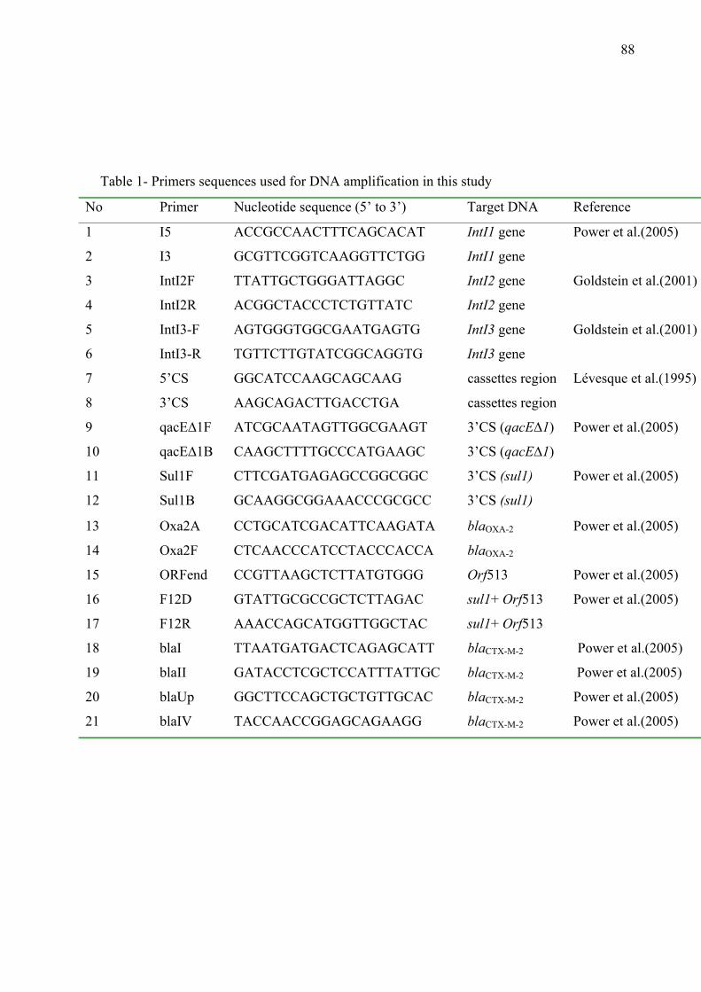
88
Table 1- Primers sequences used for DNA amplification in this study
No Primer Nucleotide sequence (5’ to 3’) Target DNA Reference
1 I5 ACCGCCAACTTTCAGCACAT IntI1 gene Power et al.(2005)
2 I3 GCGTTCGGTCAAGGTTCTGG IntI1 gene
3 IntI2F TTATTGCTGGGATTAGGC IntI2 gene Goldstein et al.(2001)
4 IntI2R ACGGCTACCCTCTGTTATC IntI2 gene
5 IntI3-F AGTGGGTGGCGAATGAGTG IntI3 gene Goldstein et al.(2001)
6 IntI3-R TGTTCTTGTATCGGCAGGTG IntI3 gene
7 5’CS GGCATCCAAGCAGCAAG cassettes region Lévesque et al.(1995)
8 3’CS AAGCAGACTTGACCTGA cassettes region
9 qacE∆1F ATCGCAATAGTTGGCGAAGT 3’CS (qacE∆1) Power et al.(2005)
10 qacE∆1B CAAGCTTTTGCCCATGAAGC 3’CS (qacE∆1)
11 Sul1F CTTCGATGAGAGCCGGCGGC 3’CS (sul1) Power et al.(2005)
12 Sul1B GCAAGGCGGAAACCCGCGCC 3’CS (sul1)
13 Oxa2A CCTGCATCGACATTCAAGATA blaOXA-2 Power et al.(2005)
14 Oxa2F CTCAACCCATCCTACCCACCA blaOXA-2
15 ORFend CCGTTAAGCTCTTATGTGGG Orf513 Power et al.(2005)
16 F12D GTATTGCGCCGCTCTTAGAC sul1+ Orf513 Power et al.(2005)
17 F12R AAACCAGCATGGTTGGCTAC sul1+ Orf513
18 blaI TTAATGATGACTCAGAGCATT blaCTX-M-2 Power et al.(2005)
19
20
21
blaII
blaUp
blaIV
GATACCTCGCTCCATTTATTGC
GGCTTCCAGCTGCTGTTGCAC
TACCAACCGGAGCAGAAGG
blaCTX-M-2
blaCTX-M-2
blaCTX-M-2
Power et al.(2005)
Power et al.(2005)
Power et al.(2005)
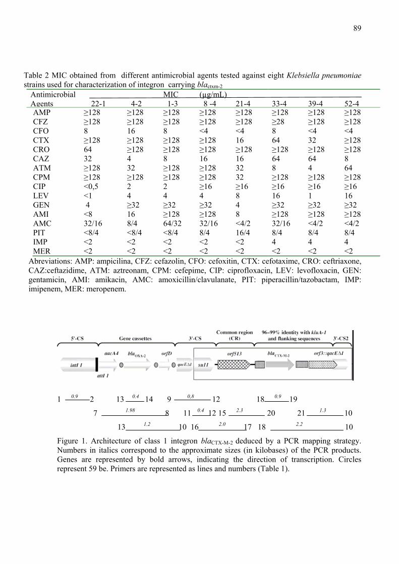
89
Table 2 MIC obtained from different antimicrobial agents tested against eight Klebsiella pneumoniae strains used for characterization of integron carrying blactxm-2
Antimicrobial Agents
22-1
4-2
MIC 1-3
(µg/mL) 8 -4
21-4
33-4
39-4
52-4
AMP ≥128 ≥128 ≥128 ≥128 ≥128 ≥128 ≥128 ≥128 CFZ ≥128 ≥128 ≥128 ≥128 ≥128 ≥28 ≥128 ≥128 CFO 8 16 8 <4 <4 8 <4 <4 CTX ≥128 ≥128 ≥128 ≥128 16 64 32 ≥128 CRO 64 ≥128 ≥128 ≥128 ≥128 ≥128 ≥128 ≥128 CAZ 32 4 8 16 16 64 64 8 ATM ≥128 32 ≥128 ≥128 32 8 4 64 CPM ≥128 ≥128 ≥128 ≥128 32 ≥128 ≥128 ≥128 CIP <0,5 2 2 ≥16 ≥16 ≥16 ≥16 ≥16 LEV <1 4 4 4 8 16 1 16 GEN 4 ≥32 ≥32 ≥32 4 ≥32 ≥32 ≥32 AMI <8 16 ≥128 ≥128 8 ≥128 ≥128 ≥128 AMC 32/16 8/4 64/32 32/16 <4/2 32/16 <4/2 <4/2 PIT <8/4 <8/4 <8/4 8/4 16/4 8/4 8/4 8/4 IMP <2 <2 <2 <2 <2 4 4 4 MER <2 <2 <2 <2 <2 <2 <2 <2 Abreviations: AMP: ampicilina, CFZ: cefazolin, CFO: cefoxitin, CTX: cefotaxime, CRO: ceftriaxone, CAZ:ceftazidime, ATM: aztreonam, CPM: cefepime, CIP: ciprofloxacin, LEV: levofloxacin, GEN: gentamicin, AMI: amikacin, AMC: amoxicillin/clavulanate, PIT: piperacillin/tazobactam, IMP: imipenem, MER: meropenem.
1 0.9 2 13 0.4 14 9 0,8 12 18 0.9 19
7 1.98 8 11 0.4 12 15 2.3 20 21 1.3 10
13 1.2 10 16 2.0 17 18 2.2 10
Figure 1. Architecture of class 1 integron blaCTX-M-2 deduced by a PCR mapping strategy. Numbers in italics correspond to the approximate sizes (in kilobases) of the PCR products. Genes are represented by bold arrows, indicating the direction of transcription. Circles represent 59 be. Primers are represented as lines and numbers (Table 1).
90
4.0 Conclusões Nossos resultados, quanto às cepas estudadas de K.pneumoniae aqui estudadas permitem concluir que: 1- Quanto aos perfis de sensibilidade, todas as amostras apresentaram-se sensíveis a Imipenem e Meropenem os quais, portanto, permanecem como antimicrobianos de escolha para o tratamento de infecções por K .pneumoniae produtora de ESBL. Piperacilina/Tazobactam apresentou melhor resultado in vitro entre as associações β-lactâmicos/ inibidores de β-lactamases utilizadas. 2- -A enzima CTX-M-2 foi a mais freqüente entre os isolados, associada ou não a outras enzimas. A enzima SHV-5 foi o tipo mais freqüente entre as ESBL do tipo SHV, coincidindo com relatos de diversos autores sobre a disseminação dessa enzima em todos os continentes. A baixa freqüência de ESBL do tipo TEM, em nossa região, sugere a necessidade de mais estudos sobre esta enzima. A presença de genes de duas enzimas com características de ESBLs em um mesmo isolado foi de 62%, mostrando assim a alta freqüência destas em ambiente hospitalar. 3- Todos os métodos baseados em PCR apresentaram alto poder de discriminação, sendo o REP-PCR o método com o mais elevado poder discriminatório, entre as técnicas baseadas em PCR, demonstrando que estas técnicas são apropriadas para estudos epidemiológicos iniciais de K .pneumoniae. A metodologia de RFLP-PFGE apresentou o melhor poder de discriminação, sugerindo assim sua utilização como análise confirmatória em estudos epidemiológicos de K.pneumoniae. 4- Integron da classe 1 foi o mais freqüente entre os isolados de K.pneumoniae e teve como característica importante a localização do gene blaCTX-M-2 em uma região incomum do integron, como já descrito em isolados da Argentina e Uruguai. A análise epidemiológica dos isolados apresentou grande diversidade e complexidade genética, sugerindo que a multiplicidade de genótipos deve-se a disseminação da enzima CTX-M-2 e genes de resistência presentes em elementos móveis, incluindo integrons.

Livros Grátis( http://www.livrosgratis.com.br )
Milhares de Livros para Download: Baixar livros de AdministraçãoBaixar livros de AgronomiaBaixar livros de ArquiteturaBaixar livros de ArtesBaixar livros de AstronomiaBaixar livros de Biologia GeralBaixar livros de Ciência da ComputaçãoBaixar livros de Ciência da InformaçãoBaixar livros de Ciência PolíticaBaixar livros de Ciências da SaúdeBaixar livros de ComunicaçãoBaixar livros do Conselho Nacional de Educação - CNEBaixar livros de Defesa civilBaixar livros de DireitoBaixar livros de Direitos humanosBaixar livros de EconomiaBaixar livros de Economia DomésticaBaixar livros de EducaçãoBaixar livros de Educação - TrânsitoBaixar livros de Educação FísicaBaixar livros de Engenharia AeroespacialBaixar livros de FarmáciaBaixar livros de FilosofiaBaixar livros de FísicaBaixar livros de GeociênciasBaixar livros de GeografiaBaixar livros de HistóriaBaixar livros de Línguas

Baixar livros de LiteraturaBaixar livros de Literatura de CordelBaixar livros de Literatura InfantilBaixar livros de MatemáticaBaixar livros de MedicinaBaixar livros de Medicina VeterináriaBaixar livros de Meio AmbienteBaixar livros de MeteorologiaBaixar Monografias e TCCBaixar livros MultidisciplinarBaixar livros de MúsicaBaixar livros de PsicologiaBaixar livros de QuímicaBaixar livros de Saúde ColetivaBaixar livros de Serviço SocialBaixar livros de SociologiaBaixar livros de TeologiaBaixar livros de TrabalhoBaixar livros de Turismo


![Phenotypic detection of ESBLs and carbapenemasesbsac.org.uk/wp-content/uploads/2018/08/18.08.22... · (Hope et al. JAC [2007]) • CXM, LEX or RAD unreliable indicators for ESBLs](https://img.pdfslide.net/doc/110x75/5f842d1072d747484f294053/phenotypic-detection-of-esbls-and-hope-et-al-jac-2007-a-cxm-lex-or-rad-unreliable.jpg)