Embed Size (px)
Citation preview
RESEARCH ARTICLE
Knockdown of histidine-rich calcium-binding protein (HRC)suppresses liver fibrosis by inhibiting the activation of hepaticstellate cellsJingmei Liu1,*, Mengke Li1,2,*, Jin Gong1, Ping Han1, YunwuWang1, Dongxiao Li1, Dean Tian1,‡ and Jiazhi Liao1,‡
ABSTRACTThe histidine-rich calcium-binding protein (HRC) is a regulator of Ca2+
homeostasis and it plays a significant role in hepatocellular carcinoma(HCC) progression. However, the relationship between HRC and liverfibrogenesis is still unknown. Our data demonstrates that HRC wasupregulated in fibrotic liver and activated hepatic stellate cells (HSCs).TGF-β treatment increased α-SMA and HRC expression dose-dependently in HSCs. Repression of HRC reduced α-SMA, CTGFand collagen expression, and inhibited HSC proliferation andmigration. In addition, we found that the anti-fibrosis effect of HRCknockdown was associated with endoplasmic reticulum (ER) stress.Silencing of HRC decreased the expression of ER stress andautophagy markers. Moreover, ER stress agonist thapsigargin (TG)enhanced, whereas ER stress antagonist 4-phenylbutyric acid(4-PBA) alleviated HSCs activation and autophagy. In conclusion,these data indicate that depletion of HRC inhibited HSC activationthrough the ER stress pathway, and HRCmay be a potential regulatorof liver fibrosis.
KEY WORDS: Liver fibrosis, Hepatic stellate cells, HRC, ER stress
INTRODUCTIONLiver fibrosis is a wound-healing response to chronic liver injury,and it also considered as a pre-cancerous condition which may resultin hepatocellular carcinoma (HCC). Activated hepatic stellate cells(HSCs) are responsible for the excessive deposition of extracellularmatrix (ECM) proteins during hepatic fibrogenesis (Friedman,2004). Prevention or reversal of HSC activation has been claimed asa potential approach to attenuate liver fibrosis (Bataller and Brenner,2005). Although many potential anti-fibrotic targets have beenidentified, effective clinical therapies are still lacking. Therefore, itis urgent to develop novel strategies that prevent the progression ofliver fibrosis.The histidine-rich calcium-binding protein (HRC) is a regulator
in Ca2+ homeostasis (Park et al., 2012). Calcium signals may be ofparticular importance in HSC biology (Kruglov et al., 2007).
Activated HSCs are capable of elevated ECM deposition, increasedproliferation and enhanced migration, which were identified as theCa2+-dependent responses (Melton et al., 2006; Soliman et al.,2009). Recently, a number of calcium-binding proteins have beenimplicated in liver fibrogenesis, such as S100A4, SPARC andannexin A2 (Zhang et al., 2010; Atorrasagasti et al., 2011, 2013).However, the role of HRC in liver fibrosis has not been investigatedso far.
Recent data indicate that ER stress plays a pivotal role in theprogression of liver diseases, including liver fibrosis (Malhi andKaufman, 2011; Pagliassotti, 2012). Inhibition of ER stressprevented hepatic fibrosis by attenuating HSC activation (Menet al., 2015). Our previous study showed that HRC promoted thegrowth of liver cancer by inhibiting ER stress-induced apoptosis(Liu et al., 2015). However, the effect of HRC on ER stress in HSCsremains unknown. It is noteworthy that ER stress induces theactivation of HSCs through autophagy, and blockage of ER stresssuppresses autophagic activity, thus inhibiting HSCs activation(Hernández-Gea et al., 2013; Men et al., 2015).
For the first time, we discovered that HRC was upregulated infibrotic liver and activated HSCs. Moreover, we demonstrated thatHRC knockdown inhibited the activation, proliferation and migrationof HSCs, which was partly attributed to ER stress and autophagy.
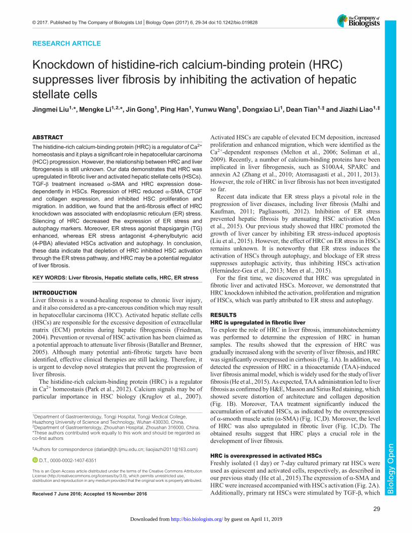
RESULTSHRC is upregulated in fibrotic liverTo explore the role of HRC in liver fibrosis, immunohistochemistrywas performed to determine the expression of HRC in humansamples. The results showed that the expression of HRC wasgradually increased along with the severity of liver fibrosis, and HRCwas significantly overexpressed in cirrhosis (Fig. 1A). In addition, wedetected the expression of HRC in a thioacetamide (TAA)-inducedliver fibrosis animalmodel, which is widely used for the study of liverfibrosis (He et al., 2015). As expected, TAAadministration led to liverfibrosis as confirmedbyH&E,Masson and SiriusRed staining, whichshowed severe distortion of architecture and collagen deposition(Fig. 1B). Moreover, TAA treatment significantly induced theaccumulation of activated HSCs, as indicated by the overexpressionof α-smooth muscle actin (α-SMA) (Fig. 1C,D). Moreover, the levelof HRC was also upregulated in fibrotic liver (Fig. 1C,D). Theobtained results suggest that HRC plays a crucial role in thedevelopment of liver fibrosis.
HRC is overexpressed in activated HSCsFreshly isolated (1 day) or 7-day cultured primary rat HSCs wereused as quiescent and activated cells, respectively, as described inour previous study (He et al., 2015).The expression of α-SMA andHRCwere increased accompanied with HSCs activation (Fig. 2A).Additionally, primary rat HSCs were stimulated by TGF-β, whichReceived 7 June 2016; Accepted 15 November 2016
1Department of Gastroenterology, Tongji Hospital, Tongji Medical College,Huazhong University of Science and Technology, Wuhan 430030, China.2Department of Gastroenterology, Zhoushan Hospital, Zhoushan 316000, China.*These authors contributed work equally to this work and should be regarded asco-first authors
‡Authors for correspondence ([email protected]; [email protected])
D.T., 0000-0002-1407-6351
This is an Open Access article distributed under the terms of the Creative Commons AttributionLicense (http://creativecommons.org/licenses/by/3.0), which permits unrestricted use,distribution and reproduction in any medium provided that the original work is properly attributed.
29
© 2017. Published by The Company of Biologists Ltd | Biology Open (2017) 6, 29-34 doi:10.1242/bio.019828
BiologyOpen
by guest on April 11, 2019http://bio.biologists.org/Downloaded from
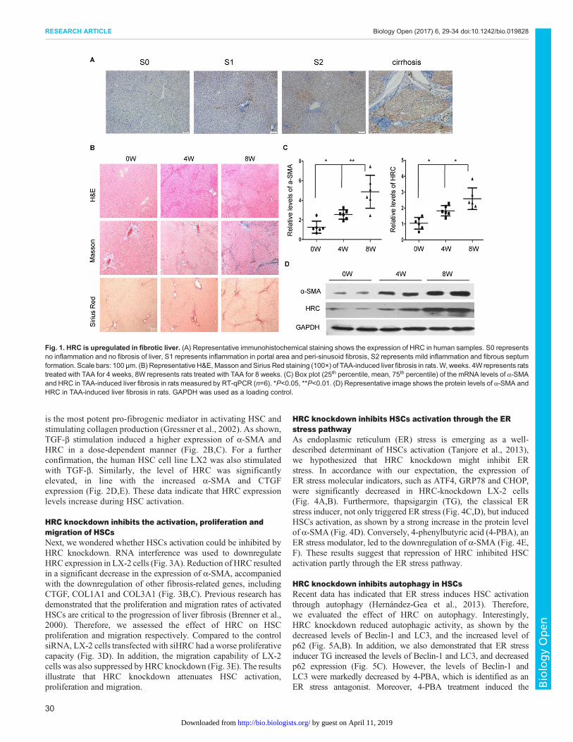
is the most potent pro-fibrogenic mediator in activating HSC andstimulating collagen production (Gressner et al., 2002). As shown,TGF-β stimulation induced a higher expression of α-SMA andHRC in a dose-dependent manner (Fig. 2B,C). For a furtherconfirmation, the human HSC cell line LX2 was also stimulatedwith TGF-β. Similarly, the level of HRC was significantlyelevated, in line with the increased α-SMA and CTGFexpression (Fig. 2D,E). These data indicate that HRC expressionlevels increase during HSC activation.
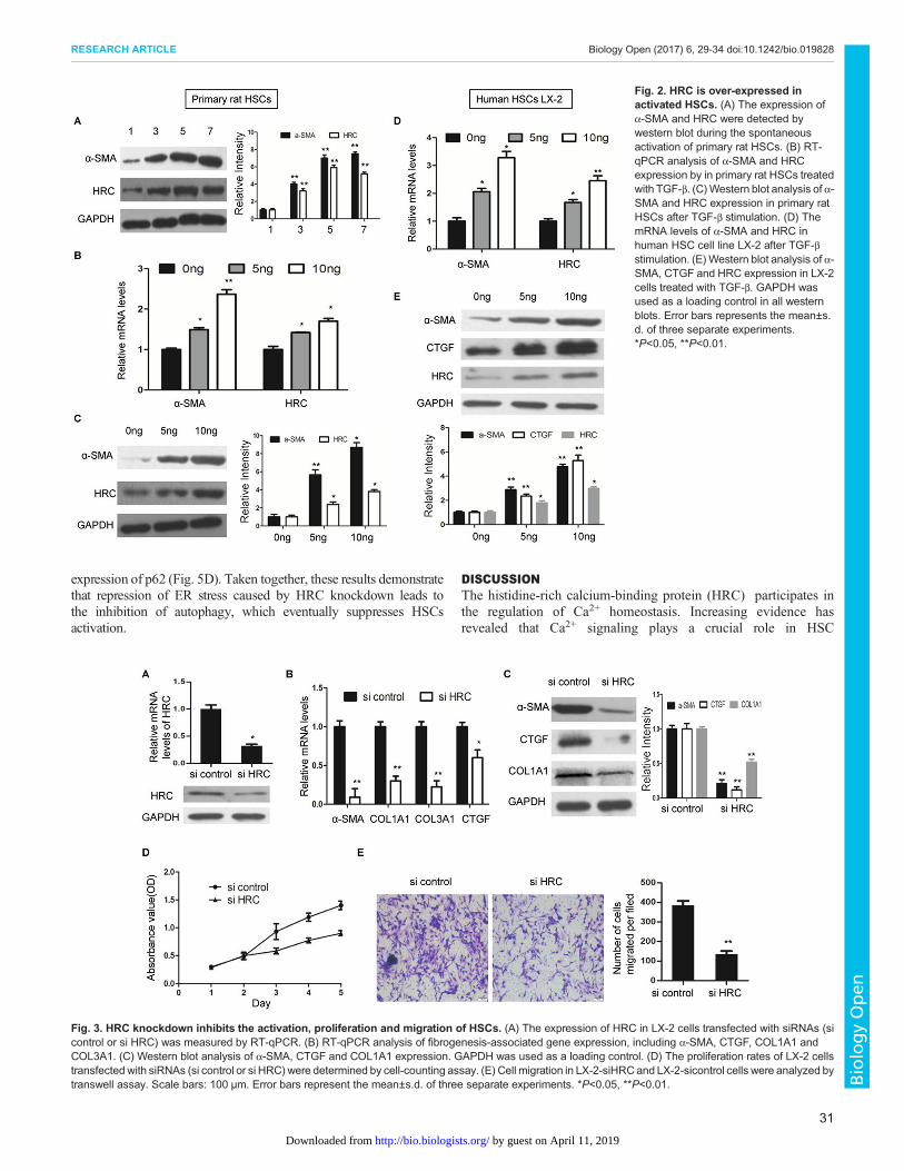
HRC knockdown inhibits the activation, proliferation andmigration of HSCsNext, we wondered whether HSCs activation could be inhibited byHRC knockdown. RNA interference was used to downregulateHRC expression in LX-2 cells (Fig. 3A). Reduction of HRC resultedin a significant decrease in the expression of α-SMA, accompaniedwith the downregulation of other fibrosis-related genes, includingCTGF, COL1A1 and COL3A1 (Fig. 3B,C). Previous research hasdemonstrated that the proliferation and migration rates of activatedHSCs are critical to the progression of liver fibrosis (Brenner et al.,2000). Therefore, we assessed the effect of HRC on HSCproliferation and migration respectively. Compared to the controlsiRNA, LX-2 cells transfected with siHRC had aworse proliferativecapacity (Fig. 3D). In addition, the migration capability of LX-2cells was also suppressed by HRC knockdown (Fig. 3E). The resultsillustrate that HRC knockdown attenuates HSC activation,proliferation and migration.
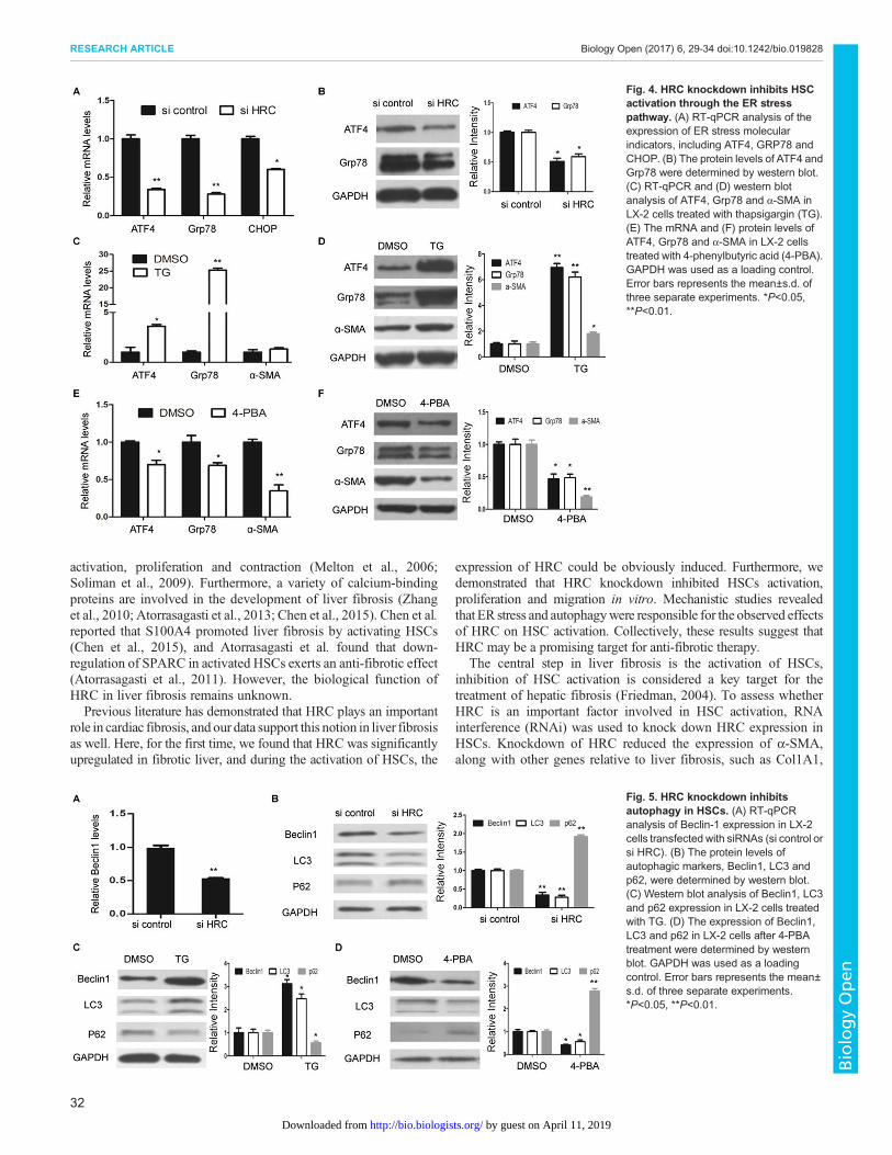
HRC knockdown inhibits HSCs activation through the ERstress pathwayAs endoplasmic reticulum (ER) stress is emerging as a well-described determinant of HSCs activation (Tanjore et al., 2013),we hypothesized that HRC knockdown might inhibit ERstress. In accordance with our expectation, the expression ofER stress molecular indicators, such as ATF4, GRP78 and CHOP,were significantly decreased in HRC-knockdown LX-2 cells(Fig. 4A,B). Furthermore, thapsigargin (TG), the classical ERstress inducer, not only triggered ER stress (Fig. 4C,D), but inducedHSCs activation, as shown by a strong increase in the protein levelof α-SMA (Fig. 4D). Conversely, 4-phenylbutyric acid (4-PBA), anER stress modulator, led to the downregulation of α-SMA (Fig. 4E,F). These results suggest that repression of HRC inhibited HSCactivation partly through the ER stress pathway.
HRC knockdown inhibits autophagy in HSCsRecent data has indicated that ER stress induces HSC activationthrough autophagy (Hernández-Gea et al., 2013). Therefore,we evaluated the effect of HRC on autophagy. Interestingly,HRC knockdown reduced autophagic activity, as shown by thedecreased levels of Beclin-1 and LC3, and the increased level ofp62 (Fig. 5A,B). In addition, we also demonstrated that ER stressinducer TG increased the levels of Beclin-1 and LC3, and decreasedp62 expression (Fig. 5C). However, the levels of Beclin-1 andLC3 were markedly decreased by 4-PBA, which is identified as anER stress antagonist. Moreover, 4-PBA treatment induced the
Fig. 1. HRC is upregulated in fibrotic liver. (A) Representative immunohistochemical staining shows the expression of HRC in human samples. S0 representsno inflammation and no fibrosis of liver, S1 represents inflammation in portal area and peri-sinusoid fibrosis, S2 represents mild inflammation and fibrous septumformation. Scale bars: 100 µm. (B) Representative H&E,Masson and Sirius Red staining (100×) of TAA-induced liver fibrosis in rats.W, weeks. 4W represents ratstreated with TAA for 4 weeks, 8W represents rats treated with TAA for 8 weeks. (C) Box plot (25th percentile, mean, 75th percentile) of the mRNA levels of α-SMAand HRC in TAA-induced liver fibrosis in rats measured by RT-qPCR (n=6). *P<0.05, **P<0.01. (D) Representative image shows the protein levels of α-SMA andHRC in TAA-induced liver fibrosis in rats. GAPDH was used as a loading control.
30
RESEARCH ARTICLE Biology Open (2017) 6, 29-34 doi:10.1242/bio.019828
BiologyOpen
by guest on April 11, 2019http://bio.biologists.org/Downloaded from
expression of p62 (Fig. 5D). Taken together, these results demonstratethat repression of ER stress caused by HRC knockdown leads tothe inhibition of autophagy, which eventually suppresses HSCsactivation.
DISCUSSIONThe histidine-rich calcium-binding protein (HRC) participates inthe regulation of Ca2+ homeostasis. Increasing evidence hasrevealed that Ca2+ signaling plays a crucial role in HSC
Fig. 2. HRC is over-expressed inactivated HSCs. (A) The expression ofα-SMA and HRC were detected bywestern blot during the spontaneousactivation of primary rat HSCs. (B) RT-qPCR analysis of α-SMA and HRCexpression by in primary rat HSCs treatedwith TGF-β. (C)Western blot analysis of α-SMA and HRC expression in primary ratHSCs after TGF-β stimulation. (D) ThemRNA levels of α-SMA and HRC inhuman HSC cell line LX-2 after TGF-βstimulation. (E)Western blot analysis of α-SMA, CTGF and HRC expression in LX-2cells treated with TGF-β. GAPDH wasused as a loading control in all westernblots. Error bars represents the mean±s.d. of three separate experiments.*P<0.05, **P<0.01.
Fig. 3. HRC knockdown inhibits the activation, proliferation and migration of HSCs. (A) The expression of HRC in LX-2 cells transfected with siRNAs (sicontrol or si HRC) was measured by RT-qPCR. (B) RT-qPCR analysis of fibrogenesis-associated gene expression, including α-SMA, CTGF, COL1A1 andCOL3A1. (C) Western blot analysis of α-SMA, CTGF and COL1A1 expression. GAPDH was used as a loading control. (D) The proliferation rates of LX-2 cellstransfected with siRNAs (si control or si HRC) were determined by cell-counting assay. (E) Cell migration in LX-2-siHRCand LX-2-sicontrol cells were analyzed bytranswell assay. Scale bars: 100 μm. Error bars represent the mean±s.d. of three separate experiments. *P<0.05, **P<0.01.
31
RESEARCH ARTICLE Biology Open (2017) 6, 29-34 doi:10.1242/bio.019828
BiologyOpen
by guest on April 11, 2019http://bio.biologists.org/Downloaded from
activation, proliferation and contraction (Melton et al., 2006;Soliman et al., 2009). Furthermore, a variety of calcium-bindingproteins are involved in the development of liver fibrosis (Zhanget al., 2010; Atorrasagasti et al., 2013; Chen et al., 2015). Chen et al.reported that S100A4 promoted liver fibrosis by activating HSCs(Chen et al., 2015), and Atorrasagasti et al. found that down-regulation of SPARC in activated HSCs exerts an anti-fibrotic effect(Atorrasagasti et al., 2011). However, the biological function ofHRC in liver fibrosis remains unknown.Previous literature has demonstrated that HRC plays an important
role in cardiac fibrosis, and our data support this notion in liver fibrosisas well. Here, for the first time, we found that HRC was significantlyupregulated in fibrotic liver, and during the activation of HSCs, the
expression of HRC could be obviously induced. Furthermore, wedemonstrated that HRC knockdown inhibited HSCs activation,proliferation and migration in vitro. Mechanistic studies revealedthat ER stress and autophagywere responsible for the observed effectsof HRC on HSC activation. Collectively, these results suggest thatHRC may be a promising target for anti-fibrotic therapy.
The central step in liver fibrosis is the activation of HSCs,inhibition of HSC activation is considered a key target for thetreatment of hepatic fibrosis (Friedman, 2004). To assess whetherHRC is an important factor involved in HSC activation, RNAinterference (RNAi) was used to knock down HRC expression inHSCs. Knockdown of HRC reduced the expression of α-SMA,along with other genes relative to liver fibrosis, such as Col1A1,
Fig. 5. HRC knockdown inhibitsautophagy in HSCs. (A) RT-qPCRanalysis of Beclin-1 expression in LX-2cells transfected with siRNAs (si control orsi HRC). (B) The protein levels ofautophagic markers, Beclin1, LC3 andp62, were determined by western blot.(C) Western blot analysis of Beclin1, LC3and p62 expression in LX-2 cells treatedwith TG. (D) The expression of Beclin1,LC3 and p62 in LX-2 cells after 4-PBAtreatment were determined by westernblot. GAPDH was used as a loadingcontrol. Error bars represents the mean±s.d. of three separate experiments.*P<0.05, **P<0.01.
Fig. 4. HRC knockdown inhibits HSCactivation through the ER stresspathway. (A) RT-qPCR analysis of theexpression of ER stress molecularindicators, including ATF4, GRP78 andCHOP. (B) The protein levels of ATF4 andGrp78 were determined by western blot.(C) RT-qPCR and (D) western blotanalysis of ATF4, Grp78 and α-SMA inLX-2 cells treated with thapsigargin (TG).(E) The mRNA and (F) protein levels ofATF4, Grp78 and α-SMA in LX-2 cellstreated with 4-phenylbutyric acid (4-PBA).GAPDH was used as a loading control.Error bars represents the mean±s.d. ofthree separate experiments. *P<0.05,**P<0.01.
32
RESEARCH ARTICLE Biology Open (2017) 6, 29-34 doi:10.1242/bio.019828
BiologyOpen
by guest on April 11, 2019http://bio.biologists.org/Downloaded from
Col3A1 and CTGF. Enhanced proliferation and migration of HSCslead to collagen deposition, and finally resulted in the developmentof fibrotic scar (Brenner et al., 2000). We next investigated thefunctional role of HRC in HSC proliferation and migration. Withinour expectations, the proliferation activity and migration ability ofHSCs were obviously suppressed by HRC knockdown.Accumulating evidence suggests that ER stress plays essential
roles in the progression of tissue fibrosis, including in liver, lung andkidney (Korfei et al., 2008; Malhi and Kaufman, 2011; Taniguchiand Yoshida, 2015). Aberrant ER stress signaling has been reportedin liver fibrosis. ER stress pathway components Grp78 and CHOPwere markedly increased in experimentally induced liver fibrosis intransgenic mice (Mencin et al., 2007). Tamaki et al. demonstratedthat liver fibrosis was greatly attenuated in CHOP-deficient micefollowing bile duct ligation (Tamaki et al., 2008). Our datademonstrated that HRC knockdown decreased the expression of ERstress markers, including Grp78, ATF4 and CHOP. Moreover, wealso confirmed that the ER stress inducer TG enhanced, while theER stress modulator 4-PBA reduced, the expression of α-SMA.These findings indicated that silencing of HRC inhibited HSCactivation by the ER stress pathway.Recent research has suggested that autophagic flux increases
during HSC activation, and the impact of ER stress on HSCactivation is partly through autophagy (Hernández-Gea et al.,2012). We further analyzed the effect of HRC knockdown onBeclin-1, LC3 and p62 expression, which represent the extent ofautophagy. The levels of Beclin-1 and LC3 were significantlydownregulated, and the level of p62 was increased by HRCknockdown, which is consistent with our expectation. In addition,we also detected autophagic makers in response to ER stress.Consistent with a previous study (Men et al., 2015), ER stressstimuli led to elevated Beclin1 and LC3 expression, and the level ofp62 was slightly decreased by TG, while a lower ER stress levelinhibited autophagic activity. These results suggested that thereduction of ER stress induced by HRC knockdown led to theinhibition of autophagy, thus suppressing HSCs activation.In conclusion, our study demonstrated that knockdown of HRC
inhibit HSC activation and liver fibrosis in vitro, and further studiesshould be carried out to clarify the role of HRC in liver fibrosis in vivo.
MATERIALS AND METHODSHuman samplesHuman samples (n=45) were divided into five groups according to theseverity of liver fibrosis, including S0 (n=5), S1 (n=10), S2 (n=10), S3 (n=2)and S4 (n=18), which were diagnosed by the pathologists at Tongji Hospitalof Tongji Medical College, Huazhong University of Science andTechnology. All procedures involving human participants were performedafter obtained their informed consent, which was also in accordancewith theMedical Ethics Committee of Tongji Hospital.
Animal models of liver fibrosisMale Sprague–Dawley (SD) rats weighting about 200 g were randomlydivided into three groups (n=6/group). Rats were treated with thioacetamide(TAA) or saline (200 mg kg−1) by intraperitoneal (i.p.) injection twice aweek for 8 weeks. All procedures performed in studies involving animalswere approved by the Institutional Laboratory Animal Care and UseCommittee by the Institutional Laboratory Animal Care and Use Committeeof Tongji hospital.
Primary HSC isolation and cultureRat primary HSCs were isolated from livers by in situ perfusion with pronaseand collagenase and single-step Nycodenz gradient centrifugation asdescribed previously (Kawada et al., 1998; Weiskirchen and Gressner, 2005).
ImmunohistochemistryHistological analysis of fibrosis was performed on fixed liver tissue,embedded in paraffin, and sectioned at a thickness of 4 μm. The 4 μm thicksections were used for Hematoxylin and Eosin (H&E), Masson and SiriusRed staining. AnHRC polyclonal antibody (Abonva, CA, USA) was used todetect the expression of HRC in fibrotic liver.
RNA extraction and real-time RT-PCRTotal RNA was extracted using TRIzol reagent (Invitrogen, CA, USA).Reverse-transcribed complementary DNA was synthesized using thePrimeScript RT reagent kit (TaKaRa, Otsu, Japan). Real-time polymerasechain reaction was performed using SYBR Premix ExTaq (TaKaRa) on anABI StepOne Real-Time PCR System (Applied Biosystem, CA, USA). Thesequences of the primers used for PCR are listed in Table S1.
Western blotWestern blot was performed as previously described (Han et al., 2014).Briefly, samples containing 30 μg of total protein were resolved on 10%polyacrylamide SDS gels and electrophoretically transferred topolyvinylidene difluoride (PVDF) membranes. The membranes wereblocked with 5% skim milk, incubated with appropriate primary antibodiesand HRP-conjugated suitable secondary antibodies, followed by detectionwith enhanced chemiluminescence reagents (Pierce Chemical, IL, USA).GAPDH was used as a loading control. The antibodies are listed in Table S2.
RNA interferenceFor RNA interference, HRC siRNAwas synthesised byRiboBio (Guangzhou,China), and then transfected into LX-2 cells using lipofectamine 2000(Invitrogen) according to the manufacturer’s instructions. The sequence ofsiHRC was designed as follows: CCACAGAGACGAGGAAGAA.
CCK-8 assayCell proliferation was analyzed by Cell Counting Kit-8 (CCK-8) assay(Dojindo Laboratories, Kumamoto, Japan) according to the manufacturer’sinstructions.
Transwell migration assayCell migration assay was performed using transwell chambers (6.5 mmdiameter and 8 µm pore size; Costar; Corning Inc.). Briefly, a total of 5×10⁴
cells in 0.2 ml media were plated in the upper chambers, 600 μl DMEMmedium containing 10% fetal calf serum (Invitrogen Gibco, CA, USA) wasadded to the lower chamber. After 24 h, cells that migrated through themembrane to the lower surfacewere fixed with 4% paraformaldehyde, stainedwith crystal violet and counted by microscopy (Olympus, NY, USA).
Statistical analysisData are presented as means±s.d. Student’s t-test was performed to assessthe significance of differences between two groups. A P-value <0.05 wasconsidered statistically significant.
AcknowledgementsWe hereby thank the Department of Pathology in Tongji hospital for help.
Competing interestsThe authors declare no competing or financial interests.
Author contributionsJ.Liu, M.L., J.G., P.H., Y.W. and D.L. performed the experiments and analysed data.J.Liu, D.T. and J.Liao designed experiments and analysed data. J.Liu andM.L. wrotethe paper.
FundingThis study is supported by the National Natural Science Foundation of China(81270507, 81572419).
Supplementary informationSupplementary information available online athttp://bio.biologists.org/lookup/doi/10.1242/bio.019828.supplemental
33
RESEARCH ARTICLE Biology Open (2017) 6, 29-34 doi:10.1242/bio.019828
BiologyOpen
by guest on April 11, 2019http://bio.biologists.org/Downloaded from
ReferencesAtorrasagasti, C., Aquino, J. B., Hofman, L., Alaniz, L., Malvicini, M., Garcia, M.,Benedetti, L., Friedman, S. L., Podhajcer, O. andMazzolini, G. (2011). SPARCdownregulation attenuates the profibrogenic response of hepatic stellate cellsinduced by TGF-beta1 and PDGF. Am. J. Physiol. Gastrointest. Liver Physiol.300, G739-G748.
Atorrasagasti, C., Peixoto, E., Aquino, J. B., Kippes, N., Malvicini, M., Alaniz, L.,Garcia, M., Piccioni, F., Fiore, E. J., Bayo, J. et al. (2013). Lack of thematricellular protein SPARC (secreted protein, acidic and rich in cysteine)attenuates liver fibrogenesis in mice. PLoS ONE 8, e54962.
Bataller, R. and Brenner, D. A. (2005). Liver fibrosis. J. Clin. Invest. 115, 209-218.Brenner, D. A., Waterboer, T., Choi, S. K., Lindquist, J. N., Stefanovic, B.,Burchardt, E., Yamauchi, M., Gillan, A. andRippe, R. A. (2000). Newaspects ofhepatic fibrosis. J. Hepatol. 32, 32-38.
Chen, L., Li, J., Zhang, J., Dai, C., Liu, X., Wang, J., Gao, Z., Guo, H., Wang, R.,Lu, S. et al. (2015). S100A4 promotes liver fibrosis via activation of hepatic stellatecells. J. Hepatol. 62, 156-164.
Friedman, S. L. (2004). Mechanisms of disease: mechanisms of hepatic fibrosisand therapeutic implications. Nat. Clin. Pract. Gastroenterol. Hepatol. 1, 98-105.
Gressner, A. M., Weiskirchen, R., Breitkopf, K. and Dooley, S. (2002). Roles ofTGF-beta in hepatic fibrosis. Front. Biosci. 7, d793-d807.
Han, P., Fu, Y., Luo, M., He, J., Liu, J., Liao, J., Tian, D. and Yan, W. (2014). BVESinhibition triggers epithelial-mesenchymal transition in human hepatocellularcarcinoma. Dig. Dis. Sci. 59, 992-1000.
He, J., Gong, J., Ding, Q., Tan, Q., Han, P., Liu, J., Zhou, Z., Tu, W., Xia, Y., Yan,W. et al. (2015). Suppressive effect of SATB1 on hepatic stellate cell activationand liver fibrosis in rats. FEBS Lett. 589, 1359-1368.
Hernandez-Gea, V., Ghiassi-Nejad, Z., Rozenfeld, R., Gordon, R., Fiel, M. I.,Yue, Z., Czaja, M. J. and Friedman, S. L. (2012). Autophagy releases lipid thatpromotes fibrogenesis by activated hepatic stellate cells in mice and in humantissues. Gastroenterology 142, 938-946.
Hernandez-Gea, V., Hilscher, M., Rozenfeld, R., Lim, M. P., Nieto, N., Werner, S.,Devi, L. A. and Friedman, S. L. (2013). Endoplasmic reticulum stress inducesfibrogenic activity in hepatic stellate cells through autophagy. J. Hepatol. 59, 98-104.
Kawada, N., Seki, S., Inoue, M. and Kuroki, T. (1998). Effect of antioxidants,resveratrol, quercetin, and N-acetylcysteine, on the functions of cultured rathepatic stellate cells and Kupffer cells. Hepatology 27, 1265-1274.
Korfei, M., Ruppert, C., Mahavadi, P., Henneke, I., Markart, P., Koch, M., Lang,G., Fink, L., Bohle, R.-M., Seeger, W. et al. (2008). Epithelial endoplasmicreticulum stress and apoptosis in sporadic idiopathic pulmonary fibrosis.Am. J. Respir. Crit. Care Med. 178, 838-846.
Kruglov, E. A., Correa, P. R. A. V., Arora, G., Yu, J., Nathanson, M. H. andDranoff, J. A. (2007). Molecular basis for calcium signaling in hepatic stellatecells. Am. J. Physiol. Gastrointest. Liver Physiol. 292, G975-G982.
Liu, J., Han, P., Li, M., Yan, W., Liu, J., He, J., Gong, J., Wang, Y. and Tian, D.(2015). Histidine-rich calcium binding protein promotes growth of hepatocellularcarcinoma in vitro and in vivo. Cancer Sci. 106, 1288-1295.
Malhi, H. and Kaufman, R. J. (2011). Endoplasmic reticulum stress in liver disease.J. Hepatol. 54, 795-809.
Melton, A. C., Datta, A. and Yee, H. F.Jr. (2006). [Ca2+]i-independent contractileforce generation by rat hepatic stellate cells in response to endothelin-1.Am. J. Physiol. Gastrointest. Liver Physiol. 290, G7-G13.
Men, R., Wen, M., Dan, X., Zhu, Y., Wang, W., Li, J., Wu, W., Liu, X. and Yang, L.(2015). Nogo-B: a potential indicator for hepatic cirrhosis and regulator in hepaticstellate cell activation. Hepatol. Res. 45, 113-122.
Mencin, A., Seki, E., Osawa, Y., Kodama, Y., De Minicis, S., Knowles, M. andBrenner, D. A. (2007). Alpha-1 antitrypsin Z protein (PiZ) increases hepaticfibrosis in a murine model of cholestasis. Hepatology 46, 1443-1452.
Pagliassotti, M. J. (2012). Endoplasmic reticulum stress in nonalcoholic fatty liverdisease. Annu. Rev. Nutr. 32, 17-33.
Park, C. S., Cha, H., Kwon, E. J., Jeong, D., Hajjar, R. J., Kranias, E. G., Cho, C.,Park, W. J. and Kim, D. H. (2012). AAV-mediated knock-down of HRCexacerbates transverse aorta constriction-induced heart failure. PLoS ONE 7,e43282.
Soliman, E. M., Rodrigues, M. A., Gomes, D. A., Sheung, N., Yu, J., Amaya,M. J., Nathanson, M. H. and Dranoff, J. A. (2009). Intracellular calcium signalsregulate growth of hepatic stellate cells via specific effects on cell cycleprogression. Cell Calcium 45, 284-292.
Tamaki, N., Hatano, E., Taura, K., Tada, M., Kodama, Y., Nitta, T., Iwaisako, K.,Seo, S., Nakajima, A., Ikai, I. et al. (2008). CHOP deficiency attenuatescholestasis-induced liver fibrosis by reduction of hepatocyte injury.Am. J. Physiol.Gastrointest. Liver Physiol. 294, G498-G505.
Taniguchi, M. and Yoshida, H. (2015). Endoplasmic reticulum stress in kidneyfunction and disease. Curr. Opin. Nephrol. Hypertens. 24, 345-350.
Tanjore, H., Lawson, W. E. and Blackwell, T. S. (2013). Endoplasmic reticulumstress as a pro-fibrotic stimulus. Biochim. Biophys. Acta 1832, 940-947.
Weiskirchen, R. and Gressner, A. M. (2005). Isolation and culture of hepaticstellate cells. Methods Mol. Med. 117, 99-113.
Zhang, L., Peng, X., Zhang, Z., Feng, Y., Jia, X., Shi, Y., Yang, H., Zhang, Z.,Zhang, X., Liu, L. et al. (2010). Subcellular proteome analysis unraveled annexinA2 related to immune liver fibrosis. J. Cell Biochem. 110, 219-228.
34
RESEARCH ARTICLE Biology Open (2017) 6, 29-34 doi:10.1242/bio.019828
BiologyOpen
by guest on April 11, 2019http://bio.biologists.org/Downloaded from