Embed Size (px)
Citation preview
Instructions for use
Title LACTATE DEHYDROGENASE AND CREATINE PHOSPHOKINASE ISOENZYMES IN TISSUES OFLABORATORY ANIMALS
Author(s) YASUDA, Jun; TATEYAMA, Kazue; SYUTO, Bunei; TOO, Kimehiko
Citation Japanese Journal of Veterinary Research, 38(1), 19-29
Issue Date 1990-02-09
DOI 10.14943/jjvr.38.1.19
Doc URL http://hdl.handle.net/2115/3192
Type bulletin (article)
File Information KJ00002377324.pdf
Hokkaido University Collection of Scholarly and Academic Papers : HUSCAP

Jpn. J. Vet. Res., 38, 19-29 (1990)
LACTATE DEHYDROGENASE AND CREATINE PHOSPHOKINASE ISOENZYMES IN TISSUES OF
LABORATORY ANIMALS
Jun YASUDAl), Kazue TATEYAMAl), Bunei SYUT02)
and Kimehiko TOOl)
(Accepted for publication: December 16, 1989)
The lactate dehydrogenase (LDH) and creatine phosphokinase (CPK) isoen
zyme distributions in tissues of the ICR mouse, Wistar rat, guinea pig and golden
hamster were analyzed by histoelectrophoresis. Tissues obtained were as follows: liver, pancreas, stomach, small intestine, heart, femoral muscle, uterus,
kidney, spleen, lymph node, cerebrum, spinal cord and erythrocyte. Histoelec
trophoresis was for the direct analysis of LDH and CPK isoenzymes in the
tissues and had high practical value compared with previous tissue-extraction
methods. In tissues of the mouse, guinea pig and golden hamster, LDH isoenzymes showed five bands. In the rat, LDH isoenzyme was separated into four
fractions. CPK isoenzyme showed three bands; BB, MB and MM. In some
tissues, the MM band was separated into two sub fractions.
Key Words: lactate dehydrogenase (LDH), creatine phosphokinase (CPK), isoenzyme, tissue, laboratory animal.
INTRODUCTION
Various biochemical markers concerning organic diseases have been applied by
means of analyzing the activities of blood enzymes in both laboratory and domesticated
animals16,l8). We have developed a new method of histoelectrophoresis for the direct analysis of hepatic LDH isoenzyme patterns in bovines, instead of that with the
serum22). Histoelectrophoresis is a modified technique of the method by Davis5
), and it has a high practical value compared with previous tissue-extract electrophoretic
methods21). The subjected organs for this method in the common veterinary clinic are
confined to liver or kidney, to which the biopsy techniques can easily be applied9,1l).
It is expected, however, that there would be many organs applicable to the direct electrophoretic method in the laboratory animals. In the present study using this
method, we examined the distribution of lactate dehydrogenase (LDH) and creatine
1) Veterinary Hospital, 2) Department of Veterinary Biochemistry, Faculty of Veterinary Medicine, Hokkaido University, Sapporo, 060, Japan

20 YASUDA. J. et al.
phosphokinase (CPK) isoenzymes in various tissues derived from four species of laboratory animals such as the mouse, rat, guinea pig and golden hamster and discussed the possibility of their value to analyze pathological conditions in experimental animals.
MATERIALS AND METRDS
Animals: The lcl: ICR mice (21 males and 20 females at 5-23 weeks of age, closed colony), the lcl: Wistar rats (20 females at 6-9 weeks of age, closed colony), the guinea pigs (6 females at 4 weeks of age) and the golden hamsters (one male and 6 females at 4-5 weeks of age) were used in this experiment. The mice and rats were housed in a SPF enviroment, and the guinea pigs and golden hamsters were in a
conventional environment. The animals were distributed from the Animal Experimentation Division, Faculty of Medicine, Sapporo Medical College. All animals employed were in apparently healthy condition. The tissues obtained from these animals were as follows: liver, pancreas, the cardiac part of the stomach (gast A), the pyloric part of the stomach (gast B), small intestine, heart ventricle, femoral muscle, uterus, renal cortex, spleen, mandibular lymph node, cerebrum, spinal cord and erythrocyte (Tabs. 1-8). LDH and CPK isoenzymes in tissues were separated by polyacrylamide disc gel (PADG) electrophoresis. A 7-cm long rod of 6% PADG for LDH analysis and 7.5% for CPK were prepared in glass tubes (3.5-mm inside diameter and 100-mm length) as described previously21,22). A small piece (ca. 1 mg) of tissue for histoelec
trophoresis was cut off with opthalmoscissors. After placing the samples on top of
the gel, electrophoresis was run at a constant current of 1 rnA/tube for 50 min with 1 mM Tris 77 mM glycine buffer at room temperature. To visualize the isoenzyme patterns, LDH-LQ and CPK-S kits (latron Laboratories, Inc.) were used. The enzyme reaction of LDH was stopped by immersing gels in 5% acetic acid solution.
The resultant zymograms were scanned with a Cliniscan integrating densitometer (Helena Laboratories Co. Ltd.) at 570 nm. The zymograms of CPK isoenzymes were directly scanned, without stopping the enzyme reaction2
1).
RESULTS
The mean values of the percentages of tissue LDH and CPK isoenzyme fractions In the animals employed are shown in Tables 1-8 and Figures 1-4, respectively.
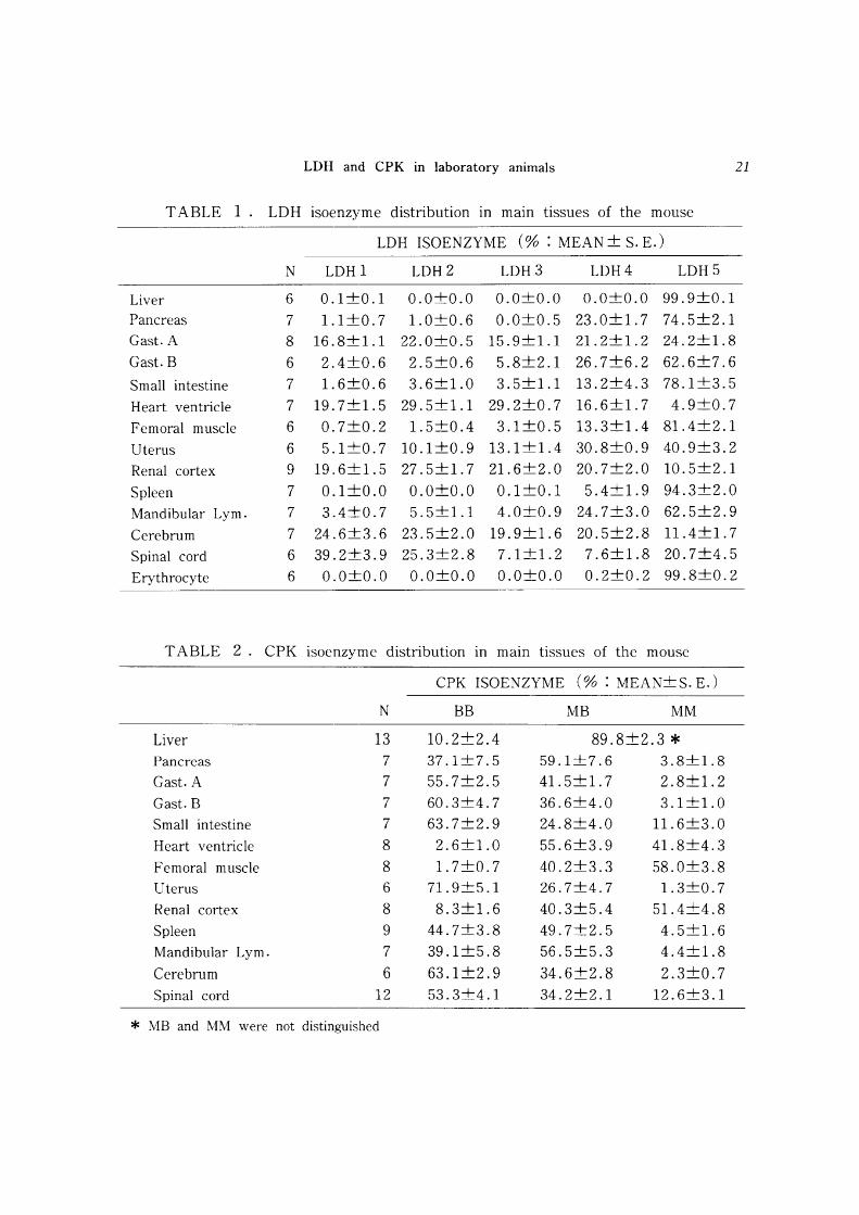
In tissues of the mouse, LDHl, LDH2 and LDH5 were the main isoenzymes only in the spinal cord. LDH4 and LDH5 were predominant isoenzymes in the pancreas,
gast B, small intestine, femoral muscle, uterus, spleen and mandibular lymph node. Liver and erythrocyte predominantly had only fraction LDH5. Gast A, heart ventricle, renal cortex and cerebrum possessed five LDH fractions i,e, LDH1 through LDH5
(Tab. 1 & Fig. 1). Pancreas, gast A and B, small intestine, uterus, spleen, mandibular lymph node, cerebrum and spinal cord mainly had CPK-BB and MB isoenzymes.
LDH and CPK in laboratory animals 21
TABLE 1 . LDH isoenzyme distribution in main tissues of the mouse
LDH ISOENZYME (% : MEAN + S. E.)
N LDH 1 LDH2 LDH3 LDH4 LDH5
Liver 6 0.1+0.1 0.0+0.0 0.0+0.0 0.0+0.0 99.9+0.1
Pancreas 7 1.1 +0.7 1.0+0.6 0.0+0.5 23.0+1. 7 74.5+2.1 Gast. A 8 16.8+1.1 22.0+0.5 15.9+1.1 21.2+1.2 24.2+1.8
Cast. B 6 2.4+0.6 2.5+0.6 5.8+2.1 26.7+6.2 62.6+7.6
Small intestine 7 1.6+0.6 3.6+1.0 3.5+1.1 13.2+4.3 78.1+3.5
Heart ventricle 7 19.7+1.5 29.5+1.1 29.2+0.7 16.6+1. 7 4.9+0.7
Femoral muscle 6 0.7+0.2 1.5+0.4 3.1+0.5 13.3+1.4 81.4+2.1
Uterus 6 5.1 +0.7 10.1+0.9 13.1+1.4 30.8+0.9 40.9+3.2
Renal cortex 9 19.6+1.5 27.5+1. 7 21.6+2.0 20.7+2.0 10.5+2.1
Spleen 7 0.1+0.0 0.0+0.0 0.1+0.1 5.4+1.9 94.3+2.0
Mandibular Lym. 7 3.4+0.7 5.5+1.1 4.0+0.9 24.7+3.0 62.5+2.9
Cerebrum 7 24.6+3.6 23.5+2.0 19.9+1.6 20.5+2.8 11.4+1.7
Spinal cord 6 39.2+3.9 25.3+2.8 7.1+1.2 7.6+1.8 20.7+4.5
Erythrocyte 6 0.0+0.0 0.0+0.0 0.0+0.0 0.2+0.2 99.8+0.2
TABLE 2. CPK isoenzyme distribution m mam tissues of the mouse
CPK ISOENZYME (% : MEAN+S. E.)
N BB MB MM
Liver 13 10.2+2.4 89.8+2.3 * Pancreas 7 37.1+7.5 59.1+7.6 3.8+1.8 Cast. A 7 55.7+2.5 41.5+1.7 2.8+1.2
Gast. B 7 60.3+4.7 36.6+4.0 3.1+1.0
Small intestine 7 63.7+2.9 24.8+4.0 11.6+3.0
Heart ventricle 8 2.6+1.0 55.6+3.9 41.8+4.3
Femoral muscle 8 1.7+0.7 40.2+3.3 58.0+3.8 Uterus 6 71.9+5.1 26.7+4.7 1.3+0.7
Renal cortex 8 8.3+1.6 40.3+5.4 51.4+4.8
Spleen 9 44.7+3.8 49.7+2.5 4.5+1.6
Mandibular Lym. 7 39.1 +5.8 56.5+5.3 4.4+1.8
Cerebrum 6 63.1+2.9 34.6+2.8 2.3+0.7
Spinal cord 12 53.3+4.1 34.2+2.1 12.6+3.1
* MB and MM were not distinguished
22 YASUDA, J. et at.
LDH 1 2 3 4 5 CPK SS MS MM ( + ) .. 'I' ... ' , '" ( -)
1
2
3
4
5
6
7
8
9
10
11
12
13
14
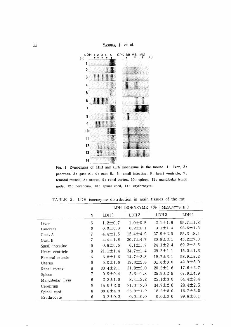
Fig. 1 Zymograms of LDH and CPK isoenzyme in the mouse. 1: liver, 2:
pancreas, 3: gast A., 4: gast B., 5: small intestine, 6: heart ventricle, 7:
femoral muscle, 8: uterus, 9: renal cortex, 10: spleen, 11: mandibular lymph
node, 12 : cerebrum, 13 : spinal cord, 14 : erythrocyte.
TABLE 3. LDH isoenzyme distribution in mam tissues of the rat
LDH ISOENZYME (% : MEAN+S. E.)
N LDH1 LDH2 LDH3 LDH4
Liver 6 1.2+0.7 1.0+0.5 2.1+1.6 95.7+1.8 Pancreas 6 0.0+0.0 0.2+0.1 3.1+1.4 96.6+1.3
Cast. A 7 4.4+1.5 12.4+4.9 27.9+2.5 55.3+8.4
Gast. B 7 4.4+1.6 20.7+4.7 30.9+3.1 45.2+7.0
Small intestine 6 0.6+0.6 6.1+1.7 24.1 +2.4 69.2+3.5
Heart ventricle 8 21.1 +1.4 34.7+1.4 29.2+1.1 15.0+1.3
Femoral muscle 6 6.8+1.6 14.7+3.8 19.7+3.1 58.9+8.2
Uterus 6 5.0+1.6 19.3+2.8 31.8+3.6 42.9+6.0
Renal cortex 8 30.4+2.1 31.8+2.0 20.2+1. 6 17.6+2.7
Spleen 7 0.9+0.4 5.3+1.8 25.9+2.9 67.9+4.9
Mandibular Lym. 6 2.3+1.0 8.4+2.2 25.1 +3.0 64.4+2.4
Cerebrum 8 15.9+2.0 21.0+2.0 34.7+2.0 28.4+2.5 Spinal cord 8 38.8+4.3 25.9+1.9 18.2+2.0 16.7+3.5
Erythrocyte 6 0.2+0.2 0.0+0.0 0.0+0.0 99.8+0.1
LDH and CPK in laboratory animals 23
Fraction CPK-MB was separated into two sub fractions m the gast B. Heart ventricle, femoral muscle and renal cortex predominantly had CPK-MB and MM fractions. It was possible to demonstrate a small amount of CPK-BB isoenzyme in the liver, but the MB band and the MM band were inseparable because of tailng (Tab. 2 & Fig. 1).
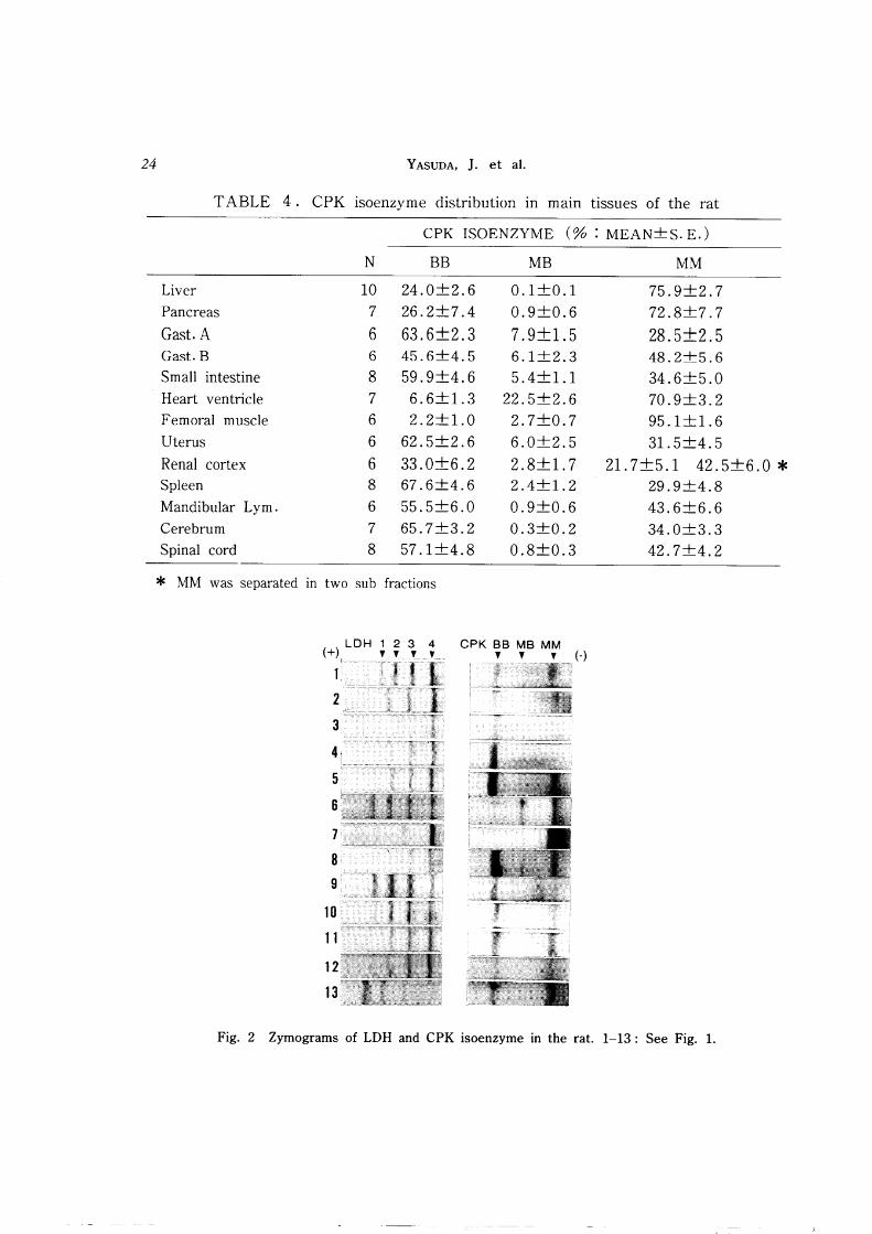
In the rat, LDH isoenzymes were separated into four fractions. LDH1, 2 and 3 bands were migrated like to the mouse, but LDH4 band in the rat were migrated between LDH4 and LDH5 bands in the mouse. Heart ventricle, renal cortex and spinal cord mainly had LDH1, LDH2 and LDH3. Liver, pancreas and erythrocyte only had LDH4. In the other tissues, LDH4 was the predominant fraction (Tab. 3 & Fig. 2). Gast A, small intestine, uterus, spleen, mandibular lymph node, cerebrum and spinal cord mainly had CPK-BB isoenzyme and had a small amount MM isoenzyme as well. While, liver, pancreas, heart ventricle, femoral muscle and renal cortex chiefly had CPK-MM isoenzyme in addition to a small amount of fraction BB (Tab. 4 & Fig. 2).
In the guinea pig, LDH1, LDH2 and LDH3 were predominant isoenzymes in the pancreas, gast A, heart ventricle, renal cortex, cerebrum and spinal cord. Erythrocyte mostly had fraction LDHl. LDH5 was the main band only in the femoral muscle. LDH3 and LDH4 were the main isoenzymes in the other tissues (Tab. 5 & Fig. 3). CPK-BB and MM were recognized in the uterus, mandibular lymph node, cerebrum and spinal cord. Heart ventricle and femoral muscle mainly had CPK-MM and had a small amount of fraction MB. Fraction CPK-MM was separated into two sub fractions. Liver, gast A and B, small intestine, renal cortex and spleen predominantly had three isoenzymes: CPK-BB, MB and MM. The MB and MM bands were inseparable in the pancreas because of tailing (Tab. 6 & Fig. 3).
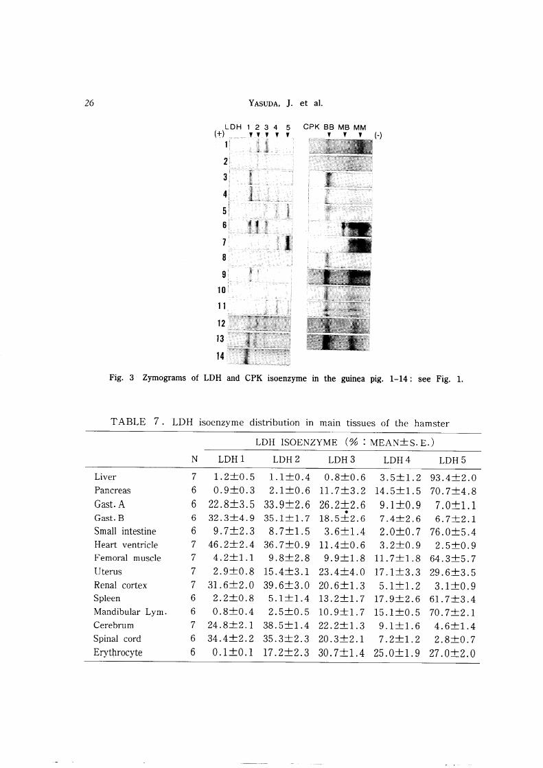
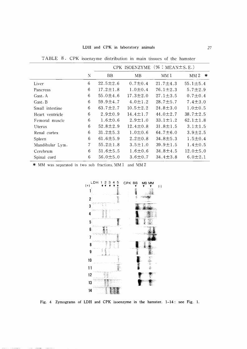
In the golden hamster, LDHI and LDH2 isoenzymes were the main fractions in the gast A and B, heart ventricle, renal cortex, cerebrum and spinal cord. On the other hand, LDH4 and LDH5 were the predominant fractions in the liver, pancreas, small intestine, femoral muscle, spleen and mandibular lymph node. LDH2, LDH3 and LDH4 were the main bands in the uterus and erythrocyte (Tab. 7 & Fig. 4). The other tissues mostly had CPK-BB and had a small amount of CPK-MM. Liver, pancreas, heart ventricle, femoral muscle and renal cortex predominantly had CPK-MM isoenzyme and had a small amount of CPK-BB. Fraction CPK-MM was separated into two sub fractions. CPK-MB bands were recognized in the gast A, small intestine, heart ventricle and uterus (Tab. 8 & Fig. 4).
DISCUSSION
The isoenzyme patterns of each tissue vary in each species of animal3, 10, 14, 15), but
the patterns of the same tissue of the closely related species are similar to each other17). Although the mouse (Mus musculus), rat (Rattus norvegicus), guinea pig
24 YASUDA, J. et al.
TABLE 4. CPK isoenzyme distribution in main tissues of the rat
CPK ISOENZYME (% : MEAN+S. E.)
N BB MB MM
Liver 10 24.0+2.6 0.1+0.1 75.9+2.7
Pancreas 7 26.2+7.4 0.9+0.6 72.8+7.7
Cast. A 6 63.6+2.3 7.9+1.5 28.5+2.5 Cast. B 6 45.6+4.5 6.1+2.3 48.2+5.6
Small intestine 8 59.9+4.6 5.4+1.1 34.6+5.0
Heart ventricle 7 6.6+1.3 22.5+2.6 70.9+3.2 Femoral muscle 6 2.2+1.0 2.7+0.7 95.1+1.6
Uterus 6 62.5+2.6 6.0+2.5 31.5+4.5
Renal cortex 6 33.0+6.2 2.8+1. 7 21.7+5.1 42.5+6.0 * Spleen 8 67.6+4.6 2.4+1.2 29.9+4.8
Mandibular Lym. 6 55.5+6.0 0.9+0.6 43.6+6.6
Cerebrum 7 65.7+3.2 0.3+0.2 34.0+3.3
Spinal cord 8 57.1+4.8 0.8+0.3 42.7+4.2
* MM was separated in two sub fractions
CPK BB MB MM , , , (-)
Fig. 2 Zymograms of LDH and CPK isoenzyme in the rat. 1-13: See Fig. 1.
LDH and CPK in laboratory animals 25
TABLE 5. LDH isoenzyme distribution in main tissues of the guinea pig
LDH ISOENZYME (% : MEAN+S. E.)
N LDH1 LDH2 LDH3 LDH4 LDH5
Liver 6 12.6+2.4 24.8+3.5 34.4+2.1 21.3+4.1 6.8+3.0
Pancreas 6 22.1 +1.0 35.0+0.9 34.1 +0.9 8.6+1.1 0.3+0.2
Gast. A 6 52.3+4.3 17.3+0.8 19.7+2.1 12.2+1.7 2.6+0.9
Gast. B 5 28.1+9.0 14.1+1.4 31.2+5.0 20.7+3.6 4.9+1.9
Small intestine 6 5.5+0.9 13.3+0.6 33.1 +2.6 32.4+2.0 15.6+4.0
Heart ventricle 6 29.1 +1.8 33.9+0.7 28.1 +1.3 8.4+0.8 0.5+0.2
Femoral muscle 6 3.7+1.0 4.0+0.5 8.8+1.1 17.6+1. 7 65.9+3.5
Uterus 6 9.2+1.9 22.9+2.1 38.3+1.8 24.3+3.4 5.3+3.4
Renal cortex 6 53.5+3.8 25.2+1.5 15.8+2.4 4.4+0.9 1.1+0.3
Spleen 6 8.6+2.4 14.9+3.7 33.4+3.2 30.0+5.1 13.1+5.5
Mandibular Lym. 3 12.2+6.0 14.7+3.5 28.6+2.7 32.2+5.2 12.4+3.5
Cerebrum 6 23.6+1.8 28.6+1.1 33.2+1.6 14.2+2.3 0.2+0.2
Spinal cord 6 44.4+2.5 27.7+1.5 21.2+1.5 7.3+2.2 0.6+0.3
Erythrocyte 6 85.4+3.3 8.9+2.1 4.5+1.8 0.9+0.5 0.3+0.1
TABLE 6. CPK isoenzyme distribution in main tissues of the guinea pig
CPK ISOENZYME (% : MEAN+S. E.)
N BB MB MM
Liver 6 24.2+1.4 11.0+2.8 64.8+2.8
Pancreas 5 11.3+2.9 88.9+3.0 * Gast. A 6 46.1 +6. 7 14.2+3.9 39.7+9.6
Gast. B 6 41.2+6.2 14.2+2.1 44.6+7.3
Small intestine 6 54.0+4.6 18.8+5.3 27.2+7.7
Heart ventricle 6 1.4+0.5 7.9+1.5 90.8+1.9
Femoral muscle 6 0.3+0.2 2.2+1.0 97.5+1.1
Uterus 6 51.2+3.6 4.8+2.8 40.9+2.9
Renal cortex 6 22.4+3.6 9.9+2.5 67.4+3.4
Spleen 6 39.4+8.2 7.0+1.8 53.4+7.4
Mandibular Lym. 6 62.4+3.5 2.0+0.9 35.7+4.0
Cerebrum 6 52.5+3.3 1.6+0.6 45.9+3.1
Spinal cord 6 53.2+6.2 2.3+0.6 44.5+5.8
* MB and MM were not distinguished
26 YASUDA, J. et al.
LDH 1 2 3 4 5 CPK SS MS MM (+) , , , , , , , , H
1
2
3
4
5 6
7
8
9
10
11
12
13
14
Fig. 3 Zymograms of LDH and CPK isoenzyme in the guinea pig. 1-14: see Fig. 1.
TABLE 7. LDH isoenzyme distribution in main tissues of the hamster
LDH ISOENZYME (% : MEAN+S. E.)
N LDH1 LDH2 LDH3 LDH4 LDH5
Liver 7 1.2+0.5 1.1 +0.4 0.8+0.6 3.5+1.2 93.4+2.0
Pancreas 6 0.9+0.3 2.1+0.6 11.7+3.2 14.5+1.5 70.7+4.8
Gast. A 6 22.8+3.5 33.9+2.6 26.2+2.6 9.1 +0.9 7.0+1.1 •
Gast. B 6 32.3+4.9 35.1 +1.7 18.5+2.6 7.4+2.6 6.7+2.1
Small intestine 6 9.7+2.3 8.7+1.5 3.6+1.4 2.0+0.7 76.0+5.4
Heart ventricle 7 46.2+2.4 36.7+0.9 11.4+0.6 3.2+0.9 2.5+0.9
Femoral muscle 7 4.2+1.1 9.8+2.8 9.9+1.8 11.7+1.8 64.3+5.7
Uterus 7 2.9+0.8 15.4+3.1 23.4+4.0 17.1+3.3 29.6+3.5
Renal cortex 7 31.6+2.0 39.6+3.0 20.6+1.3 5.1+1.2 3.1 +0.9 Spleen 6 2.2+0.8 5.1+1.4 13.2+1. 7 17.9+2.6 61. 7+3.4
Mandibular Lym. 6 0.8+0.4 2.5+0.5 10.9+1. 7 15.1 +0.5 70.7+2.1
Cerebrum 7 24.8+2.1 38.5+1.4 22.2+1.3 9.1+1.6 4.6+1.4
Spinal cord 6 34.4+2.2 35.3+2.3 20.3+2.1 7.2+1.2 2.8+0.7
Erythrocyte 6 0.1 +0.1 17.2+2.3 30.7+1.4 25.0+1.9 27.0+2.0
LDH and CPK in laboratory animals
TABLE 8. CPK isoenzyme distribution in main tissues of the hamster
CPK ISOENZYME (% : MEAN+S.E.)
N BB MB MM1 MM2 * Liver 6 22.5+2.6 0.7+0.4 21. 7+4.3 55.1 +5.4
Pancreas 6 17.2+1.8 1.0+0.4 76.1+2.3 5.7+2.9
Cast. A 6 55.0+4.6 17.3+2.0 27.1 +3.5 0.7+0.4
Cast. B 6 59.9+4.7 4.0+1.2 28.7+5.7 7.4+3.0
Small intestine 6 63.7+2.7 10.5+2.2 24.8+3.0 1.0+0.5
Heart ventricle 6 2.9+0.9 14.4+1.7 44.0+2.7 38.7+2.5
Femoral muscle 6 1.6+0.6 2.9+1.0 33.1 +1.2 62.1 +1.8
Uterus 6 52.8+2.9 12.4+0.8 31.8+1.5 3.1+1.5
Renal cortex 6 31. 2+5.3 1.0+0.6 64.7+6.0 3.9+2.5
Spleen 6 61.6+5.9 2.2+0.8 34.8+5.3 1.5+0.4
Mandibular Lym. 7 55.2+1.8 3.5+1.0 39.9+1.5 1.4+0.5
Cerebrum 6 51.6+5.5 1.6+0.6 34.8+4.5 12.0+5.0
Spinal cord 6 56.0+5.0 3.6+0.7 34.4+3.8 6.0+2.1
* MM was separated in two sub fractions. MM 1 and MM 2
LDH 1 2 3 4 5 CPK BB MB MM (+) " , , , '" (-)
11, 2
3
4
5
6
7
8
9
10
11
12
13
14
r
~ Ai. L
Fig. 4 Zymograms of LDH and CPK isoenzyme m the hamster. 1-14: see Fig. 1.
27
28 YASUDA, J. et a1.
(Cavia porcellus) and golden hamster (Mesocricetus auratus) are all belonging to Rodentia, the isoenzyme patterns of LDH and CPK in some tissues varied from each other in this experiment. Therefore, it is necessary to understand the distribution of tissue isoenzymes of each animal. In laboratory animals such as the mouse and rat, there have been many reports about parts of tissuee isoenzymes1,4,7.8,15,17>, but there
are few reports that have systematically analyzed isoenzymes of various tissues at the same time13
,19). Liver and erythrocyte LDH isoenzyme patterns of laboratory animals
obtained by the direct electrophoretic method were similar to those obtained by the extracting method 1, 17,20). CPK isoenzyme patterns in main tissues of laboratory animals were similar to those of humans and other mammals2,6,12). Analytical values
obtained by the tissue-extracting method, however, varied more than those obtained by this direct method14
).
vated during extraction. There is also evidence that enzyme activity can be inacti
Therefore, the histoelectrophoretic method is thought to be appropriate for the detailed analysis of tissue isoenzyme patterns in laboratory animals. This method has different significance from serum isoenzyme analysis and may be the
markers concerning organic damages in laboratory animals. As enzyme proteins are
direct products of gene expression, variation at the genetic level is reflected in variations of the tissue isoenzyme compositions. This technique may also be applied to elucidate ontogenetic and phylogenetic classification of inbred laboratory animals.
ACKNOWLEDGMENT
This work was supported in part by a Grant-in-aid for Scientific Research (Grant No. 63560299) from the Ministry of Education, Science and Culture, Japan. We thank Yukitoshi KITAMURA D. V. M., associate director of the Animal Experimentation Division, Faculty of Medicine, Sapporo Medical College for his kindly advice.
REFERENCES
1) ALLALOUF, D., SCHWARZUMAN, S., LEVINSKY, H., FELER, N., HART, J.t ZOHER, S. & MENACHE, R. (1986): Lactate dehydrogenase and alkaline phosphatate in regenerat
ing rat liver Res. Exp. Med., 186, 103-107
2) ANDERSON, M. G. (1976): The effect of exercise on the lactate dehydrogenase and
creatine kinase isoenzyme composition of horse serum Res. Vet. Sci., 20, 191-196
3) BEATTY, E. M. & DOXEY, D. L. (1983): Lactate dehydrogenase and creatine kinase
isoenzyme levels in the tissues and serum of normal lambs Res. Vet. Sci., 35, 325-330
4) BONAVITA, V., PONTA, F. & AMORE, G. (1962): Lactate dehydrogenase isoenzymes
in the developing rat brain Nature, 196, 576-577
5) DAVIS, B. J. (1964): Disc electrophoresis II. Method and application to human
serum proteins Ann. New York Acad. Sci. , 121, 404-427
6) DAWSON, D. M. & FINE, H. (1967): Creatine kinase in human tissues Arch. Neurol., 16, 175-180
LDH and CPK in laboratory animals
7) FINE, I. H., KAPLAN, K. O. & KUFTINEC, D. (1963): Development changes of
mammalian lactate dehydrogenases Biochem., 2, 116-121
8) GARBUS,]. HIGHMAN, B. & ALTLAND, P. D. (1964): Serum enzymes and lactate
dehydrogenase isoenzymes after exercise and training in rats Am. ]. Physiol., 207, 467-472
9) HAGER, D. A., NYLAND, T. G. & FISHER, P. (1985): Ultrasound-guided biopsy of the
canine liver, hidney and prostate Vet. Radiol., 26, 82-88
10) HINKS, M. & MASTERDS, C. J. (1966): The ontogenetic variformity of lactate
dehydrogenase in feline and cavian tissues Biochim. Biophy. Acta., 130, 458-468
11) JERAJ, K., OSBORNE, C. A. & STEVENS, J. B. (1982): Evaluation of renal biopsy in
197 dogs and cats l. Am. Vet. Med. Assoc., 181, 367-369
12) KIKUTA, Y. & ONISHI, T. (1987): Creatine kinase and its isoenzymes in dog]. lPn. Vet. Med. Assoc., 40, 26-30 (in Japanese with English abstract)
13) KITAMURA, M., IUIMA, N., HASHIMOTO, F. & HIRATSUKA, A. (1971): Hereditary
deficiency of subunit H of lactate dehydrogenase Clin. Chim. Acta., 34, 419-423
14) KITAMURA, N., NAITOU, I., HARA, Y. & SHIBANAI, D. (1985): Clinical studies on
lactate dehydrogenase in dogs and cattle I. Normal LDH isoenzyme patterns of
serum and tissues]. Vet. Med., (763) 96-103 (in Japanese)
15) MARKERT, C. L. & M 1> LLER, F. (1959): Multiple forms of enzymes; tissue,
ontogenetic and species specific patterns Proc. Natl. Acad. Sci. U. S. A., 45, 753-763
16) MATSUI, T., KOBAYASHI, Y., MlZOGUCHI, Y., MONNA, T., YAMAMOTO, S., o OTANI , S,
NAKAI, K. & MORISAWA, S. (1981): Studies on the inhibitory effects of cepharanthine
on the liver injuries induced by CCl4 poisoning lPn. l. Gastroenterol., 78, 1053-1058
(in Japanese wit English abstract)
17) OOHIRA, L, KOBAOYASHI, H., HAGIWARA, M., MATSUOKA, T., KISHINO, K., YAMA
GUCHI, Y., WATANABE, T., MARUYAMA, H., YOSHIZAWA, Y. & ISHIGAKI, T. (1975):
Liver LDH isoenzyme patterns of mammals Physico-Chem. BioI., 19, 24-207 (in
Japanese)
18) TAKEHARA, Y., YAMASAKI, M., FUJII, Y. & YASHIOKA, T. (1985): Effect of cepharan
thine on rat regenerating liver (in Japanese with English abstract) lPn. l. Surg. Metab. Nutr., 19, 323-329
19) VAN, K. ]. & WILLERBRANDS, A. F. (1966): Isoenzymes of creatine phosphokinase in
tissue extract and normal and pathological sera Clin. Chim. Acta., 13, 312-316
20) VESELL, E. S. & PHILIP, J. (1963): Isozyme of lactate dehydrogenase; sequential
alterations during development Ann. New York. Acad. Sci., 111, 243-257
21) YASUDA,]', SYUTO, B. & Too, K. (1985): Analysis of tissue creatine phosphokinase
isoenzymes by histoelectrophoresis lPn. l. Vet. Res., 33, 19-25
22) YASUDA, J., SYUTO, B., Too, K. & OOFUJI, S. (1989): Lactate dehydrogenase
isoenzyme patterns in bovine liver tissue lPn. l. Vet. Sci., 51, 733-.739
29