Embed Size (px)
Citation preview
L E W I S . N E L S O N , A N D R A C K E R
Laser Raman Spectroscopy as a Mechanistic Probe of the Phosphate Transfer from Adenosine Triphosphate in a Model System?
Aaron Lewis, Nathan Nelson,$ and Efraim Racker*
ABSTRACT: Laser Raman spectroscopy has been used to study a phosphate transfer reaction from A T P to Pi or arse- nate in dimethyl sulfoxide. The spectra support a mecha- nism involving Mg2+ binding to the (Y and phosphates of A T P leaving the third phosphate free for the transfer reac- tion. The data also indicate the formation of a relatively stable intermediate which is facilitated by the presence of dimethyl sulfoxide and a dicarboxylic acid (maleate). The
A phosphate transfer reaction in 70% dimethyl sulfoxide from A T P to either Pi or arsenate has been described (Nel- son and Racker, 1973) which resembles in some respects energy transfer reactions catalyzed by coupling factor 1 from chloroplasts (Nelson et al., 1972). Earlier studies by Lord and Thomas (1967a,b), Rimai et al. ( 1 969, 1970), and others (see Koenig, 1972; Lewis and Spoonhower, 1974) suggested that laser Raman spectroscopy might yield infor- mation on the mechanism of this reaction.
The transfer reactions in 70% dimethyl sulfoxide re- quired Mg2+ or another divalent cation (Ca2+ or Mn2+) and were markedly stimulated by dicarboxylic acids, partic- ularly maleate or malonate. The hydrolysis of A T P depend- ed on the presence of arsenate (approximately 5 m M ) suggesting a common feature with the well-known effect of arsenate in biological energy transfer reactions.
Experimental Section The experiments were performed in a final volume of 1.5
ml containing 20 m M ATP, 20 m M MgC12. 40 m M Tri- cine-maleate (pH 8), 5 m M arsenate, and 1 ml of dimethyl sulfoxide. Several controls were run to test the dependence of the spectra on various components of the above solution. Samples were made with triply distilled water obtained from the Cornell Crystal Growing facility and introduction of dust particles was avoided. The solutions were passed through 0.4-1 Millipore filters into capillaries which were sealed. These procedures successfully minimize the back- ground and thus facilitate the observation of the Raman spectra in solutions a t relatively low concentrations. To ob- tain information on reaction times a syringe attached to the cuvet was used to inject 5 m M arsenate into a solution al- ready containing the other required components. A particu-
~~~~~ ~
t From the School of Applied and Engineering Physics (A L ) and the Section of Biochemistry, Molecular and Cell Biology, (U h and E R ), Cornell University, Ithaca, New York 14850 Keceried March 1 1 , 1974 This work was supported by the Research Corporation. the Petroleum Research Fund, administered by the Americdn Chemical Society, and by National Science Foundation Grant GB-30850 A L I \
an Alfred P Sloan Fellow 3 Present address Department of Biology. Technion, Haifa, Israel
intermediate has a Raman spectrum with a band a t 1090.5 cm-I similar to the end product ADP, but is formed much more rapidly. Since the model reaction has many features in common (e.g., activation by maleate) with the transfer reactions catalyzed by coupling factors from spinach chlo- roplast, Raman spectroscopy may also prove to be a useful tool in the elucidation of biological energy transfer reac- tions.
lar vibrational frequency was monitored and recordings within 40 sec were made.
Spectra were obtained with a Spex 1401 double mono- chromator and were detected with an EM1 6256s photo- multiplier. Photons were counted at each step of the mono- chromator and were stored in the memory of a Nuclear Data Model 1100 multichannel analyzer. The spectra were then averaged and plotted out with a PDP/ 1 1 mini comput- er and a Houston plotter. Laser powers of up to 1.2 W were obtained with a Model 52G-A Coherent Radiation argon ion laser.
Results The Raman spectra seen in Figure 1 were obtained im-
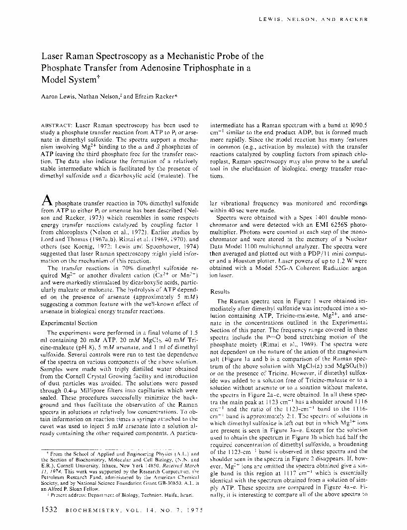
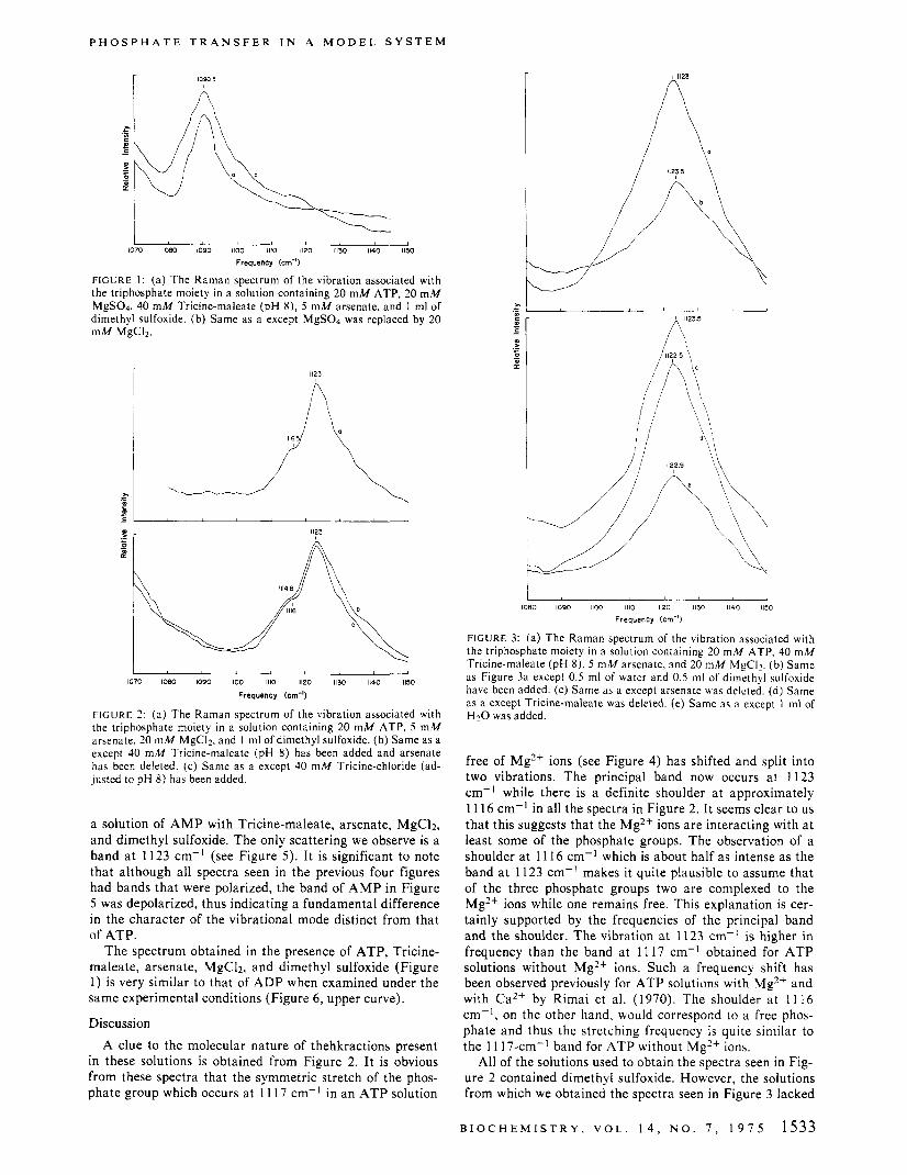
mediately after dimethyl sulfoxide was introduced into a so- lution containing ATP, Tricine-maleate, Mg2+, and arse- nate in the concentrations outlined in the Experimental Section of this paper. The frequency range covered in these spectra include the -0 bond stretching motion of the phosphate moiety (Rimai et al., 1969). The spectra were not dependent on the nature of the anion of the magnesium salt (Figure l a and b is a comparison of the Raman spec- trum of the above solution with MgCl*(a) and MgS04(b)) or on the presence of Tricine. However, if dimethyl sulfox- ide was added to a solution free of Tricine-maleate or to a solution without arsenate or to a solution without maleate, the spectra in Figure 2a-c, were obtained. I n all these spec- tra the main peak at 1 123 cm-' has a shoulder around 1 1 16 cm-' and the ratio of the 1123-cni-' band to the 1 1 16- cm- ' band is approximately 2: I . The spectra of solutions in which dimethyl sulfoxide is left out but i n Lbhich ME2+ ions are present is seen in Figure 3a-e. Except for the solution used to obtain the spectrum in Figure 3b which had half the required concentration of dimethyl sulfoxide, a broadening of the 1123-cm-' band is observed in these spectra and the shoulder seen in the spectra in Figure 2 disappears. I f , how- ever, Mg'+ ions are omitted the spectra obtained give a sin- gle band in this region at 1 1 17 cm-' which is essentially identical with the spectrum obtained from a solution of sim- ply ATP. These spectra are compared in Figure 4a-e. Fi- nally, it is interesting to compare all of the above spectra to
1532 B I O C H E M I S T R Y , V O L . 1 4 , N O . I . 1 9 7 5
P H O S P H A T E T R A N S F E R I N A M O D E L S Y S T E M
1070 1080 1090 1100 Ill0 1120 1130 1140 1150
Frequency (ern-')
FIGURE 1 : (a) The Raman spectrum of the vibration associated with the triphosphate moiety in a solution containing 20 m M ATP, 20 m M MgS04, 40 mM Tricine-maleate (pH 8), 5 mM arsenate, and 1 ml of dimethyl sulfoxide. (b) Same as a except MgS04 was replaced by 20 mM MgC12.
1123
A
1070 IOBO 1090 1100 1110 1120 1150 1140 1150
Frequency (ern-')
F I G U R E 2: (a) The Raman spectrum of the vibration associated with the triphosphate moiety in a solution containing 20 mM ATP, 5 mM arsenate, 20 mM MgC12, and 1 ml of dimethyl sulfoxide. (b) Same as a except 40 mM Tricine-maleate (pH 8) has been added and arsenate has been deleted. (c) Same as a except 40 m M Tricine-chloride (ad- justed to pH 8) has been added.
a solution of A M P with Tricine-maleate, arsenate, MgC12, and dimethyl sulfoxide. The only scattering we observe is a band a t 1123 cm-l (see Figure 5). It is significant to note that although all spectra seen in the previous four figures had bands that were polarized, the band of A M P in Figure 5 was depolarized, thus indicating a fundamental difference in the character of the vibrational mode distinct from that of ATP.
The spectrum obtained in the presence of ATP, Tricine- maleate, arsenate, MgC12, and dimethyl sulfoxide (Figure 1) is very similar to that of A D P when examined under the same experimental conditions (Figure 6, upper curve).
Discussion
A clue to the molecular nature of thehkractions present in these solutions is obtained from Figure 2. It is obvious from these spectra that the symmetric stretch of the phos- phate group which occurs a t 11 17 cm-' in an A T P solution
r i l l 23
1080 1090 1100 1110 1120 11% 1140 1150
Frequency 1crn-l)
F I G U R E 3: (a) The Raman spectrum of the vibration associated with the triphosphate moiety in a solution containing 20 m M ATP, 40 m M Tricine-maleate (pH 8), 5 m M arsenate, and 20 m M MgCI2. (b) Same as Figure 3a except 0.5 ml of water and 0.5 ml of dimethyl sulfoxide have been added. (c) Same as a except arsenate was deleted. (d) Same as a except Tricine-maleate was deleted. (e) Same as a except 1 ml of H 2 0 was added.
free of Mg2+ ions (see Figure 4) has shifted and split into two vibrations. The principal band now occurs a t 1123 cm-' while there is a definite shoulder a t approximately 11 16 cm-' in all the spectra in Figure 2. It seems clear to us that this suggests that the Mg2+ ions are interacting with a t least some of the phosphate groups. The observation of a shoulder a t 11 16 cm-l which is about half as intense as the band a t 1123 cm-' makes it quite plausible to assume that of the three phosphate groups two are complexed to the Mg2+ ions while one remains free. This explanation is cer- tainly supported by the frequencies of the principal band and the shoulder. The vibration a t 1123 cm-' is higher in frequency than the band a t 11 17 cm-' obtained for A T P solutions without Mg2+ ions. Such a frequency shift has been observed previously for A T P solutions with Mg2+ and with Ca2+ by Rimai et al. (1970). The shoulder a t 1 1 16 cm- ' , on the other hand, would correspond to a free phos- phate and thus the stretching frequency is quite similar to the 1 1 17-cm-' band for A T P without Mg2+ ions.
All of the solutions used to obtain the spectra seen in Fig- ure 2 contained dimethyl sulfoxide. However, the solutions from which we obtained the spectra seen in Figure 3 lacked
B I O C H E M I S T R Y , V O L . 1 4 , N O . 7 , 1 9 7 5 1533
L E W I S , N E L S O N , A N D R A C K E R
, 1080 1090 I100 Ill0 1120 1130 1140 1150
Frequency (cm-')
FIGLRE 4: ( a ) The Raman spectrum of the vibration associated with the triphosphate moiety in a solution containing 20 m M ATP, 40 m M Tricine-maleate (pH 8). and 5 m M arsenate. (b) Same as a except 20 m M 'UaCI and I ml of dimethyl sulfoxide were added. (c) Same as a except Tricine-maleate and arsenate were deleted. (d) Same as a ex- cept Tricine-maleate was deleted. (e) Same as a except I ml of dimeth- yl sulfoxide was added.
dimethyl sulfoxide and the resulting spectra seem quite dif- fuse. In fact, although the peak of the band is still observed a t 1123 cm-I the shoulder a t 1 1 16 cm-' cannot be detect- ed. This indicates that the dimethyl sulfoxide aids the Mg2+ ions in forming a tight complex with the phosphate groups. Solutions of A M P show a similar band at 11 23 cm-' (Fig- ure 5 ) which is definitely depolarized. In contrast the vibra- tion of A T P solutions is polarized. Thus the vibration of A M P solutions appear to have a n origin which is different from the similar vibration seen in A T P solutions. This ex- planation is supported by assignments made by Rimai e t al. (1969) on the vibrational spectra of AMP, ADP, and ATP.
If dimethyl sulfoxide is added to a solution of ATP, Tri- cine-maleate, arsenate, and MgC12 (in essence if all the key ingredients are present for the phosphate transfer reaction) the spectrum seen in Figure 1 is observed. The spectrum looks very similar to that of ADP, the end product of the ATP transfer reaction (Figure 6, upper curve). However, it can be readily deduced from the rate of A D P formation (Nelson et al., 1972) that during the short time period re- quired to take the spectrum (ca. 10 min) less than 5% of the A T P could have been converted to ADP. This amount of A D P could not be detected under the experimental condi- tions. When instead of A T P an amount of A D P correspond- ing to 10% of the A T P was added, no detectable vibrations a t 109 1 cm-l were observed (Figure 6, lower curve). More-
1070 > 1090 IOBO 1100 1110 1120 1130 1140 1150
Frequency (cm-0
FIGURE 5 : The Raman spectrum of the vibration associated with thc phosphate moiety in a solution containing 20 m M A M P , 40 m M Tri- cine-maleate ( p H 8). 5 m M arsenate, 20 m M MgC12. and I ml of di- methyl sulfoxide.
I I 2
1070 080 1090 1100 1110 I120 1130 1140 1150
Frequency (ern-')
FIGURE 6: The Raman spectrum of the vibration associated with the diphosphate moiety in a solution similar to the one used to obtain thc spectra in Figure I except that instead of 20 mM ATP. 20 m M A D P (upper curve) or 2 m M A D P (lower curve) was used.
over, when the scattering a t 1091 cm-' was monitored within 40 sec after addition of arsenate the counts per sec- ond have risen to their maximum value. At this time period only negligible amounts of A D P could have formed. Thus the intermediate formed with a band a t 1090.5 cm-' has spectral properties similar to A D P suggesting a complex formed between Mg2+ and the cy and p phosphate of A T P with the y phosphate available for transfer. This is also sug- gested by the splitting of the symmetric phosphate stretch (Figure 2 ) into two vibrations with intensity ratio of 2:1. Di- methyl sulfoxide appears to enhance the splitting in the vi- brational frequencies of the free and bound phosphate group.
Acknowledgment The authors thank Mr. John Spoonhower and Mr. Rob-
ert Cookingham for their help in recording some of the spectra reported in this paper.
References Koenig, J . (1972), J . Polym. Sci., Part D 60, 54. Lewis, A, , and Spoonhower, J. (1974), in Spectroscopy in
Biology and Chemistry, Yip, S., and Chen, S.. Ed., New York, N.Y . , Academic Press, pp. 347-376.
Lord, R. C., and Thomas, G. J . (1967a), Spertrochim. Acta, Parr A 23, 2551.
Lord, R. C., and Thomas, G. J . (1967b), Biorhim. Biophys.
1534 B I O C H E M I S T R Y . V O L . 1 4 , N O . 7 , 1 9 7 5
P O L Y A C R Y L A M I D E G E L S C O N T A I N I N G A C T I V E E S T E R S
Acta 142, 1.
Chem. 247, 6506.
Carew, E. B. (1969), Biophys. J . 9, 320.
Res. Commun. 41, 2, 3 13.
chem. Biophys. Res. Commun. 38, 23 1 .
Nelson, N., Nelson, H., and Racker, E. (1972), J . Biol.
Nelson, N., and Racker, E. (1973), Biochemistry 12, 563. Rimai, L. , Cole, T., Parsons, J. L., Hickmott, Jr., J. T., and
Rimai, L., and Heyde, M. E. (1970), Biochem. Biophys.
Rimai, L., Heyde, M. E., and Carew, E. B. (1970), Bio-
Polyacrylamide Gels Copolymerized with Active Esters. A New Medium for Affinity Systems?
Ronald L. Schnaar and Yuan Chuan Lee*
ABSTRACT: A new and versatile method for linking biologi- cally active ligands to a polyacrylamide matrix is reported. Active esters of acrylic acid (N-succinimidyl acrylate and N-phthalimidyl acrylate) were synthesized, then copolym- erized with acrylamide and N,N’-methylenebisacrylamide. Displacement of the active ester in the gel thus fofmed by various ligands containing aliphatic amino groups resulted in the formation of stable amide bonds between the ligands and the polyacrylamide gel. The affinity gel thus prepared has the following advantages: (i) resistance to chemical and microbiological degradation, (ii) ease of control of ligand
Immobilization of biologically active components has be- come an important tool in biological research, especially in its application to affinity chromatography for purification of biological molecules. Some of the variables for such im- mobilization are the insoluble support used and the type of bonding of biologically active compounds to that support. The use and comparative advantages of agarose, cellulose, dextran, glass, and polyacrylamide as insoluble carriers have been reviewed (Silman and Katchalski, 1966; Cuatre- casas and Anfinsen, 1971; Scouten, 1974). The most popu- lar among the systems currently in use are the polysaccha- rides (especially agarose beads) activated by cyanogen ha- lide treatment to accept ligands with amino terminals. However, this system has the disadvantages that the poly- saccharide is labile to attack by chemicals as well as mi- crobes, and the bonds linking the polysaccharides and the biologically active compounds are alkali labile.
Some of these disadvantages were absent in the system developed by Inman and Dintzis (1 969), in which commer- cially available preformed polyacrylamide beads (Bio-Gel) were modified to provide acyl azide and other active func- tional groups suitable for coupling ligands. Although this method led to high capacity of the carrier beads for the lig- ands, porosity was prohibitively reduced, greatly decreasing the effectiveness of the gel in macromolecule purification (Cuatrecasas, 1970; Steers et al., 1971).
+ From the Department of Biology and the McCollum-Pratt Insti- tute, The Johns Hopkins University, Baltimore. Maryland 21 218. Re- ceived November 14, 1974. Contribution No. 822 from the McCollum- Pratt Institute, The Johns Hopkins University. Supported by U S . Pub- lic Health Service. Yational Institute of Health Research Grant A M 9970, Research Career Development Award K04,79148 ( to Y.C.L.), and Training Grant GM57-17 (to R.L.S.).
level and higher levels of ligand possible, (iii) ease of control of porosity, and (iv) total displacement of the active ester under suitable conditions. Efficacy of this system was tested by preparation of 6-aminohexyl 2-acetamido-2-deoxy-p-~- glucopyranoside derivative of polyacrylamide gel by the de- scribed method. It was found to be more effective for purifi- cation of wheat germ agglutinin than the previously pub- lished affinity chromatography systems and the wheat germ hemagglutinin was obtained in crystalline form. In addition, partial resolution of isolectins was obtained by elution from the affinity gel with a p H gradient.
In a preliminary investigation, we have successfully formed polyacrylamide gels containing desired ligands by first coupling acrylic acid to the amino terminal of the lig- ands, and then copolymerizing them with acrylamide and cross-linking reagent (Lee, 1973). This approach has an ob- vious advantage of allowing easy control of porosity as well as level of ligand incorporation. I n addition, the total level of ligand that can be incorporated can be made considera- bly higher than by the use of CNBr-activated polysaccha- rides. In a similar approach that has been reported indepen- dently (HoiejS; and Kocourek, 1974), allyl glycosides were copolymerized with acrylamide to form an affinity gel sys- tem useful for purification of plant hemagglutinins. Al- though these methods overcame most of the disadvantages discussed above, the necessity of synthesizing individual lig- ands containing a double bond discourages general applica- tion of this method.
The objectionable features of the previous methods were resolved in the present studies. The acrylic acid esters of N-hydroxysuccinimide and N-hydroxyphthalimide were synthesized and copolymerized with acrylamide and A’,”- methylenebisacrylamide. The resulting “active” gels react- ed readily with ligands containing primary amino groups (Scheme I ) . Thus, the present method provides a new and versatile system of wider applicability than the earlier sys- tems. The applicability of this system to affinity chromatog- raphy is demonstrated by efficient purification of wheat germ hemagglutinin on polyacrylamide gel in which 6-ami- nohexyl 2-acetamido-2-deoxy-/3-~-glucopyranoside was in- corporated by the new method.
Experimental Section Materials. Acryloyl chloride and N-hydroxyphthalimide
were purchased from Aldrich Chemical Co. and used with-
B I O C H E M I S T R Y , V O L . 1 4 , N O . 7 , 1 9 7 5 1535


![Increased Rate of Adenosine Triphosphate …...(CANCER RESEARCH 55, 4352-4360, October 1, 1995] Increased Rate of Adenosine Triphosphate-dependent Etoposide (VP-16) Efflux in a Murine](https://img.pdfslide.net/doc/110x75/5e7e8d68c5d0407f2447f2a9/increased-rate-of-adenosine-triphosphate-cancer-research-55-4352-4360-october.jpg)