Embed Size (px)
Citation preview
Learning curve for robotic-assisted surgery for rectal cancer: useof the cumulative sum method
Tomohiro Yamaguchi • Yusuke Kinugasa • Akio Shiomi • Sumito Sato •
Yushi Yamakawa • Hiroyasu Kagawa • Hiroyuki Tomioka • Keita Mori
Received: 13 February 2014 / Accepted: 21 August 2014
� Springer Science+Business Media New York 2014
Abstract
Background Few data are available to assess the learning
curve for robotic-assisted surgery for rectal cancer. The
aim of the present study was to evaluate the learning curve
for robotic-assisted surgery for rectal cancer by a surgeon
at a single institute.
Methods From December 2011 to August 2013, a total of
80 consecutive patients who underwent robotic-assisted
surgery for rectal cancer performed by the same surgeon
were included in this study. The learning curve was ana-
lyzed using the cumulative sum method. This method was
used for all 80 cases, taking into account operative time.
Results Operative procedures included anterior resections
in 6 patients, low anterior resections in 46 patients, inter-
sphincteric resections in 22 patients, and abdominoperineal
resections in 6 patients. Lateral lymph node dissection was
performed in 28 patients. Median operative time was
280 min (range 135–683 min), and median blood loss was
17 mL (range 0–690 mL). No postoperative complications
of Clavien–Dindo classification Grade III or IV were
encountered. We arranged operative times and calculated
cumulative sum values, allowing differentiation of three
phases: phase I, Cases 1–25; phase II, Cases 26–50; and
phase III, Cases 51–80.
Conclusions Our data suggested three phases of the
learning curve in robotic-assisted surgery for rectal cancer.
The first 25 cases formed the learning phase.
Keywords Rectal cancer � Robotic-assisted surgery �Learning curve � CUSUM method � Total mesorectal
excision � Lateral lymph node dissection
Some studies have recently been published on the advan-
tages of laparoscopic rectal cancer surgery [1–3]. However,
when performing laparoscopic surgery for rectal cancer,
positivity of the circumferential resection margins (15.5 %)
and conversion rate (33.9 %) were high in a subgroup
analysis of the UK Medical Research Council (MRC) trial
of conventional versus laparoscopic-assisted surgery in
colorectal cancer (CLASICC) [2]. These findings may have
been related to the high degree of technical difficulty when
performing surgery in the narrow pelvic cavity. Limitations
include the use of straight rigid instruments within a nar-
row working space, limited degrees of freedom, an unsta-
ble camera platform with two-dimensional imaging, and
poor ergonomics of the instruments.
Since the da Vinci surgical system (Intuitive Surgical,
Sunnyvale, CA, USA) was approved by Food and Drug
Administration in 2000 and Weber et al. [4] performed the
first robotic-assisted colectomy for benign disease in 2001,
robotic-assisted surgery has gradually gained popularity.
Pigazzi et al. [5] first reported robotic-assisted total mes-
orectal excision (TME) for rectal cancer in 2006. Some of
the advantages gained with robotic-assisted surgery include
high-quality three-dimensional imaging, free-moving
multi-joint forceps, stable camera work by an operator, use
of an image stabilizer, a motion-scaling function, and
greatly improved ergonomics [6, 7].
T. Yamaguchi (&) � Y. Kinugasa � A. Shiomi � S. Sato �Y. Yamakawa � H. Kagawa � H. Tomioka
Division of Colon and Rectal Surgery, Shizuoka Cancer Center
Hospital, 1007 Shimonagakubo, Nagaizumi-cho, Sunto-gun,
Shizuoka 411-8777, Japan
e-mail: [email protected]
K. Mori
Clinical Trial Coordination Office, Shizuoka Cancer Center
Hospital, Shizuoka, Japan
123
Surg Endosc
DOI 10.1007/s00464-014-3855-5
and Other Interventional Techniques
With the adoption of new techniques, it is important to
assess the effects on the surgeon’s learning curve. The
cumulative sum (CUSUM) method was adopted by the
medical profession in the 1970s to analyze learning curves
for surgical procedures [8, 9]. Multiple reports on robotic-
assisted surgery have been published, but few reports have
evaluated the learning curve for robotic-assisted surgery
for rectal cancer [10–15]. Three reports using the CUSUM
method have suggested that the learning curve phase is
achieved after 15–35 cases [11, 12, 14]. On the other hand,
previous studies have suggested a wide ranging minimum
requirement of 40–90 cases to reach the first stabilization
of laparoscopic surgery for rectal cancer [16–20]. Robotic-
assisted surgery offers technical advantages over laparo-
scopic surgery and thus may shorten the learning curve
compared with laparoscopic surgery.
The present study attempted to evaluate the learning
curve in robotic-assisted surgery for rectal cancer based on
80 consecutive cases treated by a single surgeon at a single
institute. In Japan, TME with lateral lymph node dissection
(LLD) has been the standard treatment for patients with
lower rectal cancer [21]. We believe that our series rep-
resents the first to specifically analyze the learning curve in
robotic-assisted surgery for rectal cancer, including LLD.
Materials and methods
Patients and study design
From December 2011 to August 2013, a total of 125
patients underwent robotic-assisted surgery for rectal can-
cer at Shizuoka Cancer Center Hospital. These patients
comprised the first participants in our team’s experience
with robotic-assisted surgery. Of these, 80 consecutive
patients who underwent robotic-assisted surgery by an
expert surgeon (Y.K.) for rectal cancer were included in
this study. The rest of the robotic surgical team consisted of
robotic surgeons (T.Y. and A.S.) who performed the sur-
geries for the remaining 45 patients with rectal cancer, an
assistant surgeon (H.K.), nurses, and the anesthetic team,
all of whom were familiar with the robot setup.
We extracted data from our prospectively maintained
colorectal database, which contains information regarding
patient demographics, preoperative assessment, operative
characteristics, operative time (OT), surgeon console time
(SCT), morbidity, pathological characteristics, adjuvant
therapy, and follow-up. Indications for LLD were lower
rectal cancer with T3–4, or T1–2 rectal cancer with
metastasis to lateral lymph nodes, as described by the
Japanese Society for Cancer of the Colon and Rectum
(JSCCR) guidelines for the treatment of colorectal cancer
[22]. Until December 2011, rectal cancer surgeries with
LLD or with direct invasion to other organs were per-
formed using the open method, while the remaining rectal
cancer surgeries were performed using a laparoscopic
method. Patients with obvious suspicion of direct invasion
to other organs underwent neoadjuvant chemoradiotherapy
with TME. Patients were staged using the tumor node
metastasis (TNM) classification [23]. All study protocols
were approved by our institutional review board (25-J100-
25-1-3).
Operative technique
All procedures were performed using a systematic
approach that included colonic and pelvic phases by a
robotic approach. During the colonic phase, the inferior
mesenteric artery and vein were ligated under a medial-to-
lateral approach. When required, the splenic flexure was
taken down. The pelvic phase involved sharp dissection of
the prehypogastric nerve fascia and behind Denonvilliers’
fascia to avoid autonomic nerve injury during TME for
lower rectal cancer [24]. Total OT was defined as the
interval from first incision to closure of the incisions. SCT
was the actual time the surgeon spent at the robotic console
during the procedure, which directly corresponded to the
robotic portion of the procedure.
CUSUM method
The learning curve was analyzed using the CUSUM method
similar to that described by Bokhari et al. [12]. The CUSUM
was the running total of differences between the individual
data points and the mean of all data points. This method was
used for all 80 cases, taking into account the OT
(CUSUMOT). Patients were chronologically arranged from
the earliest to latest data of surgery. The CUSUMOT for the
first case was the difference between the OT for the first case
and the mean OT for all cases. The CUSUMOT of the second
case was the CUSUMOT of the previous case added to the
difference between the OT of the second case and the mean
OT for all cases. This same procedure was repeated for each
patient except for the last case, which was calculated as zero.
We also performed linear regression analysis and detected
the sign of the slope of regression.
Statistical analysis
All statistical analyses were performed using IBM SPSS
Statistics for Windows version 19 software (SPSS, an IBM
company, Chicago, IL, USA) and R version 3.0.1 (R
Foundation for Statistical Computing). Fisher’s exact test
or the Wilcoxon rank-sum test was used for comparisons of
two groups. Data differences between groups were con-
sidered statistically significant at the level of p \ 0.05.
Surg Endosc
123
Results
Table 1 summarizes the demographic and perioperative
characteristics and TNM staging of the 80 consecutive
patients who underwent robotic-assisted surgery for rectal
cancer. Two patients with suspected direct invasion to the
prostate underwent neoadjuvant chemoradiotherapy.
Operative procedures included anterior resection (AR) in 6
patients, low anterior resection (LAR) in 46 patients, in-
tersphincteric resection (ISR) in 22 patients, and abdomi-
noperineal resection (APR) in 6 patients. LLD was
performed in 28 patients (35.0 %). Median OT was
280 min (range 135–683 min), and median SCT was
180 min (range 55–550 min). Median blood loss was
17 mL (range 0–690 mL), and none of the patients
received intraoperative blood transfusion. No operations
were converted to laparotomy.
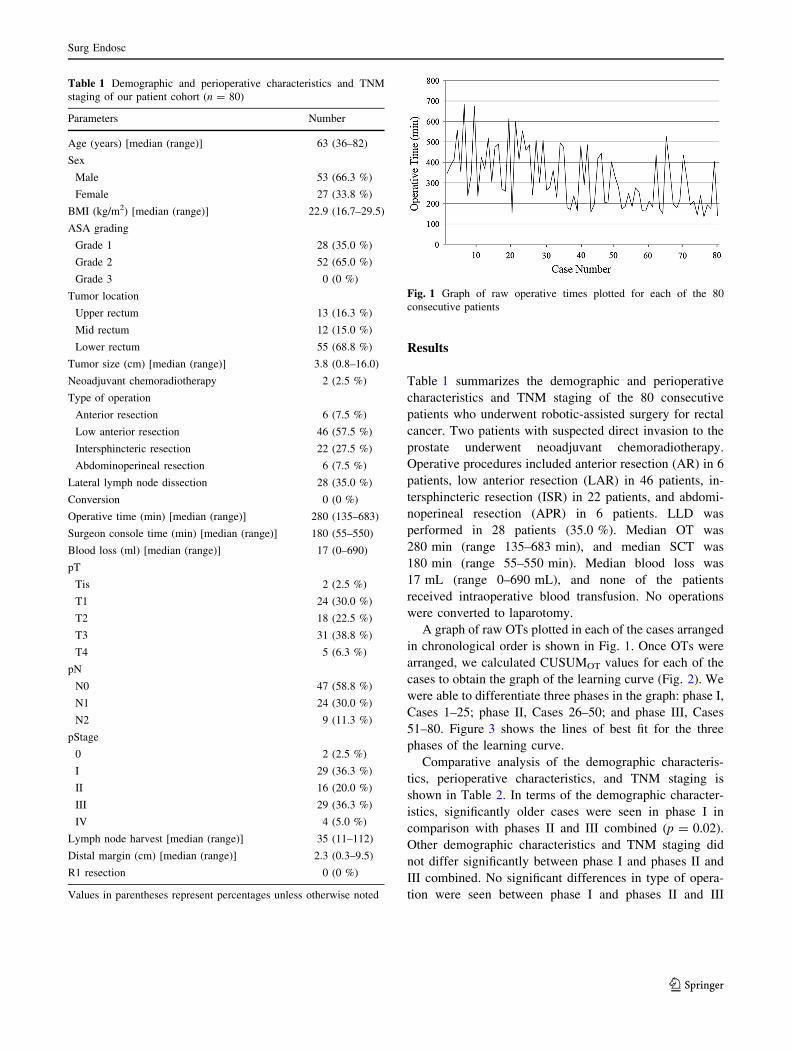
A graph of raw OTs plotted in each of the cases arranged
in chronological order is shown in Fig. 1. Once OTs were
arranged, we calculated CUSUMOT values for each of the
cases to obtain the graph of the learning curve (Fig. 2). We
were able to differentiate three phases in the graph: phase I,
Cases 1–25; phase II, Cases 26–50; and phase III, Cases
51–80. Figure 3 shows the lines of best fit for the three
phases of the learning curve.
Comparative analysis of the demographic characteris-
tics, perioperative characteristics, and TNM staging is
shown in Table 2. In terms of the demographic character-
istics, significantly older cases were seen in phase I in
comparison with phases II and III combined (p = 0.02).
Other demographic characteristics and TNM staging did
not differ significantly between phase I and phases II and
III combined. No significant differences in type of opera-
tion were seen between phase I and phases II and III
Table 1 Demographic and perioperative characteristics and TNM
staging of our patient cohort (n = 80)
Parameters Number
Age (years) [median (range)] 63 (36–82)
Sex
Male 53 (66.3 %)
Female 27 (33.8 %)
BMI (kg/m2) [median (range)] 22.9 (16.7–29.5)
ASA grading
Grade 1 28 (35.0 %)
Grade 2 52 (65.0 %)
Grade 3 0 (0 %)
Tumor location
Upper rectum 13 (16.3 %)
Mid rectum 12 (15.0 %)
Lower rectum 55 (68.8 %)
Tumor size (cm) [median (range)] 3.8 (0.8–16.0)
Neoadjuvant chemoradiotherapy 2 (2.5 %)
Type of operation
Anterior resection 6 (7.5 %)
Low anterior resection 46 (57.5 %)
Intersphincteric resection 22 (27.5 %)
Abdominoperineal resection 6 (7.5 %)
Lateral lymph node dissection 28 (35.0 %)
Conversion 0 (0 %)
Operative time (min) [median (range)] 280 (135–683)
Surgeon console time (min) [median (range)] 180 (55–550)
Blood loss (ml) [median (range)] 17 (0–690)
pT
Tis 2 (2.5 %)
T1 24 (30.0 %)
T2 18 (22.5 %)
T3 31 (38.8 %)
T4 5 (6.3 %)
pN
N0 47 (58.8 %)
N1 24 (30.0 %)
N2 9 (11.3 %)
pStage
0 2 (2.5 %)
I 29 (36.3 %)
II 16 (20.0 %)
III 29 (36.3 %)
IV 4 (5.0 %)
Lymph node harvest [median (range)] 35 (11–112)
Distal margin (cm) [median (range)] 2.3 (0.3–9.5)
R1 resection 0 (0 %)
Values in parentheses represent percentages unless otherwise noted
Fig. 1 Graph of raw operative times plotted for each of the 80
consecutive patients
Surg Endosc
123
combined, but the proportion of LLD cases differed sig-
nificantly (p = 0.04). The OT of phases II and III com-
bined was significantly shorter than that of phase I
(p = 0.0001). Blood loss in phases II and III combined was
significantly less than that in phase I (p = 0.001).
Postoperative data are presented in Table 3. No cases
showed Clavien–Dindo classification of morbidity Grade
III or IV. The frequency of Clavien–Dindo classification of
morbidity Grade II–IV did not differ between phase I and
phases II and III combined. Postoperative hospital stay was
significantly shorter in phases II and III combined than in
phase I (p = 0.02).
Discussion
When adopting a new device like the da Vinci surgical
system, proficiency and competence must be achieved.
McCulloch et al. [25] suggested that to improve the stan-
dards of clinical research in surgery, learning curves and
variations in the techniques and quality of surgery must be
measured and controlled continuously. The CUSUM
technique has been employed by the medical profession to
analyze learning curves for surgical procedures since the
1970s [8, 9]. The CUSUM method has been used to eval-
uate a practitioner’s initial and continued successful per-
formance of procedures. The main advantages of this
approach are the independence from the sample size, the
effectiveness in detecting small shifts in the system, and
the ability to allow continuous analysis in time and rapid
evaluation of data. The CUSUM method has thus been
used as an indicator of satisfactory outcomes in relation to
the acquisition of clinical skills [11].
To date, only three studies in terms of robotic-assisted
surgery for rectal cancer have reported analysis of the
learning curve using the CUSUM method [11, 12, 14]. Two
of those three studies focused on rectal cancer surgery [11,
14], but the remaining study analyzed both benign and
malignant colorectal diseases [12]. Jimenez-Rodriguez
et al. [11] included 43 rectal cancer cases and analyzed the
learning curve using the CUSUM method. They concluded
that the learning curve for robotic-assisted surgery for
rectal cancer may be divided into an initial phase of skills
acquisition, a second phase of consolidation of the tech-
nique, and a third phase when the surgeon masters the
technique and deals with more complex cases. According
to their study, the estimated learning phase for rectal cancer
was achieved after 21–23 cases. Similarly, Sng et al. [14]
reported that at least three phases were present in the
learning curve of 197 robotic-assisted surgeries for rectal
cancer patients using the CUSUM method. The first phase,
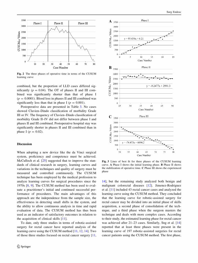
Fig. 2 The three phases of operative time in terms of the CUSUM
learning curve
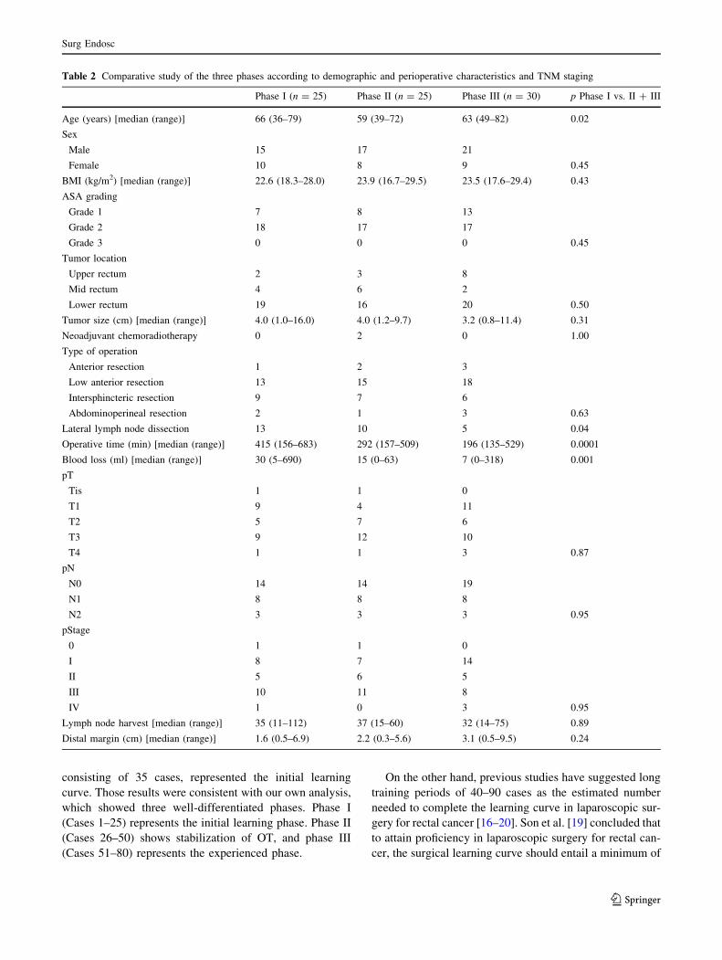
Fig. 3 Lines of best fit for three phases of the CUSUM learning
curve. A Phase I shows the initial learning phase. B Phase II shows
the stabilization of operative time. C Phase III shows the experienced
phase
Surg Endosc
123
consisting of 35 cases, represented the initial learning
curve. Those results were consistent with our own analysis,
which showed three well-differentiated phases. Phase I
(Cases 1–25) represents the initial learning phase. Phase II
(Cases 26–50) shows stabilization of OT, and phase III
(Cases 51–80) represents the experienced phase.
On the other hand, previous studies have suggested long
training periods of 40–90 cases as the estimated number
needed to complete the learning curve in laparoscopic sur-
gery for rectal cancer [16–20]. Son et al. [19] concluded that
to attain proficiency in laparoscopic surgery for rectal can-
cer, the surgical learning curve should entail a minimum of
Table 2 Comparative study of the three phases according to demographic and perioperative characteristics and TNM staging
Phase I (n = 25) Phase II (n = 25) Phase III (n = 30) p Phase I vs. II ? III
Age (years) [median (range)] 66 (36–79) 59 (39–72) 63 (49–82) 0.02
Sex
Male 15 17 21
Female 10 8 9 0.45
BMI (kg/m2) [median (range)] 22.6 (18.3–28.0) 23.9 (16.7–29.5) 23.5 (17.6–29.4) 0.43
ASA grading
Grade 1 7 8 13
Grade 2 18 17 17
Grade 3 0 0 0 0.45
Tumor location
Upper rectum 2 3 8
Mid rectum 4 6 2
Lower rectum 19 16 20 0.50
Tumor size (cm) [median (range)] 4.0 (1.0–16.0) 4.0 (1.2–9.7) 3.2 (0.8–11.4) 0.31
Neoadjuvant chemoradiotherapy 0 2 0 1.00
Type of operation
Anterior resection 1 2 3
Low anterior resection 13 15 18
Intersphincteric resection 9 7 6
Abdominoperineal resection 2 1 3 0.63
Lateral lymph node dissection 13 10 5 0.04
Operative time (min) [median (range)] 415 (156–683) 292 (157–509) 196 (135–529) 0.0001
Blood loss (ml) [median (range)] 30 (5–690) 15 (0–63) 7 (0–318) 0.001
pT
Tis 1 1 0
T1 9 4 11
T2 5 7 6
T3 9 12 10
T4 1 1 3 0.87
pN
N0 14 14 19
N1 8 8 8
N2 3 3 3 0.95
pStage
0 1 1 0
I 8 7 14
II 5 6 5
III 10 11 8
IV 1 0 3 0.95
Lymph node harvest [median (range)] 35 (11–112) 37 (15–60) 32 (14–75) 0.89
Distal margin (cm) [median (range)] 1.6 (0.5–6.9) 2.2 (0.3–5.6) 3.1 (0.5–9.5) 0.24
Surg Endosc
123
60–80 cases with multidimensional analysis using the
CUSUM method. Bege et al. [16] suggested that the learning
process for laparoscopic mesorectal excision affects the first
50 cases mostly heavily in terms of postoperative compli-
cations, but does not adversely affect oncological results
using the moving average and the CUSUM method. Lapa-
roscopic surgery for rectal cancer might be technically dif-
ficult when performing surgery in the narrow pelvic cavity.
These limitations include the use of straight rigid instruments
within a narrow working space, limited degrees of freedom,
an unstable camera platform with two-dimensional imaging,
and poor ergonomics of instruments. Robotic-assisted sur-
gery offers technical advantages such as high-quality three-
dimensional imaging, free-moving multi-joint forceps, and
image stabilization over laparoscopic surgery and thus may
shorten the learning curve compared with laparoscopic
surgery.
We believe that our series is the first to specifically ana-
lyze the learning curve for robotic-assisted surgery for rectal
cancer including LLD. A retrospective analysis conducted in
Japan reported that lateral lymph node metastasis was
present in 15.6–20.4 % of patients with lower rectal cancer
[21], so TME with LLD has been utilized as a standard
treatment for patients with lower rectal cancer, as described
by the JSCCR guidelines [22]. In terms of open mesorectal
excision with LLD, Fujita et al. [26] reported the short-term
results of mesorectal excision with and without LLD for
clinical stage II or III lower rectal cancer. OT was signifi-
cantly longer in the mesorectal excision with LLD group
(median, 360 min; interquartile range (IQR), 296–429 min)
than in the mesorectal excision alone group (median,
254 min; IQR, 210–307 min, p \ 0.0001). Reports of lap-
aroscopic TME with LLD are rare. Liang et al. [27] indicated
that laparoscopic TME with LLD was technically difficult to
perform, so laparoscopic LLD should be limited to some
selected patients. The main reasons why the procedure for
open or laparoscopic LLD remains difficult are that the
operative field of the pelvic side wall is small and the anat-
omy is complex. However, our case series included 28
patients (35.0 %) who underwent robotic-assisted LLD, and
the procedure was relatively safe and feasible because no
patients showed Clavien–Dindo classification of morbidity
Grades III–IV and no operations were converted to lapa-
rotomy. Robotic-assisted surgery offers technical advanta-
ges such as free-moving multi-joint forceps and precise
dissection can be achieved early on, so the initial learning
phase of our cases was similar to previous reports [11, 14].
Previous reports have described a trend of experienced
surgeons tending to include increasingly more difficult
cases toward the later part of their case series [11, 12, 14].
In contrast, our initial learning phase of 25 cases included
LLD in 13 cases (52.0 %), while phases II and III com-
bined consisted of 55 cases, including LLD in 15 cases
(27.3 %, p = 0.04). Our study thus differed from previous
reports in that our series included more frequently chal-
lenging cases in the earlier part of the series and we may
not encounter a phase including more complex cases in the
future. Our initial learning phase only comprised 25 cases;
therefore, we consider that the technical difficulties of each
case did not influence the learning curve.
Several limitations need to be considered for this study.
First, an expert laparoscopic surgeon (Y.K.) performed
robotic-assisted surgery. At the point of commencing
robotic-assisted surgery, he had performed over 500 lapa-
roscopic colorectal resections, including more than 200 for
rectal cancer. He was well-informed about pelvic anatomy
and laparoscopic technique. Our results therefore may not
be applicable to surgeons without this level of skill in
laparoscopic techniques. However, the shorter learning
curve may be attributed to the robotic system with
advanced technology, because we had little experience in
performing laparoscopic rectal cancer surgery with LLD at
the point of commencing robotic-assisted surgery. A sec-
ond limitation was that we did not analyze long-term
oncological and functional outcomes, such as voiding and
sexual functions. A randomized controlled trial is neces-
sary to establish the true benefits of robotic-assisted surgery
for rectal cancer compared with open and laparoscopic
surgery.
Table 3 Comparative study of the three phases according to com-
plications and postoperative hospital stay
Phase I
(n = 25)
Phase II
(n = 25)
Phase III
(n = 30)
p Phase I
vs.
II ? III
Clavien–Dindo classification
Grade 0–I 22 23 27
Grade II–IVa 3 2 3 0.70
Complicationsb
Wound infection 0 0 1 1.00
Small bowel
obstruction
0 0 0 –
Anastomotic leakage 0 0 0 –
Urinary retention 2 0 0 0.10
Urinary infection 0 1 0 1.00
Enteritis 0 1 1 1.00
Catheter-related
infection
1 0 0 0.31
Pneumonia 0 0 1 1.00
Postoperative hospital
stay (days) [median
(range)]
8 (7–12) 7 (6–12) 7 (6–15) 0.02
a There were no cases of Clavien–Dindo classification of morbidity
Grades III or IVb Clavien–Dindo classification of morbidity, Grade II–IV
Surg Endosc
123

Conclusions
Our study, using the CUSUM method, identified three
phases of the learning curve in robotic-assisted surgery for
rectal cancer. The data suggested that the initial 25 cases
formed the learning phase, the second 25 cases formed the
plateau phase, and the last 30 cases comprised the expe-
rienced phase. The learning curve for robotic-assisted
surgery may shorten that of laparoscopic surgery. These
results may impact the setting of ongoing and future trials.
Acknowledgments We wish to thank Dr. Keita Mori for his sta-
tistical help and writing assistance.
Disclosures Tomohiro Yamaguchi, Yusuke Kinugasa, Akio Shi-
omi, Sumito Sato, Yushi Yamakawa, Hiroyasu Kagawa, Hiroyuki
Tomioka, and Keita Mori have no conflicts of interest or financial ties
to disclose.
References
1. Miyajima N, Fukunaga M, Hasegawa H, Tanaka J, Okuda J,
Watanabe M (2009) Results of a multicenter study of 1,057 cases
of rectal cancer treated by laparoscopic surgery. Surg Endosc
23:113–118
2. Guillou PJ, Quirke P, Thorpe H, Walker J, Jayne DG, Smith AM,
Heath RM, Brown JM (2005) Short-term endpoints of conven-
tional versus laparoscopic-assisted surgery in patients with
colorectal cancer (MRC CLASICC trial): multicentre, random-
ised controlled trial. Lancet 365:1718–1726
3. Staudacher C, Vignali A (2010) Laparoscopic surgery for rectal
cancer: the state of the art. World J Gastrointest Surg 2:275–282
4. Weber PA, Merola S, Wasielewski A, Ballantyne GH (2002) Tele-
robotic-assisted laparoscopic right and sigmoid colectomies for benign
disease. Dis Colon Rectum 45:1689–1694 discussion 1695-1686
5. Pigazzi A, Ellenhorn JD, Ballantyne GH, Paz IB (2006) Robotic-
assisted laparoscopic LAR with TME for rectal cancer. Surg
Endosc 20:1521–1525
6. Corcione F, Esposito C, Cuccurullo D, Settembre A, Miranda N,
Amato F, Pirozzi F, Caiazzo P (2005) Advantages and limits of
robot-assisted laparoscopic surgery: preliminary experience. Surg
Endosc 19:117–119
7. Delaney CP, Lynch AC, Senagore AJ, Fazio VW (2003) Com-
parison of robotically performed and traditional laparoscopic
colorectal surgery. Dis Colon Rectum 46:1633–1639
8. de Chaput Saintonge DM, Vere DW (1974) Why don’t doctors
use cusums? Lancet 1:120–121
9. Wohl H (1977) The cusum plot: its utility in the analysis of
clinical data. N Engl J Med 296:1044–1045
10. Akmal Y, Baek JH, McKenzie S, Garcia-Aguilar J, Pigazzi A
(2012) Robot-assisted TME: is there a learning curve? Surg En-
dosc 26:2471–2476
11. Jimenez-Rodriguez RM, Diaz-Pavon JM, de la Portilla de Juan F,
Prendes-Sillero E, Dussort HC, Padillo J (2013) Learning curve
for robotic-assisted laparoscopic rectal cancer surgery. Int J
Colorectal Dis 28:815–821
12. Bokhari MB, Patel CB, Ramos-Valadez DI, Ragupathi M, Haas
EM (2011) Learning curve for robotic-assisted laparoscopic
colorectal surgery. Surg Endosc 25:855–860
13. Kim YW, Lee HM, Kim NK, Min BS, Lee KY (2012) The
learning curve for robot-assisted TME for rectal cancer. Surg
Laparosc Endosc Percutan Tech 22:400–405
14. Sng KK, Hara M, Shin JW, Yoo BE, Yang KS, Kim SH (2013)
The multiphasic learning curve for robot-assisted rectal surgery.
Surg Endosc 27:3297–3307
15. Park JS, Choi GS, Lim KH, Jang YS, Jun SH (2011) S052: a
comparison of robot-assisted, laparoscopic, and open surgery in
the treatment of rectal cancer. Surg Endosc 25:240–248
16. Bege T, Lelong B, Esterni B, Turrini O, Guiramand J, Francon D,
Mokart D, Houvenaeghel G, Giovannini M, Delpero JR (2010)
The learning curve for the laparoscopic approach to conservative
mesorectal excision for rectal cancer: lessons drawn from a single
institution’s experience. Ann Surg 251:249–253
17. Kayano H, Okuda J, Tanaka K, Kondo K, Tanigawa N (2011)
Evaluation of the learning curve in laparoscopic LAR for rectal
cancer. Surg Endosc 25:2972–2979
18. Park IJ, Choi GS, Lim KH, Kang BM, Jun SH (2009) Multidi-
mensional analysis of the learning curve for laparoscopic resec-
tion in rectal cancer. J Gastrointest Surg 13:275–281
19. Son GM, Kim JG, Lee JC, Suh YJ, Cho HM, Lee YS, Lee IK,
Chun CS (2010) Multidimensional analysis of the learning curve
for laparoscopic rectal cancer surgery. J Laparoendosc Adv Surg
Tech A 20:609–617
20. Ito M, Sugito M, Kobayashi A, Nishizawa Y, Tsunoda Y, Saito N
(2009) Influence of learning curve on short-term results after
laparoscopic resection for rectal cancer. Surg Endosc 23:403–408
21. Sugihara K, Kobayashi H, Kato T, Mori T, Mochizuki H, Kam-
eoka S, Shirouzu K, Muto T (2006) Indication and benefit of
pelvic sidewall dissection for rectal cancer. Dis Colon Rectum
49:1663–1672
22. Watanabe T, Itabashi M, Shimada Y, Tanaka S, Ito Y, Ajioka Y,
Hamaguchi T, Hyodo I, Igarashi M, Ishida H, Ishiguro M,
Kanemitsu Y, Kokudo N, Muro K, Ochiai A, Oguchi M, Ohkura
Y, Saito Y, Sakai Y, Ueno H, Yoshino T, Fujimori T, Koinuma
N, Morita T, Nishimura G, Sakata Y, Takahashi K, Takiuchi H,
Tsuruta O, Yamaguchi T, Yoshida M, Yamaguchi N, Kotake K,
Sugihara K (2012) Japanese Society for Cancer of the Colon and
Rectum (JSCCR) guidelines 2010 for the treatment of colorectal
cancer. Int J Clin Oncol 17:1–29
23. Sobin LH, Wittekind C (2009) TNM classification of malignant
tumours, 7th edn. Wiley-Liss, New York
24. Kinugasa Y, Murakami G, Suzuki D, Sugihara K (2007) Histo-
logical identification of fascial structures posterolateral to the
rectum. Br J Surg 94:620–626
25. McCulloch P, Taylor I, Sasako M, Lovett B, Griffin D (2002)
Randomised trials in surgery: problems and possible solutions.
BMJ 324:1448–1451
26. Fujita S, Akasu T, Mizusawa J, Saito N, Kinugasa Y, Kanemitsu
Y, Ohue M, Fujii S, Shiozawa M, Yamaguchi T, Moriya Y (2012)
Postoperative morbidity and mortality after mesorectal excision
with and without LLD for clinical stage II or stage III lower rectal
cancer (JCOG0212): results from a multicentre, randomised
controlled, non-inferiority trial. Lancet Oncol 13:616–621
27. Liang JT (2011) Technical feasibility of laparoscopic lateral pelvic
lymph node dissection for patients with low rectal cancer after
concurrent chemoradiation therapy. Ann Surg Oncol 18:153–159
Surg Endosc
123


![The Short-Term Results of Da Vinci Robotic Rectal …colorectal surgery [6,7]. The first laparoscopic rectal resection performed in 1990y. by Patrick Leahy [6-8]. In 2006 y. the Italian](https://img.pdfslide.net/doc/110x75/5f3ea8294ffad923680a225a/the-short-term-results-of-da-vinci-robotic-rectal-colorectal-surgery-67-the.jpg)