Embed Size (px)
Citation preview
Melanoma Management
part of
171ISSN 2045-0885Melanoma Manag. (2015) 2(2), 171–178 10.2217/MMT.15.3 © Future Medicine Ltd.
REVIEW
Lentigo maligna and lentigo maligna melanoma: contemporary issues in diagnosis and management
Karen L Connolly*,1, Kishwer S Nehal1 & Klaus J Busam2
1Dermatology Service, Memorial Sloan Kettering Cancer Center, New York, NY, USA 2Department of Pathology, Memorial Sloan Kettering Cancer Center, New York, NY, USA
*Author for correspondence: [email protected]
SUMMARY Lentigo maligna and lentigo maligna melanomas present diagnostic and treatment dilemmas due to their frequent presence within a background of sun-damaged skin, and their location on cosmetically and functionally sensitive areas. As the incidence of this entity is increasing, diagnostic and management controversies have developed. While surgery remains the gold standard of treatment, nonsurgical treatment options are also emerging for both adjunctive and primary therapy.
KEYWORDS • facial melanoma • lentigo maligna • lentigo maligna melanoma • melanoma • melanoma in situ
EpidemiologyLentigo maligna (LM), first described by Hutchinson in 1890, is the noninvasive counterpart to lentigo maligna melanoma (LMM). The latter (LMM) refers to invasive melanoma associated with a LM. LM and LMM occur on chronically sun-damaged skin, most commonly on the head and neck. Overall, LMM accounts for 5–15% of cutaneous melanomas [1–4]. As an increased incidence has been recently noted, controversies over diagnosis and appropriate management of this often challenging melanocytic neoplasm have emerged. LM is unique among melanomas, in that the natural history of this lesion is that of a slow-growing, more indolent tumor, often present for years preceding diagnosis. Even long-standing lesions often lack an invasive component, and LM has among the best 5-year survival rates among melanoma subtypes, estimated at 97.2% [5]. That said, once invasive, LMM can behave aggressively, with risk of metastasis [2]. Overall, the LM subtype accounts for about 5–10% of melanomas and 10–26% of head and neck melanomas, accounting for a larger percentage of melanomas occurring in patients over the age of 65 years [1–2,5–6].
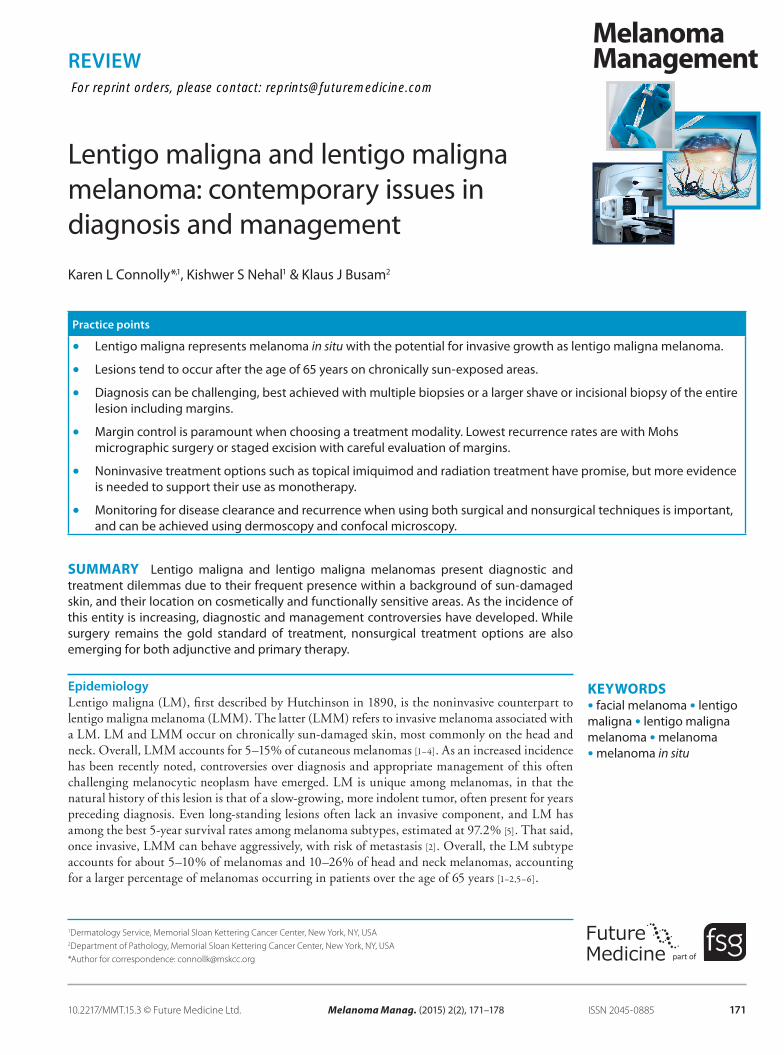
Practice points
● Lentigo maligna represents melanoma in situ with the potential for invasive growth as lentigo maligna melanoma.
● Lesions tend to occur after the age of 65 years on chronically sun-exposed areas.
● Diagnosis can be challenging, best achieved with multiple biopsies or a larger shave or incisional biopsy of the entire lesion including margins.
● Margin control is paramount when choosing a treatment modality. Lowest recurrence rates are with Mohs micrographic surgery or staged excision with careful evaluation of margins.
● Noninvasive treatment options such as topical imiquimod and radiation treatment have promise, but more evidence is needed to support their use as monotherapy.
● Monitoring for disease clearance and recurrence when using both surgical and nonsurgical techniques is important, and can be achieved using dermoscopy and confocal microscopy.
For reprint orders, please contact: [email protected]
Melanoma Manag. (2015) 2(2)172
REviEW Connolly, Nehal & Busam
future science group
Worldwide incidence of LM and LMM is increasing, demonstrated by recent reports from the USA, Australia and Denmark [1,3–4,7]. The mean age at diagnosis is 70 years [7]. Risk factors for the development of LM include his-tory of sun exposure, light skin and propensity toward development of lentigines. LM is most commonly found on the cheek (estimated at 26–48% of lesions) [7–9]. Unlike superficial spreading melanoma, LMM is more strongly associated with previous development of lentigi-nes and skin cancer history, and is not associ-ated with pre-existing nevi or propensity toward development of nevi [10,11]. Ultraviolet radiation appears to play a different role in pathogenesis than in other subtypes of melanoma, in that chronic rather than intermittent sun exposure appears to increase risk of LM [1].
Unlike other subtypes of melanoma such as superficial spreading, LMM rarely harbors BRAF mutations [12]. When present, BRAF V600K mutations are more commonly seen than BRAF V600E mutations, consistent with the increase in BRAF V600K mutations in mela-nomas arising from chronically sun-damaged skin [13]. P53 mutations are also more commonly seen in the lentigo maligna subtype (along with other melanomas occurring in sun-damaged skin) [14].
DiagnosisClinically, LM occurs almost exclusively on actinically damaged skin. Initial appearance is often that of a subtle patch. One can see a range of colors from light brown/tan, darker brown, pink or black. Other features such as asymmetry, irregular borders and report of increasing size can be helpful. Nodularity can be appreciated once a lesion develops an invasive dermal com-ponent. Clinical differential diagnosis includes solar lentigo, pigmented actinic keratosis, lichen planus-like keratosis, pigmented basal cell carcinoma and seborrheic keratoses [15].
Histologic examination is the gold standard for diagnosis of LMM. While a complete exci-sional biopsy is ideal to examine the entirety of the lesion and determine prognostic infor-mation such as maximum Breslow depth, the large clinical size of many LM as well as most frequent location on the head and neck often precludes this technique. The authors prefer a long elliptical incisional biopsy involving a mar-gin of clinically normal-appearing skin as well as a wide sampling of the lesion. An alternative
method would be a long saucerization biopsy, again sampling clinically normal skin adjacent to the lesion [16]. Other described approaches are multiple punch biopsies from within the lesion; however, this approach lends itself to sampling error [17].
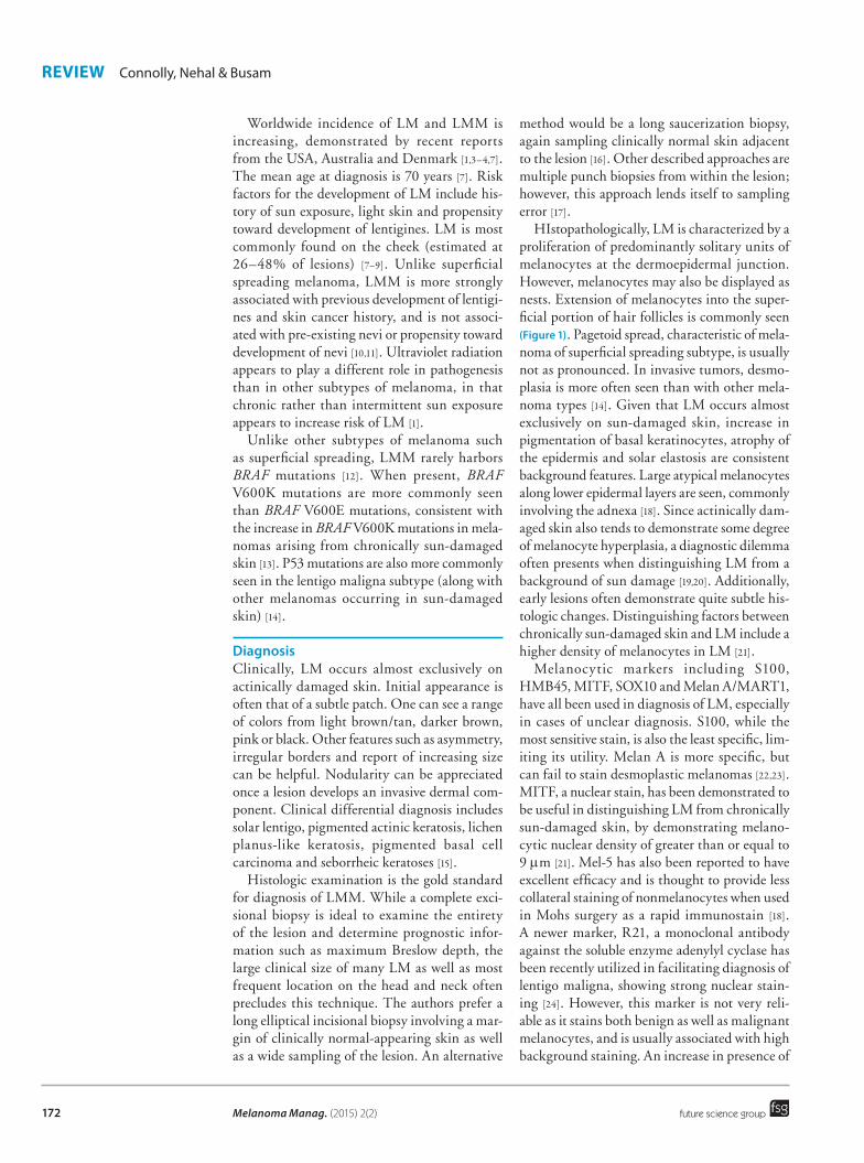
HIstopathologically, LM is characterized by a proliferation of predominantly solitary units of melanocytes at the dermoepidermal junction. However, melanocytes may also be displayed as nests. Extension of melanocytes into the super-ficial portion of hair follicles is commonly seen (Figure 1). Pagetoid spread, characteristic of mela-noma of superficial spreading subtype, is usually not as pronounced. In invasive tumors, desmo-plasia is more often seen than with other mela-noma types [14]. Given that LM occurs almost exclusively on sun-damaged skin, increase in pigmentation of basal keratinocytes, atrophy of the epidermis and solar elastosis are consistent background features. Large atypical melanocytes along lower epidermal layers are seen, commonly involving the adnexa [18]. Since actinically dam-aged skin also tends to demonstrate some degree of melanocyte hyperplasia, a diagnostic dilemma often presents when distinguishing LM from a background of sun damage [19,20]. Additionally, early lesions often demonstrate quite subtle his-tologic changes. Distinguishing factors between chronically sun-damaged skin and LM include a higher density of melanocytes in LM [21].
Melanocytic markers including S100, HMB45, MITF, SOX10 and Melan A/MART1, have all been used in diagnosis of LM, especially in cases of unclear diagnosis. S100, while the most sensitive stain, is also the least specific, lim-iting its utility. Melan A is more specific, but can fail to stain desmoplastic melanomas [22,23]. MITF, a nuclear stain, has been demonstrated to be useful in distinguishing LM from chronically sun-damaged skin, by demonstrating melano-cytic nuclear density of greater than or equal to 9 μm [21]. Mel-5 has also been reported to have excellent efficacy and is thought to provide less collateral staining of nonmelanocytes when used in Mohs surgery as a rapid immunostain [18]. A newer marker, R21, a monoclonal antibody against the soluble enzyme adenylyl cyclase has been recently utilized in facilitating diagnosis of lentigo maligna, showing strong nuclear stain-ing [24]. However, this marker is not very reli-able as it stains both benign as well as malignant melanocytes, and is usually associated with high background staining. An increase in presence of
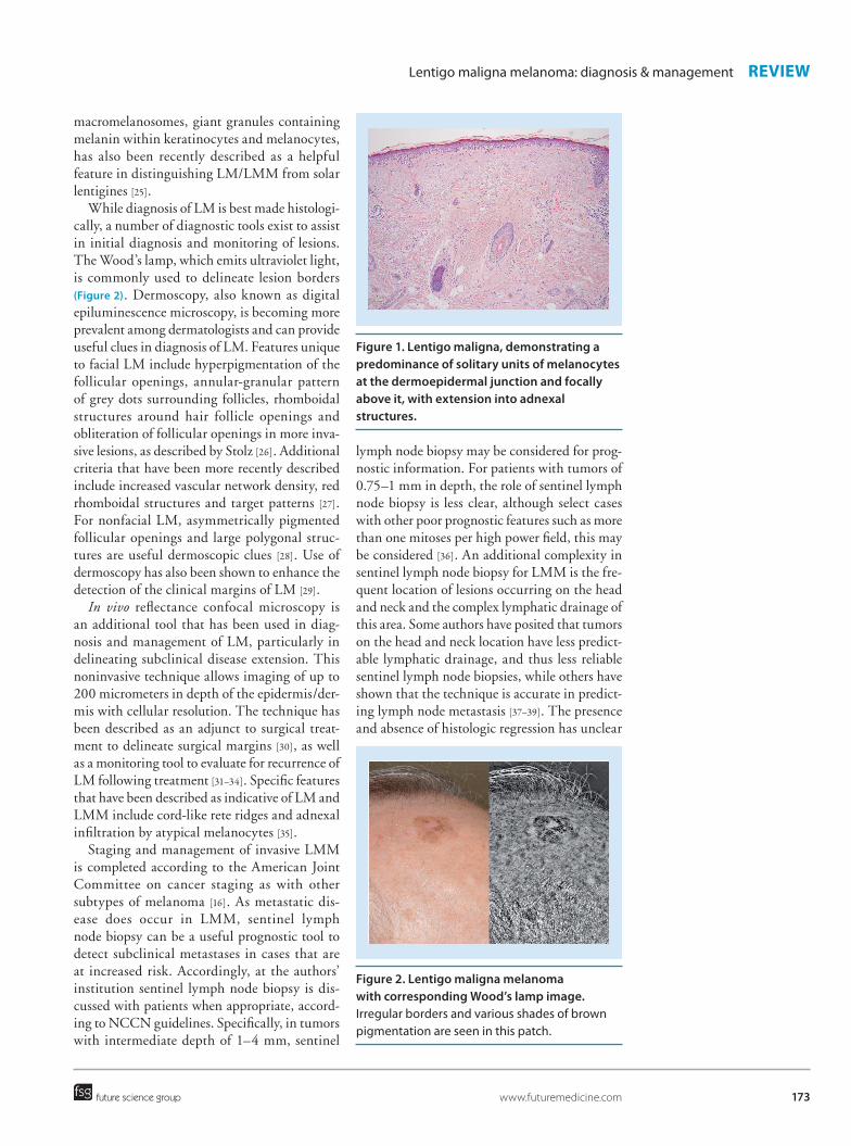
Figure 1. Lentigo maligna, demonstrating a predominance of solitary units of melanocytes at the dermoepidermal junction and focally above it, with extension into adnexal structures.
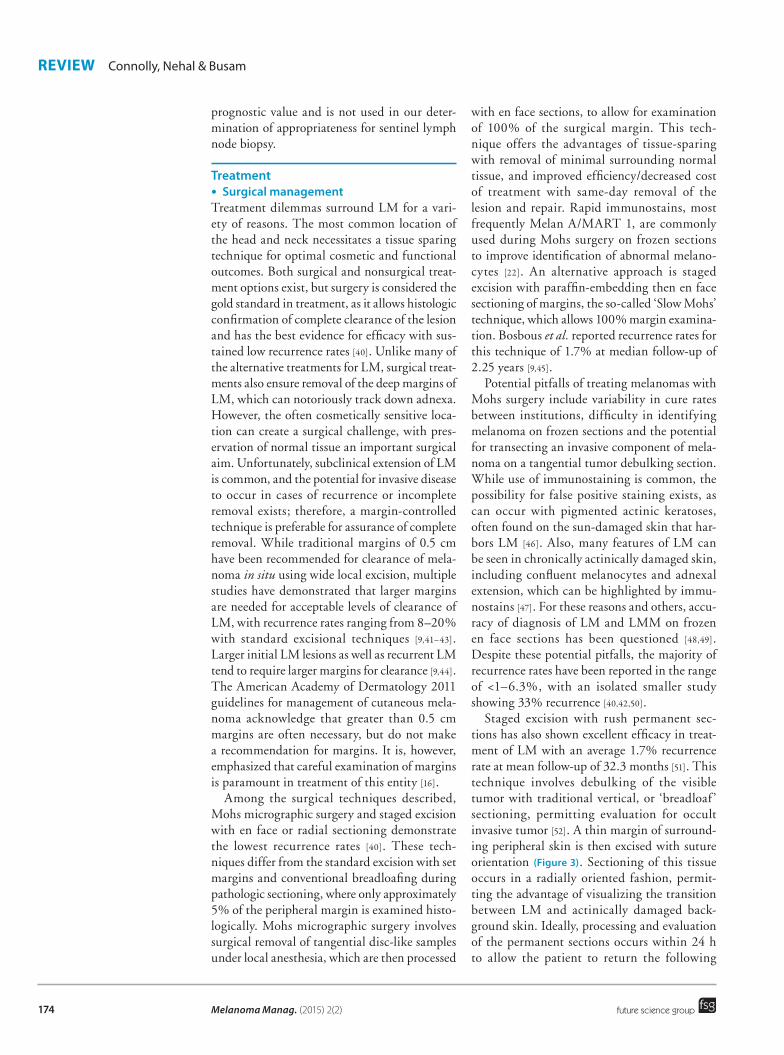
Figure 2. Lentigo maligna melanoma with corresponding Wood’s lamp image. Irregular borders and various shades of brown pigmentation are seen in this patch.
173
Lentigo maligna melanoma: diagnosis & management REviEW
future science group www.futuremedicine.com
macromelanosomes, giant granules containing melanin within keratinocytes and melanocytes, has also been recently described as a helpful feature in distinguishing LM/LMM from solar lentigines [25].
While diagnosis of LM is best made histologi-cally, a number of diagnostic tools exist to assist in initial diagnosis and monitoring of lesions. The Wood’s lamp, which emits ultraviolet light, is commonly used to delineate lesion borders (Figure 2). Dermoscopy, also known as digital epiluminescence microscopy, is becoming more prevalent among dermatologists and can provide useful clues in diagnosis of LM. Features unique to facial LM include hyperpigmentation of the follicular openings, annular-granular pattern of grey dots surrounding follicles, rhomboidal structures around hair follicle openings and obliteration of follicular openings in more inva-sive lesions, as described by Stolz [26]. Additional criteria that have been more recently described include increased vascular network density, red rhomboidal structures and target patterns [27]. For nonfacial LM, asymmetrically pigmented follicular openings and large polygonal struc-tures are useful dermoscopic clues [28]. Use of dermoscopy has also been shown to enhance the detection of the clinical margins of LM [29].
In vivo reflectance confocal microscopy is an additional tool that has been used in diag-nosis and management of LM, particularly in delineating subclinical disease extension. This noninvasive technique allows imaging of up to 200 micrometers in depth of the epidermis/der-mis with cellular resolution. The technique has been described as an adjunct to surgical treat-ment to delineate surgical margins [30], as well as a monitoring tool to evaluate for recurrence of LM following treatment [31–34]. Specific features that have been described as indicative of LM and LMM include cord-like rete ridges and adnexal infiltration by atypical melanocytes [35].
Staging and management of invasive LMM is completed according to the American Joint Committee on cancer staging as with other subtypes of melanoma [16]. As metastatic dis-ease does occur in LMM, sentinel lymph node biopsy can be a useful prognostic tool to detect subclinical metastases in cases that are at increased risk. Accordingly, at the authors’ institution sentinel lymph node biopsy is dis-cussed with patients when appropriate, accord-ing to NCCN guidelines. Specifically, in tumors with intermediate depth of 1–4 mm, sentinel
lymph node biopsy may be considered for prog-nostic information. For patients with tumors of 0.75–1 mm in depth, the role of sentinel lymph node biopsy is less clear, although select cases with other poor prognostic features such as more than one mitoses per high power field, this may be considered [36]. An additional complexity in sentinel lymph node biopsy for LMM is the fre-quent location of lesions occurring on the head and neck and the complex lymphatic drainage of this area. Some authors have posited that tumors on the head and neck location have less predict-able lymphatic drainage, and thus less reliable sentinel lymph node biopsies, while others have shown that the technique is accurate in predict-ing lymph node metastasis [37–39]. The presence and absence of histologic regression has unclear
Melanoma Manag. (2015) 2(2)174
REviEW Connolly, Nehal & Busam
future science group
prognostic value and is not used in our deter-mination of appropriateness for sentinel lymph node biopsy.
Treatment●● Surgical management
Treatment dilemmas surround LM for a vari-ety of reasons. The most common location of the head and neck necessitates a tissue sparing technique for optimal cosmetic and functional outcomes. Both surgical and nonsurgical treat-ment options exist, but surgery is considered the gold standard in treatment, as it allows histologic confirmation of complete clearance of the lesion and has the best evidence for efficacy with sus-tained low recurrence rates [40]. Unlike many of the alternative treatments for LM, surgical treat-ments also ensure removal of the deep margins of LM, which can notoriously track down adnexa. However, the often cosmetically sensitive loca-tion can create a surgical challenge, with pres-ervation of normal tissue an important surgical aim. Unfortunately, subclinical extension of LM is common, and the potential for invasive disease to occur in cases of recurrence or incomplete removal exists; therefore, a margin-controlled technique is preferable for assurance of complete removal. While traditional margins of 0.5 cm have been recommended for clearance of mela-noma in situ using wide local excision, multiple studies have demonstrated that larger margins are needed for acceptable levels of clearance of LM, with recurrence rates ranging from 8–20% with standard excisional techniques [9,41–43]. Larger initial LM lesions as well as recurrent LM tend to require larger margins for clearance [9,44]. The American Academy of Dermatology 2011 guidelines for management of cutaneous mela-noma acknowledge that greater than 0.5 cm margins are often necessary, but do not make a recommendation for margins. It is, however, emphasized that careful examination of margins is paramount in treatment of this entity [16].
Among the surgical techniques described, Mohs micrographic surgery and staged excision with en face or radial sectioning demonstrate the lowest recurrence rates [40]. These tech-niques differ from the standard excision with set margins and conventional breadloafing during pathologic sectioning, where only approximately 5% of the peripheral margin is examined histo-logically. Mohs micrographic surgery involves surgical removal of tangential disc-like samples under local anesthesia, which are then processed
with en face sections, to allow for examination of 100% of the surgical margin. This tech-nique offers the advantages of tissue-sparing with removal of minimal surrounding normal tissue, and improved efficiency/decreased cost of treatment with same-day removal of the lesion and repair. Rapid immunostains, most frequently Melan A/MART 1, are commonly used during Mohs surgery on frozen sections to improve identification of abnormal melano-cytes [22]. An alternative approach is staged excision with paraffin-embedding then en face sectioning of margins, the so-called ‘Slow Mohs’ technique, which allows 100% margin examina-tion. Bosbous et al. reported recurrence rates for this technique of 1.7% at median follow-up of 2.25 years [9,45].
Potential pitfalls of treating melanomas with Mohs surgery include variability in cure rates between institutions, difficulty in identifying melanoma on frozen sections and the potential for transecting an invasive component of mela-noma on a tangential tumor debulking section. While use of immunostaining is common, the possibility for false positive staining exists, as can occur with pigmented actinic keratoses, often found on the sun-damaged skin that har-bors LM [46]. Also, many features of LM can be seen in chronically actinically damaged skin, including confluent melanocytes and adnexal extension, which can be highlighted by immu-nostains [47]. For these reasons and others, accu-racy of diagnosis of LM and LMM on frozen en face sections has been questioned [48,49]. Despite these potential pitfalls, the majority of recurrence rates have been reported in the range of <1–6.3%, with an isolated smaller study showing 33% recurrence [40,42,50].
Staged excision with rush permanent sec-tions has also shown excellent efficacy in treat-ment of LM with an average 1.7% recurrence rate at mean follow-up of 32.3 months [51]. This technique involves debulking of the visible tumor with traditional vertical, or ‘breadloaf ’ sectioning, permitting evaluation for occult invasive tumor [52]. A thin margin of surround-ing peripheral skin is then excised with suture orientation (Figure 3). Sectioning of this tissue occurs in a radially oriented fashion, permit-ting the advantage of visualizing the transition between LM and actinically damaged back-ground skin. Ideally, processing and evaluation of the permanent sections occurs within 24 h to allow the patient to return the following
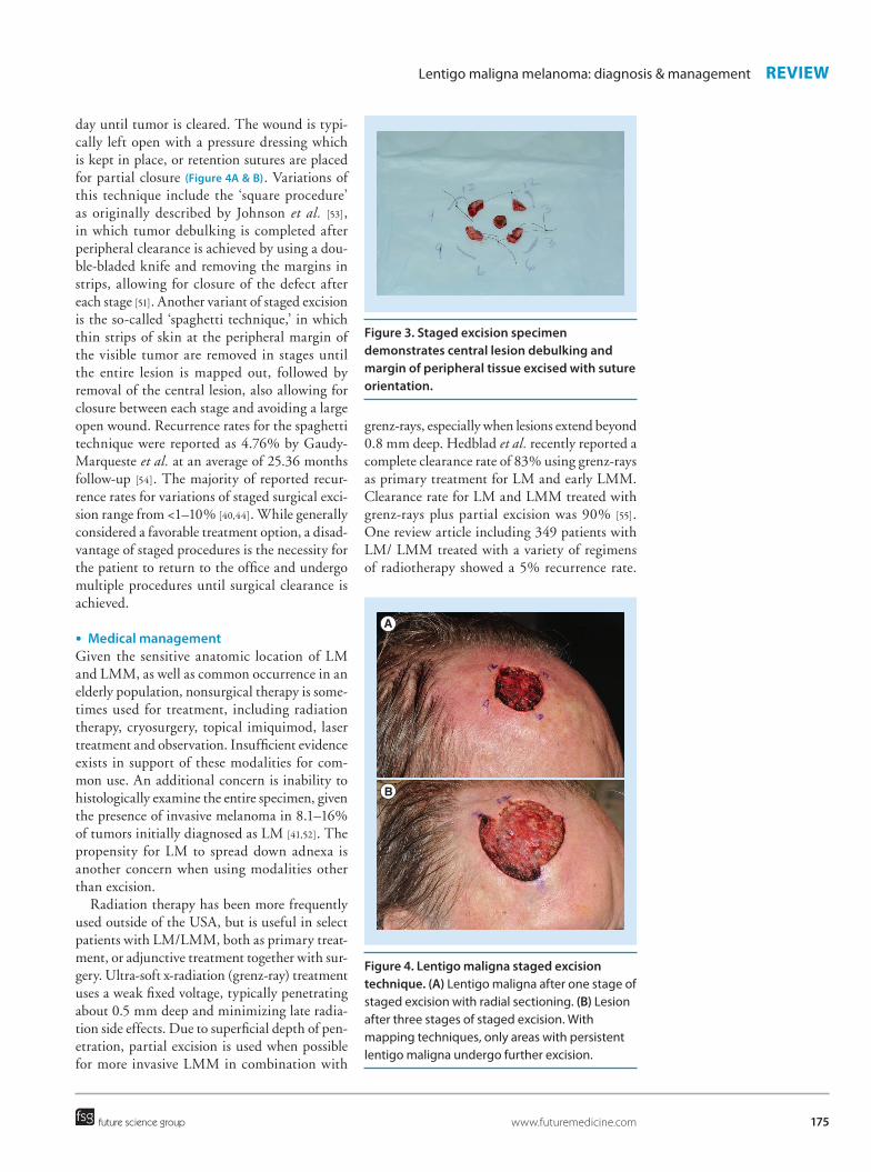
Figure 3. Staged excision specimen demonstrates central lesion debulking and margin of peripheral tissue excised with suture orientation.
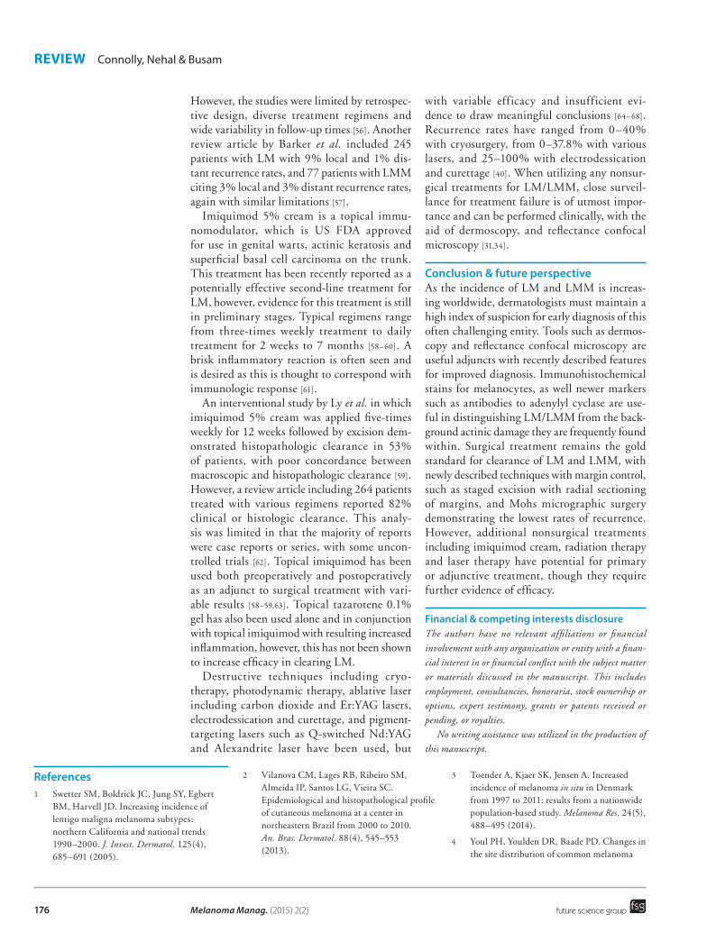
Figure 4. Lentigo maligna staged excision technique. (A) Lentigo maligna after one stage of staged excision with radial sectioning. (B) Lesion after three stages of staged excision. With mapping techniques, only areas with persistent lentigo maligna undergo further excision.
A
B
175
Lentigo maligna melanoma: diagnosis & management REviEW
future science group www.futuremedicine.com
day until tumor is cleared. The wound is typi-cally left open with a pressure dressing which is kept in place, or retention sutures are placed for partial closure (Figure 4A & B). Variations of this technique include the ‘square procedure’ as originally described by Johnson et al. [53], in which tumor debulking is completed after peripheral clearance is achieved by using a dou-ble-bladed knife and removing the margins in strips, allowing for closure of the defect after each stage [51]. Another variant of staged excision is the so-called ‘spaghetti technique,’ in which thin strips of skin at the peripheral margin of the visible tumor are removed in stages until the entire lesion is mapped out, followed by removal of the central lesion, also allowing for closure between each stage and avoiding a large open wound. Recurrence rates for the spaghetti technique were reported as 4.76% by Gaudy-Marqueste et al. at an average of 25.36 months follow-up [54]. The majority of reported recur-rence rates for variations of staged surgical exci-sion range from <1–10% [40,44]. While generally considered a favorable treatment option, a disad-vantage of staged procedures is the necessity for the patient to return to the office and undergo multiple procedures until surgical clearance is achieved.
●● Medical managementGiven the sensitive anatomic location of LM and LMM, as well as common occurrence in an elderly population, nonsurgical therapy is some-times used for treatment, including radiation therapy, cryosurgery, topical imiquimod, laser treatment and observation. Insufficient evidence exists in support of these modalities for com-mon use. An additional concern is inability to histologically examine the entire specimen, given the presence of invasive melanoma in 8.1–16% of tumors initially diagnosed as LM [41,52]. The propensity for LM to spread down adnexa is another concern when using modalities other than excision.
Radiation therapy has been more frequently used outside of the USA, but is useful in select patients with LM/LMM, both as primary treat-ment, or adjunctive treatment together with sur-gery. Ultra-soft x-radiation (grenz-ray) treatment uses a weak fixed voltage, typically penetrating about 0.5 mm deep and minimizing late radia-tion side effects. Due to superficial depth of pen-etration, partial excision is used when possible for more invasive LMM in combination with
grenz-rays, especially when lesions extend beyond 0.8 mm deep. Hedblad et al. recently reported a complete clearance rate of 83% using grenz-rays as primary treatment for LM and early LMM. Clearance rate for LM and LMM treated with grenz-rays plus partial excision was 90% [55]. One review article including 349 patients with LM/ LMM treated with a variety of regimens of radiotherapy showed a 5% recurrence rate.
Melanoma Manag. (2015) 2(2)176
REviEW Connolly, Nehal & Busam
future science group
However, the studies were limited by retrospec-tive design, diverse treatment regimens and wide variability in follow-up times [56]. Another review article by Barker et al. included 245 patients with LM with 9% local and 1% dis-tant recurrence rates, and 77 patients with LMM citing 3% local and 3% distant recurrence rates, again with similar limitations [57].
Imiquimod 5% cream is a topical immu-nomodulator, which is US FDA approved for use in genital warts, actinic keratosis and superficial basal cell carcinoma on the trunk. This treatment has been recently reported as a potentially effective second-line treatment for LM, however, evidence for this treatment is still in preliminary stages. Typical regimens range from three-times weekly treatment to daily treatment for 2 weeks to 7 months [58–60]. A brisk inflammatory reaction is often seen and is desired as this is thought to correspond with immunologic response [61].
An interventional study by Ly et al. in which imiquimod 5% cream was applied five-times weekly for 12 weeks followed by excision dem-onstrated histopathologic clearance in 53% of patients, with poor concordance between macroscopic and histopathologic clearance [59]. However, a review article including 264 patients treated with various regimens reported 82% clinical or histologic clearance. This analy-sis was limited in that the majority of reports were case reports or series, with some uncon-trolled trials [62]. Topical imiquimod has been used both preoperatively and postoperatively as an adjunct to surgical treatment with vari-able results [58–59,63]. Topical tazarotene 0.1% gel has also been used alone and in conjunction with topical imiquimod with resulting increased inflammation, however, this has not been shown to increase efficacy in clearing LM.
Destructive techniques including cryo-therapy, photodynamic therapy, ablative laser including carbon dioxide and Er:YAG lasers, electrodessication and curettage, and pigment-targeting lasers such as Q-switched Nd:YAG and Alexandrite laser have been used, but
with variable efficacy and insufficient evi-dence to draw meaningful conclusions [64–68]. Recurrence rates have ranged from 0–40% with cryosurgery, from 0–37.8% with various lasers, and 25–100% with electrodessication and curettage [40]. When utilizing any nonsur-gical treatments for LM/LMM, close surveil-lance for treatment failure is of utmost impor-tance and can be performed clinically, with the aid of dermoscopy, and reflectance confocal microscopy [31,34].
Conclusion & future perspectiveAs the incidence of LM and LMM is increas-ing worldwide, dermatologists must maintain a high index of suspicion for early diagnosis of this often challenging entity. Tools such as dermos-copy and reflectance confocal microscopy are useful adjuncts with recently described features for improved diagnosis. Immunohistochemical stains for melanocytes, as well newer markers such as antibodies to adenylyl cyclase are use-ful in distinguishing LM/LMM from the back-ground actinic damage they are frequently found within. Surgical treatment remains the gold standard for clearance of LM and LMM, with newly described techniques with margin control, such as staged excision with radial sectioning of margins, and Mohs micrographic surgery demonstrating the lowest rates of recurrence. However, additional nonsurgical treatments including imiquimod cream, radiation therapy and laser therapy have potential for primary or adjunctive treatment, though they require further evidence of efficacy.
Financial & competing interests disclosureThe authors have no relevant affiliations or financial involvement with any organization or entity with a finan-cial interest in or financial conflict with the subject matter or materials discussed in the manuscript. This includes employment, consultancies, honoraria, stock ownership or options, expert testimony, grants or patents received or pending, or royalties.
No writing assistance was utilized in the production of this manuscript.
References1 Swetter SM, Boldrick JC, Jung SY, Egbert
BM, Harvell JD. Increasing incidence of lentigo maligna melanoma subtypes: northern California and national trends 1990–2000. J. Invest. Dermatol. 125(4), 685–691 (2005).
2 Vilanova CM, Lages RB, Ribeiro SM, Almeida IP, Santos LG, Vieira SC. Epidemiological and histopathological profile of cutaneous melanoma at a center in northeastern Brazil from 2000 to 2010. An. Bras. Dermatol. 88(4), 545–553 (2013).
3 Toender A, Kjaer SK, Jensen A. Increased incidence of melanoma in situ in Denmark from 1997 to 2011: results from a nationwide population-based study. Melanoma Res. 24(5), 488–495 (2014).
4 Youl PH, Youlden DR, Baade PD. Changes in the site distribution of common melanoma
177
Lentigo maligna melanoma: diagnosis & management REviEW
future science group www.futuremedicine.com
subtypes in Queensland, Australia over time: implications for public health campaigns. Br. J. Dermatol. 168(1), 136–144 (2013).
5 Wu XC, Eide MJ, King J et al. Racial and ethnic variations in incidence and survival of cutaneous melanoma in the United States, 1999–2006. J. Am. Acad. Dermatol. 65(5 Suppl. 1), S26–S37 (2011).
6 Ciocan D, Barbe C, Aubin F et al. Distinctive features of melanoma and its management in elderly patients: a population-based study in France. JAMA Dermatol. 149(10), 1150–1157 (2013).
7 Mirzoyev SA, Knudson RM, Reed KB et al. Incidence of lentigo maligna in Olmsted County, Minnesota, 1970 to 2007. J. Am. Acad. Dermatol. 70(3), 443–448 (2014).
8 Lesage C, Barbe C, Le Clainche A, Lesage FX, Bernard P, Grange F. Sex-related location of head and neck melanoma strongly argues for a major role of sun exposure in cars and photoprotection by hair. J. Invest. Dermatol. 133(5), 1205–1211 (2013).
9 Hilari H, Llorca D, Traves V et al. Conventional surgery compared with slow Mohs micrographic surgery in the treatment of lentigo maligna: a retrospective study of 62 cases. Actas Dermosifiliogr. 103(7), 614–623 (2012).
10 Gaudy-Marqueste C, Madjlessi N, Guillot B, Avril MF, Grob JJ. Risk factors in elderly people for lentigo maligna compared with other melanomas: a double case–control study. Arch. Dermatol. 145(4), 418–423 (2009).
11 Kvaskoff M, Siskind V, Green AC. Risk factors for lentigo maligna melanoma compared with superficial spreading melanoma: a case–control study in Australia. Arch. Dermatol. 148(2), 164–170 (2012).
12 Lee JH, Choi JW, Kim YS. Frequencies of BRAF and NRAS mutations are different in histological types and sites of origin of cutaneous melanoma: a meta-analysis. Br. J. Dermatol. 164(4), 776–784 (2011).
13 Stadelmeyer E, Heitzer E, Resel M, Cerroni L, Wolf P, Dandachi N. The BRAF V600K mutation is more frequent than the BRAF V600E mutation in melanoma in situ of lentigo maligna type. J. Invest. Dermatol. 134(2), 548–550 (2014).
14 Kraft S, Granter SR. Molecular pathology of skin neoplasms of the head and neck. Arch. Pathol. Lab. Med. 138(6), 759–787 (2014).
15 Mcguire LK, Disa JJ, Lee EH, Busam KJ, Nehal KS. Melanoma of the lentigo maligna subtype: diagnostic challenges and current treatment paradigms. Plast. Reconstr.
Surg. 129(2), e288–e299 (2012).
16 Bichakjian CK, Halpern AC, Johnson TM et al. Guidelines of care for the management of primary cutaneous melanoma. American academy of dermatology. J. Am. Acad. Dermatol. 65(5), 1032–1047 (2011).
17 Mckenna JK, Florell SR, Goldman GD, Bowen GM. Lentigo maligna/lentigo maligna melanoma: current state of diagnosis and treatment. Dermatol. Surg. 32(4), 493–504 (2006).
18 Newman J, Beal M, Schram SE, Lee PK. Mohs micrographic surgery for lentigo maligna and lentigo maligna melanoma using Mel-5 immunostaining: an update from the University of Minnesota. Dermatol. Surg. 39(12), 1794–1799 (2013).
19 Hendi A, Wada DA, Jacobs MA et al. Melanocytes in non lesional sun-exposed skin: a multicenter comparative study. J. Am. Acad. Dermatol. 65(6), 1186–1193 (2011).
20 Reed JA, Shea CR. Lentigo maligna: melanoma in situ on chronically sun-damaged skin. Arch. Pathol. Lab. Med. 135(7), 838–841 (2011).
21 Black WH, Thareja SK, Blake BP, Chen R, Cherpelis BS, Glass LF. Distinction of melanoma in situ from solar lentigo on sun-damaged skin using morphometrics and MITF immunohistochemistry. Am. J. Dermatopathol. 33(6), 573–578 (2011).
22 El Tal AK, Abrou AE, Stiff MA, Mehregan DA. Immunostaining in Mohs micrographic surgery: a review. Dermatol. Surg. 36(3), 275–290 (2010).
23 Suchak R, Hameed OA, Robson A. Evaluation of the role of routine melan-A immunohistochemistry for exclusion of microinvasion in 120 cases of lentigo maligna. Am. J. Dermatopathol. 36(5), 387–391 (2014).
24 Magro CM, Yang SE, Zippin JH, Zembowicz A. Expression of soluble adenylyl cyclase in lentigo maligna: use of immunohistochemistry with anti-soluble adenylyl cyclase antibody (R21) in diagnosis of lentigo maligna and assessment of margins. Arch. Pathol. Lab. Med. 136(12), 1558–1564 (2012).
25 Sethi M, Craythorne E, Al-Arashi MY, Bhawan J, Stefanato CM. Macromelanosomes: their significantly greater presence in the margins of a lentigo maligna versus solar lentigo. Am. J. Dermatopathol. 36(6), 490–492 (2014).
26 Stolz W, Schiffner R, Burgdorf WH. Dermatoscopy for facial pigmented skin lesions. Clin. Dermatol. 20(3), 276–278 (2002).
27 Pralong P, Bathelier E, Dalle S, Poulalhon N,
Debarbieux S, Thomas L. Dermoscopy of lentigo maligna melanoma: report of 125 cases. Br. J. Dermatol. 167(2), 280–287 (2012).
28 Keir J. Dermatoscopic features of cutaneous non-facial non-acral lentiginous growth pattern melanomas. Dermatol. Pract. Concept 4(1), 77–82 (2014).
29 Robinson JK. Use of digital epiluminescence microscopy to help define the edge of lentigo maligna. Arch. Dermatol. 140(9), 1095–1100 (2004).
30 Guitera P, Moloney FJ, Menzies SW et al. Improving management and patient care in lentigo maligna by mapping with in vivo confocal microscopy. JAMA Dermatol. 149(6), 692–698 (2013).
31 Nadiminti H, Scope A, Marghoob AA, Busam K, Nehal KS. Use of reflectance confocal microscopy to monitor response of lentigo maligna to nonsurgical treatment. Dermatol. Surg. 36(2), 177–184 (2010).
32 Champin J, Perrot JL, Cinotti E et al. In vivo reflectance confocal microscopy to optimize the spaghetti technique for defining surgical margins of lentigo maligna. Dermatol. Surg. 40(3), 247–256 (2014).
33 Alarcon I, Carrera C, Alos L, Palou J, Malvehy J, Puig S. In vivo reflectance confocal microscopy to monitor the response of lentigo maligna to imiquimod. J. Am. Acad. Dermatol. 71(1), 49–55 (2014).
34 Guitera P, Haydu LE, Menzies SW et al. Surveillance for treatment failure of lentigo maligna with dermoscopy and in vivo confocal microscopy: new descriptors. Br. J. Dermatol. 170(6), 1305–1312 (2014).
35 Ahlgrimm-Siess V, Massone C, Scope A et al. Reflectance confocal microscopy of facial lentigo maligna and lentigo maligna melanoma: a preliminary study. Br. J. Dermatol. 161(6), 1307–1316 (2009).
36 Coit DG, Andtbacka R, Anker CJ et al. Melanoma, version 2.2013: featured updates to the NCCN guidelines. J. Natl. Compr. Canc. Netw. 11(4), 395–407 (2013).
37 Schmalbach CE, Nussenbaum B, Rees RS, Schwartz J, Johnson TM, Bradford CR. Reliability of sentinel lymph node mapping with biopsy for head and neck cutaneous melanoma. Arch. Otolaryngol. Head Neck Surg. 129(1), 61–65 (2003).
38 Paek SC, Griffith KA, Johnson TM et al. The impact of factors beyond Breslow depth on predicting sentinel lymph node positivity in melanoma. Cancer 109(1), 100–108 (2007).
39 Tseng WH, Martinez SR. Tumor location
Melanoma Manag. (2015) 2(2)178
REviEW Connolly, Nehal & Busam
future science group
predicts survival in cutaneous head and neck melanoma. J. Surg. Res. 167(2), 192–198 (2011).
40 Mcleod M, Choudhary S, Giannakakis G, Nouri K. Surgical treatments for lentigo maligna: a review. Dermatol. Surg. 37(9), 1210–1228 (2011).
41 Hazan C, Dusza SW, Delgado R, Busam KJ, Halpern AC, Nehal KS. Staged excision for lentigo maligna and lentigo maligna melanoma: a retrospective analysis of 117 cases. J. Am. Acad. Dermatol. 58(1), 142–148 (2008).
42 Erickson C, Miller SJ. Treatment options in melanoma in situ: topical and radiation therapy, excision and Mohs surgery. Int. J. Dermatol. 49(5), 482–491 (2010).
43 Kunishige JH, Brodland DG, Zitelli JA. Surgical margins for melanoma in situ. J. Am. Acad. Dermatol. 66(3), 438–444 (2012).
44 Bub JL, Berg D, Slee A, Odland PB. Management of lentigo maligna and lentigo maligna melanoma with staged excision: a 5-year follow-up. Arch. Dermatol. 140(5), 552–558 (2004).
45 Bosbous MW, Dzwierzynski WW, Neuburg M. Staged excision of lentigo maligna and lentigo maligna melanoma: a 10-year experience. Plast. Reconstr. Surg. 124(6), 1947–1955 (2009).
46 El Shabrawi-Caelen L, Kerl H, Cerroni L. Melan-A: not a helpful marker in distinction between melanoma in situ on sun-damaged skin and pigmented actinic keratosis. Am. J. Dermatopathol. 26(5), 364–366 (2004).
47 Bowen AR, Thacker BN, Goldgar DE, Bowen GM. Immunohistochemical staining with Melan-A of uninvolved sun-damaged skin shows features characteristic of lentigo maligna. Dermatol. Surg. 37(5), 657–663 (2011).
48 Prieto VG, Argenyi ZB, Barnhill RL et al. Are en face frozen sections accurate for diagnosing margin status in melanocytic lesions? Am. J. Clin. Pathol. 120(2), 203–208 (2003).
49 Lawrence CM, Rahim R, Charlton F, Husain A. Prospective study of formalin-fixed Mohs surgery and haematoxylin and eosin stains with control contralateral
biopsies for lentigo maligna: 5-year follow-up results. Br. J. Dermatol. 171(2), 298–303 (2014).
50 Walling HW, Scupham RK, Bean AK, Ceilley RI. Staged excision versus Mohs micrographic surgery for lentigo maligna and lentigo maligna melanoma. J. Am. Acad. Dermatol. 57(4), 659–664 (2007).
51 Abdelmalek M, Loosemore MP, Hurt MA, Hruza G. Geometric staged excision for the treatment of lentigo maligna and lentigo maligna melanoma: a long-term experience with literature review. Arch. Dermatol. 148(5), 599–604 (2012).
52 Iorizzo LJ 3rd, Chocron I, Lumbang W, Stasko T. Importance of vertical pathology of debulking specimens during Mohs micrographic surgery for lentigo maligna and melanoma in situ. Dermatol. Surg. 39(3 Pt 1), 365–371 (2013).
53 Johnson TM, Headington JT, Baker SR, Lowe L. Usefulness of the staged excision for lentigo maligna and lentigo maligna melanoma: the “square” procedure. J. Am. Acad. Dermatol. 37(5 Pt 1), 758–764 (1997).
54 Gaudy-Marqueste C, Perchenet AS, Tasei AM et al. The “spaghetti technique”: an alternative to Mohs surgery or staged surgery for problematic lentiginous melanoma (lentigo maligna and acral lentiginous melanoma). J. Am. Acad. Dermatol. 64(1), 113–118 (2011).
55 Hedblad MA, Mallbris L. Grenz ray treatment of lentigo maligna and early lentigo maligna melanoma. J. Am. Acad. Dermatol. 67(1), 60–68 (2012).
56 Fogarty GB, Hong A, Scolyer RA et al. Radiotherapy for lentigo maligna: a literature review and recommendations for treatment. Br. J. Dermatol. 170(1), 52–58 (2014).
57 Barker CA, Lee NY. Radiation therapy for cutaneous melanoma. Dermatol. Clin. 30(3), 525–533 (2012).
58 Hyde MA, Hadley ML, Tristani-Firouzi P, Goldgar D, Bowen GM. A randomized trial of the off-label use of imiquimod, 5%, cream with vs without tazarotene, 0.1%, gel for the treatment of lentigo maligna, followed by conservative staged excisions. Arch. Dermatol. 148(5), 592–596 (2012).
59 Ly L, Kelly JW, O’Keefe R et al. Efficacy of imiquimod cream, 5%, for lentigo maligna after complete excision: a study of 43 patients. Arch. Dermatol. 147(10), 1191–1195 (2011).
60 Micali G, Lacarrubba F, Nasca MR, Ferraro S, Schwartz RA. Topical pharmacotherapy for skin cancer: part II. Clinical applications. J. Am. Acad. Dermatol. 70(6), 979.e1–e12, quiz 9912 (2014).
61 Powell AM, Robson AM, Russell-Jones R, Barlow RJ. Imiquimod and lentigo maligna: a search for prognostic features in a clinicopathological study with long-term follow-up. Br. J. Dermatol. 160(5), 994–998 (2009).
62 Ellis LZ, Cohen JL, High W, Stewart L. Melanoma in situ treated successfully using imiquimod after nonclearance with surgery: review of the literature. Dermatol. Surg. 38(6), 937–946 (2012).
63 Cotter MA, Mckenna JK, Bowen GM. Treatment of lentigo maligna with imiquimod before staged excision. Dermatol. Surg. 34(2), 147–151 (2008).
64 Zalaudek I, Horn M, Richtig E, Hodl S, Kerl H, Smolle J. Local recurrence in melanoma in situ: influence of sex, age, site of involvement and therapeutic modalities. Br. J. Dermatol. 148(4), 703–708 (2003).
65 De Vries K, Rellum R, Habets JM, Prens EP. A novel two-stage treatment of lentigo maligna using ablative laser therapy followed by imiquimod. Br. J. Dermatol. 168(6), 1362–1364 (2013).
66 Lee H, Sowerby LJ, Temple CL, Yu E, Moore CC. Carbon dioxide laser treatment for lentigo maligna: a retrospective review comparing 3 different treatment modalities. Arch. Facial Plast. Surg. 13(6), 398–403 (2011).
67 Karam A, Simon M, Lemasson G, Misery L. The use of photodynamic therapy in the treatment of lentigo maligna. Pigment Cell
Melanoma Res. 26(2), 275–277 (2013).
68 Madan V, August PJ. Lentigo maligna – outcomes of treatment with Q-switched Nd:YAG and alexandrite lasers. Dermatol. Surg. 35(4), 607–611, discussion 611–602 (2009).