Embed Size (px)
Citation preview
Lessons learned from nuclear transfer (cloning)
C.L. Keefer
Department of Animal & Avian Sciences, University of Maryland, College Park, MD 20742, USA
Abstract
Somatic cell nuclear transfer (SCNT) has been accomplished in an ever-growing list of species. In each case, an enucleated
oocyte has successfully reset the nucleus of a somatic cell such that the embryonic program could progress to the production of a live
offspring. The overall efficiency of the process remains low due to a combination of biological and technical challenges, some of
which are known and others remain to be elucidated. Comparative studies between livestock and laboratory species may help
improve not only nuclear transfer efficiencies but also uncover basic underlying developmental principles.
# 2007 Elsevier Inc. All rights reserved.
www.theriojournal.com
Available online at www.sciencedirect.com
Theriogenology 69 (2008) 48–54
Keywords: Oocyte; Embryo; Reprogramming; Somatic cell nuclear transfer
1. Introduction
Nuclear transfer (cloning) in mammals was not
achieved until over three decades after the initial reports
from Briggs and King of the production of adult frog
clones using embryonic nuclei [1,2]. There is no doubt
that researchers working with mammalian eggs were
intrigued and envious of these reports of amphibian
cloning, however, various aspects of the in vitro systems
and equipment required improvement before cloning
could be attempted successfully using mammalian eggs.
The much smaller mammalian oocytes required finer
tools and better pressure control. More critically, culture
systems which allowed continued development of
embryos needed to be developed. And, of course, there
is the numbers game—the number of eggs that can be
obtained from a single frog (and which can be grown
under relatively simple conditions) is mind boggling to
someone happy to obtain 10 embryos from a super-
ovulated sheep, cow or goat. So considering the
limitations placed on working with mammalian
E-mail address: [email protected].
0093-691X/$ – see front matter # 2007 Elsevier Inc. All rights reserved.
doi:10.1016/j.theriogenology.2007.08.033
oocytes, the current success in somatic cell nuclear
transfer (SCNT) in mammals is truly amazing (Table 1).
Offspring have been obtained from an ever-growing list
of species. Therefore, we should not be disheartened by
the low frequency of success, but be astounded by the
fact that nuclear transfer actually works at all.
For a brief period of time during the late 1980s and
early 1990s, success in mammalian NT was limited
mainly to domestic livestock (sheep, cattle). During this
time period embryonic blastomeres, primarily from
morula-staged embryos, were used as nuclear donors.
While multiple clones could be produced the number of
identical offspring were limited by the number of
blastomeres per donor embryo as multiple rounds of
cloning (re-cloning) resulted in decreasing efficiencies
[3,4]. As with SCNT, gestation and perinatal losses
were observed, although at lower rates than currently
seen with SCNT. Poor neonatal viability was also
observed with large offspring syndrome (LOS),
including flexure of the tendons, being not uncommon
[5]. At that time, embryonic blastomere NT did not
work with mice. The difficulties in cloning mice were
attributed to the earlier activation of the embryonic
genome [6]. While somatic cell and blastomere NT can
C.L. Keefer / Theriogenology 69 (2008) 48–54 49
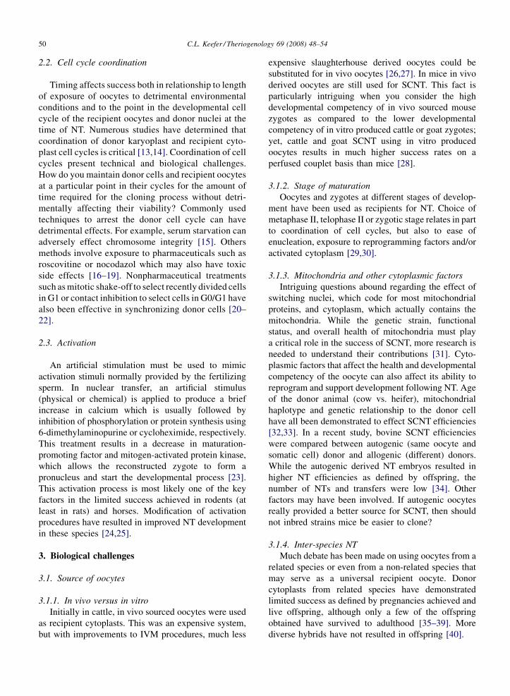
Table 1
Representative selection of NT results in domestic livestock and mice
Donor cell Percentage of
offspring per
NT transferreda
References
Blastomere
Cattle 10–25% [3,52]
Goat 30% [53]
Sheep 30% [19]
Pig 1.2–2% [54]
Mouse 57% (4-cell) [55]
28% (8-cell) [56]
Embryonic cells
Sheep 11% [57]
Mouse 13% (ESC) [58]
Fetal fibroblasts (FF)
Cattle 10–20% [14,28]
Goat 5% [59,60]
Sheep 8% [61]
Pig 1% [62]
Mouse 4% [63]
Transgenic FF
Cattle 4–29% [14,64]
Goat 3% [65,66]
Sheep 10% [67]
Pig 0.5% [68,69]
Adult cells
Cattleb 11% [17,70]
Goat 3% [65,71]
Sheep 3% [61]
Pig 1–3% [72,73]
Mouse 4–5% [74]
a Most reports are based on selected NT blastocysts transferred into
recipients and do not reflect on overall NT efficiencies (i.e., offspring
per constructed NT zygote). Results for goat and pig embryos which
are transferred at oocyte or early cleavage stages more closely reflect
their overall NT efficiency.b On occasion much higher successes for small subsets have been
achieved [75], but these were not considered representative of the
majority of reports.
now be performed using mice the success rates, at least
for adult donor cells, tend to be lower than those in
domestic species. Why? Is it due to species differences
such as earlier genomic activation or technical issues?
The introduction of the piezo system into micro-
manipulation process appeared to be a key contributor
to the current success in mice [7], although other
methods have been demonstrated to work [8,9]. Will
other simple improvements further increase the rate or
are more complex biological issues involved? There is
no doubt that techniques must be optimized for each
species, but whether or not some species are more
amenable to cloning is open to debate. Goats and pigs
seem to have fewer problems than cows and sheep in
terms of neonatal abnormalities and losses [10].
However, as techniques used and skills may vary
between groups, it is difficult to get a true comparison.
Generally, the procedure in pigs and goats involves
embryo transfer to the recipient at the zygotic or early
cleavage stages. Cattle NT embryos are maintained in
vitro to the blastocyst stage prior to embryo transfer,
while sheep NT embryos have been cultured to the
morulae and blastocyst stages either in vitro or in ligated
oviducts. Culture in a temporary recipient’s ligated
oviducts, while better than in vitro culture, does not
provide a truly normal oviductal condition and also
requires an additional retrieval and transfer procedure.
Therefore, the better results obtained with pigs and
goats may simply be a result of less time spent in vitro.
Similarly in rats, immediate transfer of reconstructed
zygotes into foster mothers resulted in live offspring
whereas cultured embryos did not [11].
Another factor affecting NT efficiencies that is
frequently ignored is the amount of NT practiced by
different groups. Groups which perform NT on a regular
and consistent basis tend to have better results. This is
probably a case of ‘‘practice makes perfect’’ in which
routines are followed consistently and speedy comple-
tion of the NT process results in minimal exposure of
oocytes to detrimental conditions.
What are the critical factors limiting NT success?
While it is hard to separate technical and biological
issues as they are so intertwined in the NT process,
some factors are known to have an effect on NT
efficiencies.
2. Technical challenges
2.1. Manipulation
NT involves a manual manipulation process in vitro
which requires exposure of oocytes and cells to
fluctuations in light, temperature, atmospheric condi-
tions, and different media. Different systems have been
used for enucleation (micromanipulation, chemical,
zona-free), transfer of the donor nucleus (electrofusion
and direct injection) and activation stimulus (electrical,
chemical and biological extracts). What may seem as a
minor alteration at any one step in the process can have
significant effects on the success rates. In particular,
species-specific requirements and sensitivities need to
be identified to achieve successful NT. This may mean
species-specific adjustments in media components,
temperatures or decreased exposure to detrimental
conditions (e.g., light intensity) [9,12].
C.L. Keefer / Theriogenology 69 (2008) 48–5450
2.2. Cell cycle coordination
Timing affects success both in relationship to length
of exposure of oocytes to detrimental environmental
conditions and to the point in the developmental cell
cycle of the recipient oocytes and donor nuclei at the
time of NT. Numerous studies have determined that
coordination of donor karyoplast and recipient cyto-
plast cell cycles is critical [13,14]. Coordination of cell
cycles present technical and biological challenges.
How do you maintain donor cells and recipient oocytes
at a particular point in their cycles for the amount of
time required for the cloning process without detri-
mentally affecting their viability? Commonly used
techniques to arrest the donor cell cycle can have
detrimental effects. For example, serum starvation can
adversely effect chromosome integrity [15]. Others
methods involve exposure to pharmaceuticals such as
roscovitine or nocodazol which may also have toxic
side effects [16–19]. Nonpharmaceutical treatments
such as mitotic shake-off to select recently divided cells
in G1 or contact inhibition to select cells in G0/G1 have
also been effective in synchronizing donor cells [20–
22].
2.3. Activation
An artificial stimulation must be used to mimic
activation stimuli normally provided by the fertilizing
sperm. In nuclear transfer, an artificial stimulus
(physical or chemical) is applied to produce a brief
increase in calcium which is usually followed by
inhibition of phosphorylation or protein synthesis using
6-dimethylaminopurine or cycloheximide, respectively.
This treatment results in a decrease in maturation-
promoting factor and mitogen-activated protein kinase,
which allows the reconstructed zygote to form a
pronucleus and start the developmental process [23].
This activation process is most likely one of the key
factors in the limited success achieved in rodents (at
least in rats) and horses. Modification of activation
procedures have resulted in improved NT development
in these species [24,25].
3. Biological challenges
3.1. Source of oocytes
3.1.1. In vivo versus in vitro
Initially in cattle, in vivo sourced oocytes were used
as recipient cytoplasts. This was an expensive system,
but with improvements to IVM procedures, much less
expensive slaughterhouse derived oocytes could be
substituted for in vivo oocytes [26,27]. In mice in vivo
derived oocytes are still used for SCNT. This fact is
particularly intriguing when you consider the high
developmental competency of in vivo sourced mouse
zygotes as compared to the lower developmental
competency of in vitro produced cattle or goat zygotes;
yet, cattle and goat SCNT using in vitro produced
oocytes results in much higher success rates on a
perfused couplet basis than mice [28].
3.1.2. Stage of maturation
Oocytes and zygotes at different stages of develop-
ment have been used as recipients for NT. Choice of
metaphase II, telophase II or zygotic stage relates in part
to coordination of cell cycles, but also to ease of
enucleation, exposure to reprogramming factors and/or
activated cytoplasm [29,30].
3.1.3. Mitochondria and other cytoplasmic factors
Intriguing questions abound regarding the effect of
switching nuclei, which code for most mitochondrial
proteins, and cytoplasm, which actually contains the
mitochondria. While the genetic strain, functional
status, and overall health of mitochondria must play
a critical role in the success of SCNT, more research is
needed to understand their contributions [31]. Cyto-
plasmic factors that affect the health and developmental
competency of the oocyte can also affect its ability to
reprogram and support development following NT. Age
of the donor animal (cow vs. heifer), mitochondrial
haplotype and genetic relationship to the donor cell
have all been demonstrated to effect SCNT efficiencies
[32,33]. In a recent study, bovine SCNT efficiencies
were compared between autogenic (same oocyte and
somatic cell) donor and allogenic (different) donors.
While the autogenic derived NT embryos resulted in
higher NT efficiencies as defined by offspring, the
number of NTs and transfers were low [34]. Other
factors may have been involved. If autogenic oocytes
really provided a better source for SCNT, then should
not inbred strains mice be easier to clone?
3.1.4. Inter-species NT
Much debate has been made on using oocytes from a
related species or even from a non-related species that
may serve as a universal recipient oocyte. Donor
cytoplasts from related species have demonstrated
limited success as defined by pregnancies achieved and
live offspring, although only a few of the offspring
obtained have survived to adulthood [35–39]. More
diverse hybrids have not resulted in offspring [40].
C.L. Keefer / Theriogenology 69 (2008) 48–54 51
3.2. Source of donor nucleus
The discussion on the source of the donor cell has
ranged from initial disbelief that it was possible to clone
using adult cells to theories that the low success rate is
contributable to the low percentage of stem cells that
may be present in adult tissues and inadvertently used as
donor cells. While cells from various adult tissues had
been used to produce offspring in sheep, cattle and
mice, the concept that a truly differentiated cell could be
reprogrammed by SCNT was proven when mature
mouse lymphocytes, B and T cells, were used as donor
cells. Researchers were able to demonstrate that nuclei
containing chromosomes that had undergone re-
arrangement, a definitive step in cell differentiation,
could result in live offspring following SCNT, albeit at
very low rates and in some cases after resorting to
chimera production to achieve success [41,42]. Other
differentiated cells, including muscle cells and post-
mitotic granulocytes, have also been used to generate
cloned calves and mouse pups, respectively [43,44].
Oback and Wells have presented a thorough discussion
on whether or not there is sufficient information to
determine if donor cell differentiation truly effects
cloning efficiencies [45].
3.3. Epigenetic state of donor nucleus
It is a demanding challenge for the recipient
cytoplast to reprogram the epigenetic code controlling
gene expression such that the donor chromatin is reset
into a pattern that is appropriate for embryonic
development. Many studies have attempted to explore
this issue by determining differences in methylation and
acetylation patterns of donor chromatin before and after
the SCNT process. Whole genomic comparisons of
methylation indicate that there is little difference
between a cloned animal and control, at least at the
global level [28]. Studies looking at specific genes often
do not find alterations in gene expression; however,
alterations in just a few key-controlling genes can be the
detrimental. Which genes are critical? Are there specific
genes which should be monitored or are genes randomly
affected, and thus unpredictable? The specific nature of
which genes might be affected also reflects on our
ability to pre-treat donor cells or oocytes. Can global
treatments, i.e., emersion in chemicals or cellular
extracts that result in alterations in methylation or
acetylation, be truly effective? Inhibition of histone
deacetylase using trychostatin A (TSA) has been shown
to improve cloning efficiencies [46,47]. While treat-
ments with 5-aza-cytidine or 5-aza-20-deoxycytidine to
decrease methylation of donor chromatin have not been
shown to improve cloning efficiencies, other treatments
including exposure to cell extracts have resulted in
improved NT blastocyst development and implantation
rates [48]. Further studies are needed to determine if
birth rates and offspring viability are also significantly
improved by such treatments.
3.4. Embryo viability and developmental potential
We should all realize by now that a blastocyst is just a
blastocyst—a zygote’s achievement of that stage of
development does not denote anything about its true
developmental potential to result in a live offspring.
Further investigation into the blastocyst’s cell number,
ICM:TE ratio, gene expression patterns and metabolic
state can give a better indicator of its potential, but there
is no clearly definitive marker of developmental
potential, thus far.
4. Conclusion
In summary, the commonalities seen in SCNT are
low overall efficiencies in offspring production, which
are due in large part to failures in initiation and
continuation of gestation. This failure appears to be due
to faulty reprogramming of trophectoderm which
results in abnormal placentation. Yet abnormalities in
placentation cannot be the sole factor as low yields of
offspring are also obtained following nuclear transfer in
species that do not have placentas, e.g., fish and
amphibians [49].
The differences in SCNT are harder to distinguish
owing to the low numbers generally reported, different
techniques, sources, and types of materials used. Are
there really species differences? I would suggest yes,
but not always in ways we might anticipate. Differences
can be found among species in patterns of demethyla-
tion during normal embryo development and patterns of
oocyte activation [50,51]. These differences may affect
the ability of certain species to reprogram donor cells
and to undergo embryonic development. Differences
and similarities need to be studied more fully in
comparative studies which may not only help improve
nuclear transfer efficiencies but also uncover basic
underlying developmental principles.
Acknowledgements
I wish to acknowledge the contributions of all those
who have contributed to the field of SCNT. As SCNT is
accomplished in an ever-growing list of animals, the
C.L. Keefer / Theriogenology 69 (2008) 48–5452
numbers of publications that mention SCNT are also
ever increasing. A recent PubMed search for the term
‘‘somatic cell nuclear transfer’’ resulted in the retrieval
of 844 references, 401 of which were from 2005 up to
2007. Further delimiting the query to ‘‘SCNT and donor
cells and reprogramming’’ resulted in 57 references
since the year 2005. So if key references were not cited
in this brief survey, my apologies to you and the authors.
References
[1] Briggs R, King T. Transplantation of living nuclei from blastula
cells into enucleated frogs’ eggs. Proc Natl Acad Sci USA
1952;38:455–63.
[2] Willadsen SM. Nuclear transplantation in sheep embryos. Nature
1986;320:63–5.
[3] Stice SL, Keefer CL. Multiple generational bovine embryo
cloning. Biol Reprod 1993;48:715–9.
[4] Peura TT, Lane MW, Lewis IM, Trounson AO. Development of
bovine embryo-derived clones after increasing rounds of nuclear
recycling. Mol Reprod Dev 2001;58:384–9.
[5] Young LE, Sinclair KD, Wilmut I. Large offspring syndrome in
cattle and sheep. Rev Reprod 1998;3:155–63.
[6] Prather RS, First NL. Cloning of embryos. J Reprod Fertil Suppl
1990;40:227–34.
[7] Wakayama T, Perry ACF, Zucotti M, Johnson KR, Yanagimachi
R. Full term development of mice from enucleated oocytes
injected with cumulus cell nuclei. Nature 1998;394:369–74.
[8] Zhou Q, Boulanger L, Renard JP. A simplified method for the
reconstruction of fully competent mouse zygotes from adult
somatic donor nuclei. Cloning 2000;2:35–44.
[9] Ribas R, Oback B, Ritchie W, Chebotareva T, Taylor J, Mauricio
AC, et al. Modifications to improve the efficiency of zona-free
mouse nuclear transfer. Cloning Stem Cells 2006;8:10–5.
[10] Rudenko L, Matheson JC. The US FDA and animal cloning: risk
and regulatory approach. Theriogenology 2007;67:198–206.
[11] Popova E, Bader M, Krivokharchenko A. Full-term development
of rat after transfer of nuclei from two-cell stage embryos. Biol
Reprod 2006;75:524–30.
[12] Hinrichs K, Choi YH, Walckenaer BE, Varner DD, Hartman DL.
In vitro-produced equine embryos: production of foals after
transfer, assessment by differential staining and effect of med-
ium calcium concentrations during culture. Theriogenology
2007;68:521–9.
[13] Campbell KH, Loi P, Otaegui PJ, Wilmut I. Cell cycle co-
ordination in embryo cloning by nuclear transfer. Rev Reprod
1996;1:40–6.
[14] Wells DN, Laible G, Tucker FC, Miller AL, Oliver JE, Xiang T,
et al. Coordination between donor cell type and cell cycle stage
improves nuclear cloning efficiency in cattle. Theriogenology
2003;59:45–59.
[15] Kues WA, Anger M, Carnwath JW, Paul D, Motlik J, Niemann H.
Cell cycle synchronization of porcine fetal fibroblasts: effects of
serum deprivation and reversible cell cycle inhibitors. Biol
Reprod 2000;62:412–9.
[16] Amano T, Tani T, Kato Y, Tsunoda Y. Mouse cloned from
embryonic stem (ES) cells synchronized in metaphase with
nocodazole. J Exp Zool 2001;289:139–45.
[17] Gibbons J, Arat S, Rzucidlo J, Miyoshi K, Waltenburg R,
Respess D, et al. Enhanced survivability of cloned calves derived
from roscovitine-treated adult somatic cells. Biol Reprod
2002;66:895–900.
[18] Tanaka H, Kanagawa H. Influence of combined activation
treatments on the success of bovine nuclear transfer using young
or aged oocytes. Anim Reprod Sci 1997;49:113–23.
[19] Liu L, Dai Y, Moor RM. Nuclear transfer in sheep embryos: the
effect of cell-cycle coordination between nucleus and cytoplasm
and the use of in vitro matured oocytes. Mol Reprod Dev
1997;47:255–64.
[20] Kasinathan P, Knott JG, Wang Z, Jerry DJ, Robl JM. Production
of calves from G1 fibroblasts. Nat Biotechnol 2001;19:1176–8.
[21] Boquest AC, Day BN, Prather RS. Flow cytometric cell cycle
analysis of cultured porcine fetal fibroblast cells. Biol Reprod
1999;60:1013–9.
[22] Lazaris A, Keyston R, Karatzas CN, Keefer CL. Transgenesis
using nuclear transfer in goats. Methods Mol Biol
2006;348:213–26.
[23] Liu L, Yang X. Interplay of maturation-promoting factor and
mitogen-activated protein kinase inactivation during metaphase-
to-interphase transition of activated bovine oocytes. Biol Reprod
1999;61:1–7.
[24] Zhou Q, Renard JP, Le Friec G, Brochard V, Beaujean N, Cherifi
Y, et al. Generation of fertile cloned rats by regulating oocyte
activation. Science 2003;302:1179.
[25] Hinrichs K, Choi YH, Love CC, Chung YG, Varner DD.
Production of horse foals via direct injection of roscovitine-
treated donor cells and activation by injection of sperm extract.
Reproduction 2006;131:1063–72.
[26] Keefer CL, Stice SL, Dobrinsky J. Effect of follicle-stimulating
hormone and luteinizing hormone during bovine in vitro matura-
tion on development following in vitro fertilization and nuclear
transfer. Mol Reprod Dev 1993;36:469–74.
[27] Barnes F, Endebrock M, Looney C, Powell R, Westhusin M,
Bondioli K. Embryo cloning in cattle: the use of in vitro matured
oocytes. J Reprod Fertil 1993;97:317–20.
[28] Yang X, Smith SL, Tian XC, Lewin HA, Renard JP, Wakayama
T. Nuclear reprogramming of cloned embryos and its implica-
tions for therapeutic cloning. Nat Genet 2007;39:295–302.
[29] Bordignon V, Smith LC. Telophase enucleation: an improved
method to prepare recipient cytoplasts for use in bovine nuclear
transfer. Mol Reprod Dev 1998;49:29–36.
[30] Polejaeva IA, Walker S, Campbell K. A double nuclear transfer
technique for cloning pigs. Methods Mol Biol 2006;348:135–50.
[31] Smith LC, Thundathil J, Filion F. Role of the mitochondrial
genome in preimplantation development and assisted reproduc-
tive technologies. Reprod Fertil Dev 2005;17:15–22.
[32] Bruggerhoff K, Zakhartchenko V, Wenigerkind H, Reichenbach
HD, Prelle K, Schernthaner W, et al. Bovine somatic cell nuclear
transfer using recipient oocytes recovered by ovum pick-up:
effect of maternal lineage of oocyte donors. Biol Reprod
2002;66:367–73.
[33] Aston KI, Li GP, Hicks BA, Sessions BR, Pate BJ, Hammon DS,
et al. The developmental competence of bovine nuclear transfer
embryos derived from cow versus heifer cytoplasts. Anim
Reprod Sci 2006;95:234–43.
[34] Yang XY, Li H, Ma QW, Yan JB, Zhao JG, Li HW, et al.
Improved efficiency of bovine cloning by autologous somatic
cell nuclear transfer. Reproduction 2006;132:733–9.
[35] Mastromonaco GF, Favetta LA, Smith LC, Filion F, King WA.
The influence of nuclear content on developmental competence
of gaur � cattle hybrid in vitro fertilized and somatic cell nuclear
transfer embryos. Biol Reprod 2007;76:514–23.
C.L. Keefer / Theriogenology 69 (2008) 48–54 53
[36] Lanza RP, Cibelli JB, Diaz F, Moraes CT, Farin PW, Farin CE,
et al. Cloning of an endangered species (Bos gaurus) using
interspecies nuclear transfer. Cloning 2000;2:79–90.
[37] Gomez MC, Pope CE, Giraldo A, Lyons LA, Harris RF, King
AL, et al. Birth of African wildcat cloned kittens born from
domestic cats. Cloning Stem Cells 2004;6:247–58.
[38] Loi P, Ptak G, Barboni B, Fulka Jr J, Cappai P, Clinton M.
Genetic rescue of an endangered mammal by cross-species
nuclear transfer using post-mortem somatic cells. Nat Biotech-
nol 2001;19:962–4.
[39] Woods GL, White KL, Vanderwall DK, Li GP, Aston KI, Bunch
TD, et al. A mule cloned from fetal cells by nuclear transfer.
Science 2003;301:1063.
[40] Yin X, Lee Y, Lee H, Kim N, Kim L, Shin H, et al. In vitro
production and initiation of pregnancies in inter-genus nuclear
transfer embryos derived from leopard cat (Prionailurus benga-
lensis) nuclei fused with domestic cat (Felis silverstris catus)
enucleated oocytes. Theriogenology 2006;66:275–82.
[41] Hochedlinger K, Jaenisch R. Monoclonal mice generated by
nuclear transfer from mature B and T donor cells. Nature
2002;415:1035–8.
[42] Inoue K, Wakao H, Ogonuki N, Miki H, Seino K, Nambu-Wakao
R, et al. Generation of cloned mice by direct nuclear transfer
from natural killer T cells. Curr Biol 2005;15:1114–8.
[43] Sung LY, Gao S, Shen H, Yu H, Song Y, Smith SL, et al.
Differentiated cells are more efficient than adult stem cells
for cloning by somatic cell nuclear transfer. Nat Genet
2006;38:1323–8.
[44] Green AL, Wells DN, Oback B. Cattle cloned from increasingly
differentiated muscle cells. Biol Reprod 2007;77:395–406.
[45] Oback B, Wells DN. Donor cell differentiation, reprogramming,
and cloning efficiency: elusive or illusive correlation? Mol
Reprod Dev 2007;74:646–54.
[46] Kishigami S, Mizutani E, Ohta H, Hikichi T, Thuan NV,
Wakayama S, et al. Significant improvement of mouse cloning
technique by treatment with trichostatin A after somatic nuclear
transfer. Biochem Biophys Res Commun 2006;340:183–9.
[47] Rybouchkin A, Kato Y, Tsunoda Y. Role of histone acetylation in
reprogramming of somatic nuclei following nuclear transfer.
Biol Reprod 2006;74:1083–9.
[48] Eilertsen KJ, Power RA, Harkins LL, Misica P. Targeting
cellular memory to reprogram the epigenome, restore potential,
and improve somatic cell nuclear transfer. Anim Reprod Sci
2007;98:129–46.
[49] Lee KY, Huang H, Ju B, Yang Z, Lin S. Cloned zebrafish by
nuclear transfer from long-term-cultured cells. Nat Biotechnol
2002;20:795–9.
[50] Young LE, Beaujean N. DNA methylation in the preimplantation
embryo: the differing stories of the mouse and sheep. Anim
Reprod Sci 2004;82/83:61–78.
[51] Malcuit C, Kurokawa M, Fissore RA. Calcium oscillations and
mammalian egg activation. J Cell Physiol 2006;206:565–73.
[52] Peura TT, Trounson AO. Recycling bovine embryos for nuclear
transfer. Reprod Fertil Dev 1998;10:627–32.
[53] Yong Z, Yuqiang L. Nuclear-cytoplasmic interaction and devel-
opment of goat embryos reconstructed by nuclear transplanta-
tion: production of goats by serially cloning embryos. Biol
Reprod 1998;58:266–9.
[54] Li GP, Tan JH, Sun QY, Meng QG, Yue KZ, Sun XS, et al.
Cloned piglets born after nuclear transplantation of embryonic
blastomeres into porcine oocytes matured in vivo. Cloning
2000;2:45–52.
[55] Kwon OY, Kono T. Production of identical sextuplet mice by
transferring metaphase nuclei from four-cell embryos. Proc Natl
Acad Sci USA 1996;93:13010–3.
[56] Greda P, Karasiewicz J, Modlinski JA. Mouse zygotes as reci-
pients in embryo cloning. Reproduction 2006;132:741–8.
[57] Wells DN, Misica PM, Day AM, Peterson AJ, Tervit HR.
Cloning sheep from cultured embryonic cells. Reprod Fertil
Dev 1998;10:615–26.
[58] Eggan K, Akutsu H, Loring J, Jackson-Grusby L, Klemm
M, Rideout III WM, et al. Hybrid vigor, fetal overgrowth,
and viability of mice derived by nuclear cloning and tetraploid
embryo complementation. Proc Natl Acad Sci USA 2001;98:
6209–14.
[59] Keefer CL, Baldassarre H, Keyston R, Wang B, Bhatia B,
Bilodeau AS, et al. Generation of dwarf goat (Capra hircus)
clones following nuclear transfer with transfected and nontrans-
fected fetal fibroblasts and in vitro-matured oocytes. Biol Reprod
2001;64:849–56.
[60] Keefer CL, Keyston R, Bhatia B, Lazaris A, Begin I, Kafidi N,
et al. Efficient production of viable goat offspring following
nuclear transfer using adult somatic cells. Biol Reprod
2000;62:192.
[61] Wilmut I, Schnieke AE, McWhir J, Kind AJ, Campbell KH.
Viable offspring derived from fetal and adult mammalian cells.
Nature 1997;385:810–3.
[62] Betthauser J, Forsberg E, Augenstein M, Childs L, Eilertsen K,
Enos J, et al. Production of cloned pigs from in vitro systems. Nat
Biotechnol 2000;18:1055–9.
[63] Saito M, Saga A, Matsuoka H. Production of a cloned mouse by
nuclear transfer from a fetal fibroblast cell of a mouse closed
colony strain. Exp Anim 2004;53:467–9.
[64] Cibelli JB, Stice SL, Golueke PJ, Kane JJ, Jerry J, Blackwell C,
et al. Cloned transgenic calves produced from nonquiescent fetal
fibroblasts. Science 1998;280:1256–8.
[65] Baldassarre H, Wang B, Keefer CL, Lazaris A, Karatzas CN.
State of the art in the production of transgenic goats. Reprod
Fertil Dev 2004;16:465–70.
[66] Reggio BC, James AN, Green HL, Gavin WG, Behboodi E,
Echelard Y, et al. Cloned transgenic offspring resulting from
somatic cell nuclear transfer in the goat: oocytes derived from
both follicle-stimulating hormone-stimulated and nonstimulated
abattoir-derived ovaries. Biol Reprod 2001;65:1528–33.
[67] Schnieke AE, Kind AJ, Ritchie WA, Mycock K, Scott AR,
Ritchie M, et al. Human factor IX transgenic sheep produced
by transfer of nuclei from transfected fetal fibroblasts. Science
1997;278:2130–3.
[68] Lai L, Park KW, Cheong HT, Kuhholzer B, Samuel M, Bonk
A, et al. Transgenic pig expressing the enhanced green fluor-
escent protein produced by nuclear transfer using colchicine-
treated fibroblasts as donor cells. Mol Reprod Dev 2002;62:
300–6.
[69] Bondioli K, Ramsoondar J, Williams B, Costa C, Fodor W.
Cloned pigs generated from cultured skin fibroblasts derived
from a H-transferase transgenic boar. Mol Reprod Dev
2001;60:189–95.
[70] Kubota C, Yamakuchi H, Todoroki J, Mizoshita K, Tabara N,
Barber M, et al. Six cloned calves produced from adult fibroblast
cells after long-term culture. Proc Natl Acad Sci USA
2000;97:990–5.
[71] Behboodi E, Memili E, Melican DT, Destrempes MM, Overton
SA, Williams JL, et al. Viable transgenic goats derived from skin
cells. Transgenic Res 2004;13:215–24.

C.L. Keefer / Theriogenology 69 (2008) 48–5454
[72] Polejaeva IA, Chen SH, Vaught TD, Page RL, Mullins J, Ball S,
et al. Cloned pigs produced by nuclear transfer from adult
somatic cells. Nature 2000;407:86–90.
[73] Tomii R, Kurome M, Ochiai T, Wako N, Ueda H, Hirakawa K,
et al. Production of cloned pigs by nuclear transfer of preadi-
pocytes established from adult mature adipocytes. Cloning Stem
Cells 2005;7:279–88.
[74] Wakayama S, Mizutani E, Kishigami S, Thuan NV, Ohta H,
Hikichi T, et al. Mice cloned by nuclear transfer from somatic
and ntES cells derived from the same individuals. J Reprod Dev
2005;51:765–72.
[75] Kato Y, Tani T, Sotomaru Y, Kurokawa K, Kato J, Doguchi H,
et al. Eight calves cloned from somatic cells of a single adult.
Science 1998;282:2095–8.