Embed Size (px)
Citation preview
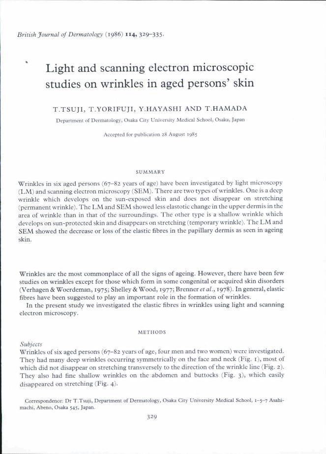
British Journal of Dermatology (1986) 114, 329-335-
Light and scanning electron microscopicstudies on wrinkles in aged persons' skin
T.TSUJI, T.YORIFUJI, Y.HAYASHI AND T.HAMADA
Dtpartmeiit of Dermatology, Osaka Ciiy University Medical School, Osaka, Japan
Accepted for publication 28 August 1985
SUMMARY
Wrinkles in six aged persons (67-82 years of age) have been investigated by light microscopy(LM) and scanning electron microscopy (SEM). There are two types of wrinkles. One is a deepwrinkle which develops on the sun-exposed skin and does not disappear on stretching(permanent wrinkle). The LM and SEM showed less elastotic change in the upper dermis in thearea of wrinkle than in that of the surroundings. The other type is a shallow wrinkle whichdevelops on sun-protected skin and disappears on stretching (temporary wrinkle). The LM andSEM showed the decrease or loss of the elastic fibres in the papillary dermis as seen in ageingskin.
Wrinkles are the most commonplace of all the signs of ageing. However, there have been fewstudies on wrinkles except for those which form in some congenital or acquired skin disorders(Verhagen& Woerdeman, 1975; Shelley & Wood, 1977; Brenner era/., 1978). In general, elasticfibres have been suggested to play an important role in the formation of wrinkles.
In the present study we investigated the elastic fibres in wrinkles using light and scanningelectron microscopy.
METHODS
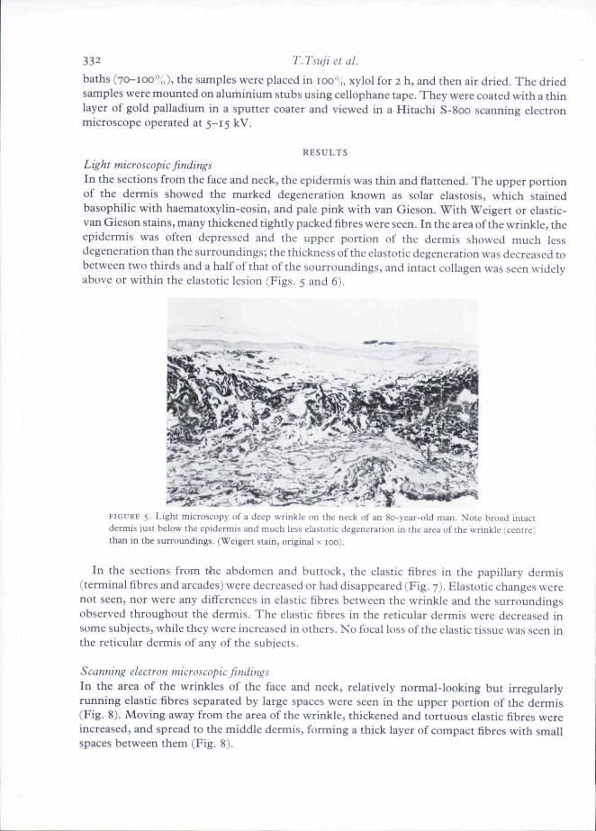
SubjectsWrinkles of six aged persons (67-82 years of age, four men and two women) were investigated.They had many deep wrinkles occurring symmetrically on the face and neck (Fig. i), most ofwhich did not disappear on stretching transversely to the direction of the wrinkle line (Fig. 2).They also had fine shallow wrinkles on the abdomen and buttocks (Fig. 3), which easilydisappeared on stretching (Fig. 4).
Correspondence; Dr T.Tsuji, Department of Dermatology, Osaka City University Medical School, 1-5-7 Asahi-machi, Abeno, Osaka 545, Japan.
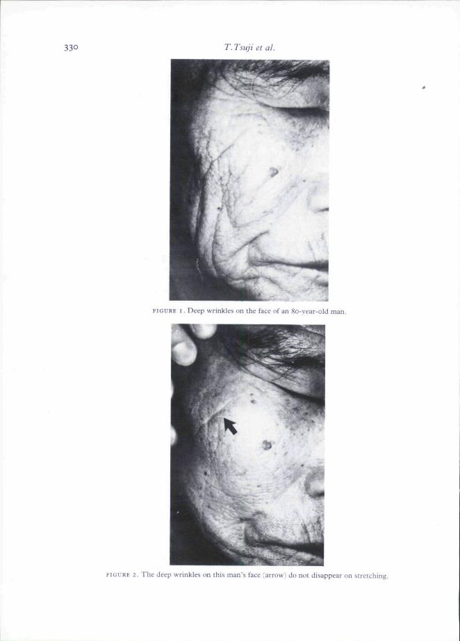
330 T. Tsuji et ai
FIGURE I, Deep wrinkles on the face of an 80-year-old man.
FIGURE 2, The deep wrinkles on this man's face (arrow) do not disappear on stretching.
Wrinkles in aged persons' skin 331
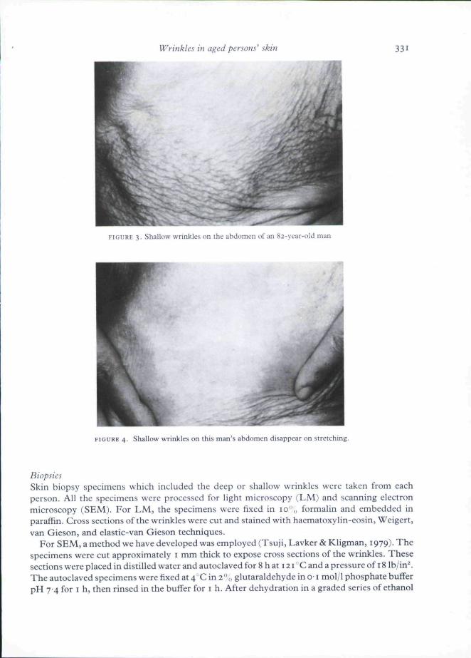
3, Shallou wrinkles (in ihc ahdomon nt an ^2-\car-old man
FIGURE 4. Shallow wrinkles on this man's abdomen disappear on stretching.
BiopsiesSkin biopsy specimens which included the deep or shallow wrinkles were taken from eachperson. All the specimens were processed for light microscopy (LM) and scanning electronmicroscopy (SEM). For LM, the specimens were flxed in 10",, formalin and embedded inparaffin. Cross sections ofthe wrinkles were cut and stained with haematoxylin-eosin, Weigert,van Gieson, and elastic-van Gieson techniques.
For SEM, a method we have developed was employed (Tsuji, Lavker & Kligman, 1979). Thespecimens were cut approximately i mm thick to expose cross sections ofthe wrinkles. Thesesections were placed in distilled water and autoclaved for 8 h at 121 C and a pressure of 18 lb/in^The autoclaved specimens were fixed at 4 C in 2'\, glutaraldehyde in o-1 mol/1 phosphate bufferpH 74 for I h, then rinsed in the buffer for i h. After dehydration in a graded series of ethanol
332 T.Tsuji et al.
baths (70-100''„), the samples were placed in 100 ,̂, xylol for 2 h, and then air dried. The driedsamples were mounted on alurhinium stubs using cellophane tape. They were coated with a thinlayer of gold palladium in a sputter coater and viewed in a Hitachi S-800 scanning electronmicroscope operated at 5-15 kV.
RESULTS
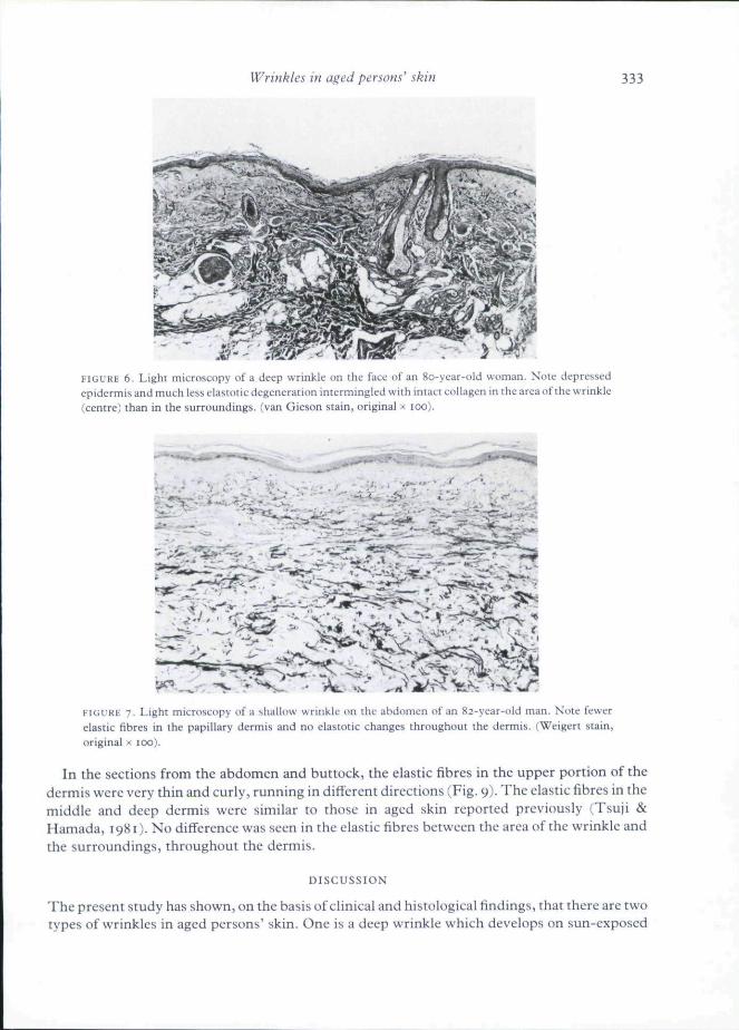
Light microscopic findingsIn the sections from the face and neck, the epidermis was thin and flattened. The upper portionof the dermis showed the marked degeneration known as solar elastosis, which stainedbasophilic with haematoxylin-eosin, and pale pink with van Gieson. With Weigert or elastic-van Gieson stains, many thickened tightly packed fibres were seen. In the area of the wrinkle, theepidermis was often depressed and the upper portion of the dermis showed much lessdegeneration than the surroundings; the thickness of the elastotic degeneration was decreased tobetween two thirds and a half of that of the sourroundings, and intact collagen was seen widelyabove or within the elastotic lesion (Figs. 5 and 6).
FIGURE 5. Light microscopy of a deep wrinkle on the neck of an 8o-ycar-old man. Note broad intactdermis just below the epidermis and much less elastotic degeneration in the area of the wrinkle ;centre)than in the surroundings. (Weigert stain, original x 100).
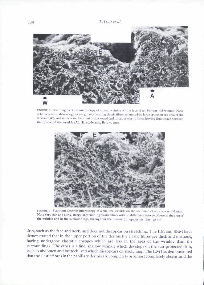
In the sections from the abdomen and buttock, the elastic fibres in the papillary dermis(terminal fibres and arcades) were decreased or had disappeared (Fig. 7). Hlastotic changes werenot seen, nor were any differences in elastic fibres between the wrinkle and the surroundingsobserved throughout the dermis. The elastic fibres in the reticular dermis were decreased insome subjects, while they were increased in others. No focal loss of the elastic tissue was seen inthe reticular dermis of any of the subjects.
Scanning electron microscopic findingsIn the area of the wrinkles of the face and neck, relatively normal-looking but irregularlyrunning elastic fibres separated by large spaces were seen in the upper portion of the dermis(Fig. 8). Moving away from the area of the wrinkle, thickened and tortuous elastic fibres wereincreased, and spread to the middle dermis, forming a thick layer of compact fibres with smallspaces between them (Fig. 8).
Wrinkles in aged persons' skin 333
FIGURE 6. Light microscopy of a deep wrinkle on the face of an 80-year-old woman. Note depressedepidermis and much less elastotic degeneration intermingled with intact collagen in the area of the wrinkle(centre) than in the surroundings, (van Gieson stain, original x too).
FIGURE 7. Light microscopy of a shallow wrinkle on the abdomen of an 82-year-old man. Note fewerelastic fibres in the papillary dermis and no elastotic changes throughout the dermis. (Weigert stain,original x 100).
In the sections from the abdomen and buttock, the elastic fibres in the upper portion of thedermis were very thin and curly, running in diflcrent directions (Fig. 9). The elastic fibres in themiddle and deep dermis were similar to those in aged skin reported previously (Tsuji &Hamada, 1981). No difference was seen in the elastic fibres between the area of the wrinkle andthe surroundings, throughout the dermis.
DISCUSSION
The present study has shown, on the basis of clinical and histological findings, that there are twotypes of wrinkles in aged persons' skin. One is a deep wrinkle which develops on sun-exposed
334 T. Tsuji et al.
FIGURE S. Scanning electron microscopy of a deep wrinkle on the face of an 80-year-old woman. Noterelatively normal-looking but irregularly running elastic fibres separated by large spaces in the area of thewrinkle fW), and an increased amount of thickened and tortuous elastic fibres leaving little space betweenthem, around the wrinkle (A). (E: epidermis. Bar:
FIGURE 9. Scanning electron microscopy of a shallow wrinkle on the abdomen of an 82-year-old man.Note very thin and curly, irregularly rurming elastic fibres with no difference between those in the area ofthe wrinkle and in the surroundings, throughout the dermis. (E: epidermis, Bar:
skin, such as the face and neck, and does not disappear on stretching. The LM and SEM havedemonstrated that in the upper portion of the dermis the elastic fibres are thick and tortuous,having undergone elastotic changes which are less in the area of the wrinkle than thesurroundings, The other is a fine, shallow wrinkle which develops on the sun-protected skin,such as abdomen and buttock, and which disappears on stretching. The LM has demonstratedthat the clastic fibres in the papillary dermis are completely or almost completely absent, and the
Wrinkles in aged persons' skin 335
SEM has shown that there are thin and curly elastic fibres in the upper portion of the dermis.There were no differences between the area of the wrinkles and the surroundings. We propose tocall the deep wrinkles 'permanent wrinkles' and the shallow ones 'temporary wrinkles'.
We suggest the following mechanism of formation of the two types of wrinkles. The facial andneck skin responds to every movement of the underlying muscles in smiling, frowning andphysical movement. The movement produces temporary but repeated wrinkling of the sameportion of the face or neck. This facility of response is achieved by the presence of a remarkablycomplex and dense intradermal elastic tissue mesh. With years of sunlight exposure, therecomes the degeneration of elastic tissue known as solar elastosis, resulting in thickened skin. Itseems reasonable to assume that the wrinkles on the face and neck are subjected to less damagethan the surroundings because of less sunlight exposure. This results in permanent wrinkles likea valley between the areas of thickened skin, and they no longer disappear on stretching. TheLM and SEM findings support this as there were marked elastotie changes surrounding the areaof the wrinkles which themselves showed less elastotic change. Thus, this type of wrinkle is dueto a biophysical rather than a histological change, but is closely associated with the abovemorphological abnormalities of the dermal elastic fibres.
On the other hand, the sun-protected skin, particularly the abdomen and buttock skin,becomes thin with a decrease in subcutaneous fat tissue with age, and wrinkles easily. Most ofthe wrinkles are fine and shallow. The movement of the underlying muscle docs not cause thewrinkles, but simply makes them evident. These are not permanent wrinkles since they easilydisappear on stretching. The LM findings in these wrinkles are identical to the changes seen inageing skin (Montagna, 1973; Montagna & Carlisle, 1979)- In addition, the SEM findingssuggest that the elastic fibres in the papillary dermis are not absent, but have become too thin tobe clearly recognized by LM.
Previous investigators have reported wrinkles due to idiopathic or non-inflammatoryelastolysis, the histology of which shows loss of mid-dermal elastic fibres (Shelley & Wood,t977; Brenner et ai, 1978), and wrinkles due to post-infiammatory elastolysis, the histology ofwhich shows almost complete loss of the upper and mid-dermal elastic fibres with the remainingelastic fibres fragmented or swollen (Verhagen & Woerdeman, 1975). These wrinkles aredifferent from either of the two types of wrinkles in aged persons' skin described here.
REFERENCES
BRENNER, W , , GSCHNAIT, F . , KONRAD, K . , HOI.I;BAK, K . & TAPPEINER, J. (1978) Non-infiammatory dermal elastoiysis.
British Journal of Dermatology, 97, 441.MONTAGNA, W . (1973) Ageing of the nipple and areola. Minerva Dermatologica, 108, 3.MONTAGNA, W . & CARLISLE, K . (1979) Structural changes in aging human skin. Joumtil of Investigative Dermatology,
73. 47-SHELLtY, W.B. & WOOD, M.G. 11977) Wrinkles due 10 idiupaihic loss of mid-dermal elasiit tissue. British Journal of
Dermatology, 97, 441.TSUJI, T . , LAVKER, R.M. 5r KLIGMAN, A.M. (1979) A new method for scanning electron microscopic visualization of
dermal clastic fibres. Journal of Microscopy, 115, 165.TSUJI, T . & HAMADA, T . (1981) Age-related changes in human dermal elastic fibres. British Journal of Dermatology, 105,
57-VERHAGKN, A.R. & WOERDEMAN, M.J. (1975) Post-inflammatory elastolysis and ctitis laxa. British Journal of
Dermatology, 92, 183.