Embed Size (px)
Citation preview
LiNi0.5Mn1.5O4 Nanowires Produced by Electrospinning Method
Rui Xu1,2, Ilias Belharouak2, Jianglan Shui2, James C.M. Li1
1Materials Science Program, University of Rochester, Rochester, NY 14627
2Chemical Science and Engineering Division, Argonne National Laboratory, 9700 South Cass Ave., Argonne, IL
60439
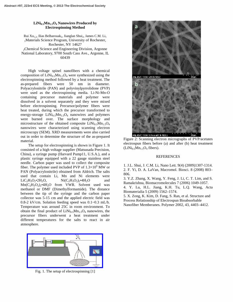
High voltage spinel nanofibers with a chemical composition of LiNi0.5Mn1.5O4 were synthesized using the electrospinning method followed by a heat treatment. The as-prepared fibers were 50 nm in diameter. Polyacrylonitrile (PAN) and polyvinylpyrrolidone (PVP) were used as the electrospinning media. Li-Ni-Mn-O containing precursor materials and polymer were dissolved in a solvent separately and they were mixed before electrospinning. Precursor/polymer fibers were heat treated, during which the precursor transformed to energy-storage LiNi0.5Mn1.5O4 nanowires and polymers were burned over. The surface morphology and microstructure of the obtained composite LiNi0.5Mn1.5O4 nanowires were characterized using scanning electron microscopy (SEM). XRD measurements were also carried out in order to determine the structure of the as-prepared material. The setup for electrospinning is shown in Figure 1. It consisted of a high voltage supplier (Matsusada Precision, China), a syringe pump (Harvard Pump11, U.S.A.), and a plastic syringe equipped with a 22 gauge stainless steel needle. Carbon paper was used to collect the composite fiber. The polymer used included PVP of 1.3×10⁶ MW or PAN (Polyacrylonitrile) obtained from Aldrich. The salts used that contain Li, Mn and Ni elements were LiC2H3O2•2H2O, Ni(C2H3O2)2•4H2O and Mn(C2H3O2)2•4H2O from VWR. Solvent used was methanol or DMF (Dimethylformamide). The distance between the tip of the syringe and the carbon paper collector was 5-15 cm and the applied electric field was 0.8-2 kV/cm. Solution feeding speed was 0.1~0.3 mL/h. Temperature was around 25C in room environment. To obtain the final product of LiNi0.5Mn1.5O4 nanowires, the precursor fibers underwent a heat treatment under different temperatures for the salts to react in air atmosphere.
Fig. 1. The setup of electrospinning [1]
Figure 2: Scanning electron micrographs of PVP/acetates electrospun fibers before (a) and after (b) heat treatment (LiNi 0.5Mn1.5O4 fibers).
REFERENCES 1. J.L. Shui, J. C.M. Li, Nano Lett. 9(4) (2009)1307-1314. 2. F. Yi, D. A. LaVan, Macromol. Biosci. 8 (2008) 803–806. 3. Y.Z. Zhang, X. Wang, Y. Feng, J. Li, C. T. Lim, and S. Ramakrishna, Biomacromolecules 7 (2006) 1049-1057. 4. Y. Lu, H.L. Jiang, K.H. Tu, L.Q. Wang, Acta Biomaterialia 5 (2009) 1562–1574. 5. X. Zong, K. Kim, D. Fang, S. Ran, et al. Structure and Process Relationship of Electrospun Bioabsorbable Nanofiber Memberanes. Polymer 2002, 43, 4403–4412.
a
b
Abstract #97, 223rd ECS Meeting, © 2013 The Electrochemical Society


![119 Nanowires 4. Nanowires - UFAMhome.ufam.edu.br/berti/nanomateriais/Nanowires.pdf · 119 Nanowires 4. Nanowires ... written about carbon nanotubes [4.57–59], which can be](https://img.pdfslide.net/doc/110x75/5abfd11e7f8b9a5d718eba2b/119-nanowires-4-nanowires-nanowires-4-nanowires-written-about-carbon-nanotubes.jpg)


![Electrospinning for Bone Tissue Engineering · solution electrospinning and melt electrospinning to produce a 3D cell-invasive scaffold has been described [20]. While melt electrospinning](https://img.pdfslide.net/doc/110x75/5e2f2481450bb928ad6e34c6/electrospinning-for-bone-tissue-engineering-solution-electrospinning-and-melt-electrospinning.jpg)