Embed Size (px)
Citation preview

LL weakness and numbness
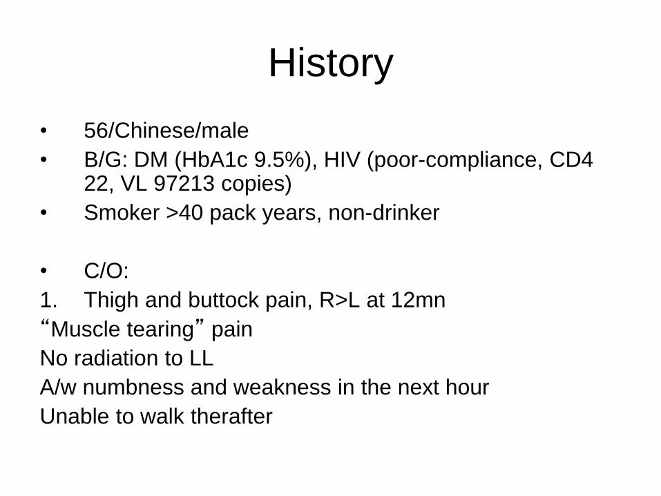
History
• 56/Chinese/male
• B/G: DM (HbA1c 9.5%), HIV (poor-compliance, CD4 22, VL 97213 copies)
• Smoker >40 pack years, non-drinker
• C/O:
1. Thigh and buttock pain, R>L at 12mn
“Muscle tearing” pain
No radiation to LL
A/w numbness and weakness in the next hour
Unable to walk therafter
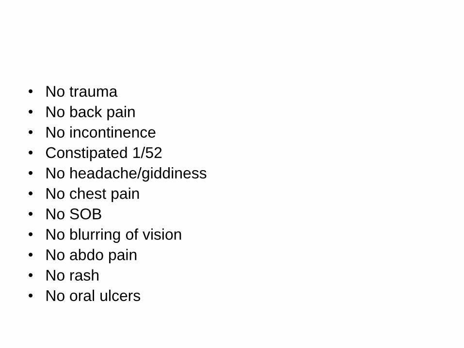
• No trauma
• No back pain
• No incontinence
• Constipated 1/52
• No headache/giddiness
• No chest pain
• No SOB
• No blurring of vision
• No abdo pain
• No rash
• No oral ulcers
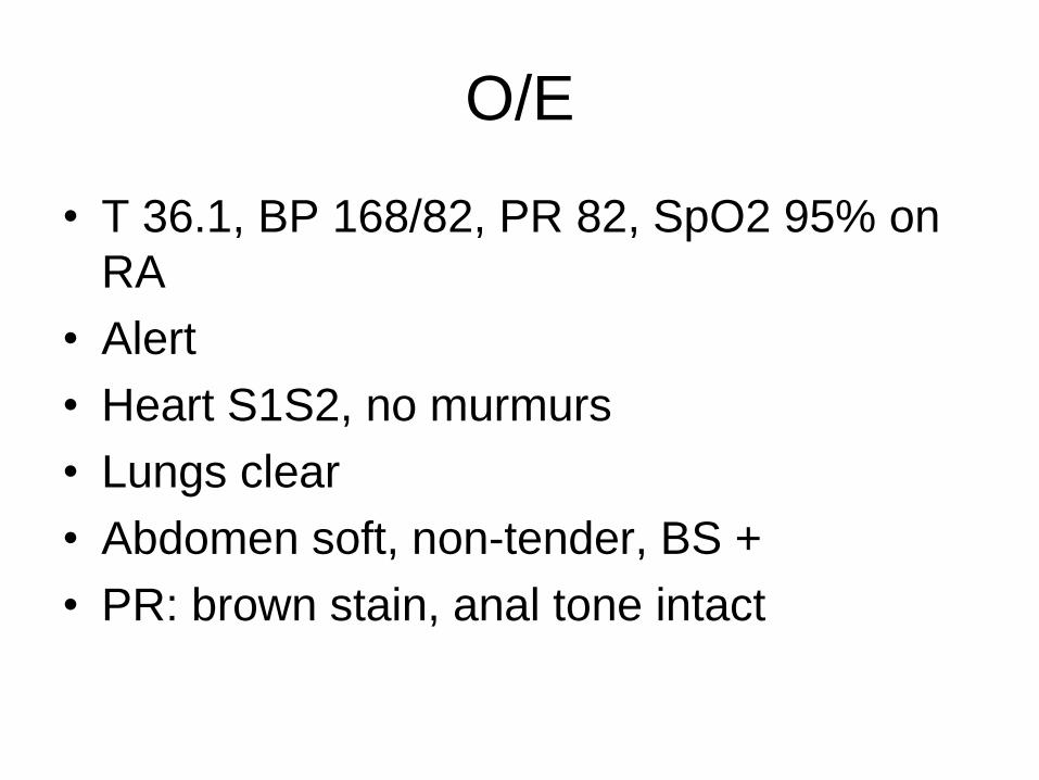
O/E
• T 36.1, BP 168/82, PR 82, SpO2 95% on
RA
• Alert
• Heart S1S2, no murmurs
• Lungs clear
• Abdomen soft, non-tender, BS +
• PR: brown stain, anal tone intact
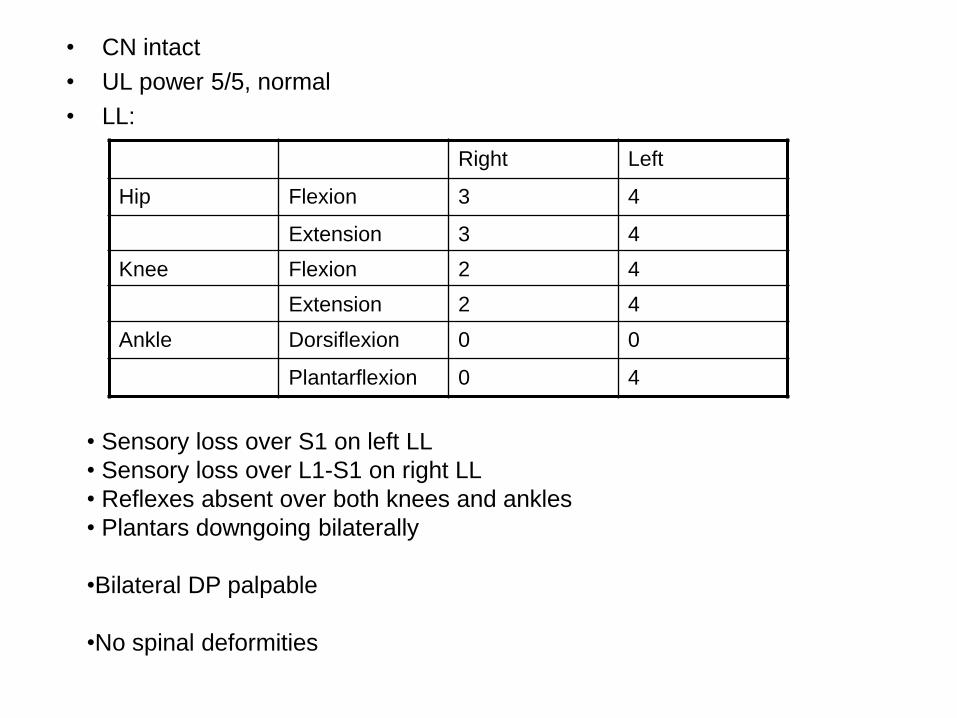
• CN intact
• UL power 5/5, normal
• LL:
Right Left
Hip Flexion 3 4
Extension 3 4
Knee Flexion 2 4
Extension 2 4
Ankle Dorsiflexion 0 0
Plantarflexion 0 4
• Sensory loss over S1 on left LL
• Sensory loss over L1-S1 on right LL
• Reflexes absent over both knees and ankles
• Plantars downgoing bilaterally
•Bilateral DP palpable
•No spinal deformities
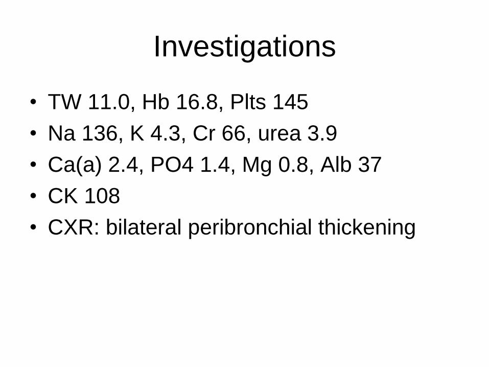
Investigations
• TW 11.0, Hb 16.8, Plts 145
• Na 136, K 4.3, Cr 66, urea 3.9
• Ca(a) 2.4, PO4 1.4, Mg 0.8, Alb 37
• CK 108
• CXR: bilateral peribronchial thickening

Differentials?
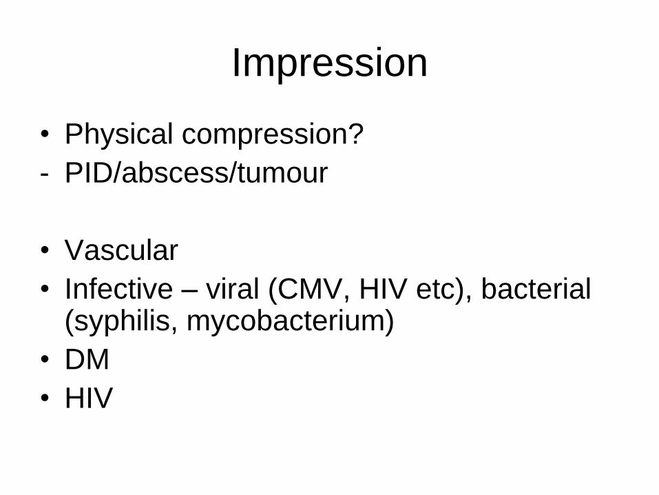
Impression
• Physical compression?
- PID/abscess/tumour
• Vascular
• Infective – viral (CMV, HIV etc), bacterial (syphilis, mycobacterium)
• DM
• HIV
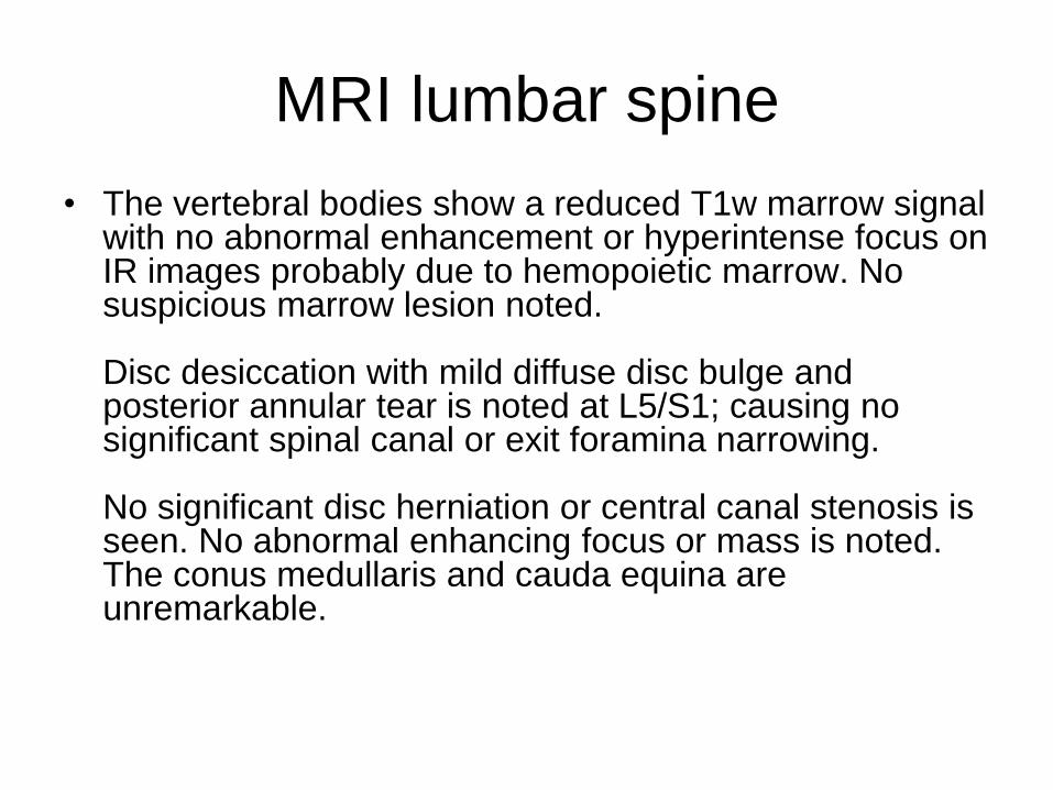
MRI lumbar spine
• The vertebral bodies show a reduced T1w marrow signal with no abnormal enhancement or hyperintense focus on IR images probably due to hemopoietic marrow. No suspicious marrow lesion noted. Disc desiccation with mild diffuse disc bulge and posterior annular tear is noted at L5/S1; causing no significant spinal canal or exit foramina narrowing. No significant disc herniation or central canal stenosis is seen. No abnormal enhancing focus or mass is noted. The conus medullaris and cauda equina are unremarkable.
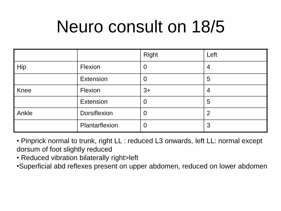
Neuro consult on 18/5
Right Left
Hip Flexion 0 4
Extension 0 5
Knee Flexion 3+ 4
Extension 0 5
Ankle Dorsiflexion 0 2
Plantarflexion 0 3
• Pinprick normal to trunk, right LL : reduced L3 onwards, left LL: normal except
dorsum of foot slightly reduced
• Reduced vibration bilaterally right>left
•Superficial abd reflexes present on upper abdomen, reduced on lower abdomen
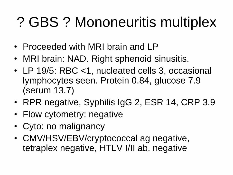
? GBS ? Mononeuritis multiplex
• Proceeded with MRI brain and LP
• MRI brain: NAD. Right sphenoid sinusitis.
• LP 19/5: RBC <1, nucleated cells 3, occasional lymphocytes seen. Protein 0.84, glucose 7.9 (serum 13.7)
• RPR negative, Syphilis IgG 2, ESR 14, CRP 3.9
• Flow cytometry: negative
• Cyto: no malignancy
• CMV/HSV/EBV/cryptococcal ag negative, tetraplex negative, HTLV I/II ab. negative
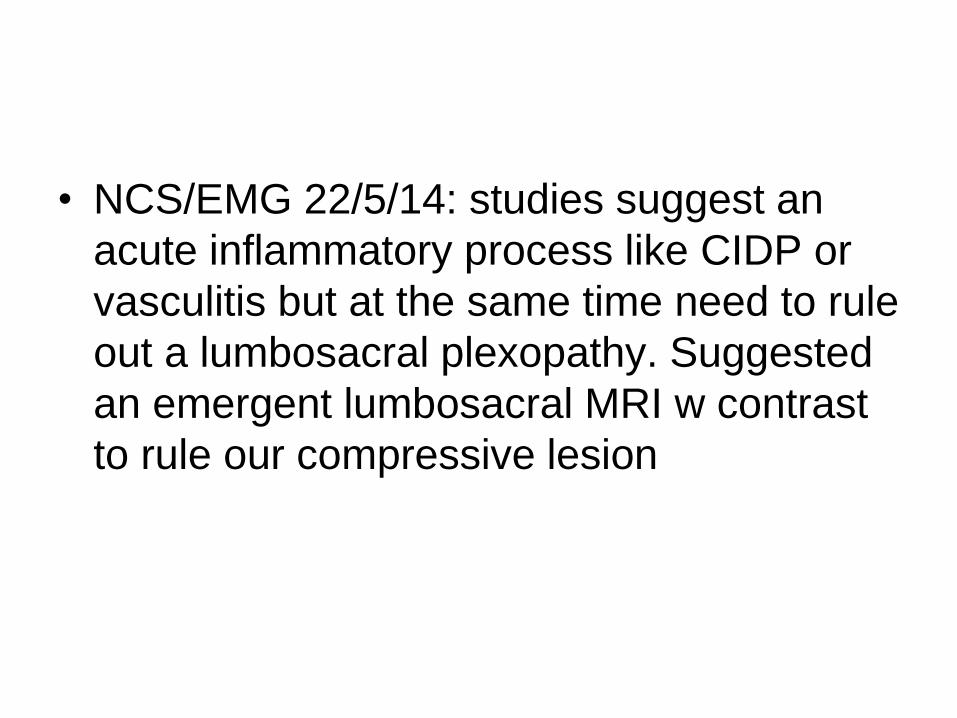
• NCS/EMG 22/5/14: studies suggest an
acute inflammatory process like CIDP or
vasculitis but at the same time need to rule
out a lumbosacral plexopathy. Suggested
an emergent lumbosacral MRI w contrast
to rule our compressive lesion
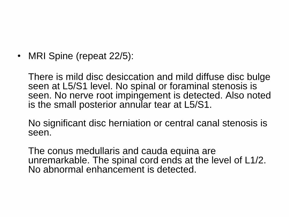
• MRI Spine (repeat 22/5):
There is mild disc desiccation and mild diffuse disc bulge seen at L5/S1 level. No spinal or foraminal stenosis is seen. No nerve root impingement is detected. Also noted is the small posterior annular tear at L5/S1. No significant disc herniation or central canal stenosis is seen. The conus medullaris and cauda equina are unremarkable. The spinal cord ends at the level of L1/2. No abnormal enhancement is detected.
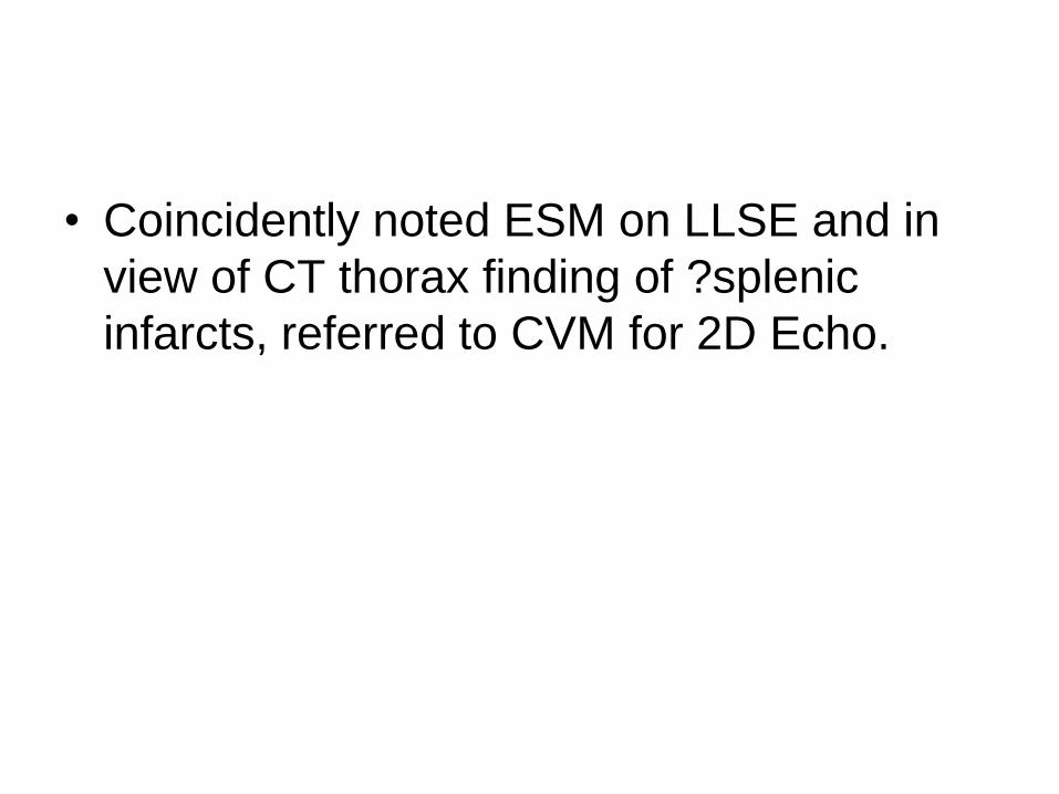
• Coincidently noted ESM on LLSE and in
view of CT thorax finding of ?splenic
infarcts, referred to CVM for 2D Echo.
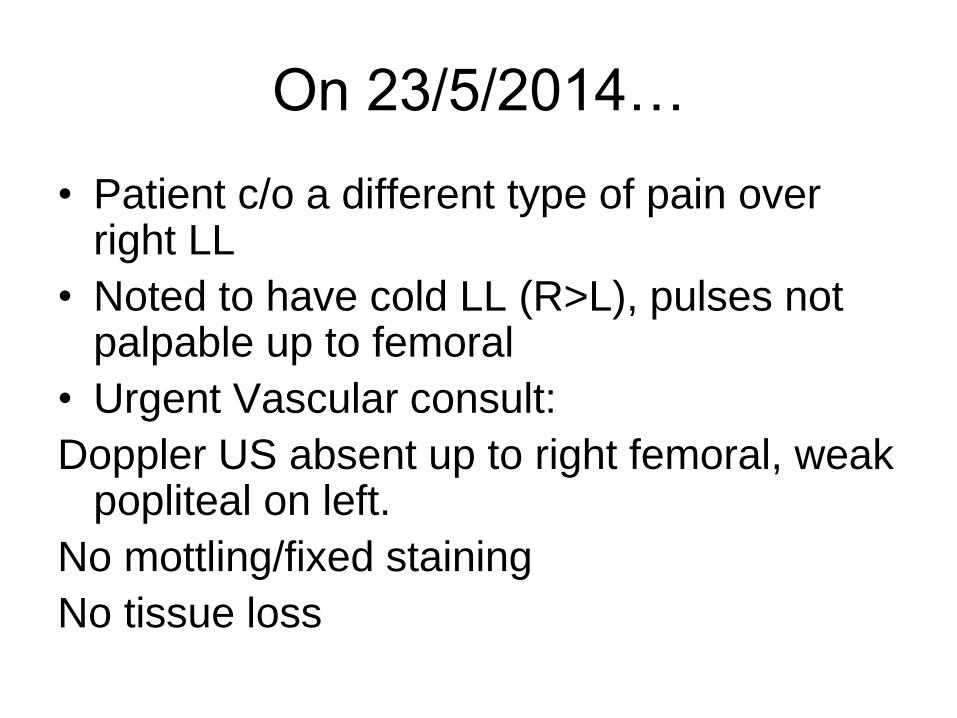
On 23/5/2014…
• Patient c/o a different type of pain over right LL
• Noted to have cold LL (R>L), pulses not palpable up to femoral
• Urgent Vascular consult:
Doppler US absent up to right femoral, weak popliteal on left.
No mottling/fixed staining
No tissue loss
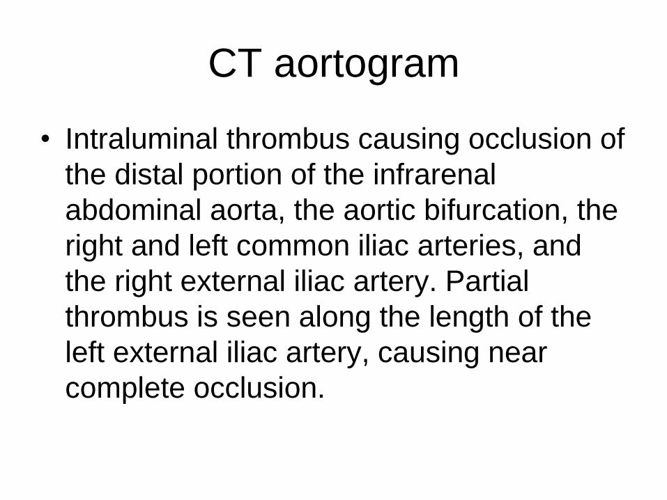
CT aortogram
• Intraluminal thrombus causing occlusion of
the distal portion of the infrarenal
abdominal aorta, the aortic bifurcation, the
right and left common iliac arteries, and
the right external iliac artery. Partial
thrombus is seen along the length of the
left external iliac artery, causing near
complete occlusion.
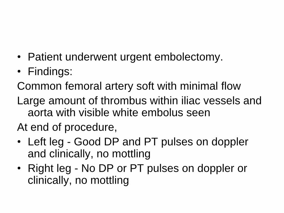
• Patient underwent urgent embolectomy.
• Findings:
Common femoral artery soft with minimal flow
Large amount of thrombus within iliac vessels and aorta with visible white embolus seen
At end of procedure,
• Left leg - Good DP and PT pulses on doppler and clinically, no mottling
• Right leg - No DP or PT pulses on doppler or clinically, no mottling
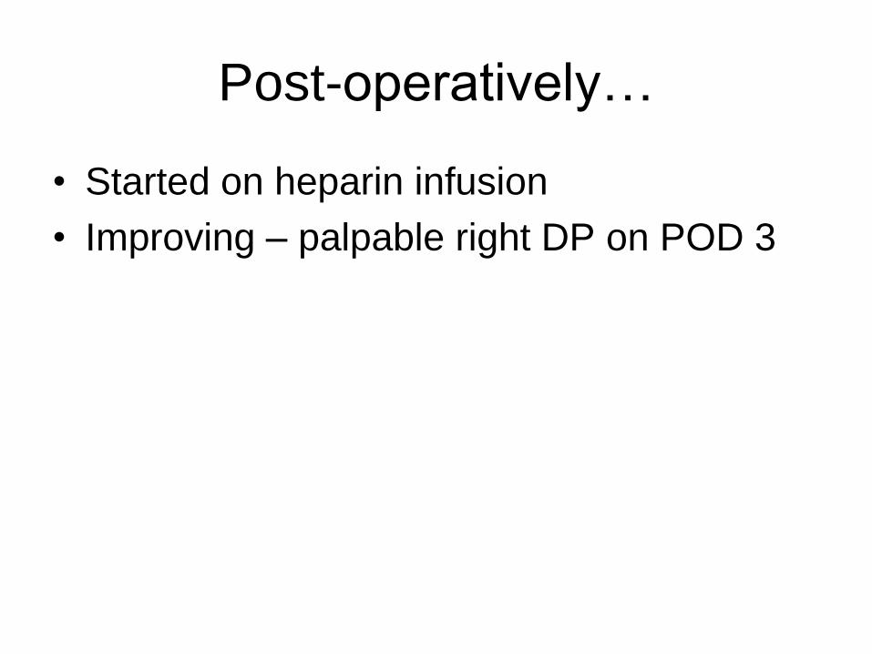
Post-operatively…
• Started on heparin infusion
• Improving – palpable right DP on POD 3
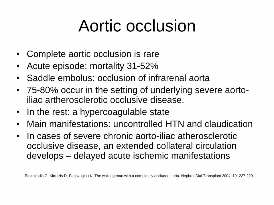
Aortic occlusion
• Complete aortic occlusion is rare
• Acute episode: mortality 31-52%
• Saddle embolus: occlusion of infrarenal aorta
• 75-80% occur in the setting of underlying severe aorto-iliac artherosclerotic occlusive disease.
• In the rest: a hypercoagulable state
• Main manifestations: uncontrolled HTN and claudication
• In cases of severe chronic aorto-iliac atherosclerotic occlusive disease, an extended collateral circulation develops – delayed acute ischemic manifestations
Efstratiadis G, Kirmizis D, Papazoglou K. The walking man with a completely occluded aorta. Nephrol Dial Transplant 2004; 19: 227-229
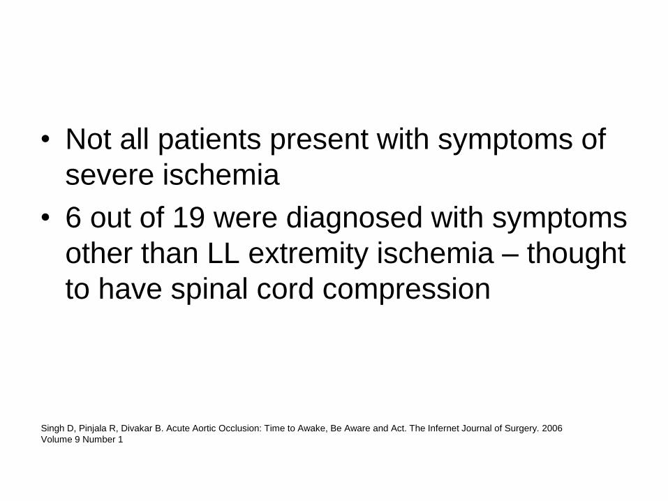
• Not all patients present with symptoms of
severe ischemia
• 6 out of 19 were diagnosed with symptoms
other than LL extremity ischemia – thought
to have spinal cord compression
Singh D, Pinjala R, Divakar B. Acute Aortic Occlusion: Time to Awake, Be Aware and Act. The Infernet Journal of Surgery. 2006
Volume 9 Number 1
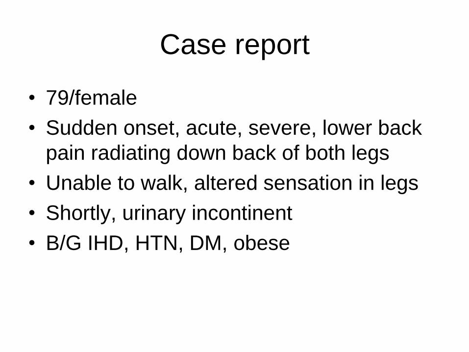
Case report
• 79/female
• Sudden onset, acute, severe, lower back
pain radiating down back of both legs
• Unable to walk, altered sensation in legs
• Shortly, urinary incontinent
• B/G IHD, HTN, DM, obese
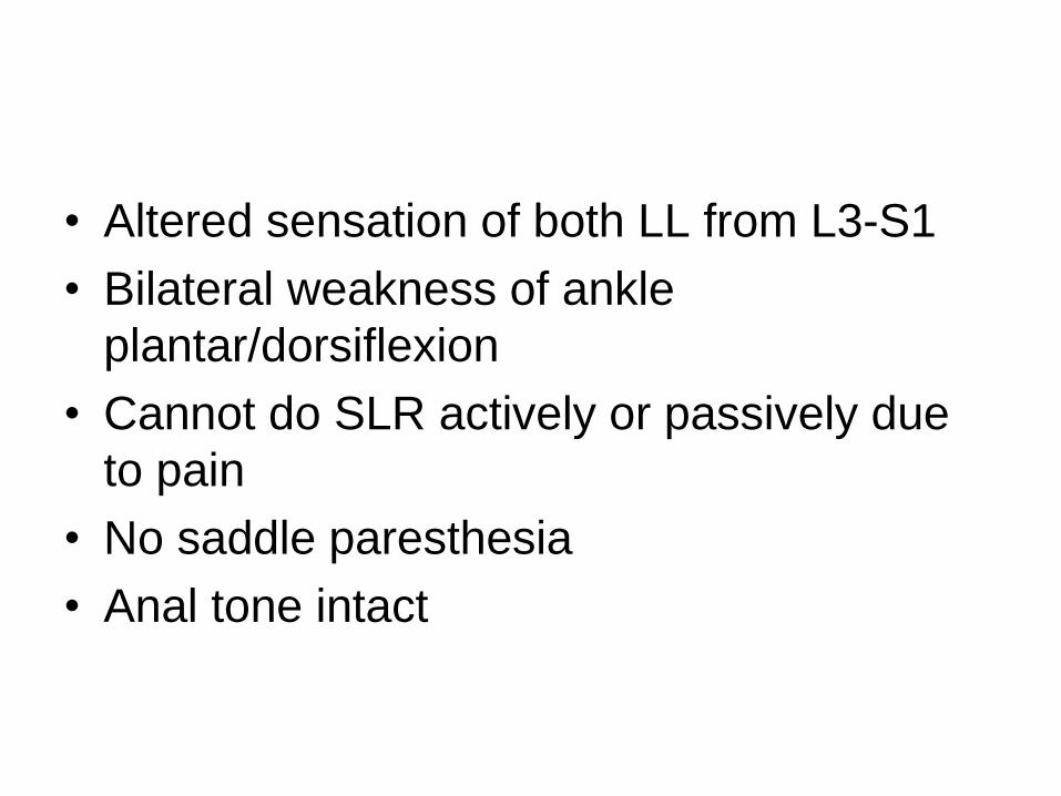
• Altered sensation of both LL from L3-S1
• Bilateral weakness of ankle
plantar/dorsiflexion
• Cannot do SLR actively or passively due
to pain
• No saddle paresthesia
• Anal tone intact
• MRI: disc protrusions multiple levels, but
no spinal cord, cauda equina or nerve root
compression
• Started on analgesia with marked
improvement, mobilising with frame and
power normalised
• 7 days after admission, noted pain and
discoloration of toes
• CRT <2s, but pulses absent
• Angiogram: saddle embolism at bifurcation
of aorta with complete occlusion of both
common iliac arteries with stenosis of the
distal aorta. Common femoral arteries
patent but poor flow. Popliteal arteries
normal
• 2DE: large ventricular mural thrombus
Shaw A, Anwar H, Targett J. Cauda equina syndrome versus saddle
embolism. Ann R Coll Surg Engl 2008; 90
• Cauda equina supplied by lower lumbar,
iliolumbar and lateral sacral arteries
• Greater radicular artery very rarely aries at
the level of L3 (1.4%) or L4-L5 (0.2%) and
this low origin may be the reason for
paralysis or cauda equina like symptoms
• Thus, a patient with reduced blood flow in
these 3 main arteries may have transient
ischemia to the cauda equina, mimicking
compression
Olearchyk AS. Saddle embolism of the aorta with sudden paraplegia.
Can J Surg 2004; 47:472-3c
• 50% of cases have no peripheral vascular
symptoms or delayed symptoms
Olearchyk AS. Saddle embolism of the aorta with sudden paraplegia.
Can J Surg 2004; 47:472-3c
Revascularization Syndrome
• Local cx: explosive swelling of the limb,
compartment syndrome, rhabdomyolysis
• General cx: acidosis, MOD
• Related to interval between ischemia and
revascularization and muscle mass