Embed Size (px)
Citation preview
50
ABSTRACT
We report a case of a 21 years old female who presented with
a history of anterior knee pain for previous 3 months. Pain
was localized to the anteromedial aspect of the left knee and
aggravated by flexion. Clinical examination revealed a 2x2
cm painful lump on the anteromedial aspect of the left
medial condyle with no effusion. Magnetic resonance
imaging revealed an anterosuperior tear of the medial
collateral ligament. The patient subsequently underwent left
knee arthroscopic examination. Two yellowish pedunculated
masses arising from the anteromedial portion of the
synovium were discovered and completely excised.
Histopathological examination of the excision biopsy
revealed pigmented villonodular synovitis (PVNS) with
marked central necrosis. One year post excision, she is well
with no signs or symptoms of recurrence. This case
highlights an uncommon cause of anterior knee pain.
Localized PVNS typically presents with mechanical
symptoms, however, pain could arise from pedicle torsion
and necrosis.
Key Words:
Pigmented Villonodular Synovitis, Anterior Knee Pain,Synovitis, Arthritis
INTRODUCTION
Anterior knee pain in adolescents is a common problem. The
pain may arise from numerous anatomical structures found at
the anterior aspect of the knee. Patellofemoral maltracking is
the most common cause of anterior knee pain 1. Other
common causes are Hoffa syndrome that is caused by
infrapatella fat pad impingement and plica syndrome caused
by inflammation and irritation of the knee plica.
Pigmented villonodular synovitis (PVNS) is a rare benign
proliferative disorder of unknown aetiology, more common
in males and mainly affecting synovial joint bursae and
tendon sheaths. First described by Jaffe et al in 1941, PVNS
may be difficult to diagnose, meaning that delay in diagnosis
is not uncommon 2. PVNS is usually monoarticular and
typically affects the knee. The lesion is usually pedunculated
or less frequently sessile in appearance and is found in the
meniscocapsular junction, the intercondylar notch region,
the anterior tibial eminence or the lateral recesses 1. It is also
seen in the fat pad, medial femoral condyle, on the medial
patellar synovial plica and inside popliteal cysts but is
extremely rare in the posterior compartment of the knee.
CASE REPORT
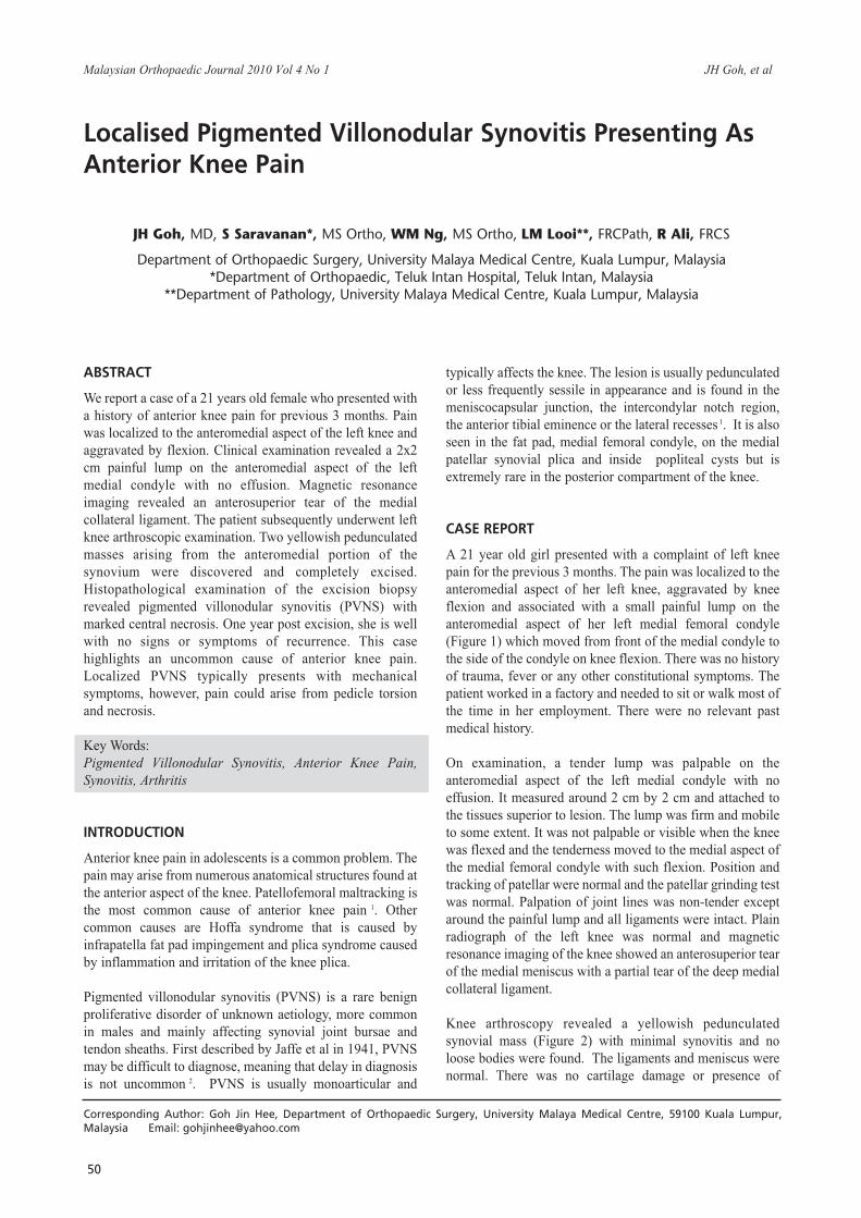
A 21 year old girl presented with a complaint of left knee
pain for the previous 3 months. The pain was localized to the
anteromedial aspect of her left knee, aggravated by knee
flexion and associated with a small painful lump on the
anteromedial aspect of her left medial femoral condyle
(Figure 1) which moved from front of the medial condyle to
the side of the condyle on knee flexion. There was no history
of trauma, fever or any other constitutional symptoms. The
patient worked in a factory and needed to sit or walk most of
the time in her employment. There were no relevant past
medical history.
On examination, a tender lump was palpable on the
anteromedial aspect of the left medial condyle with no
effusion. It measured around 2 cm by 2 cm and attached to
the tissues superior to lesion. The lump was firm and mobile
to some extent. It was not palpable or visible when the knee
was flexed and the tenderness moved to the medial aspect of
the medial femoral condyle with such flexion. Position and
tracking of patellar were normal and the patellar grinding test
was normal. Palpation of joint lines was non-tender except
around the painful lump and all ligaments were intact. Plain
radiograph of the left knee was normal and magnetic
resonance imaging of the knee showed an anterosuperior tear
of the medial meniscus with a partial tear of the deep medial
collateral ligament.
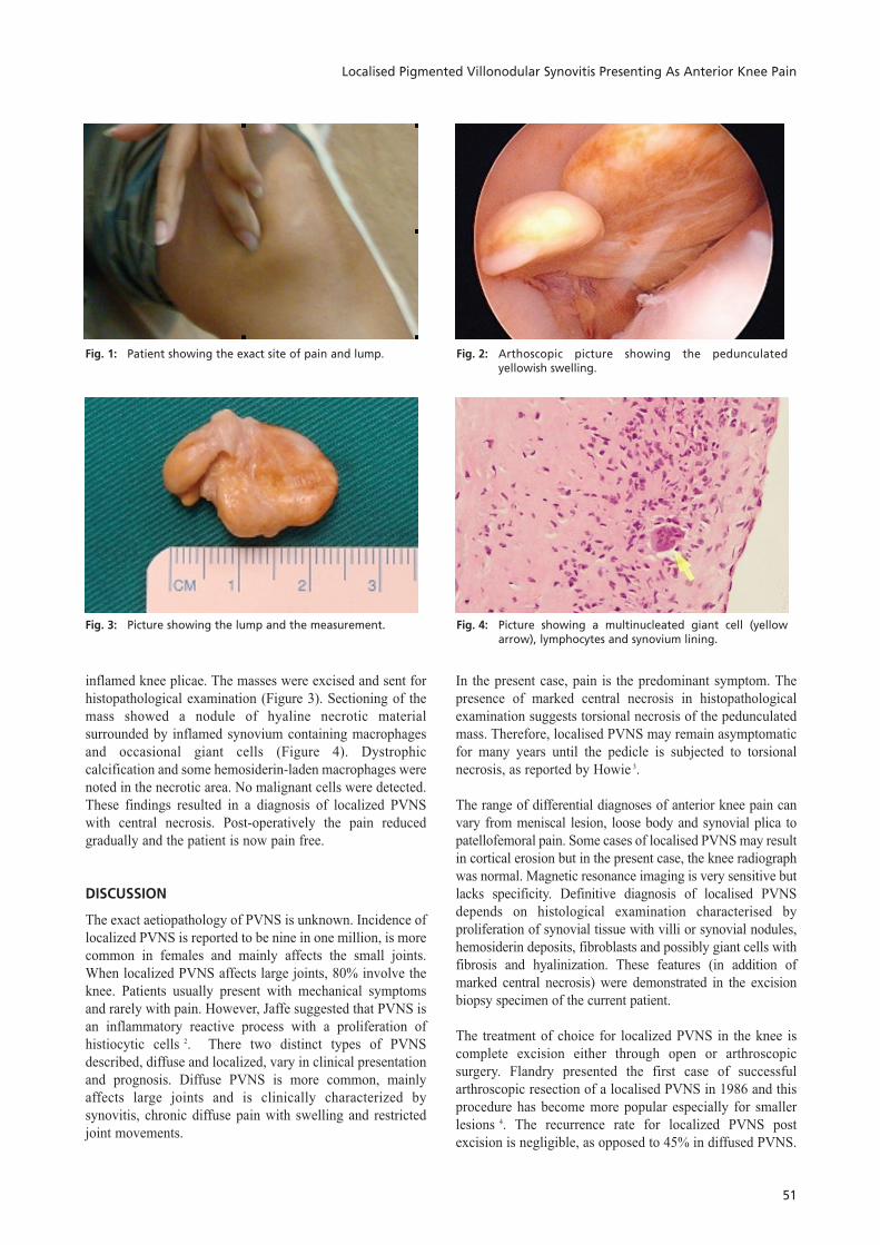
Knee arthroscopy revealed a yellowish pedunculated
synovial mass (Figure 2) with minimal synovitis and no
loose bodies were found. The ligaments and meniscus were
normal. There was no cartilage damage or presence of
Localised Pigmented Villonodular Synovitis Presenting AsAnterior Knee Pain
JH Goh, MD, S Saravanan*, MS Ortho, WM Ng, MS Ortho, LM Looi**, FRCPath, R Ali, FRCS
Department of Orthopaedic Surgery, University Malaya Medical Centre, Kuala Lumpur, Malaysia*Department of Orthopaedic, Teluk Intan Hospital, Teluk Intan, Malaysia
**Department of Pathology, University Malaya Medical Centre, Kuala Lumpur, Malaysia
Corresponding Author: Goh Jin Hee, Department of Orthopaedic Surgery, University Malaya Medical Centre, 59100 Kuala Lumpur,Malaysia Email: [email protected]
Malaysian Orthopaedic Journal 2010 Vol 4 No 1 JH Goh, et al
Localised Pigmented Villonodular Synovitis Presenting As Anterior Knee Pain
51
Fig. 1: Patient showing the exact site of pain and lump. Fig. 2: Arthoscopic picture showing the pedunculatedyellowish swelling.
Fig. 3: Picture showing the lump and the measurement. Fig. 4: Picture showing a multinucleated giant cell (yellowarrow), lymphocytes and synovium lining.
inflamed knee plicae. The masses were excised and sent for
histopathological examination (Figure 3). Sectioning of the
mass showed a nodule of hyaline necrotic material
surrounded by inflamed synovium containing macrophages
and occasional giant cells (Figure 4). Dystrophic
calcification and some hemosiderin-laden macrophages were
noted in the necrotic area. No malignant cells were detected.
These findings resulted in a diagnosis of localized PVNS
with central necrosis. Post-operatively the pain reduced
gradually and the patient is now pain free.
DISCUSSION
The exact aetiopathology of PVNS is unknown. Incidence of
localized PVNS is reported to be nine in one million, is more
common in females and mainly affects the small joints.
When localized PVNS affects large joints, 80% involve the
knee. Patients usually present with mechanical symptoms
and rarely with pain. However, Jaffe suggested that PVNS is
an inflammatory reactive process with a proliferation of
histiocytic cells 2. There two distinct types of PVNS
described, diffuse and localized, vary in clinical presentation
and prognosis. Diffuse PVNS is more common, mainly
affects large joints and is clinically characterized by
synovitis, chronic diffuse pain with swelling and restricted
joint movements.
In the present case, pain is the predominant symptom. The
presence of marked central necrosis in histopathological
examination suggests torsional necrosis of the pedunculated
mass. Therefore, localised PVNS may remain asymptomatic
for many years until the pedicle is subjected to torsional
necrosis, as reported by Howie 3.
The range of differential diagnoses of anterior knee pain can
vary from meniscal lesion, loose body and synovial plica to
patellofemoral pain. Some cases of localised PVNS may result
in cortical erosion but in the present case, the knee radiograph
was normal. Magnetic resonance imaging is very sensitive but
lacks specificity. Definitive diagnosis of localised PVNS
depends on histological examination characterised by
proliferation of synovial tissue with villi or synovial nodules,
hemosiderin deposits, fibroblasts and possibly giant cells with
fibrosis and hyalinization. These features (in addition of
marked central necrosis) were demonstrated in the excision
biopsy specimen of the current patient.
The treatment of choice for localized PVNS in the knee is
complete excision either through open or arthroscopic
surgery. Flandry presented the first case of successful
arthroscopic resection of a localised PVNS in 1986 and this
procedure has become more popular especially for smaller
lesions 4. The recurrence rate for localized PVNS post
excision is negligible, as opposed to 45% in diffused PVNS.

Malaysian Orthopaedic Journal 2010 Vol 4 No 1 JH Goh, et al
52
REFERENCES
1. Fulkerson. John P and Arendt EA. Anterior Knee Pain in Females. Clin Orthop 2000; 372. 69-73.
2. Jaffe HL, Lichtenstein L and Sutro CJ. Pigmented villonodular synovitis, bursitis and tenosynovitis. Arch Pathol. 1941; 1: 731-
65.
3. Howie CR, Smith GD, Christie J, Gregg PJ. Torsion of localized pigmented villonodular synovitis of the knee. J Bone Joint SurgBr, 1985; 67: 564-566.
4. Flandry F, Hughston JC. Pigmented villonodular synovitis. J Bone Joint Surg Am. 1987; 69: 942-9.


![Clínica Universitária de Reumatologia – Faculdade de ...§ões_Reumatologia_CHUC... · Apr 30. [Epub ahead of print] • Pigmented Villonodular Synovitis: a recurrent case with](https://img.pdfslide.net/doc/110x75/5c65198c09d3f2916e8c4235/clinica-universitaria-de-reumatologia-faculdade-de-oesreumatologiachuc.jpg)


![Multiple nodular form of localized pigmented villonodular ... · masses [11], and a mixed form, with a prominent mass and diffuse synovitis, representing a transition between DPVNS](https://img.pdfslide.net/doc/110x75/5b9b50ef09d3f291158cf882/multiple-nodular-form-of-localized-pigmented-villonodular-masses-11-and.jpg)