Embed Size (px)
Citation preview
Epilepsy & Behavior 13 (2008) 678–680
Contents lists available at ScienceDirect
Epilepsy & Behavior
journal homepage: www.elsevier .com/locate /yebeh
Long-term cognitive outcome of hemiconvulsion–hemiplegia–epilepsysyndrome affecting the left cerebral hemisphere
Seyed M. Mirsattari a,b,c,d,*, Nancy J. Wilde d, Susan E. Pigott d
a Department of Clinical Neurological Sciences, University of Western Ontario, London, Ontario, Canada N6A 5A5b Department of Medical Imaging, University of Western Ontario, London, Ontario, Canada N6A 5A5c Department of Medical Biophysics, University of Western Ontario, London, Ontario, Canada N6A 5A5d Department of Psychology, University of Western Ontario, London, Ontario, Canada N6A 5A5
a r t i c l e i n f o
Article history:Received 29 June 2008Revised 17 July 2008Accepted 21 July 2008Available online 16 August 2008
Keywords:Hemiplegia–hemiconvulsion–epilepsysyndromeFebrile convulsionsCognitive outcomeLanguageLong-term
1525-5050/$ - see front matter � 2008 Elsevier Inc. Adoi:10.1016/j.yebeh.2008.07.008
* Corresponding author. Address: Department of CUniversity of Western Ontario, B10-108, 339 WinderCanada N6A 5A5. Fax: +1 519 663 3440.
E-mail address: [email protected] (S.M. Mirsattari)
a b s t r a c t
Long-term cognitive outcome following hemiconvulsion–hemiplegia–epilepsy (HHE) syndrome has beenpoorly studied, with little attention to the implications of side of involvement in HHE. This retrospectivestudy describes language lateralization and cognitive performance in five patients with HHE syndromeaffecting the left cerebral hemisphere. All of the patients had to have intracarotid sodium amytal testing(IAT) to be included in this study. The mean age of the patients was 30.2 years (range: 13–50). All patientshad their hemiconvulsive seizures before age 1½ years (range: 6–13 months). All patients had right-sidedhemiatrophy of the body, left mesial temporal sclerosis, and seizures originating from the left temporallobe. The habitual seizures began at a mean age of 4.5 years (range: 1.5–12 years). Performance on testsof intelligence, verbal memory, and visual memory was examined. Language was represented in the rightcerebral hemisphere in three patients, the left hemisphere in one patient, and both hemispheres withpredominance on the right side in the fifth patient. Intellectual functioning was in the borderline toextremely low range among the patients with right hemispheric or bilateral representation for language.These patients were variably impaired on measures of verbal and visual memory. The patient with lefthemispheric representation for language performed in the average range on tests of intellectual function-ing and verbal memory, whereas scores on visual memory were variable. This study demonstrated thatreorganization of language to the right cerebral hemisphere or its bilateral representation is common inpatients with HHE syndrome affecting the left cerebral hemisphere, and is associated with poor cognitiveoutcome.
� 2008 Elsevier Inc. All rights reserved.
1. Introduction
Hemiconvulsion–hemiplegia–epilepsy (HHE) syndrome is anuncommon epileptic disorder that was initially described by Gas-taut et al. in 1957 [1,2]. It is characterized by the occurrence of pro-longed hemiclonic seizures followed by the development ofhemiplegia, typically in the course of a febrile illness before 4 yearsof age. Following a variable interval (mean of 1 to 2 years) [3], sub-sequent epilepsy develops, most frequently with complex partialseizures of temporal lobe origin [2,4,5]. Combinations of partialand generalized seizures may also occur [4,5]. The motor deficithas a variable course, with some patients continuing to exhibit adefinitive hemiplegia and others showing complete resolution[1,6]. The pathogenesis of HHE syndrome remains unknown. Itmay involve multiple factors including infection and genetic
ll rights reserved.
linical Neurological Sciences,mere Road, London, Ontario,
.
causes similar to benign febrile seizures [7,8]. It is accompaniedby acute cytotoxic edema of the affected hemisphere at the timeof the initial convulsive status, followed by chronic atrophicchanges of the same hemisphere [2,9,10]. Most patients developtemporal lobe epilepsy (TLE) or multifocal epilepsy later in life[5]. Magnetic resonance imaging (MRI) of the brain reveals evi-dence of abnormal diffusion within the white matter in the acutephase [11], but hippocampal sclerosis (HS) and/or hemiatrophy la-ter in life [10,11]. Most patients develop medication-resistant epi-lepsy later in life [7–9]. Surgical treatment has been shown to beeffective in the management of seizures in patients with HHE thatbecome intractable to medical therapy in the chronic state [5]. Theincidence of HHE syndrome has declined considerably in industri-alized counties over the past few decades [7,12] likely due to moreeffective management of status epilepticus [12,13] or the reducedincidence of febrile seizures as a result of immunization. However,HHE syndrome remains prevalent in developing countries [5,6,9].
The long-term cognitive outcome following HHE syndrome hasbeen poorly studied, although according to early descriptions,mental retardation is a common feature [3]. Furthermore, little
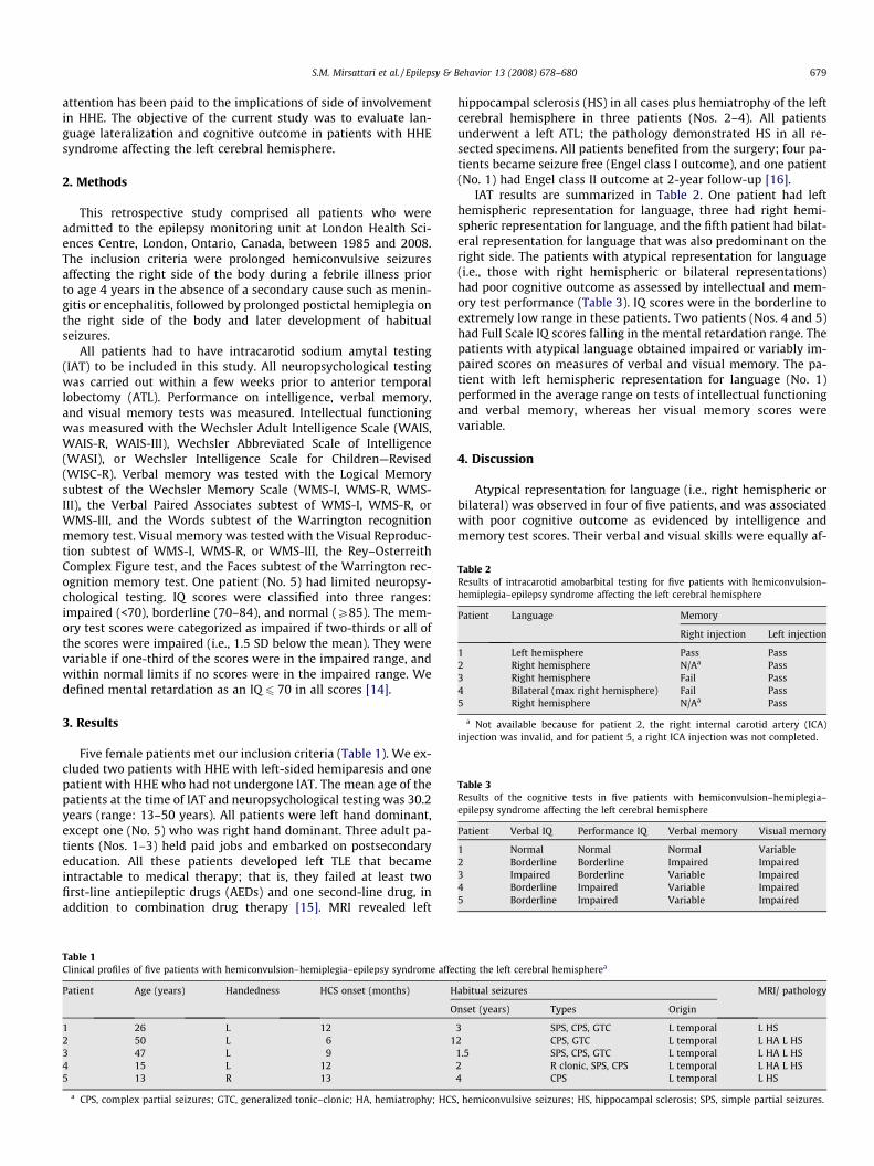
Table 2Results of intracarotid amobarbital testing for five patients with hemiconvulsion–hemiplegia–epilepsy syndrome affecting the left cerebral hemisphere
Patient Language Memory
Right injection Left injection
1 Left hemisphere Pass Pass2 Right hemisphere N/Aa Pass3 Right hemisphere Fail Pass4 Bilateral (max right hemisphere) Fail Pass5 Right hemisphere N/Aa Pass
a Not available because for patient 2, the right internal carotid artery (ICA)injection was invalid, and for patient 5, a right ICA injection was not completed.
Table 3Results of the cognitive tests in five patients with hemiconvulsion–hemiplegia–epilepsy syndrome affecting the left cerebral hemisphere
Patient Verbal IQ Performance IQ Verbal memory Visual memory
1 Normal Normal Normal Variable2 Borderline Borderline Impaired Impaired3 Impaired Borderline Variable Impaired4 Borderline Impaired Variable Impaired5 Borderline Impaired Variable Impaired
S.M. Mirsattari et al. / Epilepsy & Behavior 13 (2008) 678–680 679
attention has been paid to the implications of side of involvementin HHE. The objective of the current study was to evaluate lan-guage lateralization and cognitive outcome in patients with HHEsyndrome affecting the left cerebral hemisphere.
2. Methods
This retrospective study comprised all patients who wereadmitted to the epilepsy monitoring unit at London Health Sci-ences Centre, London, Ontario, Canada, between 1985 and 2008.The inclusion criteria were prolonged hemiconvulsive seizuresaffecting the right side of the body during a febrile illness priorto age 4 years in the absence of a secondary cause such as menin-gitis or encephalitis, followed by prolonged postictal hemiplegia onthe right side of the body and later development of habitualseizures.
All patients had to have intracarotid sodium amytal testing(IAT) to be included in this study. All neuropsychological testingwas carried out within a few weeks prior to anterior temporallobectomy (ATL). Performance on intelligence, verbal memory,and visual memory tests was measured. Intellectual functioningwas measured with the Wechsler Adult Intelligence Scale (WAIS,WAIS-R, WAIS-III), Wechsler Abbreviated Scale of Intelligence(WASI), or Wechsler Intelligence Scale for Children—Revised(WISC-R). Verbal memory was tested with the Logical Memorysubtest of the Wechsler Memory Scale (WMS-I, WMS-R, WMS-III), the Verbal Paired Associates subtest of WMS-I, WMS-R, orWMS-III, and the Words subtest of the Warrington recognitionmemory test. Visual memory was tested with the Visual Reproduc-tion subtest of WMS-I, WMS-R, or WMS-III, the Rey–OsterreithComplex Figure test, and the Faces subtest of the Warrington rec-ognition memory test. One patient (No. 5) had limited neuropsy-chological testing. IQ scores were classified into three ranges:impaired (<70), borderline (70–84), and normal (P85). The mem-ory test scores were categorized as impaired if two-thirds or all ofthe scores were impaired (i.e., 1.5 SD below the mean). They werevariable if one-third of the scores were in the impaired range, andwithin normal limits if no scores were in the impaired range. Wedefined mental retardation as an IQ 6 70 in all scores [14].
3. Results
Five female patients met our inclusion criteria (Table 1). We ex-cluded two patients with HHE with left-sided hemiparesis and onepatient with HHE who had not undergone IAT. The mean age of thepatients at the time of IAT and neuropsychological testing was 30.2years (range: 13–50 years). All patients were left hand dominant,except one (No. 5) who was right hand dominant. Three adult pa-tients (Nos. 1–3) held paid jobs and embarked on postsecondaryeducation. All these patients developed left TLE that becameintractable to medical therapy; that is, they failed at least twofirst-line antiepileptic drugs (AEDs) and one second-line drug, inaddition to combination drug therapy [15]. MRI revealed left
Table 1Clinical profiles of five patients with hemiconvulsion–hemiplegia–epilepsy syndrome affe
Patient Age (years) Handedness HCS onset (months) H
O
1 26 L 122 50 L 6 13 47 L 94 15 L 125 13 R 13
a CPS, complex partial seizures; GTC, generalized tonic–clonic; HA, hemiatrophy; HCS
hippocampal sclerosis (HS) in all cases plus hemiatrophy of the leftcerebral hemisphere in three patients (Nos. 2–4). All patientsunderwent a left ATL; the pathology demonstrated HS in all re-sected specimens. All patients benefited from the surgery; four pa-tients became seizure free (Engel class I outcome), and one patient(No. 1) had Engel class II outcome at 2-year follow-up [16].
IAT results are summarized in Table 2. One patient had lefthemispheric representation for language, three had right hemi-spheric representation for language, and the fifth patient had bilat-eral representation for language that was also predominant on theright side. The patients with atypical representation for language(i.e., those with right hemispheric or bilateral representations)had poor cognitive outcome as assessed by intellectual and mem-ory test performance (Table 3). IQ scores were in the borderline toextremely low range in these patients. Two patients (Nos. 4 and 5)had Full Scale IQ scores falling in the mental retardation range. Thepatients with atypical language obtained impaired or variably im-paired scores on measures of verbal and visual memory. The pa-tient with left hemispheric representation for language (No. 1)performed in the average range on tests of intellectual functioningand verbal memory, whereas her visual memory scores werevariable.
4. Discussion
Atypical representation for language (i.e., right hemispheric orbilateral) was observed in four of five patients, and was associatedwith poor cognitive outcome as evidenced by intelligence andmemory test scores. Their verbal and visual skills were equally af-
cting the left cerebral hemispherea
abitual seizures MRI/ pathology
nset (years) Types Origin
3 SPS, CPS, GTC L temporal L HS2 CPS, GTC L temporal L HA L HS1.5 SPS, CPS, GTC L temporal L HA L HS2 R clonic, SPS, CPS L temporal L HA L HS4 CPS L temporal L HS
, hemiconvulsive seizures; HS, hippocampal sclerosis; SPS, simple partial seizures.
680 S.M. Mirsattari et al. / Epilepsy & Behavior 13 (2008) 678–680
fected, indicating widespread dysfunction, which may be related tothe shift of language to the right hemisphere. However, other fac-tors (e.g., severity and age at HHE episode) may have also contrib-uted to the cognitive outcome. The one patient with lefthemispheric representation for language demonstrated the bestcognitive performance among these patients with HHE.
Atypical representation for language has been reported in pa-tients with various brain lesions including 4–37% of those with epi-lepsy [17–25]. Plasticity, the capability of the brain to recover oradapt after an insult, has been demonstrated for various neurolog-ical disorders including epilepsy [26–29]. Plasticity may be respon-sible for the atypical language organization found in most of thesepatients with HHE, which would imply a negative prognosis forlong-term cognitive outcome.
Mental retardation has been previously reported to be a com-mon feature of HHE [3,9]. In our group, only two of five patientshad Full Scale IQ scores falling in this range. Therefore, mentalretardation is not a uniform feature of the HHE syndrome. It is evenless likely in patients with HHE syndrome maintaining left hemi-spheric representation for language. All of the patients developedpoorly controlled TLE and were ultimately treated with ATL withfavorable outcome (Engel class I or II), similar to a recent studyin which 12 of 12 patients with HHE with TLE had Engel class I out-come [5]. Structural lesions on MRI were congruent with the earlyepisode and later side of seizure focus.
In summary, HHE syndrome affecting the left cerebral hemi-sphere can result in diffuse neuropsychological dysfunctions witha broad range of impairments that are not limited to the affectedcerebral hemisphere. However, mental retardation is not an essen-tial feature of this syndrome, and patients with retained languagein the affected hemisphere tend to have the best long-term cogni-tive outcome. This study was limited by its retrospective natureand small sample size so that formal statistical comparison wasnot possible. Other potential confounding factors such as age atHHE, age at onset of epilepsy, and medications could not be ex-cluded. A multicenter study in which a larger number of patientswith HHE affecting either hemisphere are compared with otherepilepsy populations (focal or generalized) will help to better elu-cidate the cognitive implications of this syndrome and determinetheir association with language representation using modern tech-niques such as functional MRI and magnetoencephalography.
References
[1] Gastaut H, Vigouroux M, Trevisan C, Regis H. Le syndrome ‘‘hémiconvulsion–hémiplégie–épilepsie” (syndrome H.H.E.). Rev Neurol 1957;97:37–52.
[2] Gastaut H, Poirier F, Payan H, et al. H.H.E. syndrome: hemiconvulsion,hemiplegia, epilepsy. Epilepsia 1959/1960;1:418–47.
[3] Aicardi J, Amsli J, Chevire JJ. Acute hemiplegia in infancy and childhood. DevMed Child Neurol 1969;11:162–73.
[4] Vivaldi J. Les crises hémicloniques de l’enfant: modalités évolutives (thesis).Marseilles; 1976.
[5] Kim DW, Kim KK, Chu K, Chung CK, Lee SK. Surgical treatment of delayedepilepsy in hemiconvulsion–hemiplegia–epilepsy syndrome. Neurology2008;70:2116–22.
[6] Aicardi J. Epilepsy in children. 2nd ed. New York: Raven Press; 1994.[7] Aicardi J, Chevrie JJ. Convulsive status epilepticus in infants and children: a
study of 239 cases. Epilepsia 1970;11:187–97.[8] Berg AT, Shinnar S. Complex febrile seizures. Epilepsia 1996;37:126–33.[9] Salih MAM, Kabiraj M, Al-Jarallah AS, et al. Hemiconvulsion–hemiplegia–
epilepsy syndrome: a clinical, electroencephalographic and neuroradiologicalstudy. Child’s Nerv Syst 1997;13:257–63.
[10] Auvin S, Devisme L, Maurage CL, et al. Neuropathological and MRI findings inan acute presentation of hemiconvulsion–hemiplegia: a report withpathophysiological implications. Seizure 2007;16:371–6.
[11] Toldo I, Calderone M, Boniver C, Ch Dravet, Guerrini R, Laverda AM.Hemiconvulsion–hemiplegia–epilepsy syndrome: early magnetic resonanceimaging findings and neuroradiological follow-up. Brain Dev2007;29:109–11.
[12] Roger J, Dravet C, Bureau M. (HHE) Unilateral seizures: hemiconvulsions–hemiplegia syndrome (HH) and hemiconvulsions–hemiplegia–epilepsysyndrome. Electroencephalogr Clin Neurophysiol 1982;35(Suppl.):211–21.
[13] O’Donohoe NV. Epilepsies of childhood. 3rd ed. Oxford: Butterworth/Heinemann; 1994.
[14] Bax MCO. Terminology and classification of cerebral palsy. Dev Med ChildNeurol 1964;6:295–7.
[15] Kwan P, Brodie MJ. Early identification of refractory epilepsy. N Engl J Med2000;342:314–9.
[16] Engel Jr J, Van Ness P, Rasmussen TB, Ojemann LM. Outcome with respect toepileptic seizures. In: Engel Jr J, editor. Surgical treatment of theepilepsies. New York: Raven Press; 1993. p. 609–21.
[17] Brazdil M, Zakopcan J, Kuba R, Fanfrdlova Z, Rektor I. Atypical hemisphericlanguage dominance in left temporal lobe epilepsy as a result of thereorganization of language functions. Epilepsy Behav 2003;4:414–9.
[18] Branch C, Milner B, Rasmussen T. Intracarotid sodium amytal for thelateralization of cerebral speech dominance. observations in 123 patients. JNeurosurg 1964;21:399–405.
[19] Rasmussen T, Milner B. The role of early left-brain injury in determininglateralization of cerebral speech functions. Ann NY Acad Sci 1977;299:355–69.
[20] Strauss E, Wada J. Lateral preferences and cerebral speech dominance. Cortex1983;19:165–77.
[21] Rausch R, Walsh GO. Right-hemisphere language dominance in right-handedepileptic patients. Arch Neurol 1984;41:1077–80.
[22] Loring DW, Meador KJ, Lee GP, et al. Cerebral language lateralization: evidencefrom intracarotid amobarbital testing. Neuropsychologia 1990;28:831–8.
[23] Loring DW, Meador KJ, Lee GP, et al. Crossed aphasia in a patient with complexpartial seizures: evidence from intracarotid amobarbital testing, functionalcortical mapping, and neuropsychological assessment. J Clin Exp Neuropsychol1990;12:340–54.
[24] Rausch R, Boone K, Ary CM. Right-hemisphere language dominance intemporal lobe epilepsy: clinical and neuropsychological correlates. J Clin ExpNeuropsychol 1991;13:217–31.
[25] Springer JA, Binder JR, Hammeke TA, et al. Language dominance inneurologically normal and epilepsy subjects: a functional MRI study. Brain1999;122:2033–46.
[26] Foz FB, Lucchini FLP, Palimieri S, et al. Language plasticity revealed byelectroencephalogram mapping. Pediatr Neurol 2002;26:106–14.
[27] Thulborn KR, Carpenter PA, Just MA. Plasticity of language-related brainfunction during recovery from stroke. Stroke 1999;30:749–54.
[28] Krainik A, Duffau H, Capelle L, et al. Role of the healthy hemisphere in recoveryafter resection of the supplementary motor area. Neurology 2004;62:1323–32.
[29] Kolk A, Talvik T. Cerebral lateralization and cognitive deficits after congenitalhemiparesis. Pediatr Neurol 2002;27:356–62.