Embed Size (px)
Citation preview
Lung parasitic infections;
Pneumocystosis
Jarmila Kliescikova, MD
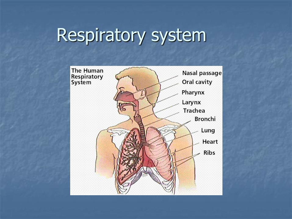
Respiratory system
Examination of the thorax
Inspection (globally, locally)
Percussion
Auscultation
Laboratory examination
Sputum
Induced sputum
BAL
Pulmonary function examination
Imaging studies
Respiratory system
Affection by parasites:
Initial port of entry
As a site of definitive multiplication and affection of host
As a transitory site of development
within host (not the port of entry)
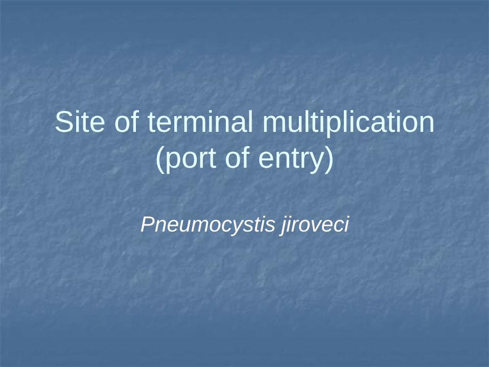
Site of terminal multiplication
(port of entry)
Pneumocystis jiroveci
Pneumocystis jiroveci
(Pneumocystis carinii)
Causative agent of pneumocystic pneumonia called in honor to czech parasitologist Otta Jírovec
Fungi like microorganism belonging to the group of yeasts (Sacharomyces cerevisae)
Distribution: cosmopolite
Associated with immunodeficiency
Pneumocystis isolated from dogs, monkeys, rats, mices, cats, sheeps…
Epidemiology
¾ of population - antibodies against P. jiroveci
Nutrition status of the population
Immunocompromised patients:
in the past 80%
at present: 10-20%
Mortality: HIV – 10-20%
Mortality increased in patients without therapy to 75-100%
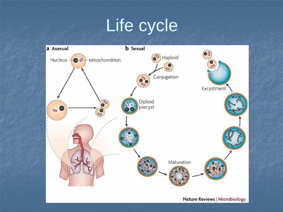
Life cycle
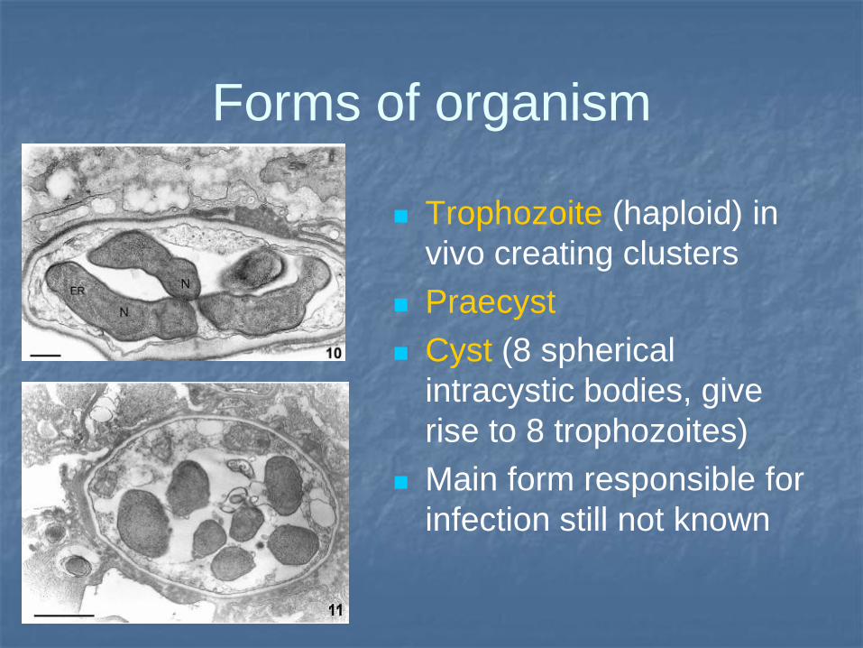
Forms of organism
Trophozoite (haploid) in
vivo creating clusters
Praecyst
Cyst (8 spherical
intracystic bodies, give
rise to 8 trophozoites)
Main form responsible for
infection still not known
Immunocompetent host
Exposition mostly at the age 3-4 years
Transmission: inhalation of infectious particles (most probably cysts)
Localisation in lungs: tight contact with type I. pneumocytes secured by presence of fibronectine
Macrophages in lungs destroy majority of pneumocysts
Pathophysiology
Destruction of basal membrane leads to
changes in permeability of
alveoli/capillaries
Changes in rate ventilation/perfusion
Situation similar to ARDS
Forms of infection
Immunocompetent individuals:
asymptomatic seroconversion
Immunocompromised population:
intersticial pneumonia (if CD4 decreased bellow
200/ul): proliferation of organism with low or
no inflammatory responce
Clinics
Fever
Non-productive mild cought
Dyspnoe; chest pain
Tachypnoe
Patients with prophylaxis: symptoms milder
BUT increased risk of dissemination;
increased risk of pneumothorax
Clinics II
Auscultation: crackles; often normal
finding
Extrapulmonary (about 1% of cases):
Lymphadenopathy
Hepatosplenomegaly
Chorioid leasions
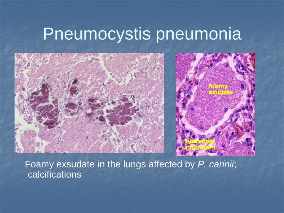
Pneumocystis pneumonia
Foamy exsudate in the lungs affected by P. carinii; calcifications
Diagnostics
Increased LDH: over 220 (non-specific)
Puls oxymetry: desaturation
Blood gases: hypoxemy, decreased CO2
RTG: intersticial pneumonia
High resolution CT
Bronchoscopy (associated with BAL)
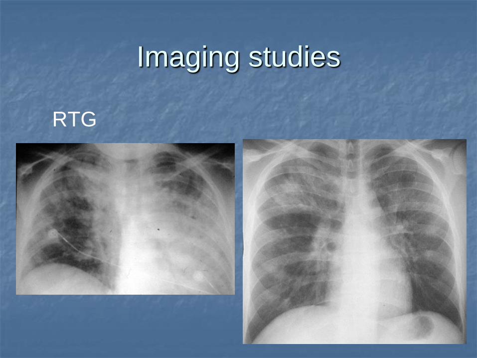
Imaging studies
RTG
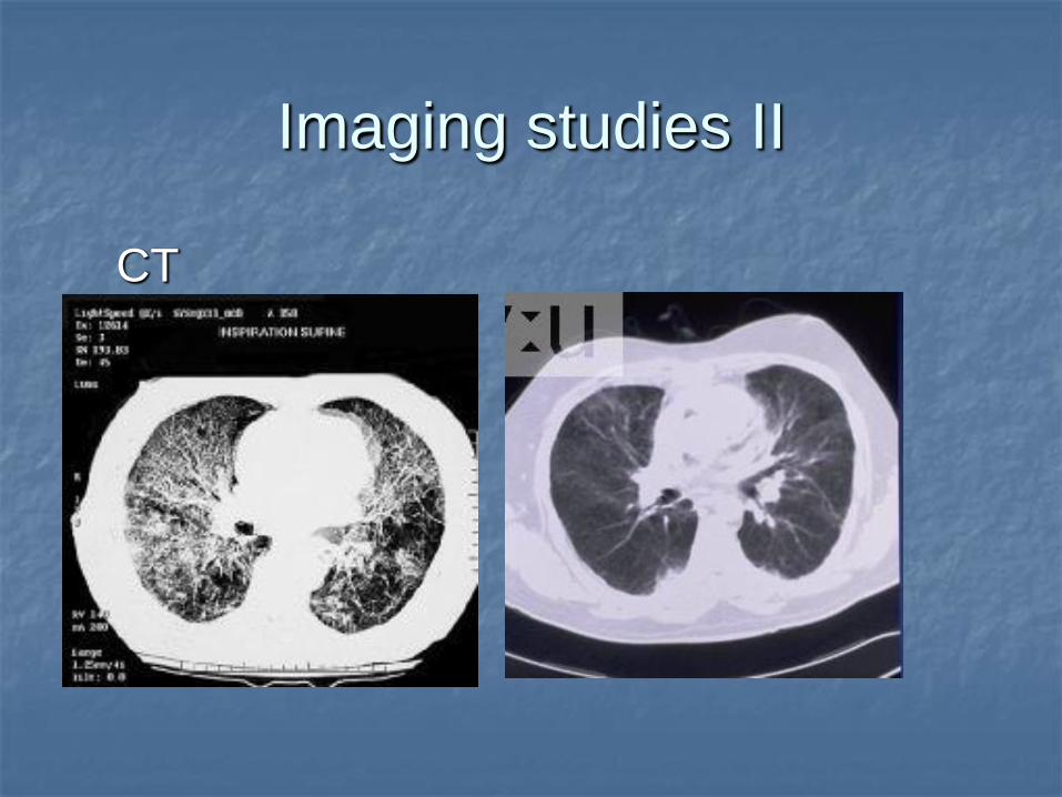
Imaging studies II
CT
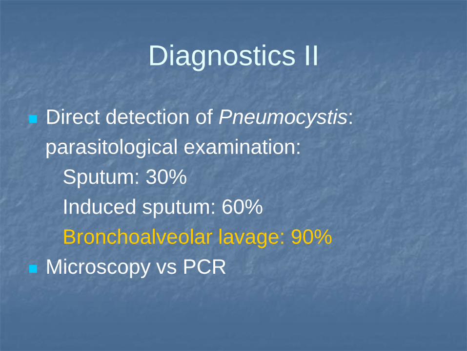
Diagnostics II
Direct detection of Pneumocystis:
parasitological examination:
Sputum: 30%
Induced sputum: 60%
Bronchoalveolar lavage: 90%
Microscopy vs PCR
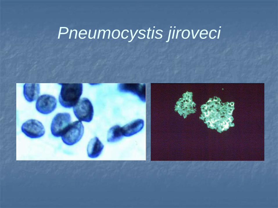
Pneumocystis jiroveci
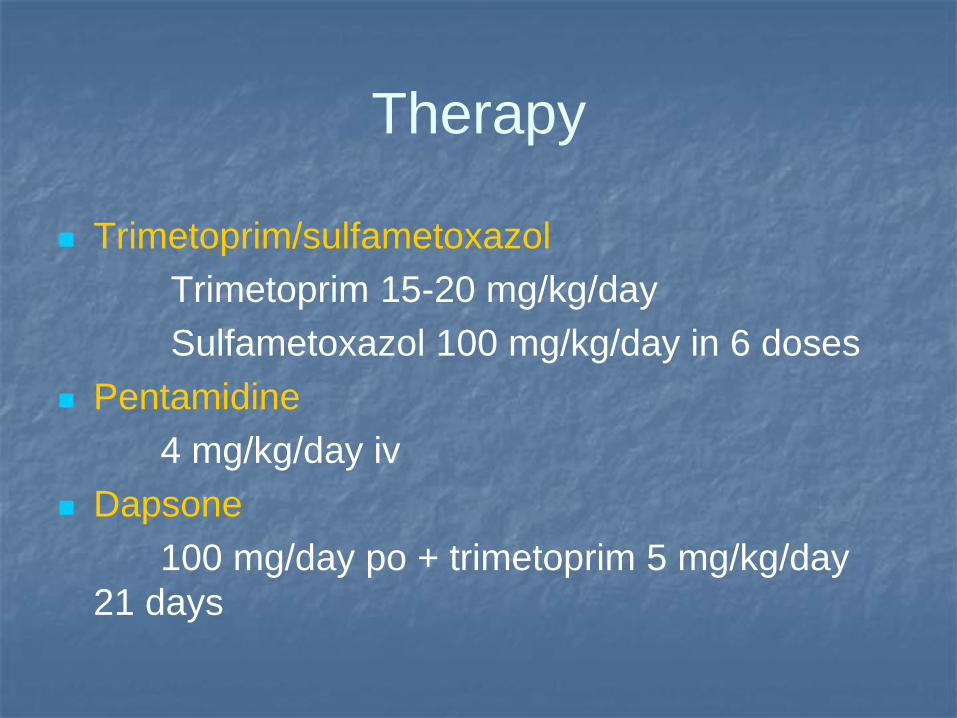
Therapy
Trimetoprim/sulfametoxazol
Trimetoprim 15-20 mg/kg/day
Sulfametoxazol 100 mg/kg/day in 6 doses
Pentamidine
4 mg/kg/day iv
Dapsone
100 mg/day po + trimetoprim 5 mg/kg/day
21 days
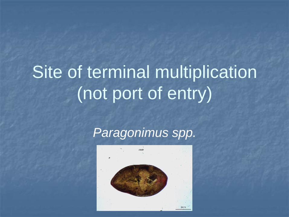
Site of terminal multiplication
(not port of entry)
Paragonimus spp.
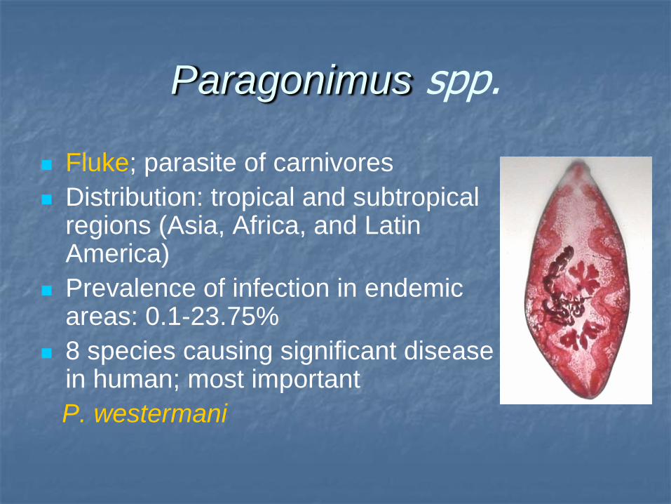
Paragonimus spp.
Fluke; parasite of carnivores
Distribution: tropical and subtropical regions (Asia, Africa, and Latin America)
Prevalence of infection in endemic areas: 0.1-23.75%
8 species causing significant disease in human; most important
P. westermani
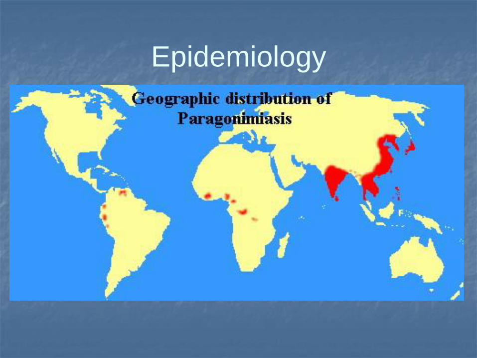
Epidemiology
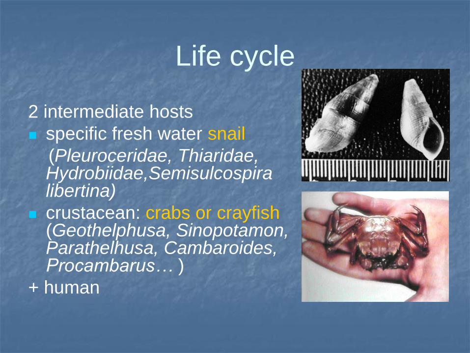
Life cycle
2 intermediate hosts
specific fresh water snail
(Pleuroceridae, Thiaridae, Hydrobiidae,Semisulcospira libertina)
crustacean: crabs or crayfish (Geothelphusa, Sinopotamon, Parathelhusa, Cambaroides, Procambarus… )
+ human
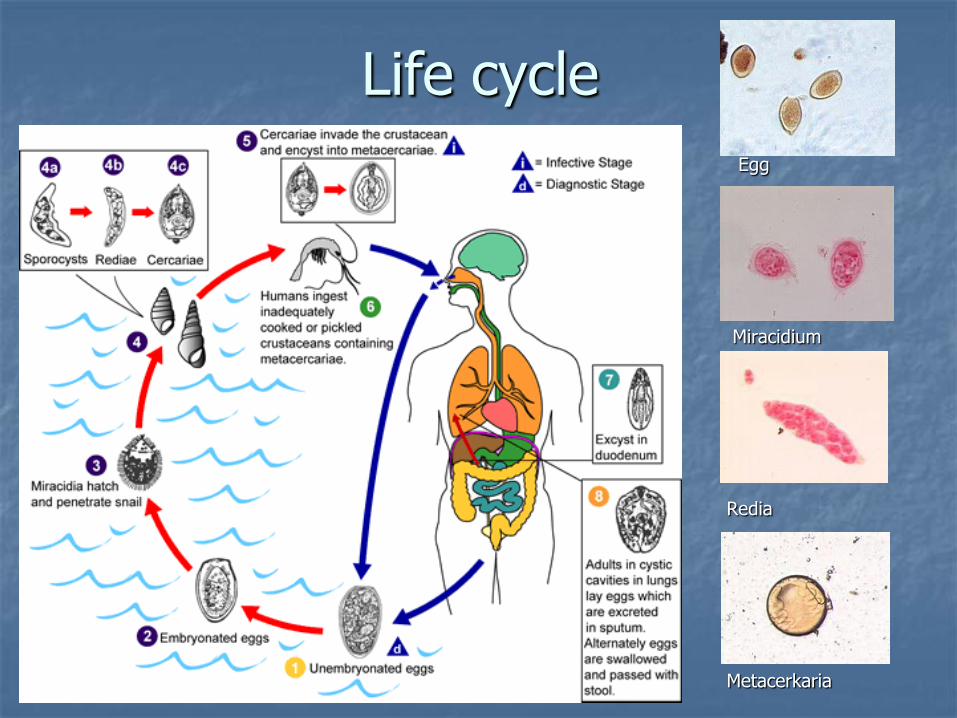
Life cycle
Egg
Miracidium
Redia
Metacerkaria
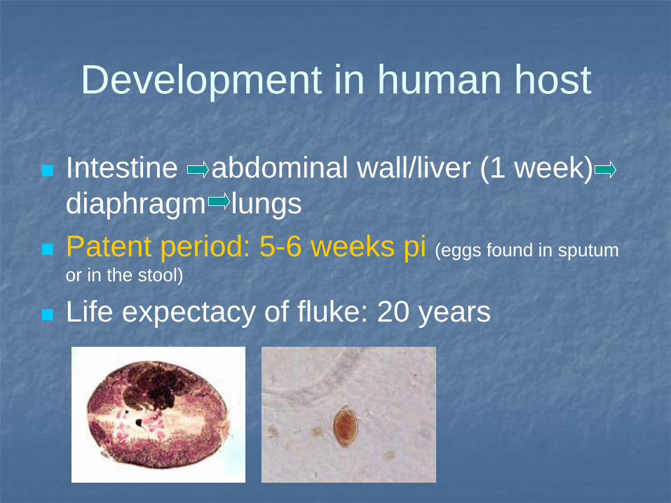
Development in human host
Intestine abdominal wall/liver (1 week)
diaphragm lungs
Patent period: 5-6 weeks pi (eggs found in sputum
or in the stool)
Life expectacy of fluke: 20 years
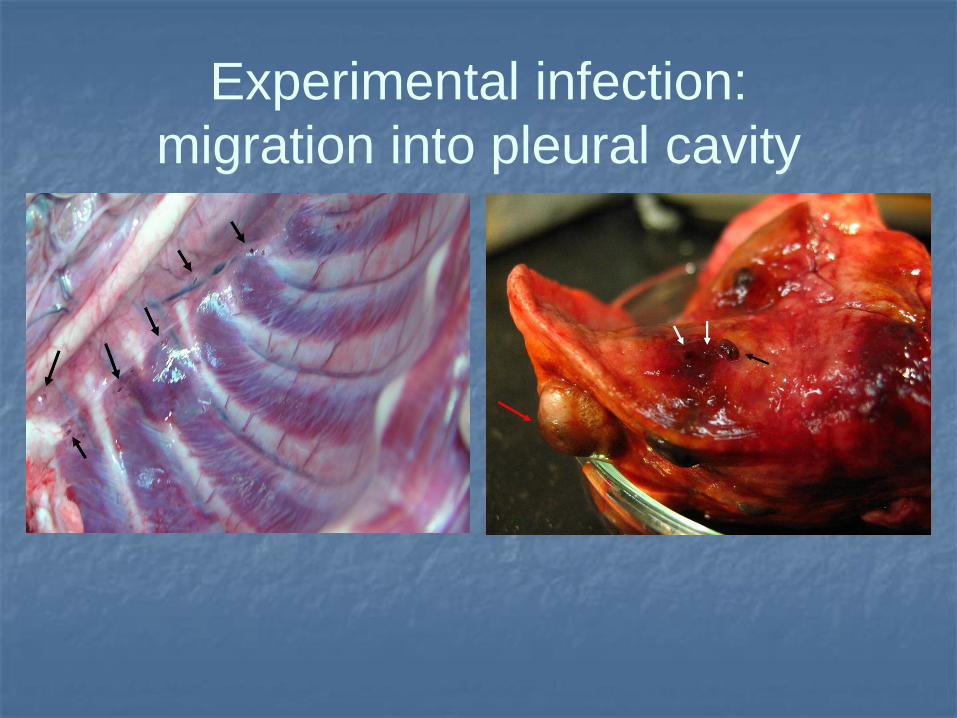
Experimental infection:
migration into pleural cavity
Clinics
Incubation period: 2-20 days
20% of patients are asymptomatic
Acute phase: intestinenal phase
respiratory phase
Chronic phase: pulmonary vs
extrapulmonary symptoms
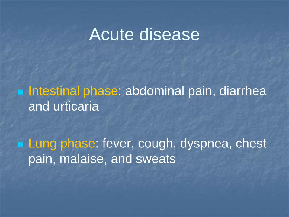
Acute disease
Intestinal phase: abdominal pain, diarrhea
and urticaria
Lung phase: fever, cough, dyspnea, chest
pain, malaise, and sweats
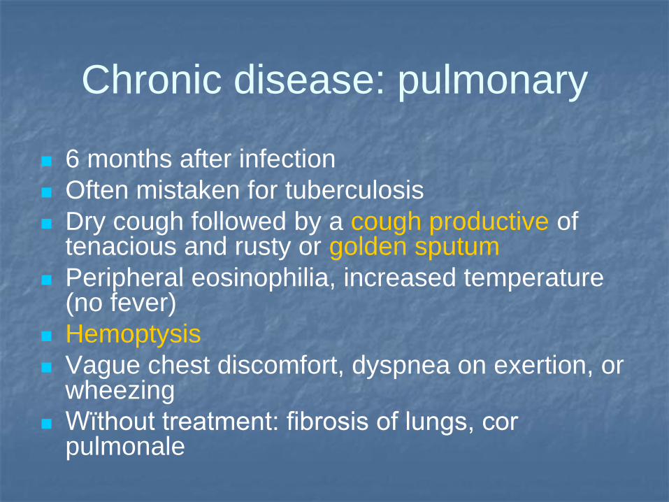
Chronic disease: pulmonary
6 months after infection
Often mistaken for tuberculosis
Dry cough followed by a cough productive of tenacious and rusty or golden sputum
Peripheral eosinophilia, increased temperature (no fever)
Hemoptysis
Vague chest discomfort, dyspnea on exertion, or wheezing
Wïthout treatment: fibrosis of lungs, cor pulmonale

Pathology
Cyst of fluke in trachea Flukes in lungs (exp. inf. dog)
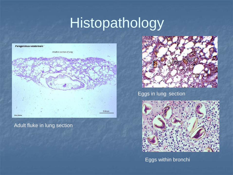
Histopathology
Eggs in lung section
Adult fluke in lung section
Eggs within bronchi
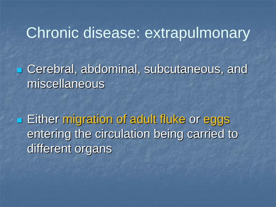
Chronic disease: extrapulmonary
Cerebral, abdominal, subcutaneous, and
miscellaneous
Either migration of adult fluke or eggs
entering the circulation being carried to
different organs
Extrapulmonary disease
Cysts in the intestinal wall, liver, spleen, abdominal wall, peritoneal cavity, or mesenteric lymph nodes: bloody diarrhea or abdominal pain.
Cerebral form: mainly in children
(up to 1%): meningoencephalitis (headache, vomiting, seizures, or weakness, Jacksons epilepsy)
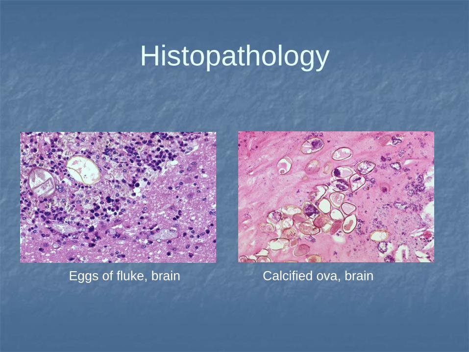
Histopathology
Eggs of fluke, brain Calcified ova, brain
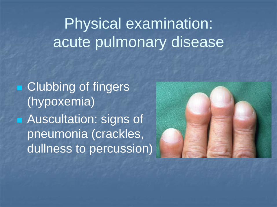
Physical examination:
acute pulmonary disease
Clubbing of fingers
(hypoxemia)
Auscultation: signs of
pneumonia (crackles,
dullness to percussion)
Physical examination:
chronic pulmonary disease
Similar to chronic bronchitis or
bronchiectasis
Profuse expectoration, pleuritic chest pain,
dyspnea, cough, occasional hemoptysis
Physical examination:
extrapulmonary disease
Cerebral: palsy, hemiplegia, seizures, and paraplegia
Ocular: impaired visual acuity: optic atrophy, papilledema, and hemianopsia
Spinal: monoplegia, paraplegia, lower extremity paresthesias, or sensory loss
Abdominal: palpable masses
Kidneys: hematuria
Subcutaneous: migratory swelling or subcutaneous nodules containing immature flukes (often in lower abdominal and inguinal region)
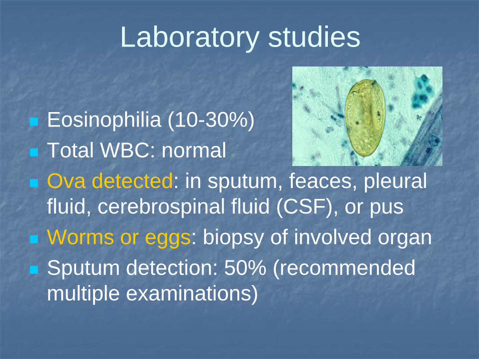
Laboratory studies
Eosinophilia (10-30%)
Total WBC: normal
Ova detected: in sputum, feaces, pleural
fluid, cerebrospinal fluid (CSF), or pus
Worms or eggs: biopsy of involved organ
Sputum detection: 50% (recommended
multiple examinations)
Imaging studies
RTG: ring shadows, representing cavitating lesions, fibrosis, nodules or linear infiltrates with calcified foci, loculated pleural effusions, and pleural thickening
soap bubble sign of frontal lobes
CT/NMR: cerebral calcification, cystic lesions, or hydrocephalus
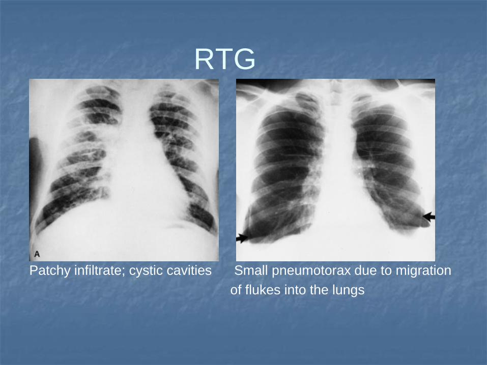
RTG
Patchy infiltrate; cystic cavities Small pneumotorax due to migration
of flukes into the lungs
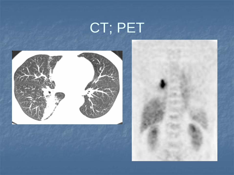
CT; PET
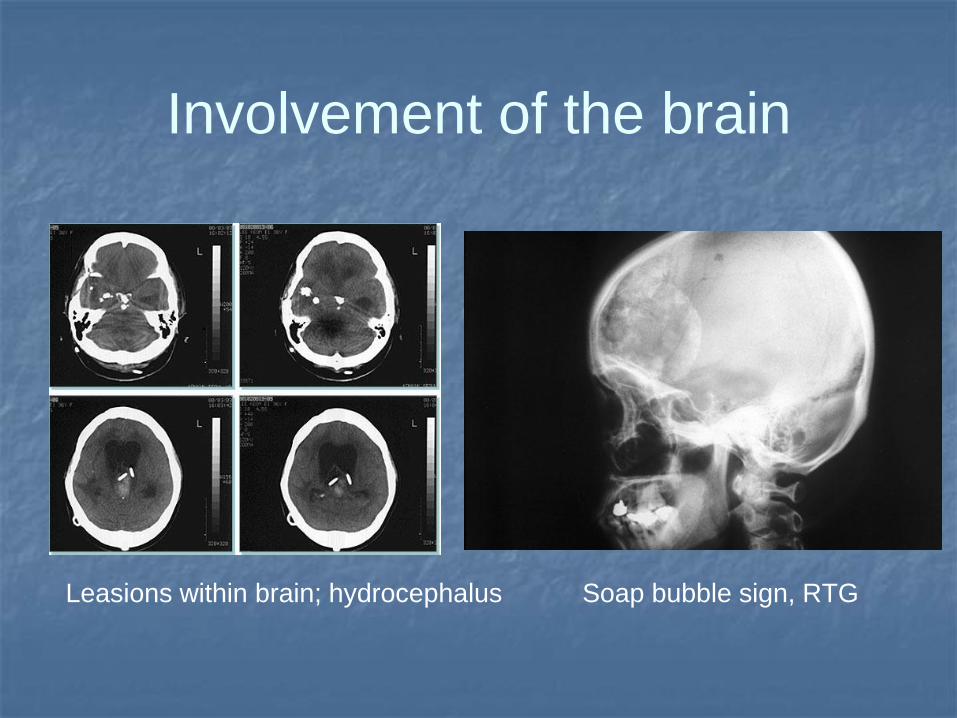
Involvement of the brain
Leasions within brain; hydrocephalus Soap bubble sign, RTG
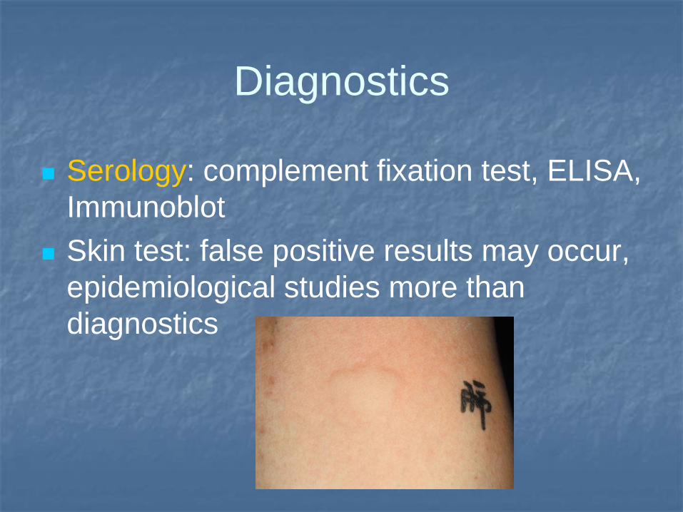
Diagnostics
Serology: complement fixation test, ELISA,
Immunoblot
Skin test: false positive results may occur,
epidemiological studies more than
diagnostics
Diagnostics
CSF: numerous eosinophils
Thoracentesis: serosanguineous, has more than 1000 red cells with accompanying eosinophilia; low glucose
Lung biopsy: multiple worms or eggs
Adults found in cysts (mostly right lung): granulation tissue with fibroblasts, mononuclear cells, plasma cells, lymphoid cells, and eosinophils; Charcot-Leyden crystals
Therapy
Praziquantel: 25 mg/kg PO tid for 2 d
Extrapulmonary lesions should be surgically
excised.
An intraventricular shunt may be needed to
manage hydrocephalus
Persistent seizures in cerebral involvement
Prognosis: good, with therapeutic cure rates
between 90 and 100%
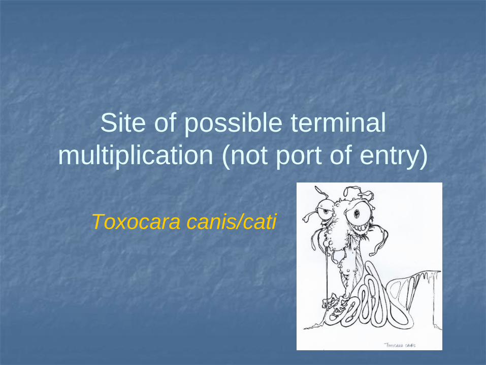
Site of possible terminal
multiplication (not port of entry)
Toxocara canis/cati
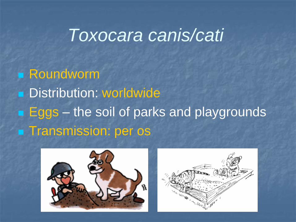
Toxocara canis/cati
Roundworm
Distribution: worldwide
Eggs – the soil of parks and playgrounds
Transmission: per os
Epidemiology
Epidemiology: 2-5% positive rate in urban
Western countries
14.2-37% in rural areas of Western countries
Tropical countries:
63.2% in Bali,
86% in Saint Lucia (West Indies), and
92.8% in La Reunion (French Overseas
Territories, Indian Ocean)
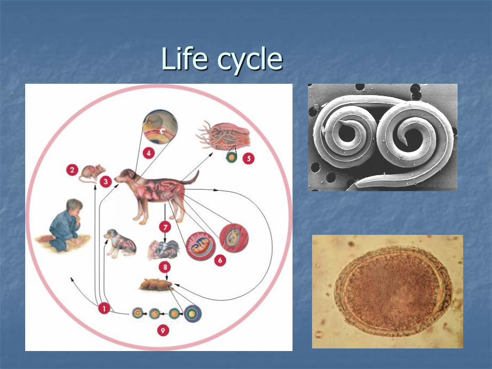
Life cycle
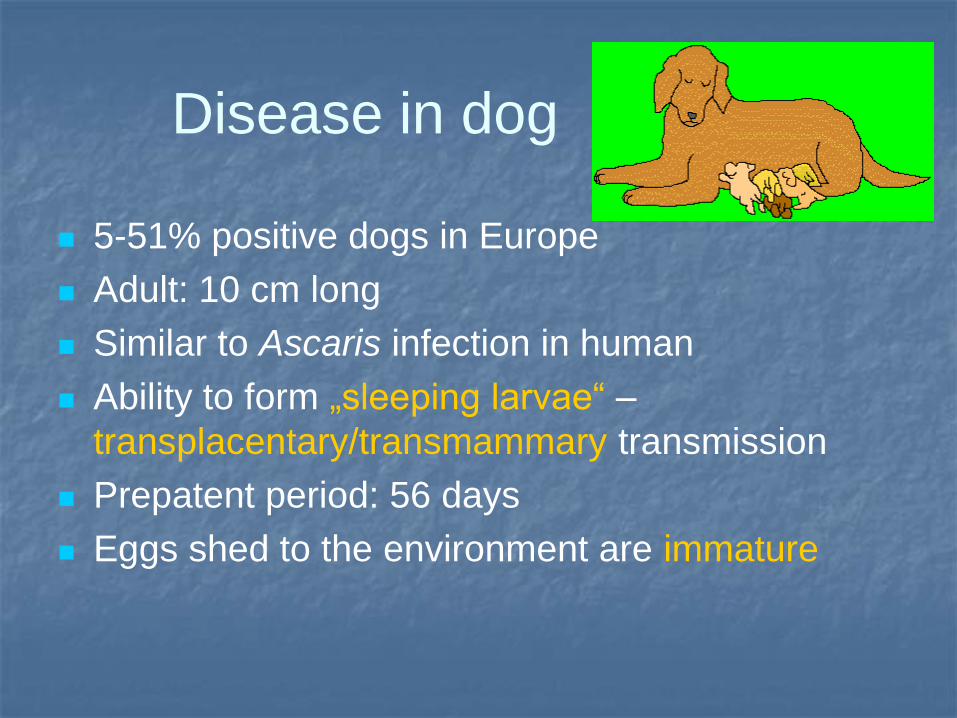
Disease in dog
5-51% positive dogs in Europe
Adult: 10 cm long
Similar to Ascaris infection in human
Ability to form „sleeping larvae“ –
transplacentary/transmammary transmission
Prepatent period: 56 days
Eggs shed to the environment are immature
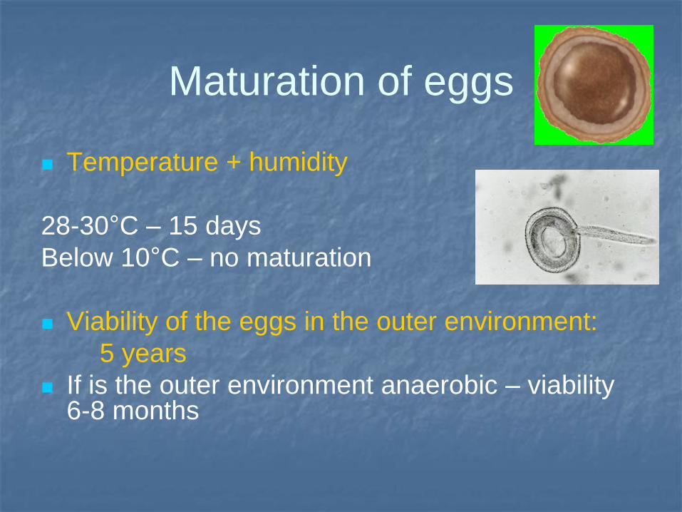
Maturation of eggs
Temperature + humidity
28-30°C – 15 days
Below 10°C – no maturation
Viability of the eggs in the outer environment:
5 years
If is the outer environment anaerobic – viability 6-8 months
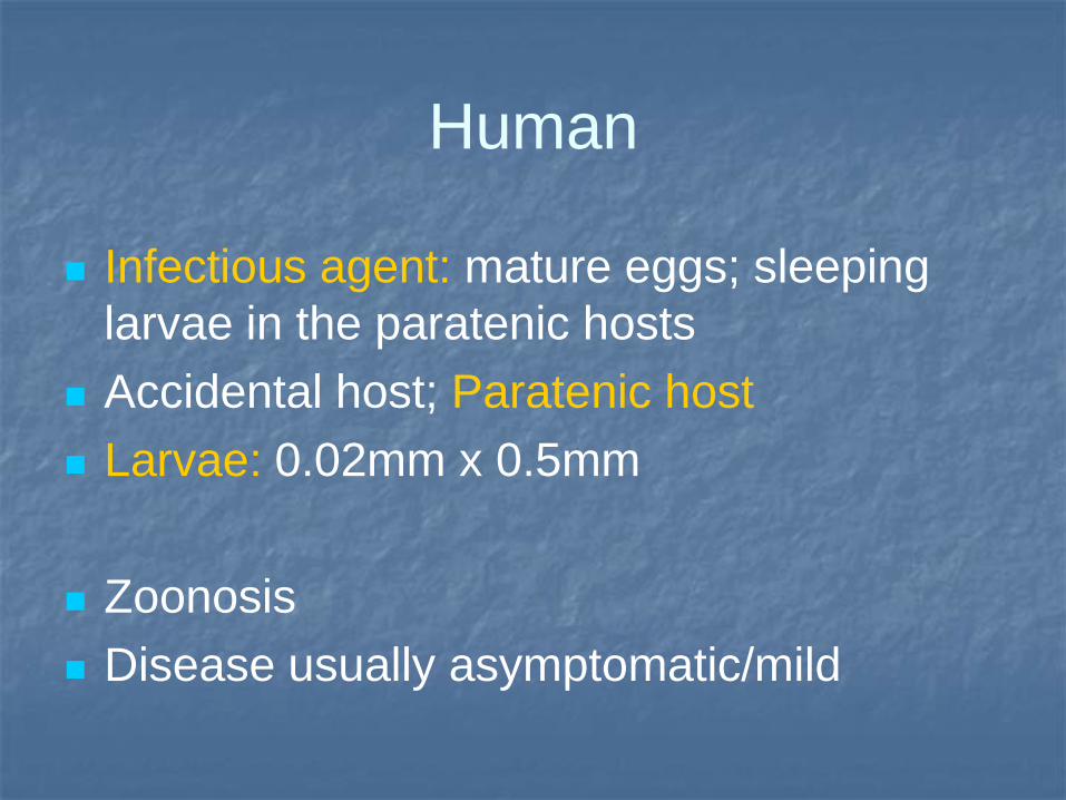
Human
Infectious agent: mature eggs; sleeping
larvae in the paratenic hosts
Accidental host; Paratenic host
Larvae: 0.02mm x 0.5mm
Zoonosis
Disease usually asymptomatic/mild
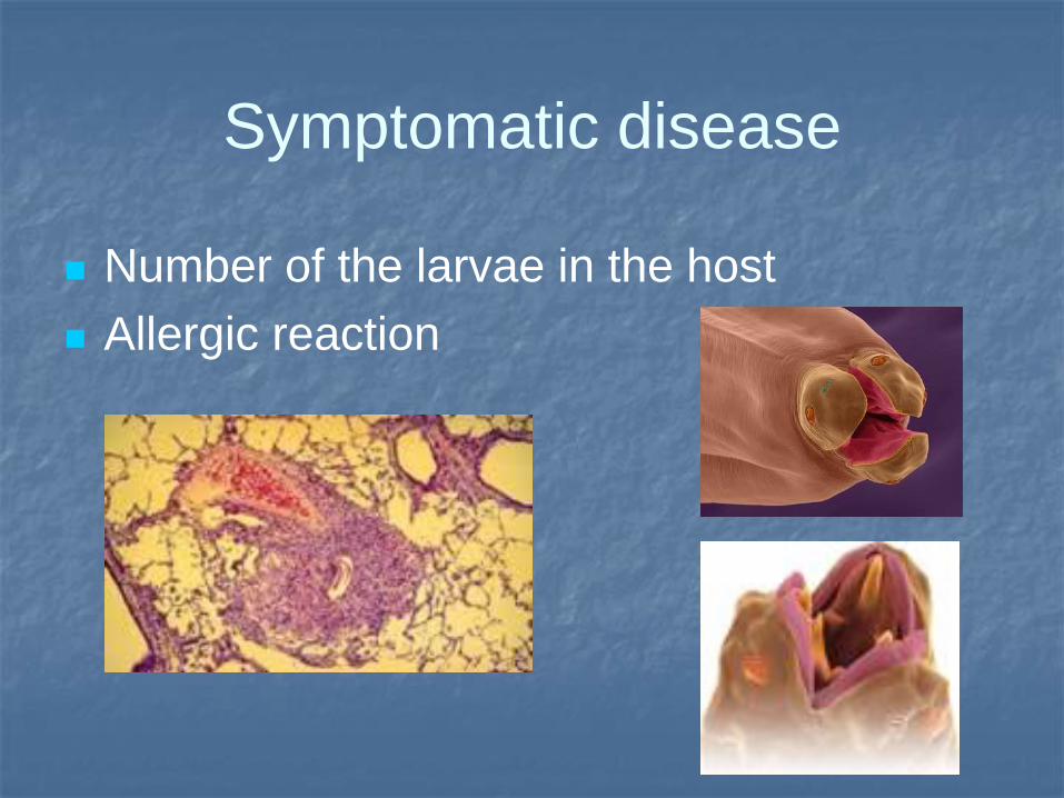
Symptomatic disease
Number of the larvae in the host
Allergic reaction
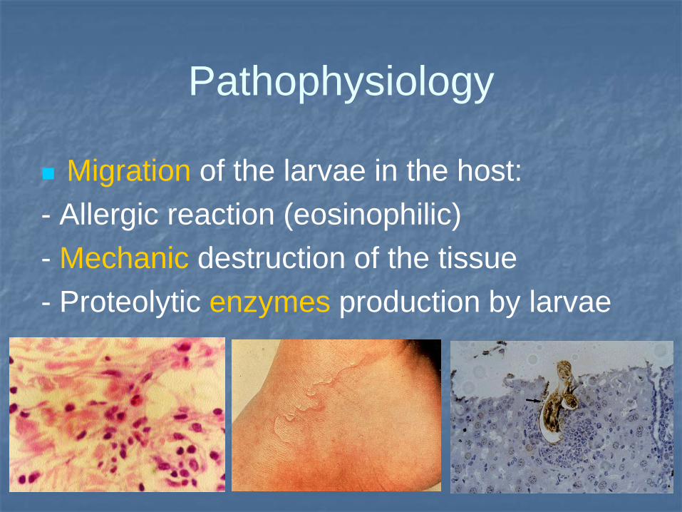
Pathophysiology
Migration of the larvae in the host:
- Allergic reaction (eosinophilic)
- Mechanic destruction of the tissue
- Proteolytic enzymes production by larvae
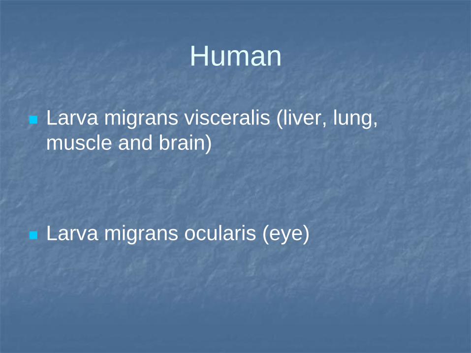
Human
Larva migrans visceralis (liver, lung,
muscle and brain)
Larva migrans ocularis (eye)
Anamnesis
Living with or raising dogs and cats
Eating without hand washing
Infection from contact with soil from a
yard, sandbox, park, or playground
Larva migrans visceralis
Diarrhoea, abdominal pain, anorexia, nausea, fatigue
Pruritus, rash
Liver
Lungs: Cought, temperature (38°C), bronchospasm, wheezing
Brain: Difficulty sleeping, abdominal pain, headaches, and behavioral problems, seizures, temperature
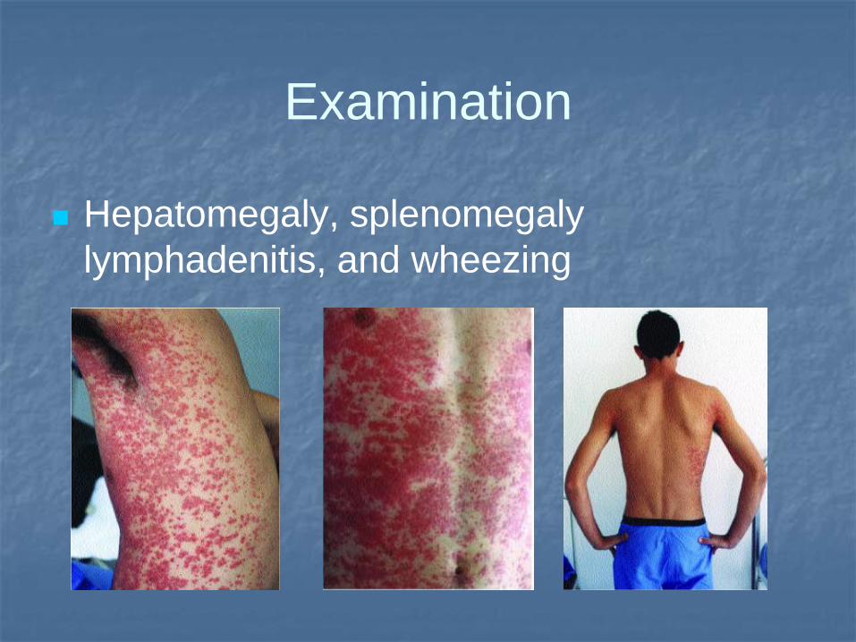
Examination
Hepatomegaly, splenomegaly
lymphadenitis, and wheezing
Larva migrans visceralis: laboratory
Elevation of the leukocytes
Eosinophilia (20-90%)
Diagnostics: Serology (ELISA)
Biopsy
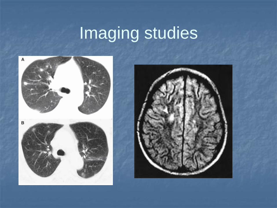
Imaging studies
Therapy
Dont treat positive titres if person
asymptomatic!!!!
Mebendazole (Vermox) - 25 mg/kg/d PO
single dose for 4 wk
Albendazole (Albenza) - 10 mg/kg/d PO
single dose for 4 wk
Site of possible terminal
multiplication (not port of entry)
Echinococcus
granulosus/multilocularis
Transitory site of development
Ascaris lumbricoides, Strongyloides stercoralis, Ancylostoma duodenale, Necator americanus,
Toxocara canis/cati, Schistosomiasis, Echinococcosis
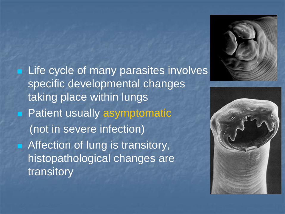
Life cycle of many parasites involves
specific developmental changes
taking place within lungs
Patient usually asymptomatic
(not in severe infection)
Affection of lung is transitory,
histopathological changes are
transitory
Migration of parasites: eosinophilia
Lung phase: (pneumonia): damage of
cappillaries and alveoli -
cought, chest pain, subfebrilia
blood in the sputum
Sputum positive for detection of larvae of the
parasites (if examinated)
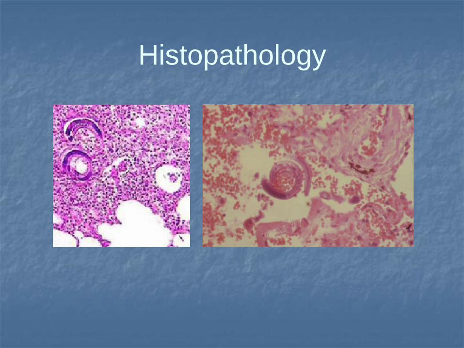
Histopathology
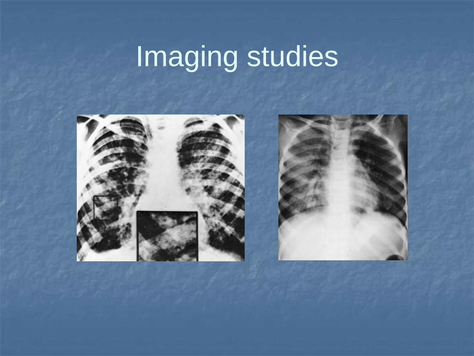
Imaging studies
Symptoms lasting for particular time
After finish of development parasite
migrates to definitive pathological site
(intestine, portal venous system, …)
Therapy: specific (low detection);
corticosteroids


![[ANELOSIMUS PARASITIC ARCHITECTURE]](https://img.pdfslide.net/doc/110x75/568bf0fb1a28ab893391962f/anelosimus-parasitic-architecture.jpg)