Embed Size (px)
Citation preview
sensors
Review
Magnetic Particles Coupled to Disposable ScreenPrinted Transducers for Electrochemical Biosensing
Paloma Yáñez-Sedeño *, Susana Campuzano and José M. Pingarrón
Departamento de Química Analítica, Facultad de CC. Químicas, Universidad Complutense de Madrid,E-28040 Madrid, Spain; [email protected] (S.C.); [email protected] (J.M.P.)* Correspondence: [email protected]; Tel.: +34-91-394-4317
Academic Editor: Jesus Iniesta ValcarcelReceived: 2 September 2016; Accepted: 22 September 2016; Published: 25 September 2016
Abstract: Ultrasensitive biosensing is currently a growing demand that has led to the developmentof numerous strategies for signal amplification. In this context, the unique properties of magneticparticles; both of nano- and micro-size dimensions; have proved to be promising materials to becoupled with disposable electrodes for the design of cost-effective electrochemical affinity biosensingplatforms. This review addresses, through discussion of selected examples, the way that nano- andmicro-magnetic particles (MNPs and MMPs; respectively) have contributed significantly to thedevelopment of electrochemical affinity biosensors, including immuno-, DNA, aptamer and otheraffinity modes. Different aspects such as type of magnetic particles, assay formats, detectiontechniques, sensitivity, applicability and other relevant characteristics are discussed. Researchopportunities and future development trends in this field are also considered.
Keywords: magnetic materials; screen-printed electrodes; electrochemical affinity biosensors
1. Introduction
Applications of superparamagnetic iron oxide particles with sizes between a few nanometersand micrometers units are numerous and extend to fields as varied as clinical analysis, food andenvironmental safety, drug delivery, or molecular imaging. The possibility of easy functionalizationwith diverse reactive moieties such as carboxyl, amine, aldehyde, hydroxyl, thiol or tosyl), as well as theability for interact with biomolecules (e.g., antibodies, oligonucleotides, protein A or G, streptavidin),and the development of efficient methods for the synthesis of magnetic nanoparticles with improvedproperties when associated with different materials, makes them a useful and versatile tool to recognizea variety of targets. Moreover, the magnetic properties of these particles allow enhancing the isolationand preconcentration of the analyte from complex samples, and providing a useful technology byimplementing various types of magnetic devices including biosensors or microfluidic platformsamong others.
Electrochemical affinity biosensors are usually prepared by immobilization of the bioreceptoronto an electrode surface whose selection is crucial to achieve an efficient and stable incorporation ofthe recognition element, as well as a high sensitivity. The development of these biosensors typicallyrequires various steps and several calibration points. Moreover, small sample volumes are analyzed.These reasons make screen printed electrodes (SPEs) highly attractive and preferable over other typesof electrodes for such applications [1]. SPEs are suitable to be fabricated with different materials, to bedrawn in diverse geometries and arrays [2], and characterized by the possibility for mass productionand low fabrication cost. In addition, the small size of these electrodes enables drops to be usedfor the assays and, importantly in the context of this review, the planar shape of SPEs facilitatesthe incorporation of magnetic bioconjugates by simple attraction using a magnet positioned underthe electrode.
Sensors 2016, 16, 1585; doi:10.3390/s16101585 www.mdpi.com/journal/sensors
Sensors 2016, 16, 1585 2 of 32
Coupling of electrochemical transduction with the use of magnetic nano/microcarriers hasgreatly contributed to significant improvements in the performance of electrochemical biosensors.The application of superparamagnetic particles based on iron oxide functionalized with diversereactive groups for the easy, fast and selective capture of a specific target molecule from a complexsample, and further coupling with disposable electrochemical sensors, is nowadays a well-establishedmethodology [3]. These particles enable the efficient immobilization of capture bioreceptors, allowfaster assay kinetics, minimize unspecific adsorptions frequently occurring in complex samples,and avoid the need for delicate electrode preparation to enable control of density and orientation ofrecognition probes at the disposable electrode surface [4]. Other applications take advantage also ofthe easy handling, modification and manipulation of these magnetic particles for the preparation ofmagnetic bioconjugates useful as advanced labels for amplification signal purposes.
This review discusses briefly the main characteristics and advantages offered by micro- andnano-magnetic particles in electrochemical biosensing making special emphasis on recent advancesin the field of electrochemical affinity platforms involving the coupling of these magnetic materialsand disposable electrochemical transducers. The highlighted approaches are classified accordingto the size of the magnetic particles used, as well as to its use as electrode modifiers or carriers ofsignaling molecules and type of biosensing platform. Single- or multiplexed capabilities of the magneticparticles-based disposable biaffinity platforms are also differentiated. In addition, the bottlenecks andpossible research directions are also pointed out.
2. Micromagnetic Particles
Micro-magnetic particles (MMPs) consisted of a dispersion of Fe2O3 and Fe3O4 magnetic materialcoated by a polymeric thin shell that define also a surface area for interaction by coupling or adsorptionwith a variety of molecules.
Currently there are commercially available MMPs modified with various functional groups (tosyl,amine, carboxyl or epoxy) and ligands such as antibodies, nucleic acids, proteins or small moleculesfor specific applications. Hence, MMPs can be tailored-modified in an easy way with a whole range ofligands in bioaffinity reactions [5].
These highly reactive materials of relatively low volume constitute a versatile tool in thedevelopment of biosensors, as they exhibit a large surface area for binding in an oriented wayrecognition elements or specific analytes, which can be easily isolated from the solution with asmall magnet and promptly redispersed when the magnet is placed out. In addition to significantlyaccelerate the kinetics of the recognition processes, the reagents that are not specifically linked orunwanted sample components can be removed easily and efficiently by magnetically controlledwashing, thus giving them utility for purification and preconcentration purposes and suitability forautomation [6]. The use of MMPs greatly facilitates the preparation of biosensors and detectionsteps [7], leading to strategies that generally exhibit better detection limits (LODs) and lower matrixeffects than conventional integrated configurations [8,9]. Additional advantages of the use of MMPs arethe small sample volumes of solution required for their modification, the large quantity of recognitionelement that can be immobilized in a single step, and that MMPs bioconjugates can be stored forseveral weeks without loss of activity. This last advantage is crucial when a large number of sampleshave to be analyzed, and could lead to a meaningful reduction of the analysis time.
Probably, the major drawback associated with the use of MMPs-based electrochemical sensorsis the lack of uncomplicated, low cost and practical magnetic electrodes for MMPs manipulation.Direct sensing has been appropriately solved by using planar disposable electrodes with small magnetsplaced below their surface. This makes it possible to concentrate MMPs on the active area of theworking electrode in the best conditions for the electrochemical measurement once the affinity reactionstook place [1,10].
Sensors 2016, 16, 1585 3 of 32
The most important characteristics of the electrochemical bioaffinity sensors involving the use ofMMPs and disposable electrodes, discussed in Sections 2.1–2.4, are summarized in Tables S1–S3 (in theSupporting Information).
2.1. Magnetic Microparticles-Screen Printed Electrodeselectrochemical Immunosensors
Moreno-Guzmán et al. [11] developed an electrochemical immunosensor based on MMPs andscreen-printed carbon electrodes (SPCEs) for the determination of cortisol, an important stressbiomarker of great relevance in sport medicine. The specific anti-cortisol antibody was immobilizedonto Protein A (Prot A)-MMPs, and cortisol quantification was implemented by a direct competitiveimmunoassay involving cortisol antigen labeled with alkaline phosphatase (AP) and 1-naphthylphosphate as AP substrate. Differential pulse voltammetry (DPV) was the electrochemical techniqueused to detect the generated 1-naphthol. A calibration plot with a linear range between 5.0 × 10⁻³ and150 ng·mL−1 cortisol, and a LOD of 3.5 pg·mL−1 were obtained. The usefulness of this immunosensingapproach was demonstrated by application to certified human sera containing cortisol at two differentconcentration levels.
The same group developed also a similar immunosensing approach for testosterone withhorseradish peroxidase (HRP)-labeled antigen and amperometric detection at SCPEs using the systemH2O2/HQ [12]. This immunosensor exhibited a linear calibration plot between 5.0 × 10⁻³ and50 ng·mL−1 testosterone and a LOD of 1.7 pg·mL−1, and was successfully applied to the determinationof the hormone in spiked serum.
With the objective of specifically detect Streptococcus pneumoniae, disposable amperometricmagnetoimmunosensors using ProtA-MMPs, and a sandwich format with the same antibody(produced in the laboratory) used unconjugated as capture (Ab) and HRP-labeled as detector (HRP-Ab),were reported. Amperometric detection of H2O2 at tetrathiafulvalene (TTF)-modified Au-SPEswas employed [13]. LODs of 1.5104 and 6.3 × 10⁵ colony forming unit (cfu) mL−1 for modelcapsulated (Dawn) and non-encapsulated (R6) Streptococcus pneumoniae selected strains, were achieved,respectively, with no need for pre-concentration or pre-enrichment steps. Results demonstrated a goodselectivity of the immunosensor against other closely related streptococci, and the analytical utility ofthe developed methodology for application to inoculated urine.
A sandwich immunoassay was developed for myoglobin (a protein marker for muscle damage)using immunoconjugates of silver nanoparticles as labels and immunoconjugates of MMPs asmicrocarriers for selective capture of the target protein [14]. The sandwich immunoconjugates wheremyoglobin was linked to both tosyl activated-MMPs and silver nanoparticles were magneticallycaptured on the surface of SPCEs. To perform the electrochemical transduction, ammonium thiocyanatewas added to break the captured conjugates and stabilize silver. Finally, the silver nanoparticles weredissolved to form silver ions and, after nucleation and accumulation steps of silver on the electrodesurface, the stripping voltammetric signal corresponding to silver removal from the electrode surfacewas recorded and related to the myoglobin concentration. This approach provided a dynamic rangeranging between 0.2–20 ng·mL−1 (10 pM–1 nM), far below the normal human serum myoglobinlevels (30–90 ng·mL−1) and the elevated levels found within an hour of onset of myocardial infarction(200–900 ng·mL−1).
Moreno-Guzmán et al. [15] designed a sandwich immunoassay for prolactin (PRL), a relevanthormone in lactation and reproduction, using streptavidin (Strep)-MMPs, SPCEs and a detectorantibody labeled with AP. Prolactin quantification was accomplished by DPV of generated 1-naphtol byhydrolysis of 1-naphtyl phosphate in the presence of AP. A linear range between 10 and 2000 ng·mL−1
prolactin (slope value of 7.0 nA·mL ng−1) and a LOD of 3.74 ng·mL−1 were found. The applicabilityof this approach was demonstrated by analyzing spiked certified human serum.
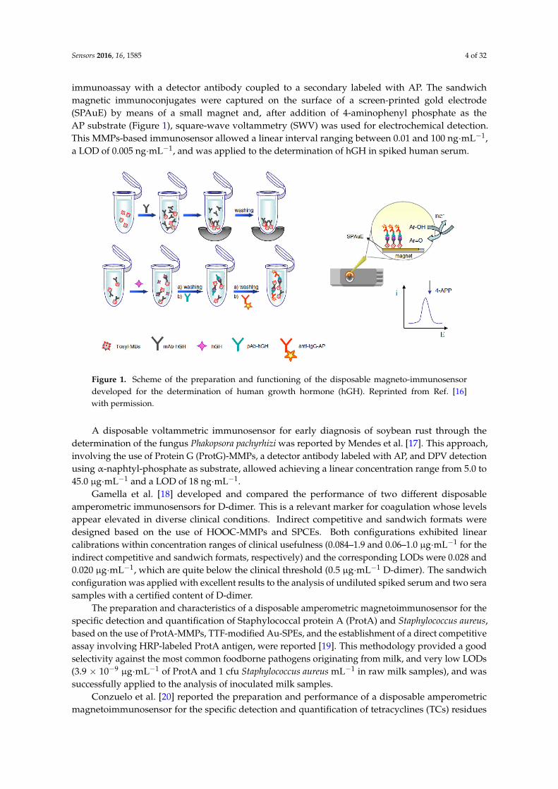
One year after, this same group prepared an electrochemical magnetoimmunosensor forthe detection of human growth hormone (hGH) [16] involving the covalen immobilization of amonoclonal capture antibody on tosyl-activated MMPs, and the establishment of a sandwich-type
Sensors 2016, 16, 1585 4 of 32
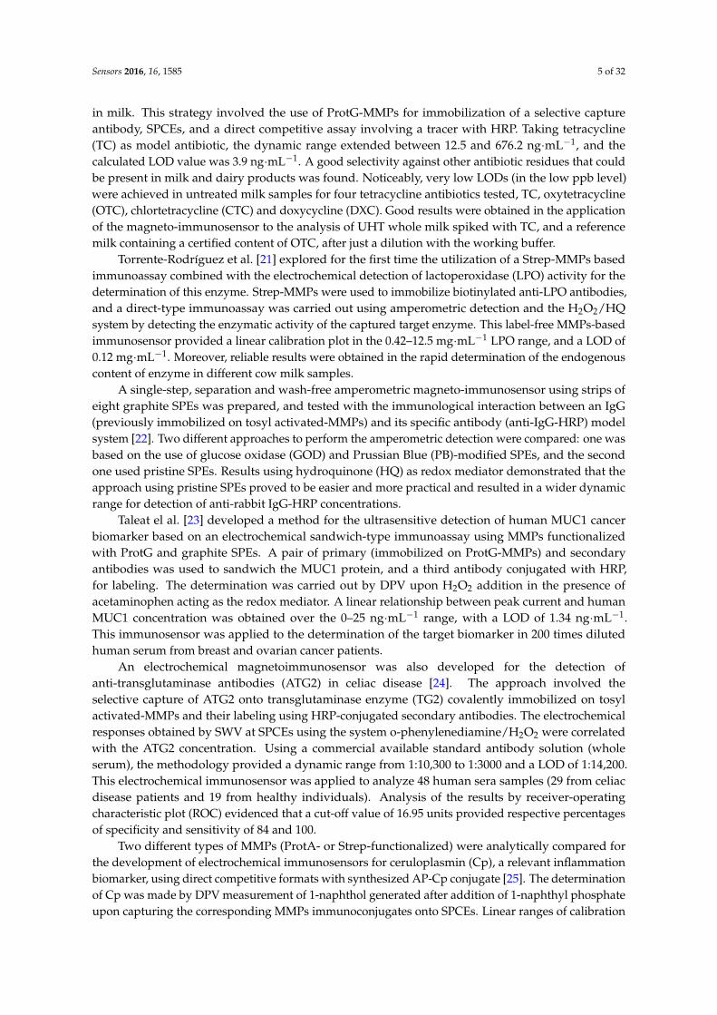
immunoassay with a detector antibody coupled to a secondary labeled with AP. The sandwichmagnetic immunoconjugates were captured on the surface of a screen-printed gold electrode(SPAuE) by means of a small magnet and, after addition of 4-aminophenyl phosphate as theAP substrate (Figure 1), square-wave voltammetry (SWV) was used for electrochemical detection.This MMPs-based immunosensor allowed a linear interval ranging between 0.01 and 100 ng·mL−1,a LOD of 0.005 ng·mL−1, and was applied to the determination of hGH in spiked human serum.
Figure 1. Scheme of the preparation and functioning of the disposable magneto-immunosensordeveloped for the determination of human growth hormone (hGH). Reprinted from Ref. [16]with permission.
A disposable voltammetric immunosensor for early diagnosis of soybean rust through thedetermination of the fungus Phakopsora pachyrhizi was reported by Mendes et al. [17]. This approach,involving the use of Protein G (ProtG)-MMPs, a detector antibody labeled with AP, and DPV detectionusing α-naphtyl-phosphate as substrate, allowed achieving a linear concentration range from 5.0 to45.0 µg·mL−1 and a LOD of 18 ng·mL−1.
Gamella et al. [18] developed and compared the performance of two different disposableamperometric immunosensors for D-dimer. This is a relevant marker for coagulation whose levelsappear elevated in diverse clinical conditions. Indirect competitive and sandwich formats weredesigned based on the use of HOOC-MMPs and SPCEs. Both configurations exhibited linearcalibrations within concentration ranges of clinical usefulness (0.084–1.9 and 0.06–1.0 µg·mL−1 for theindirect competitive and sandwich formats, respectively) and the corresponding LODs were 0.028 and0.020 µg·mL−1, which are quite below the clinical threshold (0.5 µg·mL−1 D-dimer). The sandwichconfiguration was applied with excellent results to the analysis of undiluted spiked serum and two serasamples with a certified content of D-dimer.
The preparation and characteristics of a disposable amperometric magnetoimmunosensor for thespecific detection and quantification of Staphylococcal protein A (ProtA) and Staphylococcus aureus,based on the use of ProtA-MMPs, TTF-modified Au-SPEs, and the establishment of a direct competitiveassay involving HRP-labeled ProtA antigen, were reported [19]. This methodology provided a goodselectivity against the most common foodborne pathogens originating from milk, and very low LODs(3.9 × 10⁻⁹ µg·mL−1 of ProtA and 1 cfu Staphylococcus aureus mL−1 in raw milk samples), and wassuccessfully applied to the analysis of inoculated milk samples.
Conzuelo et al. [20] reported the preparation and performance of a disposable amperometricmagnetoimmunosensor for the specific detection and quantification of tetracyclines (TCs) residues
Sensors 2016, 16, 1585 5 of 32
in milk. This strategy involved the use of ProtG-MMPs for immobilization of a selective captureantibody, SPCEs, and a direct competitive assay involving a tracer with HRP. Taking tetracycline(TC) as model antibiotic, the dynamic range extended between 12.5 and 676.2 ng·mL−1, and thecalculated LOD value was 3.9 ng·mL−1. A good selectivity against other antibiotic residues that couldbe present in milk and dairy products was found. Noticeably, very low LODs (in the low ppb level)were achieved in untreated milk samples for four tetracycline antibiotics tested, TC, oxytetracycline(OTC), chlortetracycline (CTC) and doxycycline (DXC). Good results were obtained in the applicationof the magneto-immunosensor to the analysis of UHT whole milk spiked with TC, and a referencemilk containing a certified content of OTC, after just a dilution with the working buffer.
Torrente-Rodríguez et al. [21] explored for the first time the utilization of a Strep-MMPs basedimmunoassay combined with the electrochemical detection of lactoperoxidase (LPO) activity for thedetermination of this enzyme. Strep-MMPs were used to immobilize biotinylated anti-LPO antibodies,and a direct-type immunoassay was carried out using amperometric detection and the H2O2/HQsystem by detecting the enzymatic activity of the captured target enzyme. This label-free MMPs-basedimmunosensor provided a linear calibration plot in the 0.42–12.5 mg·mL−1 LPO range, and a LOD of0.12 mg·mL−1. Moreover, reliable results were obtained in the rapid determination of the endogenouscontent of enzyme in different cow milk samples.
A single-step, separation and wash-free amperometric magneto-immunosensor using strips ofeight graphite SPEs was prepared, and tested with the immunological interaction between an IgG(previously immobilized on tosyl activated-MMPs) and its specific antibody (anti-IgG-HRP) modelsystem [22]. Two different approaches to perform the amperometric detection were compared: one wasbased on the use of glucose oxidase (GOD) and Prussian Blue (PB)-modified SPEs, and the secondone used pristine SPEs. Results using hydroquinone (HQ) as redox mediator demonstrated that theapproach using pristine SPEs proved to be easier and more practical and resulted in a wider dynamicrange for detection of anti-rabbit IgG-HRP concentrations.
Taleat el al. [23] developed a method for the ultrasensitive detection of human MUC1 cancerbiomarker based on an electrochemical sandwich-type immunoassay using MMPs functionalizedwith ProtG and graphite SPEs. A pair of primary (immobilized on ProtG-MMPs) and secondaryantibodies was used to sandwich the MUC1 protein, and a third antibody conjugated with HRP,for labeling. The determination was carried out by DPV upon H2O2 addition in the presence ofacetaminophen acting as the redox mediator. A linear relationship between peak current and humanMUC1 concentration was obtained over the 0–25 ng·mL−1 range, with a LOD of 1.34 ng·mL−1.This immunosensor was applied to the determination of the target biomarker in 200 times dilutedhuman serum from breast and ovarian cancer patients.
An electrochemical magnetoimmunosensor was also developed for the detection ofanti-transglutaminase antibodies (ATG2) in celiac disease [24]. The approach involved theselective capture of ATG2 onto transglutaminase enzyme (TG2) covalently immobilized on tosylactivated-MMPs and their labeling using HRP-conjugated secondary antibodies. The electrochemicalresponses obtained by SWV at SPCEs using the system o-phenylenediamine/H2O2 were correlatedwith the ATG2 concentration. Using a commercial available standard antibody solution (wholeserum), the methodology provided a dynamic range from 1:10,300 to 1:3000 and a LOD of 1:14,200.This electrochemical immunosensor was applied to analyze 48 human sera samples (29 from celiacdisease patients and 19 from healthy individuals). Analysis of the results by receiver-operatingcharacteristic plot (ROC) evidenced that a cut-off value of 16.95 units provided respective percentagesof specificity and sensitivity of 84 and 100.
Two different types of MMPs (ProtA- or Strep-functionalized) were analytically compared forthe development of electrochemical immunosensors for ceruloplasmin (Cp), a relevant inflammationbiomarker, using direct competitive formats with synthesized AP-Cp conjugate [25]. The determinationof Cp was made by DPV measurement of 1-naphthol generated after addition of 1-naphthyl phosphateupon capturing the corresponding MMPs immunoconjugates onto SPCEs. Linear ranges of calibration
Sensors 2016, 16, 1585 6 of 32
plots and LODs were respectively 0.1–1000 µg·mL−1 and 0.040 µg·mL−1 (Prot A-MMPs), and0.025–20 µg·mL−1 and 0.018 µg·mL−1 (Strept-MMPs). The Strept-MMPs-based immunosensor wasapplied with satisfactory results to the determination of Cp in spiked human serum.
Esteban-Fernández de Ávila et al. developed highly sensitive MMPs-based immunosensingstrategies for human C-reactive protein (CRP) [26] and human cardiac troponin T (cTnT) [27]determination using sandwich formats onto HOOC-MMPs and Strep-MMPs, respectively, as well asimmunoreagents labeled with HRP and amperometric detection involving H2O2/TMB at Au-SPEs.A wide range of linearity (0.07–1000 ng·mL−1) was exhibited by the CRP magnetoimmunosensor, witha LOD value (0.021 ng·mL−1) far below the clinical cut-off (1000 ng·mL−1) corresponding to the severityof risk for cardiovascular disease. This device was successfully applied to the determination of CRP inan international CRP serum standard. Regarding cTnT, the developed methodology achieved a LODvalue of 0.017 ng·mL−1 and was also evaluated by application to serum samples with good results.
Same authors also developed an amperometric magnetoimmunosensor involving an indirectcompetitive format for detection of the amino-terminal pro-B-type natriuretic peptide(NT-proBNP) [28]. The antigen was covalently immobilized onto HOOC-MMPs and furtherincubated in a mixture solution containing the antigen at variable concentrations, and a fixedconcentration of the HRP-labeled antibody detector. The amperometric detection was performedafter magnetic capturing of the immunoconjugate-bearing MBs at Au-SPEs and using the systemH2O2/TMB. The analytical characteristics of the developed method using this magnetoimmunosensorin 10-times diluted human serum samples, were especially attractive. Thus, a linear range(0.12–42.9 ng·mL−1) and a LOD of 0.02 ng·mL−1, useful in clinical diagnosis of chronic heart failure inthe elderly, as well as for classifying patients at risk of death after heart transplantation, were achieved.Moreover, as in other cases, this immunosensor was successfully applied to the analysis of spikedhuman serum.
Human interleukin-6 (IL-6) is a cytokine playing a prominent role in the inflammatory response.A magnetoimmunosensor design for this protein involving covalent immobilization of anti-IL-6antibodies onto HOOC-functionalized MMPs and the performance of a sandwich-type immunoassaywith signal amplification using poly-HRP-streptavidin conjugates was also developed [29].By amperometric detection involving the H2O2/HQ system at SPCEs, a linear calibration plot inthe 1.75 to 500 pg·mL−1 concentration range, and a LOD of 0.39 pg·mL−1 IL-6 were obtained.This immunosensor was validated by analysis of spiked urine and saliva samples providing resultsthat statistically agreed with those obtained with a commercial ELISA kit but in a much shorter time.
Two different amperometric immunosensors were described by Campuzano et al. [30] forfibrinogen determination based on a novel specific nanobody expressed in Escherichia coli, and MMPs.Direct and indirect competitive magnetoimmunosensing configurations using His-Tag-Isolation-MMPsand COOH-MMPs for the immobilization of the recombinant nanobody or the antigen, respectively,were tested and compared. In the direct competitive format, fibrinogen and biotinylated-fibrinogencompeted for the binding sites of the immobilized nanobody binding sites. Furthermore, the indirectapproach involved competition between free fibrinogen in solution and immobilized fibrinogenfor binding to the specific biotinylated nanobody at a fixed amount. In both cases, the capturedbiotinylated antigen or nanobody was labeling with Strep-HRP, and detection was accomplished byamperometry at SPCEs with the system H2O2/HQ. Although the indirect competitive immunoassayallowed a better analytical performance and a LOD of 0.044 µg·mL−1, the analytical utility of bothapproaches was proved by application to an international standard for fibrinogen plasma. Moreover,the same authors developed also other amperometric bioplatform for fibrinogen based on an indirectcompetitive format with an HRP-labeled commercial antibody and biotinylated fibrinogen immobilizedonto Strep-MMPs [31]. Using also amperometric detection involving H2O2/HQ at SPCEs, thismethodology demonstrated an excellent analytical behavior showing a dynamic range between0.004 and 0.8 µg·mL−1 and providing a LOD value of 0.8 ng·mL−1. This magnetoimmunosensor was
Sensors 2016, 16, 1585 7 of 32
used to determine fibrinogen in a commercial plasma sample with certified content, taking a totalassay time of 80 min, after a simple sample treatment consisted just of a dilution in the buffer assay.
An electrochemical immunosensor for the determination of tumor necrosis factor alpha (TNFα)was also reported [32]. Sandwich immunocomplexes were magnetically captured on the surface ofworking SPCEs and, upon addition of HQ and H2O2, the amperometric responses were measured.This MMPs-based bioscaffold provided LODs of 2.0 (36 fM) for standard solutions, and 5.8 pg·mL−1
(105 fM) for spiked human serum, these values lying well in the range of clinical relevance. Using asimilar approach, same authors also developed other amperometric magnetoimmunosensor for thebreast cancer biomarker ErbB2 [33]. The method exhibited a very low LOD value (26 pg·mL−1) whichfar below the cut-off established for this biomarker in serum (15 ng·mL−1). Moreover, good resultswere obtained by application of the immunosensor to the determination of the target protein in humanserum and cell lysates in the absence of matrix effect. In addition, the developed bioplatform madepossible the direct assessment of ErbB2 status in intact breast cancer cells, thus demonstrating thatthe new magnetoimmunosensing bioscaffold constitutes a useful and truthful analytical tool in thediagnosis of breast cancer by either ErbB2 protein determination in serum or detection of breast cancercell status.
An electrochemical immunosensor for ghrelin (GHRL) determination was also developed byusing a direct competitive format involving GHRL and biotinylated-GHRL, Strep-AP as enzymatictracer and ProtG-MMPs as solid support [34]. By measuring the DPV signal of 1-naphtol producedupon 1-naphtyl phosphate addition at SPCEs, a calibration plot with a linear range between 10−3 and103 ng·mL−1 and a LOD value of 7 pg·mL−1 (much smaller than those provided by commercialELISAs) were achieved. Results presented demonstrated the usefulness of this immunosensor byanalyzing spiked human saliva samples.
Jodrá et al. [35] described an amperometric magnetoimmunosensor for Ochratoxin A (OTA).The implemented strategy was founded on the direct competitive assay between OTA and anHRP-labeled derivative on selective capture antibody immobilized on ProtG-MMPs, as well as theamperometric transduction at SPCEs using the system H2O2/HQ. The method achieved a dynamicconcentration range between 1.3 and 153.8 µg·L−1 and a LOD of 0.32 µg·L−1, well below the legislativerequirements of this mycotoxin in soluble coffee samples (10 µg·kg−1). These authors developed asimultaneous simplified calibration and coffee analysis strategy which provided recoveries of 90% inspiked coffees.
This same group developed also a very similar MMPs-based immunosensing strategy for thedetection of fumonisins B1 (FB1), B2 (FB2) and B3 (FB3) [36]. Direct competitive formats andProtG-MMPs as solid support were utilized to perform the immunoreactions using FB1 as modelfumonisin. Amperometric detection at SPCE with the system H2O2/HQ, allowed estimated LOD valueand dynamic range of 0.33 µg·L−1 and 0.73–11.2 µg·L−1, respectively. The reliability and accuracy ofthis strategy was investigated by application to the analysis of reference material of certified maize,and 10-fold diluted spiked beer samples, getting recovery dates of 85%–96%.
Lipoprotein (a) (Lp(a)) biomarker is a relevant predictor of risk for cardiovascular disease.Kaçar et al. [37] described a very sensitive electrochemical magnetoimmunosensor for rapid detectionof Lp(a) in human serum using a sandwich configuration involving HOOC-MMPs, together with aselective capture antibody, a biotin-labeled detector antibody, and a Strep-HRP conjugate. The resultingMMPs bearing the sandwiched immunoconjugates were captured on the surface of a SPCE workingelectrode, and the extent of the affinity reaction was monitored amperometrically using the H2O2/HQsystem. The method exhibited a wide linear response range (0.01–0.5 µg·mL−1) and a LOD of4 ng·mL−1. This value was significantly below the minimum cut-off value of 300 mg·L−1 establishedin serum to prognosticate the probability of cardiovascular risk, and demonstrated the utility for thedetermination of Lp(a) in a reference serum containing a certified quantity of protein.
A label-free ProtG-MMPs-based immunosensing approach was developed for the determinationof acetaminophen (APAP) [38]. APAP, selectively captured onto ProtG-MMPs modified with a specific
Sensors 2016, 16, 1585 8 of 32
antibody, was detected by DPV at SPCEs. The developed approach, which allowed obtaining a LODof 1.76 µM and a linear concentration range between 5.28 µM and 0.75 mM, was utilized for thedetermination of APAP in two pharmaceutical products.
Pt-SPEs and HOOC-MMPs were combined by Cadková [39] to design a voltammetricimmunosensor for monitoring the serious food allergen ovalbumin (OVA). Using a sandwich formatand a detector antibody conjugated with HRP, the electrochemical signal was monitored by LSV(linear sweep voltammetry) using the system H2O2/thionine. This newly established method exhibitedhigh sensitivity and demonstrated its suitability for OVA quantification in the concentration range of11 to 222 nM providing a LOD of 5 nM.
Novel magnetoimmunosensing platforms were developed for the sensitive and selectivedetermination of Ara h 1 [40] and Ara h 2 [41], two peanut allergenic proteins. These platforms arebased on sandwich formats with HRP-labeled immunoreagents onto HOOC-MMPs and amperometricdetection (H2O2/HQ) at SPCEs. The developed immunosensors exhibited calibrations with wideranges of linearity (20.8–1000.0 ng·mL−1 for Ara h 1 and 87–10,000 pg·mL−1 for Ara h 2) and low LODswith respective values of 6.3 ng·mL−1 and 26 pg·mL−1. The analytical utility of these approaches wasdemonstrated by the accurate determination of content of the endogenous target protein in variousfood extracts. Moreover, results presented demonstrated the feasibility of these platforms to detect thepeanut presence in undiluted saliva samples [40] as well as traces of the peanut allergen (0.0005% or5.0 mg·kg−1) in spiked wheat flour [41].
Same authors designed also interesting immunoassay platforms based on MMPs for the detectionof β-lactoglobulin (β-LG) [42] and α-lactalbumin (α-LA) [43], two of the main milk allergenic proteins.Both immunosensing platforms, using also sandwich formats with HRP-labeled immunoreagentsonto HOOC-MMPs and amperometric detection at SPCEs, provided excellent analytical performanceswith wide linear ranges (2.8–100 ng·mL−1 for β-LG and 37.0–5000 pg·mL−1 for α-LA) and low LODs(0.8 ng·mL−1 for β-LG and 11.0 pg·mL−1 for α-LA). The immunosensors were successfully applied forthe detection of the target proteins in different types of milk with no matrix effect after just sampledilution, providing results in good agreement with those given by commercial ELISA methods.
Vidal et al. [9] recently reported a multiple electrochemical immunosensor based on acompetitive assay for fast determination of unmetabolized cocaine in urine, saliva and human serum.The immunosensor consisted of an array of eight SPCEs, and used immunoconjugates of anti-cocainepolyclonal antibodies onto ProtG-MMPs, as well as direct competitive formats between the analyteand HRP-labeled analyte. The amperometric detection was performed using the H2O2/HQ system.This multi-electrochemical competitive immunosensor allowed LODs of 0.09 (PBS), 0.36 (urine),0.09 (saliva), and 0.63 (human serum) ng·mL−1 of cocaine.
The determination of estrogen receptor α (ERα) protein, a relevant breast cancerhormonal receptor, was the objective of an electrochemical magnetoimmunosensor developed byEletxigerra et al. [44]. Specifically functionalized HOOC-MMPs with sandwich immunocomplexescomprising HRP immunoreagents, and amperometric detection at SPCEs using the H2O2/HQsystem, resulted in highly selective and sensitive ERα detection with a LOD value of 19 pg·mL−1.The demonstrated capabilities of this magnetoimmunosensing platform for ERα quantitation in spikedhuman serum and cell lysates with no matrix effect, as well to assess ERα in intact breast cancer cells,make it competitive in terms of simplicity, rapidity and reliability, with conventional strategies appliedin clinical practice for diagnosis, monitoring and follow-up of metastatic breast cancer.
Very recently, an amperometric MMPs-based immunosensing strategy was developed for miR-205determination. This approach involved the use of ProtG-MMPs modified with the antibody AbS9.6specific for the DNA/RNA heteroduplexes as selective microcarriers to capture the hybrids previouslyformed in solution by homogeneous hybridization between the biotinylated complementary DNAprobe and the target miRNA [45]. This immunosensing approach exhibited a dynamic range from8.2 to 250 pM and a LOD of 2.4 pM (60 amol) of the synthetic target miRNA. The usefulness of themethod was validated by analysis of total RNA (RNAt) extracted from human tumor tissues and
Sensors 2016, 16, 1585 9 of 32
cancer cell lines, which fully demonstrated its potential to carry out the determination of maturemiRNAs in this type of complex samples.
A rapid disposable magneto-actuated immunosensor was also developed for determination pfendoglin, a relevant biomarker in cancer and rheumatoid arthritis, in serum samples [46]. A specificantibody was immobilized onto HOOC-MBs to selectively capture the target protein. Then, theconjugate was sandwiched with a secondary HRP-labeled antibody, and the immunocomplexesattached to the MMPs were amperometrically detected at SPCEs using the HQ/H2O2 system. Theimmunosensing platform could detect 5 pmoles of endoglin in 25 µL of sample (0.2 ng·mL−1) in 30 minproviding results statistically similar to those given by a commercial ELISA kit for the determinationof endogenous endoglin in human serum.
2.2. Magnetic Microparticles-Screen Printed Electrodes Electrochemical DNA/RNA Biosensors
MMPs benefits have been also coupled with those of SPEs for the preparation of DNA (or RNA)electrochemical biosensors. Some illustrative examples are commented below.
A disposable amperometric MMPs-based DNA sensor coupled to asymmetric PCR (aPCR) wasdeveloped for the ultrasensitive determination of Streptococcus pneumoniae [47]. This approach wasbased on the selective hybridization of specific biotynilated capture DNA probe, complementary toa specific region of the pneumococcal lytA gene. Strep-MMPs were modified with the biotinylatedsynthetic target or the predominantly 235-base single-stranded (ss) amplicon generated by direct aPCR(daPCR) from bacterial cultures. In a final step, the biotinylated hybrid attached to the Strep-MMPs waslabeled with Strep-HRP, and determination was performed by amperometric detection using H2O2
at TTF-Au-SPEs. While a LOD value of 5.1 nM for a 20-mer synthetic target DNA was obtainedwithout any amplification protocol, a LOD of 1.1 nM was estimated for the ss-aPCR amplicon.Results demonstrated that daPCR products could be prepared with as few as 2 cfus of S. pneumoniae.Furthermore, by application of this methodology, no cross-reaction with Streptococcus mitis (a closelyrelated streptococcus) was observed, and data also demonstrated the possibility of discriminationbetween samples of non-inoculated blood and urine and samples inoculated with S. pneumoniae ata very low concentration (103 CFU·mL−1). Indeed, this approach was successfully validated with109 clinical samples of diverse origins providing both sensitivity and specificity of around 90% [48].
Erdem et al. [49] developed a sensitive and selective enzyme-linked sensor technology forelectrochemical detection of microRNAs (miRNAs) using a multi-channel array of 16 screen-printedcarbon electrodes (MUX-SPCE16s) and Strep-MMPs. Strep-MMPs modified with specific biotinylatedDNA capture probes were used for selective capture of the biotinylated miRNA target and,after labeling with Strep-AP the resultant biotinylated hybrid, the oxidation signal of α-naphtholgenerated by hydrolysis of α-naphthylphosphate in the presence of AP was measured by LSV at theMUX-SPCE16s. It is important to mention than in this case only the supernatant of the hydrolysisreaction and not the modified MMPs was transferred to the MUX-SPCE16s to perform the LSVdetection. Using miRNA-15a as model target, the developed methodology provided a calibrationplot with a linear interval ranging between 2.5 and 10.0 µg·mL−1, achieving a LOD of 0.114 µg·mL−1
(34.20 fmole in 3 µL sample).Pingarrón´s group has developed recently different attractive strategies for miRNAs detection
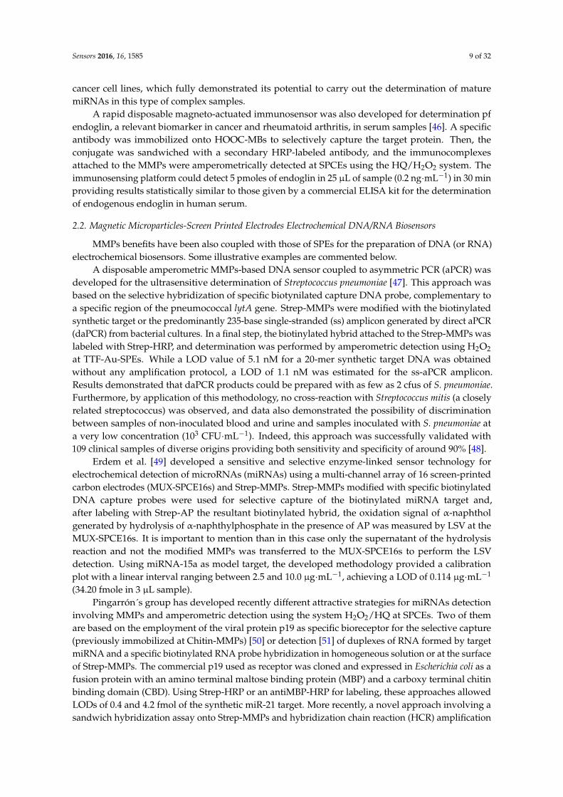
involving MMPs and amperometric detection using the system H2O2/HQ at SPCEs. Two of themare based on the employment of the viral protein p19 as specific bioreceptor for the selective capture(previously immobilized at Chitin-MMPs) [50] or detection [51] of duplexes of RNA formed by targetmiRNA and a specific biotinylated RNA probe hybridization in homogeneous solution or at the surfaceof Strep-MMPs. The commercial p19 used as receptor was cloned and expressed in Escherichia coli as afusion protein with an amino terminal maltose binding protein (MBP) and a carboxy terminal chitinbinding domain (CBD). Using Strep-HRP or an antiMBP-HRP for labeling, these approaches allowedLODs of 0.4 and 4.2 fmol of the synthetic miR-21 target. More recently, a novel approach involving asandwich hybridization assay onto Strep-MMPs and hybridization chain reaction (HCR) amplification
Sensors 2016, 16, 1585 10 of 32
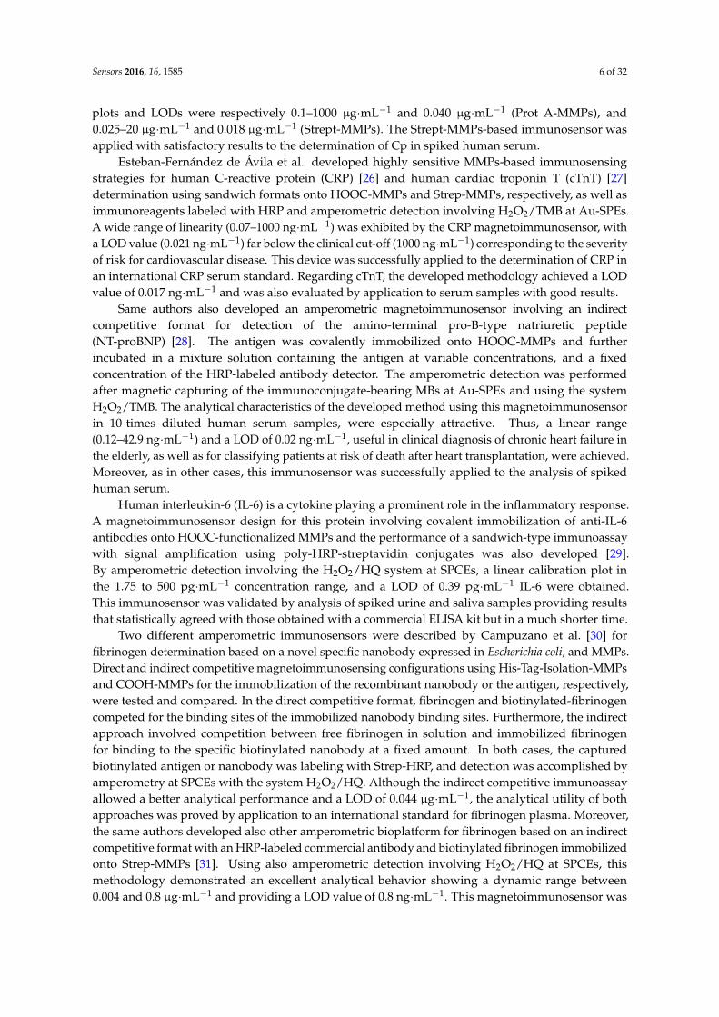
has been developed [52]. The target miRNA-21 contains 11-base complementary sequences to both thebiotinylated capture probe (b-LCp) and the detector probe (Dp) (Figure 2). This latter acts also as theinitiator strand of an HCR amplification in the presence of two biotinylated hairpin sequences leadingto the formation of a long nicked double-helix structure bearing a large number of biotin moleculesused to capture multiple HRP enzymes in a highly ordered way thus allowing an efficient amplificationof the final amperometric signal. This HCR-sandwich based approach allowed a linear increase inthe amperometric signal from 0.2 to 5.0 nM and a LOD value of 0.06 nM (5.0 fmol in 25 µL sample).The applicability of these three different approaches has been demonstrated for the determinationof the mature miRNAs target directly in raw RNAt extracted from human tissues and cancer cells.It is worth mentioning that none of these approaches for miRNA determination requires previousreverse transcription into cDNA, which implies a lower cost and a shorter analysis time.
Figure 2. Strategies developed by Pingarrón´s group for miRNA determination based on the couplingof micro-magnetic particles (MMPs) and amperometric detection at screen-printed carbon electrodes(SPCEs) using p19 protein as capture bioreceptor (1) [50] as detector bioreceptor (2) [51]; and asandwich hybridization coupled to an HCR amplification strategy (3) [52]. Adapted from Refs. [50–52]with permission.
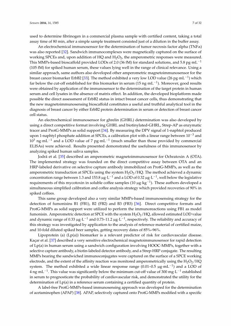
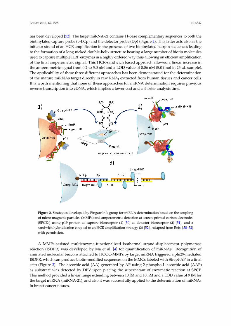
A MMPs-assisted multienzyme-functionalized isothermal strand-displacement polymerasereaction (ISDPR) was developed by Ma et al. [4] for quantification of miRNAs. Recognition ofaminated molecular beacons attached to HOOC-MMPs by target miRNA triggered a phi29-mediatedISDPR, which can produce biotin-modified sequences on the MMCs labeled with Strept-AP in a finalstep (Figure 3). The ascorbic acid (AA) generated by AP using 2-phospho-L-ascorbic acid (AAP)as substrate was detected by DPV upon placing the supernatant of enzymatic reaction at SPCE.This method provided a linear range extending between 10 fM and 10 nM and a LOD value of 9 fM forthe target miRNA (miRNA-21), and also it was successfully applied to the determination of miRNAsin breast cancer tissues.
Sensors 2016, 16, 1585 11 of 32
Figure 3. Representative diagram for the isothermal strand-displacement polymerase reaction (ISDPR)amplification (A) and the fabrication process of the multienzyme functionalized MMPs and oxidationof ascorbic acid (AA) at an SPCE (B). Reprinted from Ref. [4] with permission.
2.3. Other Magnetic Microparticles-Screen Printed Electrodes Electrochemical Affinity Approaches
Gamella et al. [53] developed an amperometric affinity sensor based on His-Tag-Isolation-MMPsfor the 30-min determination of β-lactam antibiotics in milk using the functionalized microparticles toimmobilize a recombinant Histidine-tagged penicillin-binding protein (His-PBP). A direct competitiveassay was performed involving a tracer with HRP and amperometric detection at SPCEs usingthe H2O2/HQ system. This methodology provided LOD values in the low parts-per-billion levelin untreated milk samples for the six antibiotics tested, and was able to detect in a selective wayexclusively the active form of β-lactam antibiotics showing high affinities for both cephalosporinsand penicillins.
An original magneto-immuno-PCR electrochemical approach was also reported for direct andhighly sensitive detection of Streptococcus pneumoniae [54]. In order to amplify a characteristic235-bp region of the gene coding for the major pneumococcal autolysin (lytA), the developedmethodology involved the use of direct asymmetric PCR (DaPCR) of the bacteria attached to captureantibody-ProtA-MMPs. Subsequently, hybridization of biotinylated amplicons and the predominantlysingle-stranded generated was performed onto Strep-MMPs modified with a specific biotinylated20-mer capture probe. After labeling the resultant biotinylated hybrid with Strep-HRP, amperometricdetection was performed using H2O2 at TTF-modified SPAuEs upon magnetic capture of the modifiedMMPs. Using this methodology, calibration plots were constructed for R6 and Dawn serotypes, andthe LOD values were of 132 and 130 cfu·mL−1, respectively, these being about 100–1000 times lowerthan those provided by using this same methodology without PCR amplification [13].
Sensors 2016, 16, 1585 12 of 32
2.4. Multiplexing Using Magnetic Microparticles-Screen Printed Electrodes
SPEs technology is particularly well suited for multiplexing purposes with commercial availabilityof several low cost electrochemical platforms that can be considered as an attractive alternative forpoint-of-care analysis.
Erdem et al. [55] described a label-free voltammetric method for simultaneous determinationof three different microRNAs deregulated in Alzheimer disease (miRNA-16, miRNA-15a andmiRNA-660). Strep-MMPs modified with biotinylated inosine substituted DNA probes were usedfor selective capture of the target miRNAs and, after passive adsorption of the hybrid released fromthe MMPs onto the working electrodes of the MUX-SPCE16s, the oxidation response of guanine wasmeasured using DPV. With miRNA-16 as the model, a LOD of 4.3 pmole in 3 µL sample was estimatedand the guanine response gradually increased till 80 µg·mL−1.
Amino-terminal pro-B-type natriuretic peptide (NT-proBNP) and C-reactive protein (CRP)are two very promising biomarkers for cardiac risk prediction whose clinically relevant cut-offconcentrations differ by three orders of magnitude. For their simultaneous determination in humanserum, a rapid magnetoimmunosensor was developed [56]. HOOC-MMPs were used to covalentlyimmobilize a specific capture antibody or the target antigen, and the quantification of CRP andNT-proBNP was performed by using sandwich and indirect competitive configurations, respectively,as well as HRP-labeled tracers and amperometric detection (H2O2/TMB) at SPdCEs. The developedmethodology showed linear and dynamic ranges of 2.0–100 ng·mL−1 and 2.5–504 ng·mL−1 respectivelyfor CRP and NT-proBNP, and LODs of 0.47 ng·mL−1 for both biomarkers, and it was validated byapplication to a CRP serum international standard also spiked with NT-proBNP.
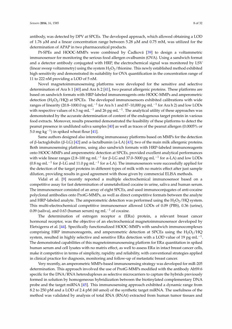
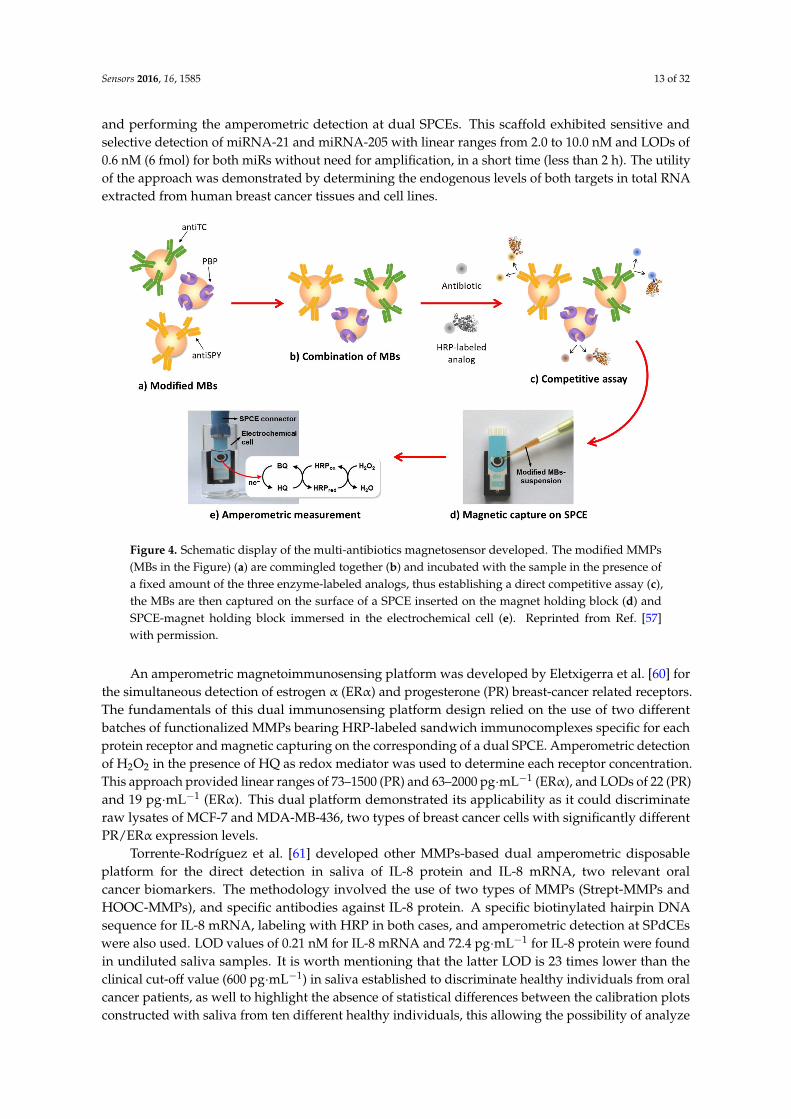
An interesting strategy for the rapid multiplexed screening of antibiotic residues in milk, appliedto cephalosporins (CPHs), sulfonamides (SAs) and TCs, was reported by Conzuelo et al. [57].This involved the use of SPCEs, a mixture of MMPs modified with 3-target specific, and theestablishment of direct competitive assays with HRP-labeled tracers (Figure 4). Specific antibodiesimmobilized onto ProtG-MMPs and His6-tagged penicillin-binding protein (PBP) immobilized ontoHis-tag isolation-MMPs were used as selective capture microcarriers for SAs, TCs and CPHs,respectively. The amperometric responses measured at SPCEs using the H2O2/HQ system allowed toevaluate the extent of affinity reactions. Noticeably, this methodology makes possible discriminationbetween raw milk and uncontaminated UHT samples and samples containing antibiotic residues atthe maximum residue limits (MRLs) in only 5 min, by applying a simple and short pretreatment.
A method for quantification of genetically modified organisms (GMO) was also developedby Manzanares-Palenzuela et al. [58]. Round-Up Ready Soybean (RRS) was selected as a modelGMO. This is an herbicide-resistant form of soybean that was genetically engineered representingone of the most successful achievements of crop biotechnology. Thus, in 2013, it was extendingas more than 75% of worldwide soybean plantations. Two DNA sequences, a fragment of theendogenous lectin (Lec) gen for soybean and an event-specific sequence from RRS, were targeted viasandwich hybridization onto specific biotinylated DNA probes-modified Strep-MMPs and detectionprobes labeled with digoxigenin (Dig) or fluorescein isothiocyanate (FITC). Dual enzymatic labelingusing Fab fragments of anti-FITC and anti-Dig conjugated to HRP or AP, respectively was alsoperformed. Electrochemical measurements of enzymes activity were made in parallel at individualSPCEs using chronoamperometry (HRP, H2O2/TMB system) and DPV (AP, 1-naphthyl phosphatesubstrate). The affinity assay provided a linear range of 2–250 pM for both targets, and LOD values of650 and 190 fM, respectively, for the event and the taxon-specific targets. These results demonstratedthe applicability of this method for the determination of GMO at levels below the European labelingthreshold (0.9%) in foods.
Based on the methodology previously developed by the same group for the determination ofa single miRNA [50], Torrente-Rodríguez et al. [59] implemented the first affinity sensor able tosimultaneously detect in one single experiment, two different miRNAs expression. The approachimplied preparation of two distinct batches of p19-protein modified MBs, one for each target miRNA,
Sensors 2016, 16, 1585 13 of 32
and performing the amperometric detection at dual SPCEs. This scaffold exhibited sensitive andselective detection of miRNA-21 and miRNA-205 with linear ranges from 2.0 to 10.0 nM and LODs of0.6 nM (6 fmol) for both miRs without need for amplification, in a short time (less than 2 h). The utilityof the approach was demonstrated by determining the endogenous levels of both targets in total RNAextracted from human breast cancer tissues and cell lines.
Figure 4. Schematic display of the multi-antibiotics magnetosensor developed. The modified MMPs(MBs in the Figure) (a) are commingled together (b) and incubated with the sample in the presence ofa fixed amount of the three enzyme-labeled analogs, thus establishing a direct competitive assay (c),the MBs are then captured on the surface of a SPCE inserted on the magnet holding block (d) andSPCE-magnet holding block immersed in the electrochemical cell (e). Reprinted from Ref. [57]with permission.
An amperometric magnetoimmunosensing platform was developed by Eletxigerra et al. [60] forthe simultaneous detection of estrogen α (ERα) and progesterone (PR) breast-cancer related receptors.The fundamentals of this dual immunosensing platform design relied on the use of two differentbatches of functionalized MMPs bearing HRP-labeled sandwich immunocomplexes specific for eachprotein receptor and magnetic capturing on the corresponding of a dual SPCE. Amperometric detectionof H2O2 in the presence of HQ as redox mediator was used to determine each receptor concentration.This approach provided linear ranges of 73–1500 (PR) and 63–2000 pg·mL−1 (ERα), and LODs of 22 (PR)and 19 pg·mL−1 (ERα). This dual platform demonstrated its applicability as it could discriminateraw lysates of MCF-7 and MDA-MB-436, two types of breast cancer cells with significantly differentPR/ERα expression levels.
Torrente-Rodríguez et al. [61] developed other MMPs-based dual amperometric disposableplatform for the direct detection in saliva of IL-8 protein and IL-8 mRNA, two relevant oralcancer biomarkers. The methodology involved the use of two types of MMPs (Strept-MMPs andHOOC-MMPs), and specific antibodies against IL-8 protein. A specific biotinylated hairpin DNAsequence for IL-8 mRNA, labeling with HRP in both cases, and amperometric detection at SPdCEswere also used. LOD values of 0.21 nM for IL-8 mRNA and 72.4 pg·mL−1 for IL-8 protein were foundin undiluted saliva samples. It is worth mentioning that the latter LOD is 23 times lower than theclinical cut-off value (600 pg·mL−1) in saliva established to discriminate healthy individuals from oralcancer patients, as well to highlight the absence of statistical differences between the calibration plotsconstructed with saliva from ten different healthy individuals, this allowing the possibility of analyze
Sensors 2016, 16, 1585 14 of 32
all saliva samples using a single calibration plot. Furthermore, the dual electrochemical biosensorwas successfully used for the direct analysis of spiked raw saliva, where the both biomarkers weredetermined, and also to analyze saliva samples from seven healthy individuals for detecting theendogenous content of IL-8 protein. Providing results were in good agreement with those obtained byconventional ELISA methodology.
Very recently, a novel MMPs-based immunosensing approach was also described for the rapidand simultaneous determination of Ara h 1 and Ara h 2, the main peanut allergenic proteins [62].It involves sandwich-type immunoassays onto HOOC-MMPs and amperometric detection (H2O2/HQ)at SPdCES. This methodology exhibited LOD values of 18.0 and 0.07 ng·mL−1, respectively, for Arah 1 and Ara h 2, requiring an assay time as short as 2 h. The utility of the reported approach wasinvestigated by analyzing various food extracts to determine the endogenous concentration of bothallergens, as well as wheat flour spiked samples to detect trace amounts of peanut allergen (0.0001% or1.0 mg/kg). The developed platform provided better LODs than those of commercial allergen specificELISA kits.
3. Magnetic Nanoparticles
Properties of magnetic nanobeads (φ ≤ 100 nm) in terms of structure, biological reactivity orbiorecognition strategies, are similar to those from their microsized counterparts. However, theirhigher surface/volume ratio provides much more binding sites for biomolecules anchoring, these oftengiving better S/N ratios [63]. Moreover, the use of nanomaterials avoids the size mismatch existingbetween magnetic microbeads and nanometric biological reagents, also providing biocompatibility.Furthermore, due to their unique properties, magnetic nanoparticles (MNPs) make possible fasterelectron transfer between redox systems and the electrode surface, and can act as electrocatalysts formolecules such as hydrogen peroxide, implied in biochemical reactions of interest [64]. In addition,MNPs are used not only for the preparation of bioconjugates on the electrode surface but also as labelsfor signal amplification.
Due to their biodegradability and high biocompatibility, magnetite (Fe3O4) and maghemite(γ-Fe2O3), are the main representative materials of MNPs used for biological applications [65].Diverse physical, chemical, and microbial methods have been proposed for the synthesis of lowcost, uniformly sized particles exhibiting large surface areas and diameters usually ranging beweenunits and various tens of nanometers [66]. Core-shell nanomagnetics and nanocomposites have alsobeen used for electrochemical biosensing. Among them, core-shell Fe3O4@AuNPs and Fe3O4@SiO2
are the most common [67]. In the first case, the presence of gold nanoparticles assures theexcellent conductivity and adsorption ability of magnetic material and, furthermore, SiO2 surfaceenhances the stability of nanoparticles and provides a good surface for bioreagents binding. Otheroxides such as TiO2 have also been used to prepare core-shell magnetic nanoparticles with specificproperties. Metal-doped iron oxides (MFe2O4, with M = Co or Mn, among others), with enhancedmagnetic properties of the resulting spinel metal ferrites, have also been proposed. Moreover, it iscurrently becoming common the use of polymers for coating the surface of nanoparticles in orderto increase the immobilization capacity, as well as the combination of magnetic nanoparticles withcarbon nanomaterials such as carbon nanotubes or graphene to take advantage of properties of theresulting hybrids.
3.1. Magnetic Nanoparticles-Screen Printed Electrodes Electrochemical Immunosensors
The special properties of gold nanoparticles, particularly their high conductivity and the abilityto stably adsorb biomolecules, make them the most used nanomaterial in combination with magneticnanoparticles for the preparation of electrochemical affinity biosensors. One example is the useof core/shell Fe3O4@AuNPs conjugated with anti-Salmonella antibodies for the development of anelectrochemical immunosensor for S. typhimurium involving labeling with CdS nanocrystals for thedetection by stripping SWV. A thermal decomposition method by reaction of Fe(III) acetylacetonate
Sensors 2016, 16, 1585 15 of 32
with 1,2-hexadecanediol in the presence of both oleic acid and oleylamine was used to obtainmagnetite nanoparticles [68], that were further coated by gold in toluene at 85 ◦C. The resultingMNPs were functionalized with a mixed monolayer of 2-mercaptoethanol and 12-mercaptododecanoicacid and used to covalently immobilize the capture antibody. Separately, CdS nanocrystalswere used to immobilize also anti-Salmonella antibodies, and a sandwich immunoassay involvingSPCE/Fe3O4@AuNPs-Ab and CdS-Ab was performed. The anodic stripping current from cadmiumregistered by SWV provided a sigmoidal calibration plot between 10 and 106 cells·mL−1, with a LODvalue of 13 cells·mL−1 for an incubation time of 20 min. The method was used for analyzing spikedmilk samples [69]. It should be noted that this method demonstrated the possibility of determine thebacteria in milk at low concentrations and in less than 1 h.
Gan et al. [70] prepared an electrochemical immunosensor for carcinoembryonic antigen (CEA)using Fe3O4@AuNPs to assemble HRP-anti-CEA and BSA. The magnetoconjugate was immobilizedonto a SPCE modified with a carboxylated MWCNTs-thionine (Thi)-Nafion composite and, throughone-step immunoassay format, the immunosensor was incubated with CEA. Immunoconjugationwith the antigen produced a decrease in the catalytic efficiency of HRP for the oxidation by H2O2
of immobilized thionine. This provided a decrease in the electrochemical current which, under theoptimized conditions, could be proportionally related to the concentration of the antigen between0.1–5.0 and 5.0–80 ng·mL−1 with a LOD value of 0.03 ng·mL−1. With the aim of obtaining a label-free,simple, fast, reproducible and non-toxic immunosensor for tumor biomarkers, AuNPs and Fe3O4 wereimmobilized onto MWCNTs previously functionalized with redox-active hemin and the polyelectrolytePDDA (poly(dimethyldiallylammonium)). Alpha fetoprotein (AFP) was used as the model antigen,whereas anti-AFP was adsorbed on the AuNPs surface, and the nanocomposite was incorporated toSPCE. In the presence of AFP antigen, the immunosensing event affected the electron transfer of hemingiven a decrease of the DPV peak current, this providing a linear response for AFP between 0.1 and200 ng·mL−1 with and LOD of 0.04 ng·mL−1 [71]. The developed immunosensor constitute a goodexample of a one-step immunoassay device providing good sensitivity and selectivity. However, it hasonly been applied to AFP standard solutions.
The same group developed a similar immunosensor for high-sensitivity (hs)-CRPusing Fe3O4@AuNPs functionalized with 2-aminoethanethiol to immobilize HRP-anti-hs-CRP.SPCEs modified with iron phtalocyanine (FePc) and chitosan were used to magnetically attractHRP-anti-hs-CRP/Fe3O4@AuNPs conjugates, so that in this case, the immunoconjugation with theantigen inhibited the catalytic efficiency of HRP to the H2O2 reduction of FePc. This inhibition wasproportional to the concentration of the antigen within 1.2 to 200 ng·mL−1 concentration range, and theLOD was 0.5 ng·mL−1 [72]. An important characteristic of the immunosensor is the reusability as itcan be regenerated, this greatly decreases the cost of detection.
Clenbuterol (CLB) is a β-agonist that can promote muscle growth reducing body fat in foodanimals. CLB residues accumulate in meat and liver can produce adverse effects on humans, thus,the misuse of CLB must be controlled. An electrochemical immunosensor was described for thedetermination of CLB in pork meat using SPCEs modified with graphene sheets—Nafion films andcore-shell Fe3O4@AuNPs. BSA-CLB biomagnetic conjugates were immobilized on the electrodeand an indirect competitive immunoassay was performed with anti-CLB and free CLB/BSA-CLB.The electrochemical detection was carried out using Fe(CN)6
3− as the electroactive probe bymeasuring the decrease in the current signal as increases the steric hindrance produced by theformation of anti-CLB-CLB-BSA complex on the surface of SPCE. An increase in current proportionalto the concentration of CLB (less CLA-BSA was immobilized) was observed over the range of0.5 to 200 ng·mL−1 CLB, and a LOD value of 0.22 ng·mL−1 was obtained. Compared with otherelectrochemical immunosensors, this provided a better sensitivity but a narrower dynamic range.The immunosensor was employed to determine CLB in spiked pork meat [73].
A very similar strategy was also used by the same group for developing a voltammetricimmunosensor for the determination of chloramphenicol (CAP). CAP is a broad-spectrum antibiotic
Sensors 2016, 16, 1585 16 of 32
used in veterinary medicine, which can cause serious toxic effects to humans. Although its applicationis banned in different countries, detection of CAP residues in food is still necessary. As in theprevious protocol, SPCEs modified with graphene sheets-Nafion films and core-shell Fe3O4@AuNPswere employed, BSA-CAP biomagnetic conjugates were immobilized on the electrode, and an indirectcompetitive immunoassay was performed with anti-CAP and free CAP/BSA-CAP. The electrochemicaldetection by DPV using Fe(CN)6
3− provided a calibration plot over the range from 2.0 to 200 ng·mL−1
and a LOD of 0.82 ng·mL−1. The method was applied to spiked milk [74], and the results comparedsatisfactorily with those provided by liquid chromatography.
Fe3O4@AuNPs in combination with SPCEs were also used by Zhang et al. [75] to constructan immunosensor for detecting microcystin-(leucine-arginine) (MCLR), the most toxic species ofthe cyanotoxins. The capture antibody (anti-MCLR) was immobilized on the electrode surface byadsorption onto AuNPs, and the immunosensor utilized the direct competitive immunoassay formatbetween the MCLR and HRP-MCLR. Electrochemical detection was made by DPV with H2O2 inthe presence of HQ. Decrease in the peak current responses was proportionally related to MCLRlevel in the 0.79–12.9 ng·mL−1 concentration range. This result together with the LOD, 0.38 µg L−1,demonstrated the utility of the developed immunosensor for the analysis of water samples, as theWHO provisional guideline value is 1 µg·L−1. Noting that, by its characteristics, this immunosensor iscompetitive with chromatographic techniques recommended for MCLR determination. Furthermore,the reported method was applied to the analysis of tap and river spiked waters with good results.
Various natural and synthetic polymers have been used as modifiers of magnetic nanoparticlessurface in order to take advantages of their attractive properties. Among them, electronic conductingpolymers such as polyaniline (PANI) offer special interest due to the ease for preparing core-shellMNPs and their ability to immobilize biomolecules. PANI-coated γ-Fe2O3 were used to immobilizeelectrostatically a specific antibody for Escherichia coli O157:H7, and for the preparation of asandwich-type electrochemical immunosensor using carbohydrate-capped AuNPs labeled with apolyclonal antibody. The electrochemical detection was performed by measuring the DPV oxidationsignal from gold nanoparticles at a SPCE. A linear range of 10–106 cfu·mL−1 and a LOD of 10 cfu·mL−1
were obtained [76].PANI@γ-Fe2O3 MNPs were also used to fabricate an immunosensor for the determination of
surface glycoprotein hemagglutinin (HA) from the Influenza A virus (FLUAV) H5N1(A/Vietnam/1203/04).Antibodies against target HA were immobilized onto MNPs and the resulting anti-HA–MNPs wereshown to interact with glycans preincubated in mouse serum with HA. Glutaraldehyde, AuNPs andstreptavidin-modified SPCEs were used to immobilize the biotinylated glycan/HA/anti-HA-MNPscomplex. Cyclic volammetry was used to measure the charge transfer between PANI and the electrodesurface, which significantly increased in the presence of target [77].
Another immunosensor was prepared by Pingarron´s group for the determination ofLegionella pneumophila SG1 using SPCEs and MNPs prepared with dopamine (DA) electropolymerizedon Fe3O4. After immobilization of a specific antibody (Ab) onto Fe3O4@pDA NPs, a sandwichimmunoassay was carried out in the presence of bacteria using the HRP-labeled antibody (Ab-HRP).Once the resulting conjugates were captured on the electrode surface by applying a magnetic field,the amperometric response was measured after H2O2 addition in the presence of HQ. The LODvalue achieved was 104 cfu·mL−1 with no need for pre-concentration or pre-enrichment treatments.This method was applied to the analysis of water samples [78]. Relevant aspects of this work are thegood stability during 30 days of the Ab-Fe3O4@pDA NPs and the possibility of detecting bacteriaat 10 CFU·mL−1 level in less than three hours after a preconcentration step.
MNPs prepared by combination of iron oxides with other metal oxides possess improvedproperties for bioconjugation. Oxides that exhibit specific affinity to certain functional groups of interestare selected in this type of applications. For example, Fe3O4@TiO2 nanoparticles were used to developtwo different configurations of electrochemical immunosensors for determining phosphorylatedbutyrylcholinesterase (OP-BChE), a biomarker of the exposure to organophosphorous pesticides (OP),
Sensors 2016, 16, 1585 17 of 32
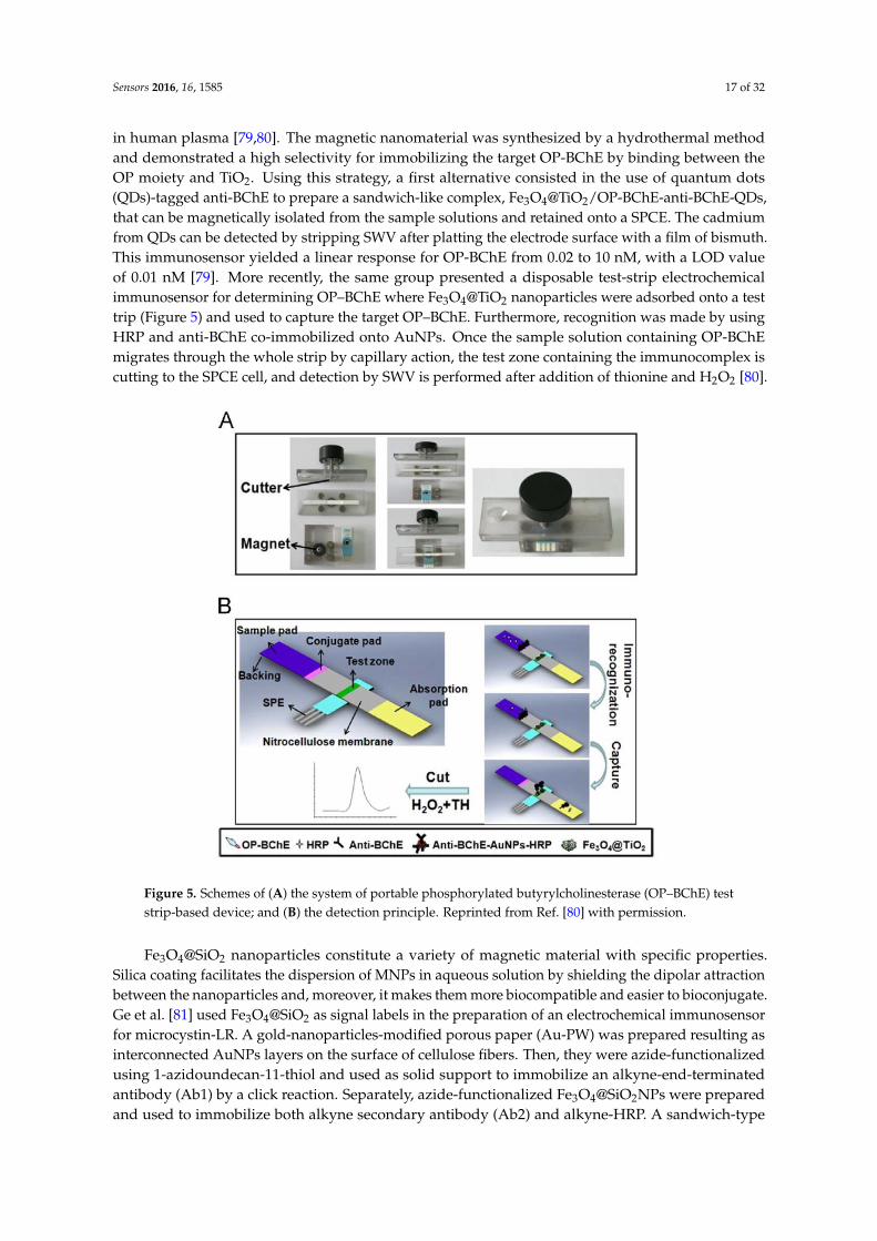
in human plasma [79,80]. The magnetic nanomaterial was synthesized by a hydrothermal methodand demonstrated a high selectivity for immobilizing the target OP-BChE by binding between theOP moiety and TiO2. Using this strategy, a first alternative consisted in the use of quantum dots(QDs)-tagged anti-BChE to prepare a sandwich-like complex, Fe3O4@TiO2/OP-BChE-anti-BChE-QDs,that can be magnetically isolated from the sample solutions and retained onto a SPCE. The cadmiumfrom QDs can be detected by stripping SWV after platting the electrode surface with a film of bismuth.This immunosensor yielded a linear response for OP-BChE from 0.02 to 10 nM, with a LOD valueof 0.01 nM [79]. More recently, the same group presented a disposable test-strip electrochemicalimmunosensor for determining OP–BChE where Fe3O4@TiO2 nanoparticles were adsorbed onto a testtrip (Figure 5) and used to capture the target OP–BChE. Furthermore, recognition was made by usingHRP and anti-BChE co-immobilized onto AuNPs. Once the sample solution containing OP-BChEmigrates through the whole strip by capillary action, the test zone containing the immunocomplex iscutting to the SPCE cell, and detection by SWV is performed after addition of thionine and H2O2 [80].
Figure 5. Schemes of (A) the system of portable phosphorylated butyrylcholinesterase (OP–BChE) teststrip-based device; and (B) the detection principle. Reprinted from Ref. [80] with permission.
Fe3O4@SiO2 nanoparticles constitute a variety of magnetic material with specific properties.Silica coating facilitates the dispersion of MNPs in aqueous solution by shielding the dipolar attractionbetween the nanoparticles and, moreover, it makes them more biocompatible and easier to bioconjugate.Ge et al. [81] used Fe3O4@SiO2 as signal labels in the preparation of an electrochemical immunosensorfor microcystin-LR. A gold-nanoparticles-modified porous paper (Au-PW) was prepared resulting asinterconnected AuNPs layers on the surface of cellulose fibers. Then, they were azide-functionalizedusing 1-azidoundecan-11-thiol and used as solid support to immobilize an alkyne-end-terminatedantibody (Ab1) by a click reaction. Separately, azide-functionalized Fe3O4@SiO2NPs were preparedand used to immobilize both alkyne secondary antibody (Ab2) and alkyne-HRP. A sandwich-type
Sensors 2016, 16, 1585 18 of 32
immunoassay was employed and the electrochemical detection was carried out by placing the reactionzone of the Au-PW support on the active surface of a SPCE. The linear dependence ranged between0.01 and 200 µg·mL−1, and the LOD value was 0.004 µg·mL−1.
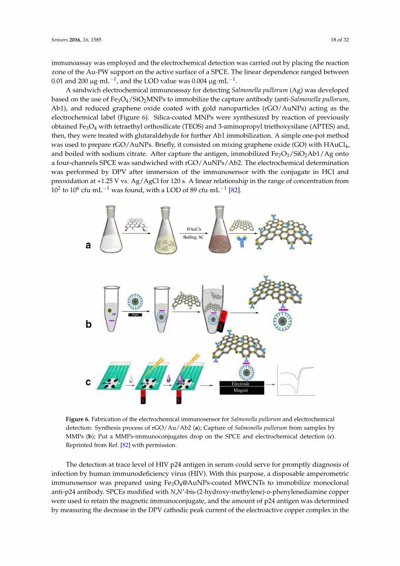
A sandwich electrochemical immunoassay for detecting Salmonella pullorum (Ag) was developedbased on the use of Fe3O4/SiO2MNPs to immobilize the capture antibody (anti-Salmonella pullorum,Ab1), and reduced graphene oxide coated with gold nanoparticles (rGO/AuNPs) acting as theelectrochemical label (Figure 6). Silica-coated MNPs were synthesized by reaction of previouslyobtained Fe3O4 with tetraethyl orthosilicate (TEOS) and 3-aminopropyl triethoxysilane (APTES) and,then, they were treated with glutaraldehyde for further Ab1 immobilization. A simple one-pot methodwas used to prepare rGO/AuNPs. Briefly, it consisted on mixing graphene oxide (GO) with HAuCl4,and boiled with sodium citrate. After capture the antigen, immobilized Fe2O3/SiO2Ab1/Ag ontoa four-channels SPCE was sandwiched with rGO/AuNPs/Ab2. The electrochemical determinationwas performed by DPV after immersion of the immunosensor with the conjugate in HCl andpreoxidation at +1.25 V vs. Ag/AgCl for 120 s. A linear relationship in the range of concentration from102 to 106 cfu·mL−1 was found, with a LOD of 89 cfu·mL−1 [82].
Figure 6. Fabrication of the electrochemical immunosensor for Salmonella pullorum and electrochemicaldetection: Synthesis process of rGO/Au/Ab2 (a); Capture of Salmonella pullorum from samples byMMPs (b); Put a MMPs-immunoconjugates drop on the SPCE and electrochemical detection (c).Reprinted from Ref. [82] with permission.
The detection at trace level of HIV p24 antigen in serum could serve for promptly diagnosis ofinfection by human immunodeficiency virus (HIV). With this purpose, a disposable amperometricimmunosensor was prepared using Fe3O4@AuNPs-coated MWCNTs to immobilize monoclonalanti-p24 antibody. SPCEs modified with N,N’-bis-(2-hydroxy-methylene)-o-phenylenediamine copperwere used to retain the magnetic immunoconjugate, and the amount of p24 antigen was determinedby measuring the decrease in the DPV cathodic peak current of the electroactive copper complex in the
Sensors 2016, 16, 1585 19 of 32
presence of H2O2 after the immunoreaction took place. A linear dependence with p24 concentration inthe 0.6–160 ng·mL−1 range together with a LOD of 0.32 ng·L−1 were obtained. This approach was usedto analyze serum samples of patients with AIDS, and the results obtained in the determination of p24were in good agreement with those from ELISA method [83]. Some advantages of this configurationare the renewability of the electrode surface, and the use of the copper complex as the catalyst forH2O2 reduction instead HRP, with no need of redox mediator.
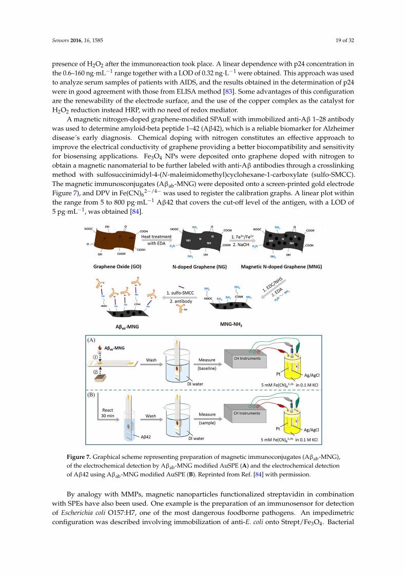
A magnetic nitrogen-doped graphene-modified SPAuE with immobilized anti-Aβ 1–28 antibodywas used to determine amyloid-beta peptide 1–42 (Aβ42), which is a reliable biomarker for Alzheimerdisease´s early diagnosis. Chemical doping with nitrogen constitutes an effective approach toimprove the electrical conductivity of graphene providing a better biocompatibility and sensitivityfor biosensing applications. Fe3O4 NPs were deposited onto graphene doped with nitrogen toobtain a magnetic nanomaterial to be further labeled with anti-Aβ antibodies through a crosslinkingmethod with sulfosuccinimidyl-4-(N-maleimidomethyl)cyclohexane-1-carboxylate (sulfo-SMCC).The magnetic immunosconjugates (Aβab-MNG) were deposited onto a screen-printed gold electrodeFigure 7), and DPV in Fe(CN)6
2−/4− was used to register the calibration graphs. A linear plot withinthe range from 5 to 800 pg·mL−1 Aβ42 that covers the cut-off level of the antigen, with a LOD of5 pg·mL−1, was obtained [84].
Figure 7. Graphical scheme representing preparation of magnetic immunoconjugates (Aβab-MNG),of the electrochemical detection by Aβab-MNG modified AuSPE (A) and the electrochemical detectionof Aβ42 using Aβab-MNG modified AuSPE (B). Reprinted from Ref. [84] with permission.
By analogy with MMPs, magnetic nanoparticles functionalized streptavidin in combinationwith SPEs have also been used. One example is the preparation of an immunosensor for detectionof Escherichia coli O157:H7, one of the most dangerous foodborne pathogens. An impedimetricconfiguration was described involving immobilization of anti-E. coli onto Strept/Fe3O4. Bacterial
Sensors 2016, 16, 1585 20 of 32
cells from samples were isolated and concentrated onto MNBs and placed onto a screen-printedinterdigitated electrode. Impedance measurements could detect E. coli O157:H7 within a linear rangeof 104–107 cfu·mL−1, with a LOD that corresponded to approximately 1400 cells in the used volume of25 µL. This method demonstrated its utility by application to ground beef samples [85].
3.2. Magnetic Nanoparticles-Screen Printed Electrodes Electrochemical DNA Biosensors
MNPs also offer a potent tool for DNA-based electrochemical biosensing characterized by thelow cost, high sensitivity and simple fabrication of the designed devices. One example is thedisposable DNA biosensor using gold Fe3O4@AuNPs reported by Loaiza et al. [86] for detectingspecific hybridization processes. Aβab-MNG/SPAuE was used to attach a thiolated 19-mer captureprobe and, then, Strept-HRP was bound to the biotinylated target. The resulting magnetic complexwas captured on the surface of a homemade SPCE, and the hybridization event was detected by SWVusing hydroquinone as a mediator after the addition of H2O2. A low LOD (31 pM) was found for a50-mer synthetic target without the need of PCR amplification.
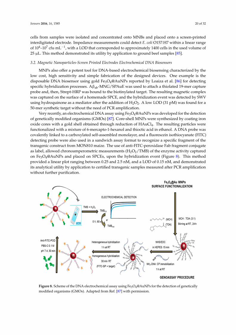
Very recently, an electrochemical DNA assay using Fe3O4@AuNPs was developed for the detectionof genetically modified organisms (GMOs) [87]. Core-shell MNPs were synthesized by coating ironoxide cores with a gold shell obtained through reduction of HAuCl4. The resulting particles werefunctionalized with a mixture of 6-mercapto-1-hexanol and thioctic acid in ethanol. A DNA probe wascovalently linked to a carboxylated self-assembled monolayer, and a fluorescein isothiocyanate (FITC)detecting probe were also used in a sandwich assay format to recognize a specific fragment of thetransgenic construct from MON810 maize. The use of anti-FITC-peroxidase Fab fragment conjugateas label, allowed chronoamperometric measurements (H2O2/TMB) of the enzyme activity capturedon Fe3O4@AuNPs and placed on SPCEs, upon the hybridization event (Figure 8). This methodprovided a linear plot ranging between 0.25 and 2.5 nM, and a LOD of 0.15 nM, and demonstratedits analytical utility by application to certified transgenic samples measured after PCR amplificationwithout further purification.
Figure 8. Scheme of the DNA electrochemical assay using Fe3O4@AuNPs for the detection of geneticallymodified organisms (GMOs). Adapted from Ref. [87] with permission.
Sensors 2016, 16, 1585 21 of 32
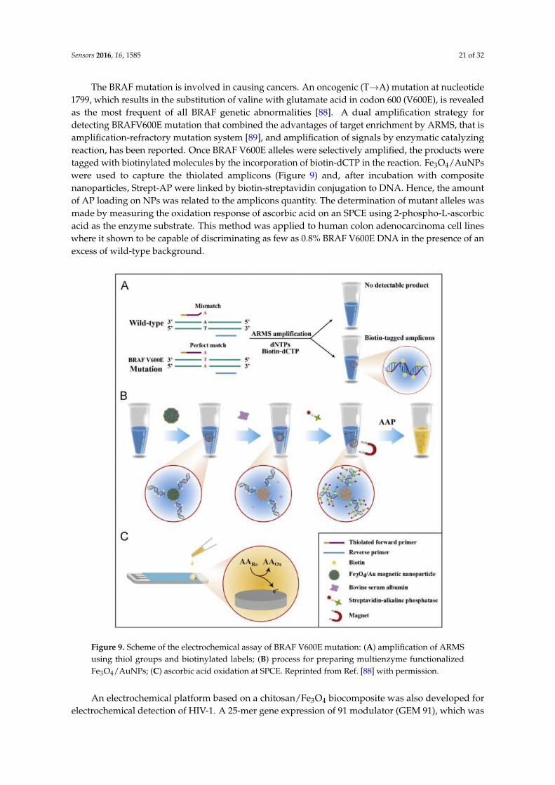
The BRAF mutation is involved in causing cancers. An oncogenic (T→A) mutation at nucleotide1799, which results in the substitution of valine with glutamate acid in codon 600 (V600E), is revealedas the most frequent of all BRAF genetic abnormalities [88]. A dual amplification strategy fordetecting BRAFV600E mutation that combined the advantages of target enrichment by ARMS, that isamplification-refractory mutation system [89], and amplification of signals by enzymatic catalyzingreaction, has been reported. Once BRAF V600E alleles were selectively amplified, the products weretagged with biotinylated molecules by the incorporation of biotin-dCTP in the reaction. Fe3O4/AuNPswere used to capture the thiolated amplicons (Figure 9) and, after incubation with compositenanoparticles, Strept-AP were linked by biotin-streptavidin conjugation to DNA. Hence, the amountof AP loading on NPs was related to the amplicons quantity. The determination of mutant alleles wasmade by measuring the oxidation response of ascorbic acid on an SPCE using 2-phospho-L-ascorbicacid as the enzyme substrate. This method was applied to human colon adenocarcinoma cell lineswhere it shown to be capable of discriminating as few as 0.8% BRAF V600E DNA in the presence of anexcess of wild-type background.
Figure 9. Scheme of the electrochemical assay of BRAF V600E mutation: (A) amplification of ARMSusing thiol groups and biotinylated labels; (B) process for preparing multienzyme functionalizedFe3O4/AuNPs; (C) ascorbic acid oxidation at SPCE. Reprinted from Ref. [88] with permission.
An electrochemical platform based on a chitosan/Fe3O4 biocomposite was also developed forelectrochemical detection of HIV-1. A 25-mer gene expression of 91 modulator (GEM 91), which was
Sensors 2016, 16, 1585 22 of 32
capable of inhibit HIV-1 replication was covalently immobilized onto Chit/Fe3O4NPs by means ofa phosphoramidate reaction between phosphate group at 5’ terminal of GEM sequence and aminegroup of CS. The hybridization process was detected by SWV with methylene blue (MB) as theredox indicator [90]. Pal et al. [91] used magnetite NPs coated with polyaniline for electrochemicallydetecting the anthrax Sterne strain (34F2) of Bacillus anthracis. An electrochemical sandwich assaywas used consisting of a DNA detector probe labeled with PANI@γ-Fe2O3NPs and a biotinylatedDNA capture probe. Once hybridization with DNA targets took place, the Fe3O4@PANI/detectorprobe–DNA/capture probe–biotin hybrid was magnetically immobilized onto a Strept-modifiedSPCE. The electrochemical detection was made by measuring the characteristic voltammetric peaks ofpolyaniline from PANI@γ-Fe2O3NPs. The LOD was 0.01 ng µL−1 DNA.
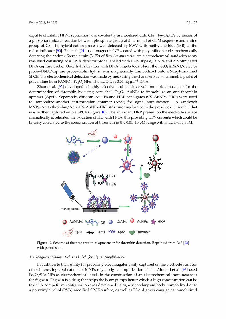
Zhao et al. [92] developed a highly selective and sensitive voltammetric aptasensor for thedetermination of thrombin by using core–shell Fe3O4–AuNPs to immobilize an anti-thrombinaptamer (Apt1). Separately, chitosan–AuNPs and HRP conjugates (CS–AuNPs–HRP) were usedto immobilize another anti-thrombin aptamer (Apt2) for signal amplification. A sandwichMNPs–Apt1/thrombin/Apt2–CS–AuNPs–HRP structure was formed in the presence of thrombin thatwas further captured onto a SPCE (Figure 10). The abundant HRP present on the electrode surfacedramatically accelerated the oxidation of HQ with H2O2, this providing DPV currents which could belinearly correlated to the concentration of thrombin in the 0.01–10 pM range with a LOD of 5.5 fM.
Figure 10. Scheme of the preparation of aptasensor for thrombin detection. Reprinted from Ref. [92]with permission.
3.3. Magnetic Nanoparticles as Labels for Signal Amplification
In addition to their utility for preparing bioconjugates easily captured on the electrode surfaces,other interesting applications of MNPs rely as signal amplification labels. Ahmadi et al. [93] usedFe3O4@AuNPs as electrochemical labels in the construction of an electrochemical immunosensorfor digoxin. Digoxin is a drug that helps the heart pumps better which a high concentration can betoxic. A competitive configuration was developed using a secondary antibody immobilized ontoa polyvinylalcohol (PVA)-modified SPCE surface, as well as BSA-digoxin conjugates immobilized
Sensors 2016, 16, 1585 23 of 32
onto MNPs, and anti-digoxin antibody. DPV was employed for quantitative detection of digoxin inserum after immersing the electrode in HCl for the electrochemical oxidation of gold, and furthervoltammetric reduction of the generated AuCl4−. This strategy allowed determining digoxin withinthe range from 0.5 to 5 ng·mL−1 with a LOD of 0.05 ng·mL−1.
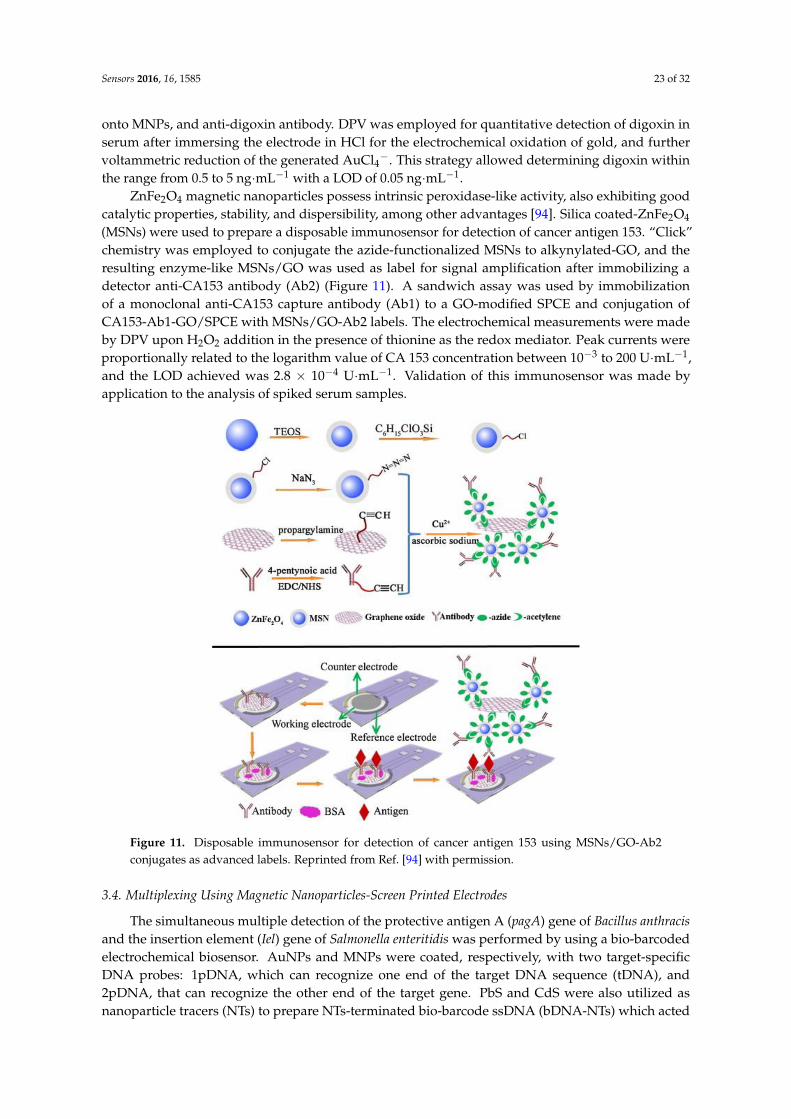
ZnFe2O4 magnetic nanoparticles possess intrinsic peroxidase-like activity, also exhibiting goodcatalytic properties, stability, and dispersibility, among other advantages [94]. Silica coated-ZnFe2O4
(MSNs) were used to prepare a disposable immunosensor for detection of cancer antigen 153. “Click”chemistry was employed to conjugate the azide-functionalized MSNs to alkynylated-GO, and theresulting enzyme-like MSNs/GO was used as label for signal amplification after immobilizing adetector anti-CA153 antibody (Ab2) (Figure 11). A sandwich assay was used by immobilizationof a monoclonal anti-CA153 capture antibody (Ab1) to a GO-modified SPCE and conjugation ofCA153-Ab1-GO/SPCE with MSNs/GO-Ab2 labels. The electrochemical measurements were madeby DPV upon H2O2 addition in the presence of thionine as the redox mediator. Peak currents wereproportionally related to the logarithm value of CA 153 concentration between 10−3 to 200 U·mL−1,and the LOD achieved was 2.8 × 10⁻⁴ U·mL−1. Validation of this immunosensor was made byapplication to the analysis of spiked serum samples.
Figure 11. Disposable immunosensor for detection of cancer antigen 153 using MSNs/GO-Ab2conjugates as advanced labels. Reprinted from Ref. [94] with permission.
3.4. Multiplexing Using Magnetic Nanoparticles-Screen Printed Electrodes
The simultaneous multiple detection of the protective antigen A (pagA) gene of Bacillus anthracisand the insertion element (Iel) gene of Salmonella enteritidis was performed by using a bio-barcodedelectrochemical biosensor. AuNPs and MNPs were coated, respectively, with two target-specificDNA probes: 1pDNA, which can recognize one end of the target DNA sequence (tDNA), and2pDNA, that can recognize the other end of the target gene. PbS and CdS were also utilized asnanoparticle tracers (NTs) to prepare NTs-terminated bio-barcode ssDNA (bDNA-NTs) which acted
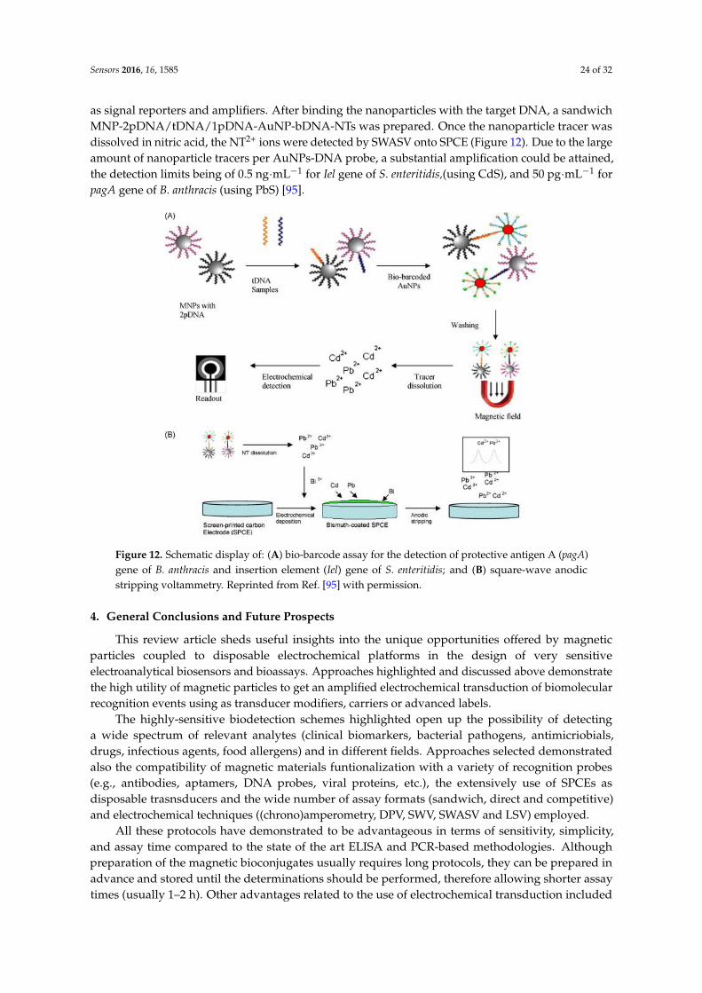
Sensors 2016, 16, 1585 24 of 32
as signal reporters and amplifiers. After binding the nanoparticles with the target DNA, a sandwichMNP-2pDNA/tDNA/1pDNA-AuNP-bDNA-NTs was prepared. Once the nanoparticle tracer wasdissolved in nitric acid, the NT2+ ions were detected by SWASV onto SPCE (Figure 12). Due to the largeamount of nanoparticle tracers per AuNPs-DNA probe, a substantial amplification could be attained,the detection limits being of 0.5 ng·mL−1 for Iel gene of S. enteritidis,(using CdS), and 50 pg·mL−1 forpagA gene of B. anthracis (using PbS) [95].
Figure 12. Schematic display of: (A) bio-barcode assay for the detection of protective antigen A (pagA)gene of B. anthracis and insertion element (Iel) gene of S. enteritidis; and (B) square-wave anodicstripping voltammetry. Reprinted from Ref. [95] with permission.
4. General Conclusions and Future Prospects
This review article sheds useful insights into the unique opportunities offered by magneticparticles coupled to disposable electrochemical platforms in the design of very sensitiveelectroanalytical biosensors and bioassays. Approaches highlighted and discussed above demonstratethe high utility of magnetic particles to get an amplified electrochemical transduction of biomolecularrecognition events using as transducer modifiers, carriers or advanced labels.
The highly-sensitive biodetection schemes highlighted open up the possibility of detectinga wide spectrum of relevant analytes (clinical biomarkers, bacterial pathogens, antimicriobials,drugs, infectious agents, food allergens) and in different fields. Approaches selected demonstratedalso the compatibility of magnetic materials funtionalization with a variety of recognition probes(e.g., antibodies, aptamers, DNA probes, viral proteins, etc.), the extensively use of SPCEs asdisposable trasnsducers and the wide number of assay formats (sandwich, direct and competitive)and electrochemical techniques ((chrono)amperometry, DPV, SWV, SWASV and LSV) employed.
All these protocols have demonstrated to be advantageous in terms of sensitivity, simplicity,and assay time compared to the state of the art ELISA and PCR-based methodologies. Althoughpreparation of the magnetic bioconjugates usually requires long protocols, they can be prepared inadvance and stored until the determinations should be performed, therefore allowing shorter assaytimes (usually 1–2 h). Other advantages related to the use of electrochemical transduction included
Sensors 2016, 16, 1585 25 of 32
the use of portable and cost-effective instrumentation, able to perform multiplexed detection whichcan be claimed as important practical advantages in the implementation of user-friendly devices fordecentralized and routine analysis.
Although MMPs and MNPs have demonstrated to improve significantly the analyticalperformance of electrochemical affinity biosensors, comparative studies demonstrated that. MNPshave larger surface-to-volume ratio providing more chemical reactive sites for the attachment of higheramounts of smaller size (<1 µm) biomolecules at their surface. Despite this improved reactivity, similaror slightly better analytical performance with MNPs compared to MMPs has been reported in standardsolutions [96]. However lower efficiency for immunomagnetic separation was achieved in complexsamples (such as whole milk) using MNPs attributable to these nanoparticles might be more affectedby matrix effect [5]. Moreover, MNPs are easily aggregated and required longer times for magneticactuation [96]. The wide commercial availability of MMPs modified with various functional groupsgreatly facilitates the design of many different approaches test depending on the particular application.Moreover, although the bigger size of the MMPs can pose some limitations in the final sensitivityof the approach because the labeling reagent was not in direct contact with the electrode surface [1],currently the use of small magnets below the surface of the working electrode of the SPEs allowsto concentrate the MBs on the electrode surface and minimize this effect. An additional advantageof MNPs is the easy modification by coating with different materials to give hybrid nanoparticlesthat can be used for further functionalization with specific components such as catalytically activespecies [97]. Furthermore, MNPs can also be utilized as signal reporters; Fe3O4 nanoparticles haveshown peroxidase-like activity [98] and can be used as artificial enzymes for signal amplification.In this sense, although the use of magnetic materials-bioconjugates as label for biosensing has beenmuch less explored that their use to modify transducers, lessons learned in the few reported approachesusing MNPs (no examples found using MMPs) will provide useful starting points in this direction.It is worth mentioning also that the selected examples demonstrated that while commercial MMPs areused generally without further modification apart from the bioreceptor immobilization, the MNPsoffer a greater versatility for modification. Apart from the common Au core/shell modification, MNPshave been modified with natural and synthetic polymers (PANI, DA), other metal oxides (TiO2, SiO2)and nanomaterials (MWCNTs).
It is also worth mentioning that in order to increase even more the sensitivity of these magneticparticles-based disposable electrochemical bioplatforms some approaches based on their couplingwith nanomaterial-based signal or target amplification strategies have been described also.
Although electrochemical disposable electrochemical biosensors using magnetic particles havedemonstrated very attractive advantages for enhancing and superseding the capabilities of currentanalytical methodologies mainly in terms of simplicity, assay time and analysis of complex samples,this field, especially with MNPs, is still new and there are many points to be addressed.
Further research is required for the development of protocols for synthesis, functionalization andbioconjugation of MNPs with high level of uniformity and reproducibility to avoid significant variationin measurements. Moreover, to broaden the scope of bioapplications and open new alternatives,the construction of new hybrid magnetic materials with ideal building blocks such as functionalnanoparticles or biomolecules is also highly desirable.
Other important challenge is testing with real applications. Although in most of cases very lowLODs are achieved improving those obtained with other conventional approaches, a big portion ofthe reports deal with pure analytes in buffer solutions. Therefore, researchers should also make moreeffort on applying these attractive designs to the analysis of complex biological matrices and solvethe problem of MNPs aggregation in these media. Validation tests to demonstrate the applicability,trueness and selectivity in real samples are also required. In this context, simplified protocols able todecrease the time required for the assay would help an easier and wider applicability.
Moreover, most of these biosensor devices still remain in the proof of concept or prototypestages of development and their transfer to practical applications is a challenging process which
Sensors 2016, 16, 1585 26 of 32
so far has rarely resulted in market implementation. Taking into account the disposable natureof the electrochemical transducers used, it will be very helpful also the implementation of thesemagnetic bioassays in microfluidic systems allowing the determination to be performed with minimalmanipulation. Solving drawbacks associated with non-specific binding, the reproducibility of themeasurements, and the long-term stability of the developed devices are also needed.
Although some approaches for dual detection have been reported, the simultaneous determinationof multiple targets in complex samples still remains unsolved and the design of novel approaches formultiplexed analysis of a large number of analytes needs yet to be explored.
Despite these challenging issues which remain to be addressed, taking into account thefast advancement and great advantages offered by electrochemical biosensors using disposableelectrochemical platforms and MMPs or MNPs-bioconjugates to develop sensitive, simple and fastdetection systems that may provide robust tools for biosensor research, it is expected that theseapproaches will reach an increasing importance within the electroanalytical research area in the nearfuture in diverse scientific fields such as in quantification of biotargets, and early detection of geneticmutations or diseases.
Supplementary Materials: The following are available online at http://www.mdpi.com/1424-8220/16/10/1585/s1, Table S1, Table S2 and Table S3.
Acknowledgments: Financial support of the CTQ2015-70023-R and CTQ2015-64402-C2-1-R (Spanish Ministeriode Economía y Competitividad Research Projects) and S2013/MT3029 (NANOAVANSENS Program from theComunidad de Madrid) are gratefully acknowledged.
Author Contributions: The three authors contributed equally to the writing of this manuscript.
Conflicts of Interest: The authors declare no conflict of interest.
References
1. Ricci, F.; Adornetto, G.; Palleschi, G. A review of experimental aspects of electrochemical immunosensors.Electrochim. Acta 2012, 84, 74–83. [CrossRef]
2. Arduini, F.; Micheli, L.; Moscone, D.; Palleschi, G.; Piermarini, S.; Ricci, F.; Volpe, G. Electrochemicalbiosensors based on nanomodified screen-printed electrodes: Recent applications in clinical analysis.Trends Anal. Chem. 2016, 79, 114–126. [CrossRef]
3. Brugnera, M.F.; Bundalian, R.; Laube, T.; Julián, E.; Luquin, M.; Boldrin-Zanoni, M.V.; Pividori, M.I.Magneto-actuated immunoassay for the detection of Mycobacterium fortuitum in hemodialysis water.Talanta 2016, 153, 38–44. [CrossRef] [PubMed]
4. Ma, W.; Situ, B.; Lv, W.; Li, B.; Yin, X.; Vadgama, P.; Zheng, L.; Wang, W. Electrochemical determinationof microRNAs based on isothermal strand-displacement polymerase reaction coupled with multienzymefunctionalized magnetic micro-carriers. Biosens. Bioelectron. 2016, 80, 344–351. [CrossRef] [PubMed]
5. Pividori, M.I.; Ben Aissa, A.; Brandao, D.; Carinelli, S.; Alegret, S. Magneto Actuated Biosensors forFoodborne Pathogens and Infection Diseases Affecting Global Health. In Biosensors for Security andBioterrorism Applications; Biosensors Advanced Sciences and Technologies for Security Applications;Nikolelis, D.P., Nikoleli, G.-P., Eds.; Springer: Switzerland, 2016.
6. Erdem, A.; Ariksoysal, D.O.; Karadeniz, H.; Kara, P.; Sengonul, A.; Sayiner, A.A.; Ozsoz, M. Electrochemicalgenomagnetic assay for the detection of hepatitis B virus DNA in polymerase chain reaction amplicons byusing disposable sensor technology. Electrochem. Commun. 2005, 7, 815–820. [CrossRef]
7. Centi, S.; Laschi, S.; Mascini, M. Improvement of analytical performances of a disposable electrochemicalimmunosensor by using magnetic beads. Talanta 2007, 73, 394–399. [CrossRef] [PubMed]
8. Zacco, E.; Adrian, J.; Galve, R.; Marco, M.P.; Alegret, S.; Pividori, M.I. Electrochemical magnetoimmunosensing of antibiotic residues in milk. Biosens. Bioelectron. 2007, 22, 2184–2191. [CrossRef] [PubMed]
9. Vidal, J.C.; Bertol, J.R.; Bonel, L.; Asturias, L.; Arcos-Martínez, M.J.; Castillo, J.R. A Multi-electrochemicalCompetitive Immunosensor for Sensitive Cocaine Determination in Biological Samples. Electroanalysis 2016,28, 685–694. [CrossRef]
10. Li, F.; Zhou, R.; Zhao, K.; Chen, H.; Hu, Y. Magnetic beads-based electrochemical immunosensor for detectionof pseudorabies virus antibody in swine serum. Talanta 2011, 87, 302–306. [CrossRef] [PubMed]
Sensors 2016, 16, 1585 27 of 32
11. Moreno-Guzmán, M.; Eguílaz, M.; Campuzano, S.; González-Cortés, A.; Yáñez-Sedeño, P.; Pingarrón, J.M.Disposable immunosensor for cortisol using functionalized magnetic particles. Analyst 2010, 135, 1926–1933.[CrossRef] [PubMed]
12. Eguílaz, M.; Moreno-Guzmán, M.; Campuzano, S.; González-Cortés, A.; Yáñez-Sedeño, P.; Pingarrón, J.M.An electrochemical immunosensor for testosterone using functionalized magnetic beads and screen-printedcarbon electrodes. Biosens. Bioelectron. 2010, 26, 517–522. [CrossRef] [PubMed]
13. Campuzano, S.; Esteban-Fernández de Ávila, B.; Yuste, J.; Pedrero, M.; García, J.L.; García, P.;García, E.; Pingarrón, J.M. Disposable amperometric magnetoimmunosensors for the specific detectionof Streptococcus pneumoniae. Biosens. Bioelectron. 2010, 26, 1225–1230. [CrossRef] [PubMed]
14. Szymanski, M.; Porter, R.; Dep, G.V.; Wang, Y.; Haggett, B.G.D. Silver nanoparticles and magnetic beadswith electrochemical measurement as a platform for immunosensing devices. Phys. Chem. Chem. Phys. 2011,13, 5383–5387. [CrossRef] [PubMed]
15. Moreno-Guzmán, M.; González-Cortés, A.; Yáñez-Sedeño, P.; Pingarrón, J.M. A disposable electrochemicalimmunosensor for prolactin involving affinity reaction on streptavidin-functionalized magnetic particles.Anal. Chim. Acta 2011, 692, 125–130. [CrossRef] [PubMed]
16. Serafín, V.; Úbeda, N.; Agüí, L.; Yáñez-Sedeño, P.; Pingarrón, J.M. Ultrasensitive determination of humangrowth hormone (hGH) with a disposable electrochemical magneto-immunosensor. Anal. Bioanal. Chem.2012, 403, 939–946. [CrossRef] [PubMed]
17. Mendes, R.K.; Laschi, S.; Stach-Machado, D.R.; Kubota, L.T.; Marrazza, G. A disposable voltammetricimmunosensor based on magnetic beads for early diagnosis of soybean rust. Sens. Actuators B Chem. 2012,166–167, 135–140. [CrossRef]
18. Gamella, M.; Campuzano, S.; Conzuelo, F.; Reviejo, A.J.; Pingarrón, J.M. AmperometricMagnetoimmunosensors for Direct Determination of D-Dimer in Human Serum. Electroanalysis 2012,24, 2235–2243. [CrossRef]
19. Esteban-Fernández de Ávila, B.; Pedrero, M.; Campuzano, S.; Escamilla-Gómez, V.; Pingarrón, J.M.Sensitive and rapid amperometric magnetoimmunosensor for the determination of Staphylococcus aureus.Anal. Bioanal. Chem. 2012, 403, 917–925. [CrossRef] [PubMed]
20. Conzuelo, F.; Gamella, M.; Campuzano, S.; Reviejo, A.J.; Pingarrón, J.M. Disposable amperometricmagneto-immunosensor for direct detection of tetracyclines antibiotics residues in milk. Anal. Chim. Acta2012, 737, 29–36. [CrossRef] [PubMed]
21. Torrente-Rodríguez, R.M.; Campuzano, S.; Gamella, M.; Conzuelo, F.; Reviejo, A.J.; Pingarrón, J.M. Label-FreeAmperometric Magnetoimmunosensors for Direct Determination of Lactoperoxidase in Milk. Electroanalysis2013, 25, 967–974. [CrossRef]
22. Volpe, G.; Sozzo, U.; Piermarini, S.; Delibato, E.; Palleschi, G.; Moscone, D. Towards the developmentof a single-step immunosensor based on an electrochemical screen-printed electrode strip coupled withimmunomagnetic beads. Anal. Bioanal. Chem. 2013, 405, 655–663. [CrossRef] [PubMed]
23. Taleat, Z.; Cristea, C.; Marrazza, G.; Sandulescu, R. Electrochemical Sandwich Immunoassay for theUltrasensitive Detection of Human MUC1 Cancer Biomarker. Int. J. Electrochem. 2013, 2013, 740265.[CrossRef]
24. Kergaravat, S.V.; Beltramino, L.; Garnero, N.; Trotta, L.; Wagener, M.; Pividori, M.I.; Hernandez, S.R.Electrochemical magneto immunosensor for the detection of anti-TG2 antibody in celiac disease.Biosens. Bioelectron. 2013, 48, 203–209. [CrossRef] [PubMed]
25. Ojeda, I.; Moreno-Guzmán, M.; González-Cortés, A.; Yánez-Sedeño, P.; Pingarrón, J.M. ElectrochemicalMagnetic Immunosensors for the Determination of Ceruloplasmin. Electroanalysis 2013, 25, 2166–2174.[CrossRef]
26. Esteban-Fernández de Ávila, B.; Escamilla-Gómez, V.; Campuzano, S.; Pedrero, M.; Salvador, J.P.;Marco, M.P.; Pingarrón, J.M. Ultrasensitive amperometric magnetoimmunosensor for human C-reactiveprotein quantification in serum. Sens. Actuators B Chem. 2013, 188, 212–220. [CrossRef]
27. Esteban-Fernández de Ávila, B.; Escamilla-Gómez, V.; Campuzano, S.; Pedrero, M.; Pingarrón, J.M.Disposable Electrochemical Magnetoimmunosensor for the Determination of Troponin T Cardiac Marker.Electroanalysis 2013, 25, 51–58. [CrossRef]
Sensors 2016, 16, 1585 28 of 32
28. Esteban-Fernández de Ávila, B.; Escamilla-Gómez, V.; Campuzano, S.; Pedrero, M.; Pingarrón, J.M.Disposable amperometric magnetoimmunosensor for the sensitive detection of the cardiac biomarkeramino-terminal pro-B-type natriuretic peptide in human serum. Anal. Chim. Acta 2013, 784, 18–24. [CrossRef][PubMed]
29. Ojeda, I.; Moreno-Guzmán, M.; González-Cortés, A.; Yáñez-Sedeño, P.; Pingarrón, J.M. Electrochemicalmagnetoimmunosensor for the ultrasensitive determination of interleukin-6 in saliva and urine usingpoly-HRP streptavidin conjugates as labels for signal amplification. Anal. Bioanal. Chem. 2014, 406,6363–6371. [CrossRef] [PubMed]
30. Campuzano, S.; Salema, V.; Moreno-Guzmán, M.; Gamella, M.; Yáñez-Sedeño, P.; Fernández, L.A.;Pingarrón, J.M. Disposable amperometric magnetoimmunosensors using nanobodies as biorecognitionelement. Determination of fibrinogen in plasma. Biosens. Bioelectron. 2014, 52, 255–260. [CrossRef] [PubMed]
31. Torrente-Rodríguez, R.M.; Campuzano, S.; Gamella, M.; Conzuelo, F.; Reviejo, A.J.; Pingarrón, J.M.Amperometric disposable magnetoimmunosensor for the determination of fibrinogen in plasma. Chem. Sens.2014, 4, 7.
32. Eletxigerra, U.; Martinez-Perdiguero, J.; Merino, S.; Villalonga, R.; Pingarrón, J.M.; Campuzano, S.Amperometric magnetoimmunoassay for the direct detection of tumor necrosis factor alpha biomarker inhuman serum. Anal. Chim. Acta 2014, 838, 37–44. [CrossRef] [PubMed]
33. Eletxigerra, U.; Martinez-Perdiguero, J.; Merino, S.; Barderas, R.; Torrente-Rodríguez, R.M.; Villalonga, R.;Pingarrón, J.M.; Campuzano, S. Amperometric magnetoimmunosensor for ErbB2 breast cancer biomarkerdetermination in human serum, cell lysates and intact breast cancer cells. Biosens. Bioelectron. 2015, 70, 34–41.[CrossRef] [PubMed]
34. Martínez-García, G.; Serafín, V.; Agüí, L.; Yáñez-Sedeño, P.; Pingarrón, J.M. Electrochemical Immunosensorfor the Determination of Total Ghrelin Hormone in Saliva. Electroanalysis 2015, 27, 1119–1126. [CrossRef]
35. Jodrá, A.; Hervás, M.; López, M.A.; Escarpa, A. Disposable electrochemical magneto immunosensor forsimultaneous simplified calibration and determination of Ochratoxin A in coffee samples. Sens. ActuatorsB Chem. 2015, 221, 777–783. [CrossRef]
36. Jodrá, A.; López, M.A.; Escarpa, A. Disposable and reliable electrochemical magnetoimmunosensor forFumonisins simplified determination in maize-based foodstuffs. Biosens. Bioelectron. 2015, 64, 633–638.[CrossRef] [PubMed]
37. Kaçar, C.; Torrente-Rodríguez, R.M.; Pedrero, M.; Campuzano, S.; Kilic, E.; Pingarrón, J.M. Amperometricmagnetoimmunoassay for the determination of lipoprotein(a). Microchim. Acta 2015, 182, 1457–1464.[CrossRef]
38. Hosu, O.; Tertis, M.; Sandulescu, R.; Cristea, C. Protein G magnetic beads based immunosensor for sensitivedetection of acetaminophen. Farmacia 2015, 63, 140–145.
39. Cadkova, M.; Metelka, R.; Holubová, L.; Horák, D.; Dvoráková, V.; Bílková, Z.; Korecká, L. Magneticbeads-based electrochemical immunosensor for monitoring allergenic food proteins. Anal. Biochem. 2015,484, 4–8. [CrossRef] [PubMed]
40. Ruiz-Valdepeñas Montiel, V.; Campuzano, S.; Pellicanò, A.; Torrente-Rodríguez, R.M.; Reviejo, A.J.;Cosio, M.S.; Pingarrón, J.M. Sensitive and selective magnetoimmunosensing platform for determination ofthe food allergen Ara h 1. Anal. Chim. Acta 2015, 880, 52–59.
41. Ruiz-Valdepeñas Montiel, V.; Pellicanò, A.; Campuzano, S.; Torrente-Rodríguez, R.M.; Reviejo, A.J.;Cosio, M.S.; Pingarrón, J.M. Electrochemical detection of peanuts at trace levels in foods using amagnetoimmunosensor for the allergenic protein Ara h 2. Sens. Actuators B Chem. 2016, 236, 825–833.[CrossRef]
42. Ruiz-Valdepeñas Montiel, V.; Campuzano, S.; Conzuelo, F.; Torrente-Rodríguez, R.M.; Gamella, M.;Reviejo, A.J.; Pingarrón, J.M. Electrochemical magnetoimmunosensing platform for determination of themilk allergen β-lactoglobulin. Talanta 2015, 131, 156–162. [CrossRef] [PubMed]
43. Ruiz-Valdepeñas Montiel, V.; Campuzano, S.; Torrente-Rodríguez, R.M.; Reviejo, A.J.; Pingarrón, J.M.Electrochemical magnetic beads-based immunosensing platform for the determination of α-lactalbumin inmilk. Food Chem. 2016, 213, 595–601. [CrossRef] [PubMed]
Sensors 2016, 16, 1585 29 of 32
44. Eletxigerra, U.; Martinez-Perdiguero, J.; Merino, S.; Barderas, R.; Ruiz-Valdepeñas Montiel, V.; Villalonga, R.;Pingarrón, J.M.; Campuzano, S. Estrogen receptor α determination in serum, cell lysates and breast cancercells using an amperometric magnetoimmunosensing platform. Sens. Bio. Sens. Res. 2016, 7, 71–76.[CrossRef]
45. Torrente-Rodríguez, R.M.; Ruiz-Valdepeñas Montiel, V.; Campuzano, S.; Farchado-Dinia, M.; Barderas, R.;San Segundo-Acosta, P.; Montoya, J.J.; Pingarrón, J.M. Fast Electrochemical miRNAs Determination inCancer Cells and Tumor Tissues with Antibody-Functionalized Magnetic Microcarriers. ACS Sens. 2016, 1,896–903. [CrossRef]
46. Torrente-Rodríguez, R.M.; Campuzano, S.; Ruiz-Valdepeñas-Montiel, V.; Pedrero, M.;Fernández-Aceñero, M.J.; Barderas, R.; Pingarrón, J.M. Rapid endoglin determination in serumsamples using anamperometric magneto-actuated disposable immunosensing platform. J. Pharm.Biomed. Anal. 2016, 129, 288–293. [CrossRef] [PubMed]
47. Campuzano, S.; Pedrero, M.; García, J.L.; García, E.; García, P.; Pingarrón, J.M. Development of amperometricmagnetogenosensors coupled to asymmetric PCR for the specific detection of Streptococcus pneumoniae.Anal. Bioanal. Chem. 2011, 399, 2413–2420. [CrossRef] [PubMed]
48. Sotillo, A.; Pedrero, M.; de Pablos, M.; García, J.L.; García, E.; García, P.; Pingarrón, J.M.; Mingorance, J.;Campuzano, S. Clinical evaluation of a disposable amperometric magneto-genosensor for the detection andidentification of Streptococcus pneumoniae. J. Microbiol. Meth. 2014, 103, 25–28. [CrossRef] [PubMed]
49. Erdem, A.; Congur, G.; Eksin, E. Multi channel screen printed array of electrodes for enzyme-linkedvoltammetric detection of MicroRNAs. Sens. Actuators B Chem. 2013, 188, 1089–1095. [CrossRef]
50. Campuzano, S.; Torrente-Rodríguez, R.M.; López-Hernández, E.; Conzuelo, F.; Granados, R.;Sánchez-Puelles, J.M.; Pingarrón, J.M. Magnetobiosensors Based on Viral Protein p19 for MicroRNADetermination in Cancer Cells and Tissues. Angew. Chem. Int. Ed. 2014, 53, 6168–6171. [CrossRef][PubMed]
51. Torrente-Rodríguez, R.M.; Campuzano, S.; López-Hernández, E.; Granados, R.; Sánchez-Puelles, J.M.;Pingarrón, J.M. Direct Determination of miR-21 in Total RNA Extracted from Breast Cancer Samples UsingMagnetosensing Platforms and the p19 Viral Protein as Detector Bioreceptor. Electroanalysis 2014, 26,2080–2087. [CrossRef]
52. Torrente-Rodríguez, R.M.; Campuzano, S.; Ruiz-Valdepeñas Montiel, V.; Montoya, J.J.; Pingarrón, J.M.Sensitive electrochemical determination of miRNAs based on a sandwich assay onto magnetic microcarriersand hybridization chain reaction amplification. Biosens. Bioelectron. 2016, 86, 516–521. [CrossRef] [PubMed]
53. Gamella, M.; Campuzano, S.; Conzuelo, F.; Esteban-Torres, M.; de las Rivas, B.; Reviejo, A.J.; Muñoz, R.;Pingarrón, J.M. An amperometric affinity penicillin-binding protein magnetosensor for the detection ofβ-lactam antibiotics in milk. Analyst 2013, 138, 2013–2022. [CrossRef] [PubMed]
54. Campuzano, S.; Esteban-Fernández de Ávila, B.; Pedrero, M.; García, J.L.; García, E.; Pingarrón, J.M.;García, P. Electrochemical magneto-immuno-PCR approach for direct and highly sensitive detection ofStreptococcus pneumonia. Chem. Sens. 2011, 1, 8.
55. Erdem, A.; Congur, G. Label-free voltammetric detection of MicroRNAs at multi-channel screen printedarray of electrodes comparison to graphite sensors. Talanta 2014, 118, 7–13. [CrossRef] [PubMed]
56. Esteban-Fernández de Ávila, B.; Escamilla-Gómez, V.; Lanzone, V.; Campuzano, S.; Pedrero, M.;Compagnone, D.; Pingarrón, J.M. Multiplexed Determination of Amino-Terminal Pro-B Type NatriureticPeptide and C-Reactive Protein Cardiac Biomarkers in Human Serum at a Disposable ElectrochemicalMagnetoimmunosensor. Electroanalysis 2014, 26, 254–261. [CrossRef]
57. Conzuelo, F.; Ruiz-Valdepeñas Montiel, V.; Campuzano, S.; Gamella, M.; Torrente-Rodríguez, R.M.;Reviejo, A.J.; Pingarrón, J.M. Rapid screening of multiple antibiotic residues in milk using disposableamperometric magnetosensors. Anal. Chim. Acta 2014, 820, 32–38. [CrossRef] [PubMed]
58. Manzanares-Palenzuela, C.; de-los-Santos-Álvarez, N.; Lobo-Castañón, M.J.; López-Ruiz, B. Multiplexelectrochemical DNA platform for femtomolar-level quantification of genetically modified soybean.Biosens. Bioelectron. 2015, 68, 259–265. [CrossRef] [PubMed]
59. Torrente-Rodríguez, R.M.; Campuzano, S.; López-Hernández, E.; Ruiz-Valdepeñas Montiel, V.; Barderas, R.;Granados, R.; Sánchez-Puelles, J.M.; Pingarrón, J.M. Simultaneous detection of two breast cancer-relatedmiRNAs in tumor tissues using p19-based disposable amperometric magnetobiosensing platforms.Biosens. Bioelectron. 2015, 66, 385–391. [CrossRef] [PubMed]
Sensors 2016, 16, 1585 30 of 32
60. Eletxigerra, U.; Martínez-Perdiguero, J.; Merino, S.; Barderas, R.; Ruiz-Valdepeñas Montiel, V.; Villalonga, R.;Pingarrón, J.M.; Campuzano, S. Electrochemical Magnetoimmunosensor for Progesterone ReceptorDetermination. Application to the Simultaneous Detection of Estrogen and Progesterone Breast-cancerRelated Receptors in Raw Cell Lysates. Electroanalysis 2016, 28, 1787–1794. [CrossRef]
61. Torrente-Rodríguez, R.M.; Campuzano, S.; Ruiz-Valdepeñas Montiel, V.; Gamella, M.; Pingarrón, J.M.Electrochemical bioplatforms for the simultaneous determination of interleukin (IL)-8 mRNA and IL-8protein oral cancer biomarkers in raw saliva. Biosens. Bioelectron. 2016, 77, 543–548. [CrossRef] [PubMed]
62. Ruiz-Valdepeñas Montiel, V.; Torrente-Rodríguez, R.M.; Campuzano, S.; Pellicanò, A.; Reviejo, A.J.;Cosio, M.S.; Pingarrón, J.M. Simultaneous Determination of the Main Peanut Allergens in Foods UsingDisposable Amperometric Magnetic Beads-Based Immunosensing Platforms. Chemosensors 2016, 4, 11.[CrossRef]
63. Teles, F.S. Biosensors and rapid diagnostic tests on the frontier between analytical and clinical chemistry forbiomolecular diagnosis of dengue disease: A review. Anal. Chim. Acta 2011, 687, 28–42. [CrossRef] [PubMed]
64. Hasanzadeh, M.; Shadjou, N.; de la Guardia, M. Iron and iron-oxide magnetic nanoparticles assignal-amplification elements in electrochemical biosensing. Trends Anal. Chem. 2015, 72, 1–9. [CrossRef]
65. Reddy, L.H.; Arias, J.L.; Nicolas, J.; Couvreur, P. Magnetic Nanoparticles: Design and Characterization,Toxicity and Biocompatibility, Pharmaceutical and Biomedical Applications. Chem. Rev. 2012, 112, 5818–5878.[CrossRef] [PubMed]
66. Urbanova, V.; Magro, M.; Gedanken, A.; Baratella, D.; Vianello, F.; Zboril, R. Nanocrystalline Iron Oxides,Composites, and Related Materials as a Platform for Electrochemical, Magnetic, and Chemical Biosensors.Chem. Mater. 2014, 26, 6653–6673. [CrossRef]
67. Xu, Y.; Wang, E. Electrochemical biosensors based on magnetic micro/nano particles. Electrochim. Acta 2012,84, 62–73. [CrossRef]
68. Sun, S.; Zeng, H.; Robinson, D.B.; Raoux, S.; Rice, P.M.; Wang, S.X.; Li, G. Monodisperse MFe2O4 (M = Fe,Co, Mn) nanoparticles. J. Am. Chem. Soc. 2004, 126, 273–279. [CrossRef] [PubMed]
69. Freitas, M.; Viswanathan, S.; Nouws, H.P.A.; Oliveira, M.B.P.P.; Delerue-Matos, C. Iron oxide/goldcore/ shell nanomagnetic probes and CdS biolabels for amplified electrochemical immunosensing ofSalmonella typhimurium. Biosens. Bioelectron. 2014, 51, 195–200. [CrossRef] [PubMed]
70. Gan, N.; Meng, L.-H.; Li, T.-H.; Cao, Y.-T.; Zheng, L. A magnet-controlled and renewable amperometricimmunosensor for carcinoembryonic antigen based on magnetic Fe3O4(core)/Au(shell) nanoparticlesmodified screen-printed carbon electrode. Asian J. Chem. 2011, 23, 3261–3267.
71. Meng, L.; Gan, N.; Li, T.; Cao, Y.; Hu, F.; Zheng, L. A three-dimensional, magnetic and electroactive nanoprobefor amperometric determination of tumor biomarkers. Int. J. Mol. Sci. 2011, 12, 362–375. [CrossRef] [PubMed]
72. Gan, N.; Meng, L.; Hu, F.; Cao, Y.; Wu, Y.; Jia, L.; Zheng, L. A renewable amperometricimmunosensor for hs-CRP based on functionalized Fe3O4@Au magnetic nanoparticles attracted on Fephtalocyanine/chitosan-membrane modified screen printed carbon electrode by a magnet. Appl. Mech. Mater.2012, 110–116, 519–526.
73. Yang, X.; Wu, F.; Chen, D.-Z.; Lin, H.-W. An electrochemical immunosensor for rapid determinationofclenbuterol by using magnetic nanocomposites to modify screenprinted carbon electrode based oncompetitive immunoassay mode. Sens. Actuators B Chem. 2014, 192, 529–535. [CrossRef]
74. He, G.; Yang, X.; Hu, Y.; Hu, Y.; Zhang, F. A sensitive and selective amperometric immunosensor forchloramphenicol detection based on magnetic nanocomposites modify screen-printed carbon electrodes as adisposable platform. Int. J. Electrochem. Sci. 2014, 9, 6962–6974.
75. Zhang, J.-G.; Kang, T.-F.; Xue, R.; Sue, X. An Immunosensor for Microcystins Based on Fe3O4 @Au magneticnanoparticle modified screen-printed electrode. Chin. J. Anal. Chem. 2013, 41, 1353–1358. [CrossRef]
76. Wang, Y.; Alocilja, E.C. Gold nanoparticle-labeled biosensor for rapid and sensitive detection of bacterialpathogens. J. Biol. Eng. 2015, 9, 16. [CrossRef] [PubMed]
77. Kamikawa, T.L.; Mikolajczyk, M.G.; Kennedy, M.; Zhang, P.; Wang, W.; Scott, D.E.; Alocilja, E.C.Nanoparticle-based biosensor for the detection of emerging pandemic influenza strains. Biosens. Bioelectron.2010, 26, 1346–1352. [CrossRef] [PubMed]
Sensors 2016, 16, 1585 31 of 32
78. Martín, M.; Salazar, P.; Jiménez, C.; Lecuona, M.; Ramos, M.J.; Ode, J.; Alcoba, J.; Roche, R.; Villalonga, R.;Campuzano, S.; et al. Rapid Legionella pneumophila determination based on a disposable coreeshellFe3O4@poly(dopamine) magnetic nanoparticles immunoplatform. Anal. Chim. Acta 2015, 887, 51–58.[CrossRef] [PubMed]
79. Zhang, X.; Wang, H.; Yang, C.; Du, D.; Lin, Y. Preparation, characterization of Fe3O4 at TiO2 magneticnanoparticles and their application for immunoassay of biomarker of exposure to organophosphoruspesticides. Biosens. Bioelectron. 2013, 41, 669–674. [CrossRef] [PubMed]
80. Ge, X.; Zhang, W.; Lin, Y.; Du, D. Magnetic Fe3O4@TiO2 nanoparticles-based test strip immunosensingdevice for rapid detection of phosphorylated butyrylcholinesterase. Biosens. Bioelectron. 2013, 50, 486–491.[CrossRef] [PubMed]
81. Ge, S.; Liu, W.; Ge, L.; Yan, M.; Yan, J.; Huang, J.; Yu, J. In situ assembly of porous Au-paper electrodeand functionalization of magnetic silica nanoparticles with HRP via click chemistry for microcystin-LRimmunoassay. Biosens. Bioelectron. 2013, 49, 111–117. [CrossRef] [PubMed]
82. Fei, J.; Dou, W.; Zhao, G. Amperometric immunoassay for the detection of Salmonella pullorum using ascreen—Printed carbon electrode modified with gold nanoparticle-coated reduced graphene oxide andimmunomagnetic beads. Microchim. Acta 2016, 183, 757–764. [CrossRef]
83. Gan, N.; Luo, N.-X.; Li, T.-H.; Zheng, L.; Ni, M.-J. A non-enzyme amperometric immunosensor for rapiddetermination of human immunodeficiency virus p24 based on magnetism controlled carbon nanotubesmodified printed electrode. Chin. J. Anal. Chem. 2010, 38, 1556–1562. [CrossRef]
84. Li, S.-S.; Lin, C.-W.; Wei, K.-C.; Huang, C.-Y.; Hsu, P.-H.; Liu, H.-L.; Lu, Y.-J.; Lin, S.-C.;Yang, H.-W.; Ma, C.-C.M. Non-invasive screening for early Alzheimer’s disease diagnosis by a sensitivelyimmunomagnetic biosensor. Sci. Rep. 2016, 6, 25155. [CrossRef] [PubMed]
85. Wang, R.; Lum, J.; Callaway, Z.; Lin, J.; Bottje, W.; Li, Y. A label-free impedance immunosensor usingscreen-printed interdigitated electrodes and magnetic nanobeads for the detection of E. coli O157:H7.Biosensors 2015, 5, 792–803. [CrossRef] [PubMed]
86. Loaiza, Ó.A.; Jubete, E.; Ochoteco, E.; Cabañero, G.; Grande, H.; Rodríguez, J. Gold coated ferric oxidenanoparticles based disposable magnetic genosensors for the detection of DNA hybridization processes.Biosens. Bioelectron. 2011, 26, 2194–2200. [CrossRef] [PubMed]
87. Freitas, M.; SáCouto, M.; Barroso, M.F.; Pereira, C.; de-los-Santos-Álvarez, N.; Miranda-Ordieres, A.J.;Lobo-Castañón, M.J.; Delerue-Matos, C. Highly Monodisperse Fe3O4@Au Superparamagnetic Nanoparticlesas Reproducible Platform for Genosensing Genetically Modified Organisms. ACS Sens. 2016, in Press.[CrossRef]
88. Situ, B.; Cao, N.; Li, B.; Liu, Q.; Lin, L.; Dai, Z.; Zou, X.; Cai, Z.; Wang, Q.; Yan, X.; et al. Sensitiveelectrochemical analysis of BRAF V600E mutation based on an amplification-refractory mutation systemcoupled with multienzyme functionalized Fe3O4/AuNPs. Biosens. Bioelectron. 2013, 43, 257–263. [CrossRef][PubMed]
89. Dempsey, E.; Barton, D.E.; Ryan, F. Detection of Five Common CFTR Mutations by Rapid-Cycle Real-TimeAmplification Refractory Mutation System PCR. Clin. Chem. 2004, 50, 773–775. [CrossRef] [PubMed]
90. Tran, L.D.; Nguyen, B.H.; Hieu, N.V.; Tran, H.V.; Nguyen, H.L.; Nguyen, P.X. Electrochemical detection ofshort HIV sequences on chitosan/Fe3O4 nanoparticle based screen printed electrodes. Mater. Sci. Eng. C2011, 31, 477–485. [CrossRef]
91. Pal, S.; Alocilja, E.C. Electrically active magnetic nanoparticles as novel concentrator and electrochemicalredox transducer in Bacillus anthracis DNA detection. Biosens. Bioelectron. 2010, 26, 1624–1630. [CrossRef][PubMed]
92. Zhao, J.; Lin, F.; Yi, Y.; Huang, Y.; Li, H.; Zhang, Y.; Yao, S. Dual amplification strategy of highly sensitivethrombin amperometric aptasensor based on chitosan–Au nanocomposites. Analyst 2012, 137, 3488–3495.[CrossRef] [PubMed]
93. Ahmadi, A.; Shirazi, H.; Pourbagher, N.; Akbarzadeh, A.; Omidfar, K. An electrochemical immunosensor fordigoxin using core–shell gold coated magnetic nanoparticles as labels. Mol. Biol. Rep. 2014, 41, 1659–1668.[CrossRef] [PubMed]
94. Ge, S.; Sun, M.; Liu, W.; Li, S.; Wang, X.; Chu, C.; Yan, M.; Yu, J. Disposable electrochemical immunosensorbased on peroxidase-likemagnetic silica–graphene oxide composites for detection of cancer antigen 153.Sens. Actuators B Chem. 2014, 192, 317–326. [CrossRef]
Sensors 2016, 16, 1585 32 of 32
95. Zhang, D.; Huarng, M.C.; Alocilja, E.C. A multiplex nanoparticle-based bio-barcoded DNA sensor for thesimultaneous detection of multiple pathogens. Biosens. Bioelectron. 2010, 26, 1736–1742. [CrossRef] [PubMed]
96. Brandão, D.; Liébana, S.; Campoy, S.; Alegret, S.; Pividori, M.I. Immunomagnetic separation of Salmonellawith tailored magnetic micro and nanocarriers. A comparative study. Talanta 2015, 143, 198–204. [CrossRef][PubMed]
97. Lu, A.-H.; Salabas, E.L.; Schüth, F. Magnetic nanoparticles: Synthesis, protection, functionalization, andapplication. Angew. Chem. Int. Ed. 2007, 46, 1222–1244. [CrossRef] [PubMed]
98. Gao, L.; Zhuang, J.; Nie, L.; Zhang, J.; Zhang, Y.; Gu, N.; Wang, T.; Feng, J.; Yang, D.; Perrett, S. Intrinsicperoxidase-like activity of ferromagnetic nanoparticles. Nat. Nanotechnol. 2007, 2, 577–583. [CrossRef][PubMed]
© 2016 by the authors; licensee MDPI, Basel, Switzerland. This article is an open accessarticle distributed under the terms and conditions of the Creative Commons Attribution(CC-BY) license (http://creativecommons.org/licenses/by/4.0/).