Embed Size (px)
DESCRIPTION
Malignant Renal tumors. DR.Gehan Mohamed. Malignant renal tumors. It may be: primary tumors : i.e arise from kidney tissue itself Secondary tumors: represent a metastasis from any malignant tumor present anywhere in the body and reach kidney. - PowerPoint PPT Presentation
Citation preview
Malignant Renal tumors Malignant Renal tumors
DR.Gehan MohamedDR.Gehan Mohamed
Malignant renal tumorsMalignant renal tumors- It may be:It may be:
- primary tumors : i.e arise from kidney tissue itselfprimary tumors : i.e arise from kidney tissue itself - Secondary tumors: represent a metastasis from any Secondary tumors: represent a metastasis from any
malignant tumor present anywhere in the body and malignant tumor present anywhere in the body and reach kidney.reach kidney.
Primary malignant renal tumors include: Primary malignant renal tumors include:
A- malignant epithelial tumors :A- malignant epithelial tumors :1- Renal cell carcinoma (hypernephroma = renal cell 1- Renal cell carcinoma (hypernephroma = renal cell adenocarcinoma) represents 80-85% ,it is subdivided into :adenocarcinoma) represents 80-85% ,it is subdivided into :
a- Clear cell renal cell carcinomaa- Clear cell renal cell carcinoma
b- Papillary renal cell carcinoma b- Papillary renal cell carcinoma
c- chromophobe renal cell carcinoma c- chromophobe renal cell carcinoma d - Collecting duct carcinomad - Collecting duct carcinoma
2- Transitional cell carcinoma 8% arise from transitional epithelium 2- Transitional cell carcinoma 8% arise from transitional epithelium which line renal pelvis.which line renal pelvis.
B- malignant mesodermal tumors B- malignant mesodermal tumors (i.e arise from smooth muscles or (i.e arise from smooth muscles or blood vessels or fat present in the kidney ) : blood vessels or fat present in the kidney ) : which called Renal sarcomas e.g leiomyosarcoma (malignant which called Renal sarcomas e.g leiomyosarcoma (malignant tumor arise from smooth muscle) ,generally poorer outcome .tumor arise from smooth muscle) ,generally poorer outcome .
C- mixed malignant epithelial and mesenchymal tumors C- mixed malignant epithelial and mesenchymal tumors : e.g : e.g Nephroblastoma (Wilms’ tumor in children)Nephroblastoma (Wilms’ tumor in children)
We will study in details :We will study in details :
RENAL CELL CARCINOMA(RCC) :more common in adult age.RENAL CELL CARCINOMA(RCC) :more common in adult age.
Wilms tumor (nephroblastoma): more common in children (2-5 years)Wilms tumor (nephroblastoma): more common in children (2-5 years)
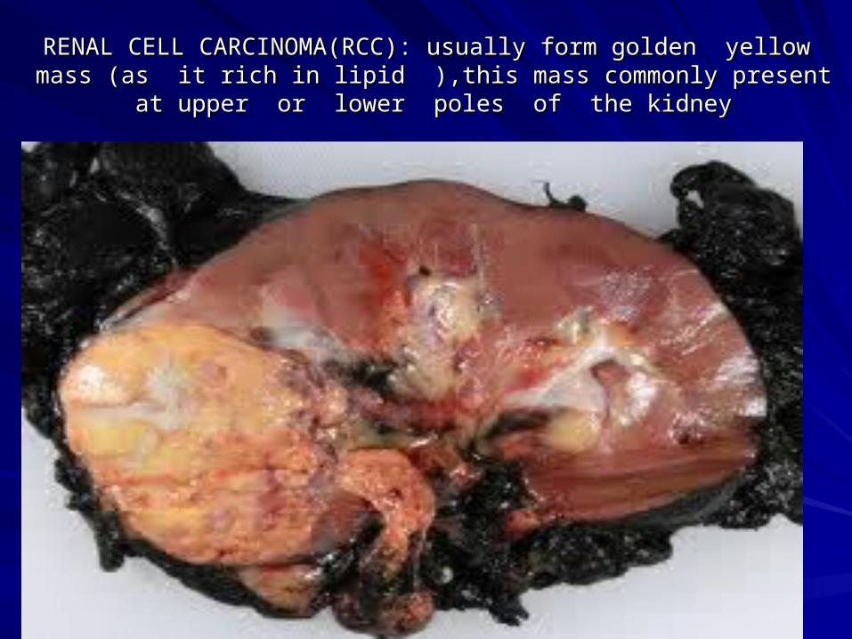
RENAL CELL CARCINOMA(RCC): usually form golden yellow mass (as it RENAL CELL CARCINOMA(RCC): usually form golden yellow mass (as it rich in lipid ),this mass commonly present at upper or lower poles of the rich in lipid ),this mass commonly present at upper or lower poles of the
kidneykidney
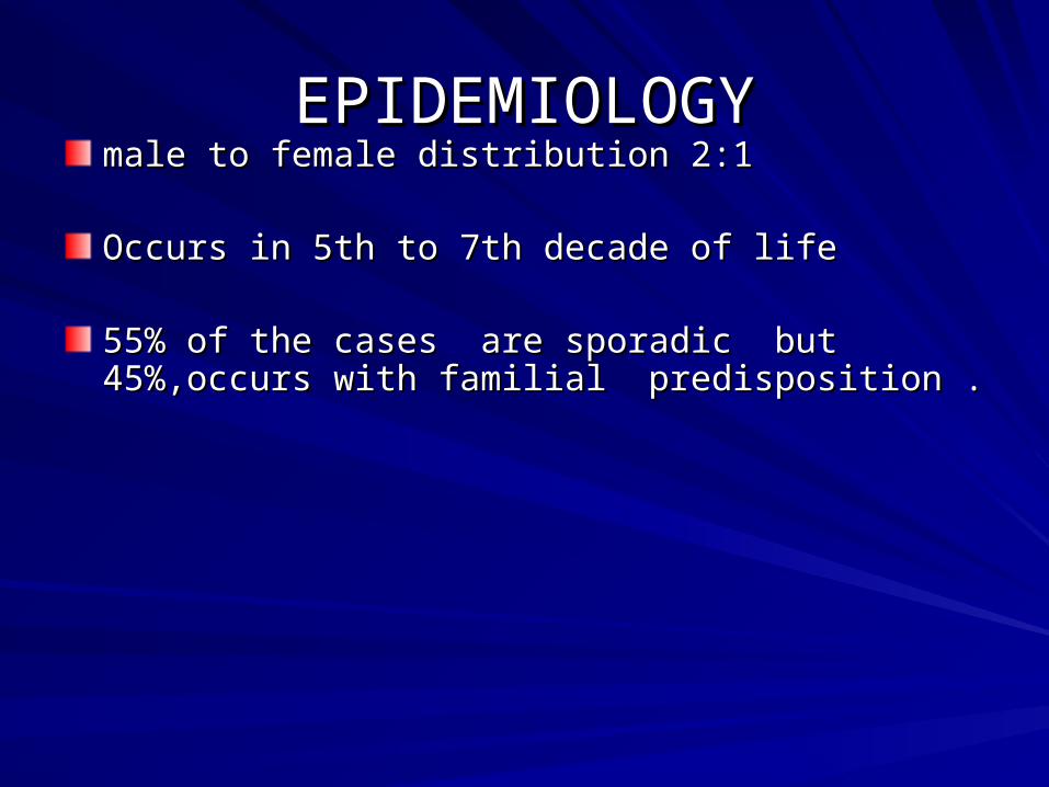
EPIDEMIOLOGYEPIDEMIOLOGYmale to female distribution 2:1male to female distribution 2:1
Occurs in 5th to 7th decade of life Occurs in 5th to 7th decade of life
55% of the cases are sporadic but 45%,occurs with 55% of the cases are sporadic but 45%,occurs with familial predisposition . familial predisposition .
Risk factors for RCCRisk factors for RCC
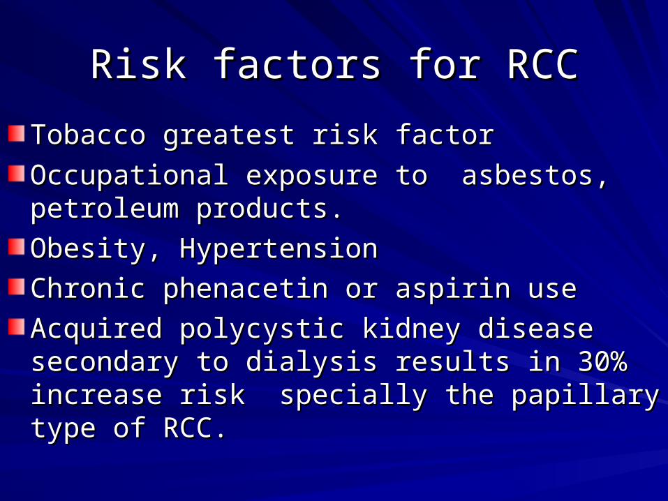
Tobacco greatest risk factor Tobacco greatest risk factor
Occupational exposure to asbestos, petroleum Occupational exposure to asbestos, petroleum products.products.
Obesity, HypertensionObesity, Hypertension
Chronic phenacetin or aspirin use Chronic phenacetin or aspirin use
Acquired polycystic kidney disease secondary to Acquired polycystic kidney disease secondary to dialysis results in 30% increase risk specially dialysis results in 30% increase risk specially the papillary type of RCC. the papillary type of RCC.
Diagnosis Diagnosis Clinical presentationClinical presentation
investigationsinvestigations
Clinical presentationClinical presentation
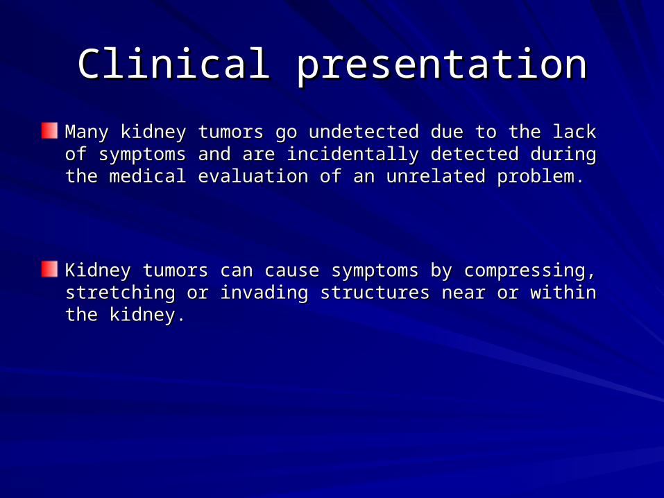
Many kidney tumors go undetected due to the lack of symptoms and Many kidney tumors go undetected due to the lack of symptoms and are incidentally detected during the medical evaluation of an are incidentally detected during the medical evaluation of an unrelated problem. unrelated problem.
Kidney tumors can cause symptoms by compressing, stretching or Kidney tumors can cause symptoms by compressing, stretching or invading structures near or within the kidney. invading structures near or within the kidney.
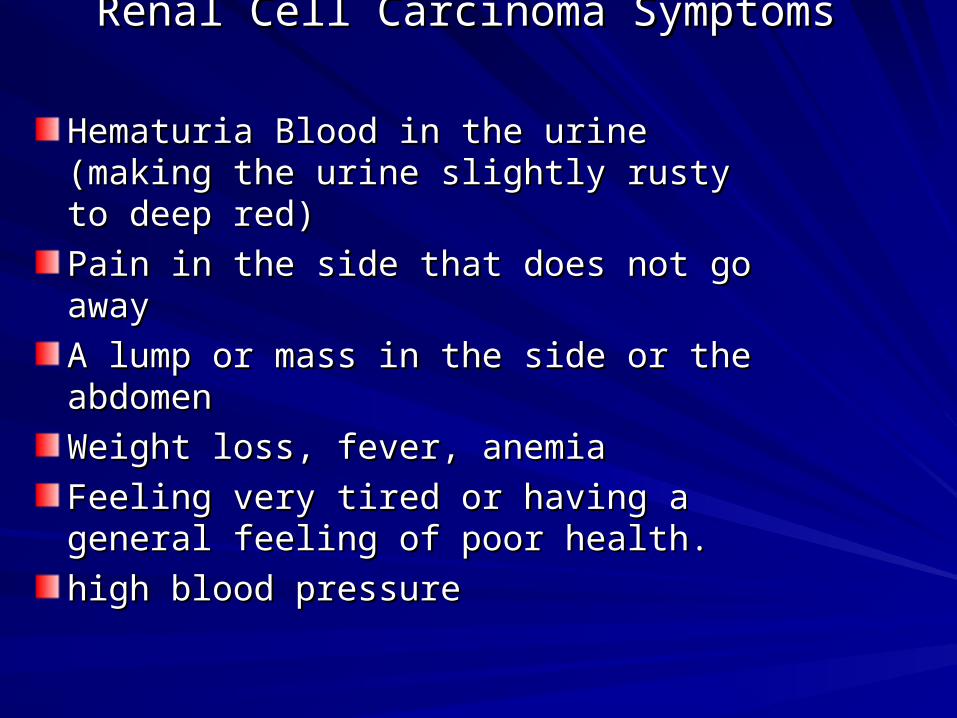
Renal Cell Carcinoma Symptoms Renal Cell Carcinoma Symptoms
Hematuria Blood in the urine (making the Hematuria Blood in the urine (making the urine slightly rusty to deep red) urine slightly rusty to deep red)
Pain in the side that does not go away Pain in the side that does not go away
A lump or mass in the side or the A lump or mass in the side or the abdomen abdomen
Weight loss, fever, anemia Weight loss, fever, anemia
Feeling very tired or having a general Feeling very tired or having a general feeling of poor health.feeling of poor health.
high blood pressurehigh blood pressure
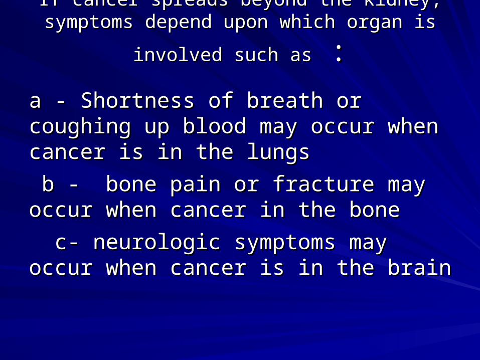
If cancer spreads beyond the kidney, symptoms If cancer spreads beyond the kidney, symptoms
depend upon which organ is involved such asdepend upon which organ is involved such as : :
a - Shortness of breath or coughing up blood a - Shortness of breath or coughing up blood may occur when cancer is in the lungsmay occur when cancer is in the lungs
b - bone pain or fracture may occur when b - bone pain or fracture may occur when cancer in the bone cancer in the bone
c- neurologic symptoms may occur when c- neurologic symptoms may occur when cancer is in the braincancer is in the brain
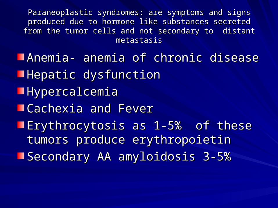
Paraneoplastic syndromes: are symptoms and signs produced due to Paraneoplastic syndromes: are symptoms and signs produced due to hormone like substances secreted from the tumor cells and not hormone like substances secreted from the tumor cells and not
secondary to distant metastasissecondary to distant metastasis
Anemia- anemia of chronic disease Anemia- anemia of chronic disease
Hepatic dysfunctionHepatic dysfunction
Hypercalcemia Hypercalcemia
Cachexia and FeverCachexia and Fever
Erythrocytosis as 1-5% of these tumors Erythrocytosis as 1-5% of these tumors produce erythropoietinproduce erythropoietin
Secondary AA amyloidosis 3-5%Secondary AA amyloidosis 3-5%
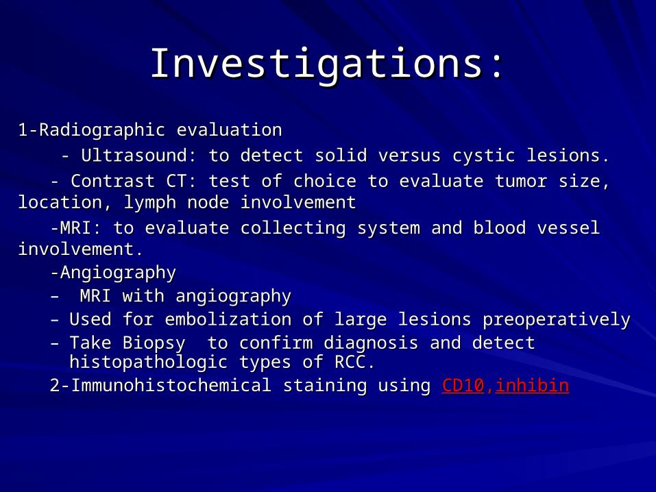
Investigations:Investigations:
1-Radiographic evaluation1-Radiographic evaluation
- Ultrasound: to detect solid versus cystic lesions.- Ultrasound: to detect solid versus cystic lesions.
- Contrast CT: test of choice to evaluate tumor size, location, lymph node - Contrast CT: test of choice to evaluate tumor size, location, lymph node involvementinvolvement
-MRI: to evaluate collecting system and blood vessel involvement.-MRI: to evaluate collecting system and blood vessel involvement. -Angiography -Angiography
– MRI with angiography MRI with angiography – Used for embolization of large lesions preoperativelyUsed for embolization of large lesions preoperatively– Take Biopsy to confirm diagnosis and detect histopathologic types of Take Biopsy to confirm diagnosis and detect histopathologic types of
RCC. RCC. 2-Immunohistochemical staining using 2-Immunohistochemical staining using CD10CD10,,inhibininhibin
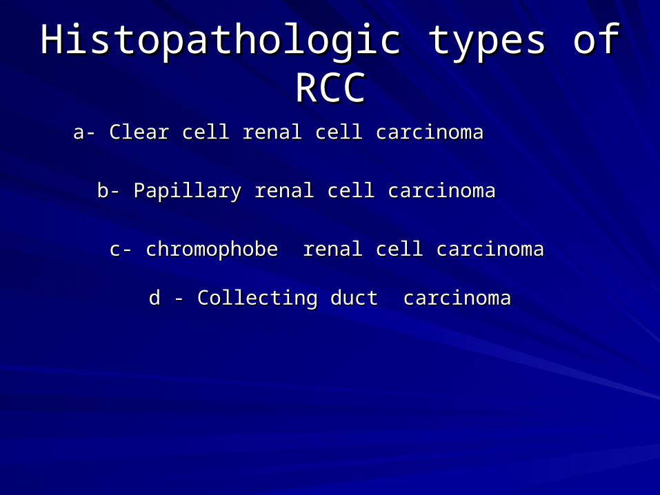
Histopathologic types of RCCHistopathologic types of RCC
a- Clear cell renal cell carcinomaa- Clear cell renal cell carcinoma
b- Papillary renal cell carcinoma b- Papillary renal cell carcinoma
c- chromophobe renal cell carcinoma c- chromophobe renal cell carcinoma d - Collecting duct carcinomad - Collecting duct carcinoma
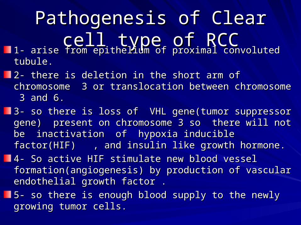
Pathogenesis of Clear cell type Pathogenesis of Clear cell type of RCCof RCC
1- arise from epithelium of proximal convoluted tubule.1- arise from epithelium of proximal convoluted tubule.
2- there is deletion in the short arm of chromosome 3 or 2- there is deletion in the short arm of chromosome 3 or translocation between chromosome 3 and 6.translocation between chromosome 3 and 6.
3- so there is loss of VHL gene(tumor suppressor gene) 3- so there is loss of VHL gene(tumor suppressor gene) present on chromosome 3 so there will not be inactivation of present on chromosome 3 so there will not be inactivation of hypoxia inducible factor(HIF) , and insulin like growth hypoxia inducible factor(HIF) , and insulin like growth hormone.hormone.
4- So active HIF stimulate new blood vessel 4- So active HIF stimulate new blood vessel formation(angiogenesis) by production of vascular endothelial formation(angiogenesis) by production of vascular endothelial growth factor . growth factor .
5- so there is enough blood supply to the newly growing tumor 5- so there is enough blood supply to the newly growing tumor cells.cells.
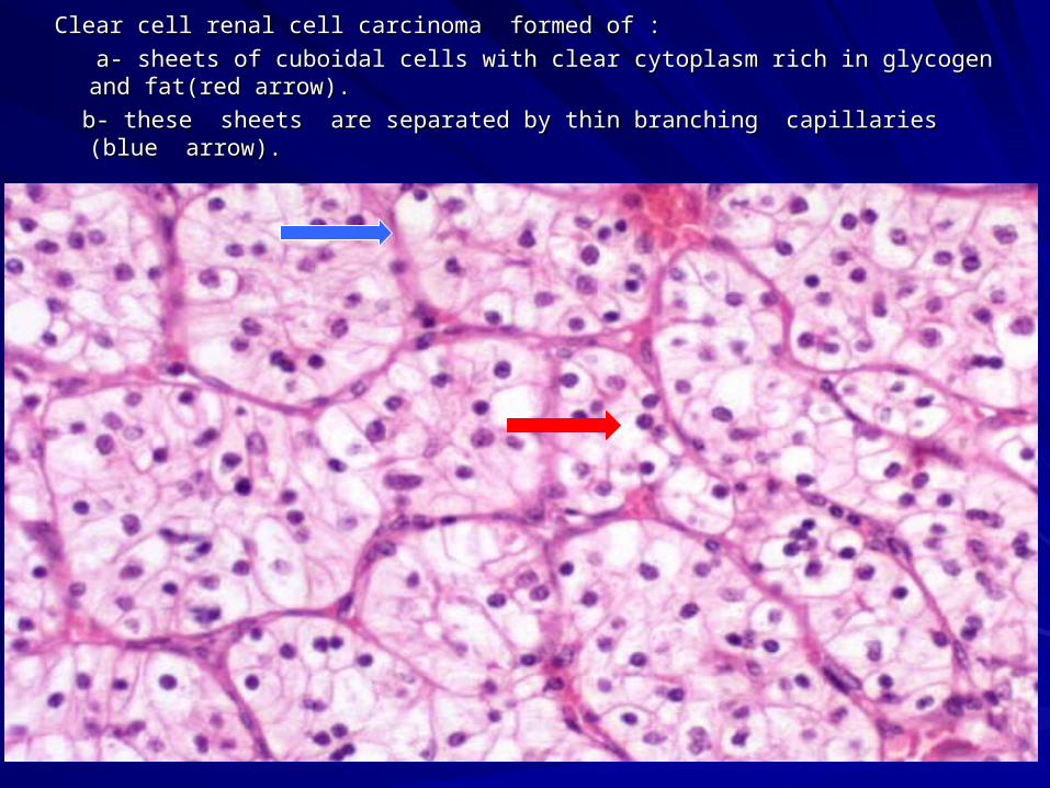
Clear cell renal cell carcinoma formed of :Clear cell renal cell carcinoma formed of :
a- sheets of cuboidal cells with clear cytoplasm rich in glycogen and fat(red arrow).a- sheets of cuboidal cells with clear cytoplasm rich in glycogen and fat(red arrow).
b- these sheets are separated by thin branching capillaries (blue arrow).b- these sheets are separated by thin branching capillaries (blue arrow).
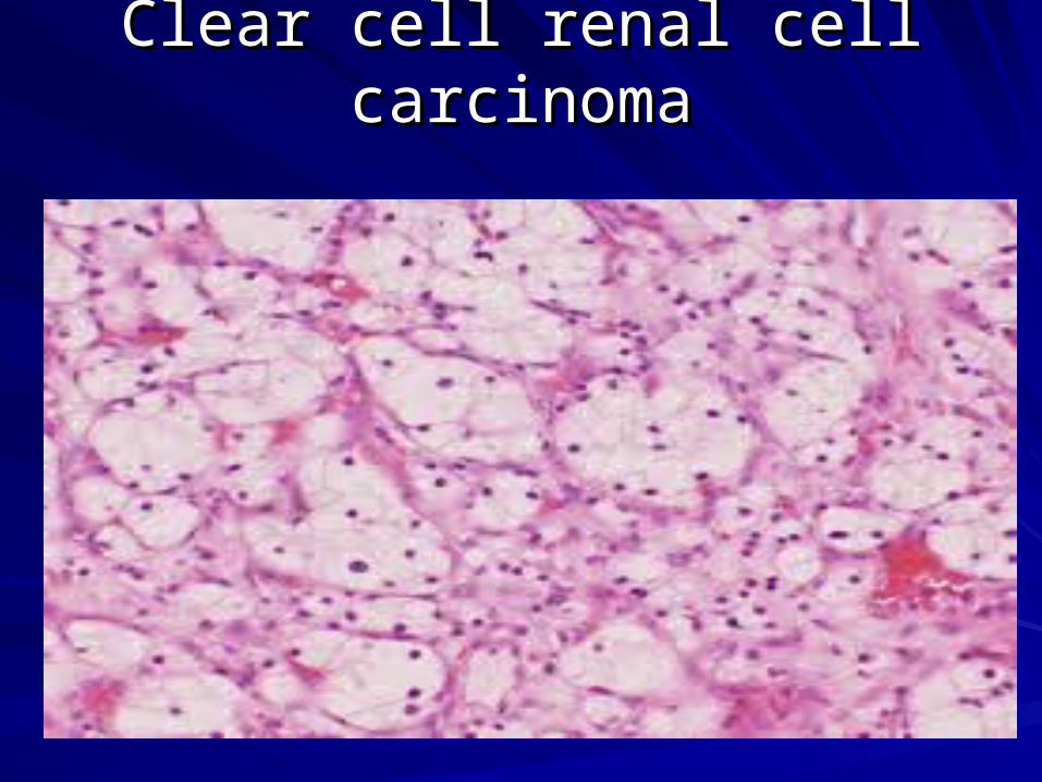
Clear cell renal cell carcinomaClear cell renal cell carcinoma
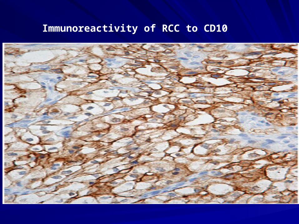
Immunoreactivity of RCC to CD10
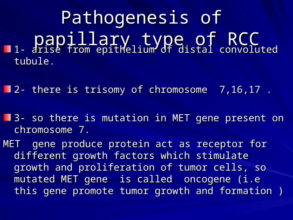
Pathogenesis of papillary type Pathogenesis of papillary type of RCCof RCC
1- arise from epithelium of distal convoluted tubule.1- arise from epithelium of distal convoluted tubule.
2- there is trisomy of chromosome 7,16,17 .2- there is trisomy of chromosome 7,16,17 .
3- so there is mutation in MET gene present on chromosome 3- so there is mutation in MET gene present on chromosome 7.7.
MET gene produce protein act as receptor for different growth MET gene produce protein act as receptor for different growth factors which stimulate growth and proliferation of tumor cells, factors which stimulate growth and proliferation of tumor cells, so mutated MET gene is called oncogene (i.e this gene so mutated MET gene is called oncogene (i.e this gene promote tumor growth and formation )promote tumor growth and formation )
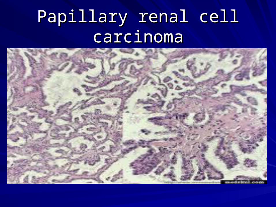
Papillary renal cell carcinomaPapillary renal cell carcinoma
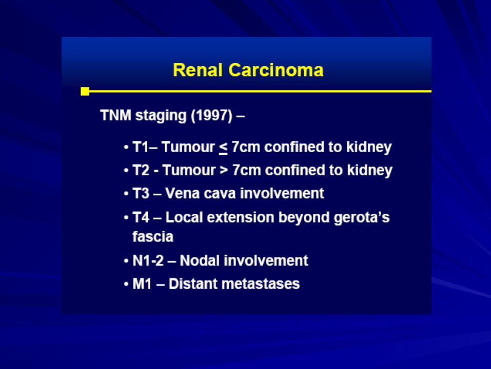
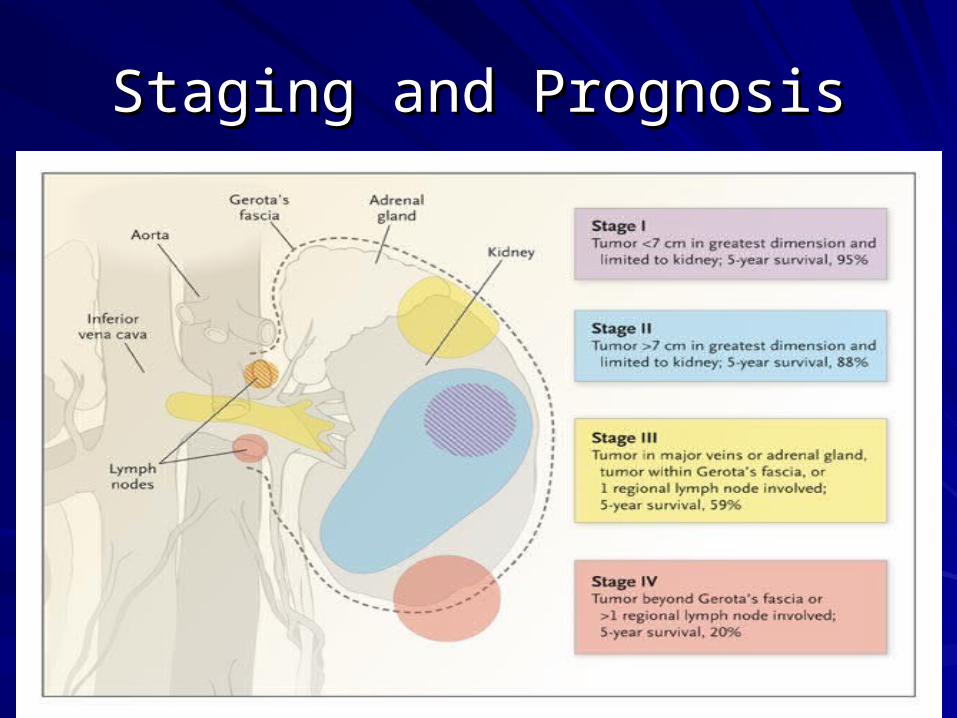
Staging and PrognosisStaging and Prognosis
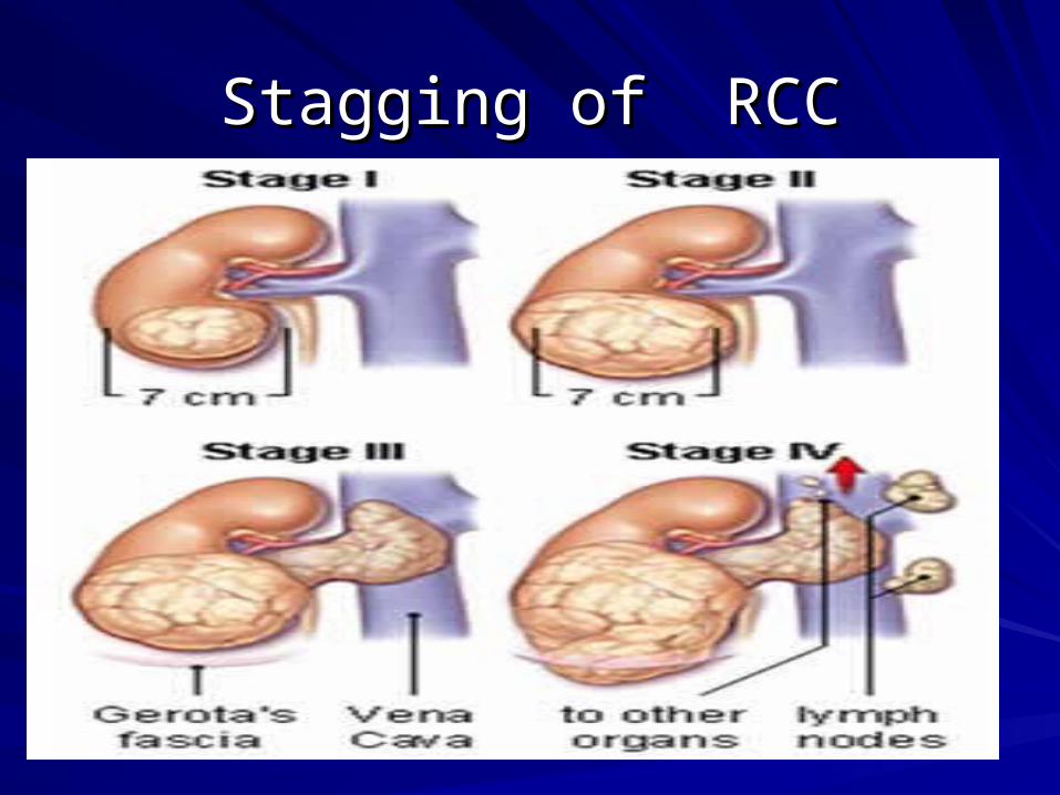
Stagging of RCCStagging of RCC

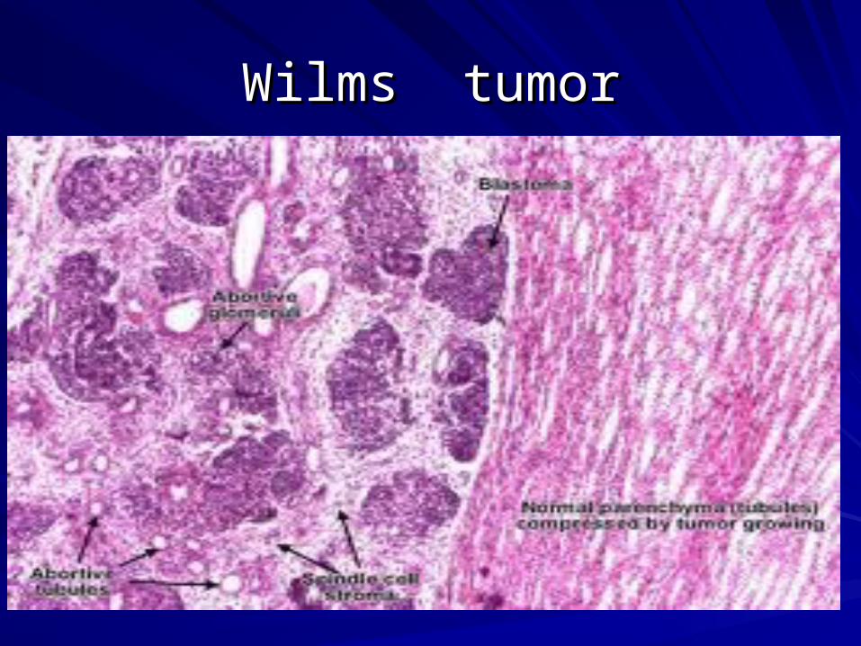
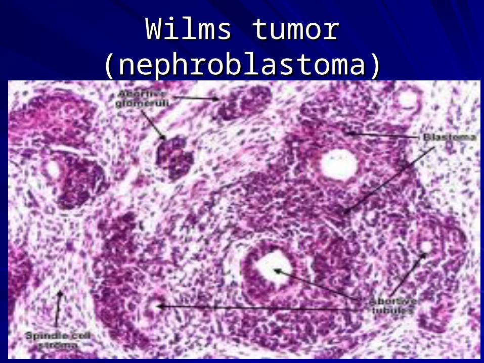
Wilms tumor (nephroblastoma)Wilms tumor (nephroblastoma)
Wilms tumorWilms tumorIt is a Malignant renal tumorIt is a Malignant renal tumor
There is Wilms tumor gene mutation. There is Wilms tumor gene mutation.
common in children 2-5 years.common in children 2-5 years.
It arises from nephrogenic rests(NR)It arises from nephrogenic rests(NR)
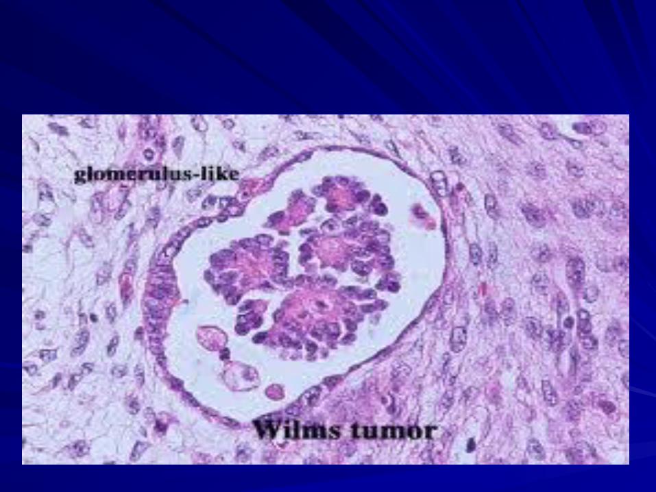
Microscopic picture characterized by triphasic Microscopic picture characterized by triphasic pattern:pattern:
a- dark blue immature blastema cellsa- dark blue immature blastema cells
b- stromal cells b- stromal cells
c- epithelial elements in form of abortive c- epithelial elements in form of abortive glomeruli and abortive tubules. glomeruli and abortive tubules.
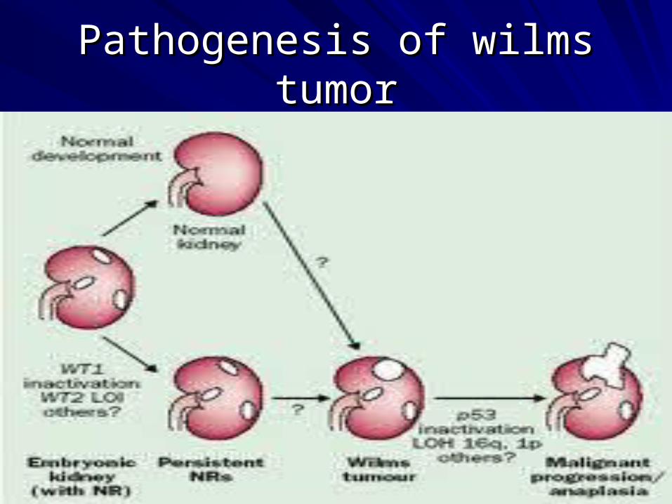
Pathogenesis of wilms tumorPathogenesis of wilms tumor
Wilms tumorWilms tumor
Wilms tumor (nephroblastoma)Wilms tumor (nephroblastoma)