Embed Size (px)
Citation preview
345A.N. Kingsnorth and K.A. LeBlanc (eds.), Management of Abdominal Hernias, DOI 10.1007/978-1-84882-877-3_22, © Springer Science+Business Media London 2013
Introduction
Approximately 200,000 ventral hernias are repaired in the US yearly [ 1 ] . This common problem has been approached in a myriad of ways, each with various technical aspects that contribute to the long term success or failure of the repair. Laparoscopic Incisional Ventral and Hernia repair (LIVH) as fi rst described by LeBlanc in 1993 [ 2 ] , builds upon the strengths of various techniques that improve overall out-come. The signi fi cant mesh overlap in the rectro-rectus repair with transfascial fi xation fi rst described by Rives and Stoppa [ 3, 4 ] is technically similar to what is achieved in LIVH Repair.
Though some still commonly perform primary suture repair of ventral hernias, it has been shown to have a recurrence rate of 54–63% [ 5, 6 ] . When primary suture repair was compared to open mesh repair, open mesh repair was found to have a recur-rence rate of 32% [ 6 ] . Though some advocate the recurrence rate to be equivalent between open mesh repair and LIVH [ 5, 7 ] , multiple other studies show LIVH to be superior in the rate of hernia relapse [ 8, 9 ] . Three prospective trials comparing laparo-scopic ventral hernia repair to open mesh repair show the recur-rence rate for LIVH to be 2–3.3% in comparison to open mesh repair which is reported to be 1.1–10% in these studies [ 7, 9, 10 ] . LIVH has been shown to be superior to open mesh repair in postoperative wound complications, hospital length of stay, and identi fi cation of multiple defects [ 7– 12 ] .
The repair of incisional and ventral hernias by the laparo-scopic approach should be performed by high-volume laparo-scopic surgeons. The surgeon should be adept at performing the more common laparoscopic operations and also be comfortable
to perform the more complex laparoscopic procedures. The assistance of another surgeon during this operation is felt to be of great bene fi t, if not mandatory, on most occasions. This chapter will present the concepts, technical aspects, and results of the LIVH as it is currently performed. There are variations of the technique that are presented within this chapter, as is com-mon to every surgical procedure. This methodology is continu-ing to evolve and undoubtedly will be modi fi ed as newer prosthetic biomaterials and instrumentation are developed in the future. One such advancement is the laparoscopic approach to component separation. Multiple studies have shown that myo-fascial advancement can be achieved with minimal fl ap dissec-tion and improved wound outcome [ 13, 14 ] .
Preoperative Evaluation
In general, if a patient is a medically appropriate candidate for open hernioplasty, then he or she could be considered a candidate for the laparoscopic approach. Patients that have signi fi cant cardiac decompensation may experience physio-logical abnormalities during the procedure because of the insuf fl ation, and resulting decrease in the venous return. Lower insuf fl ation pressures may decrease the hemodynamic fl uctuations [ 15 ] .
Generally almost all hernias are candidates for the LIVH. Even the smaller hernias in obese individuals could be repaired with this technique. Recurrence rates have been shown to be higher in obese patients [ 16– 18 ] . Yet the bene fi ts of less wound complications and the ability to identify the occult defects that are missed during an open approach make LIVH a viable option for obese patients. One may opt to use the open approach in a thin patient if it is apparent that the defect is 3 cm or less [ 16 ] .
A very large fascial defect that nearly encompasses the entire anterior abdominal wall may pose a dif fi cult problem. A laparoscopic approach, however, may be feasible. The deci-sion to attempt the laparoscopic method should be based upon the experience of the surgeon, the number of prior operative
Laparoscopic Incisional and Ventral Hernia Repair
Patrice R. Carter and Karl A. LeBlanc
22
P. R. Carter Department of Surgery , Adventist LaGrange Hospital , LaGrange , IL , USA
K. A. LeBlanc (*) Surgeons Group of Baton Rouge/Our Lady of the Lake Physician Group, Baton Rouge, LA, USA e-mail: [email protected]
346 P.R. Carter and K.A. LeBlanc
procedures, mesh repairs, the type of prosthetic utilized in any previous repair(s), and the location of the potential sites. However, there are currently no “hard and fast” rules about this issue. In those patients with very large defects, a reason-able option would be to commence the operation laparoscopi-cally and convert to an open repair if that appears to be the best alternative. More often than not, this proves to be unnecessary. A probable exception to this sequence is those individuals that exhibit a “loss of domain” of the abdominal contents. In these patients it is usually impossible to actually enter the abdomen behind the abdominal wall musculature because this muscula-ture has been displaced laterally. In these cases, conversion to the open method would occur earlier rather than later. More commonly, however, prudence dictates that the entire proce-dure should be of the open type rather than even attempting the laparoscopic approach.
Absolute contraindications to the use of the laparoscopic method would be the presence of an acute surgical abdomen. A relative contraindication is intra-abdominal infection from any source. The use of a prosthetic biomaterial in the site of an overt infection may preclude the use of such a product. However, pri-mary closure of the hernia defect with the assistance of a laparo-scopic suture passer and biologic mesh, [ 19 ] may have a role in such instances though an open repair may be indicated for gross contamination. Similarly, while the presence of incarcerated bowel does not prevent the performance of the procedure, stran-gulation of the bowel necessitates an open hernioplasty.
Because the most common incision of the abdomen is placed in the midline, most incisional hernias occur in the midline. When a surgeon begins to perform laparoscopic incisional hernioplasty, it is recommended that he or she should repair midline defects initially to gain con fi dence in use of the laparoscopic technique. Once this is accomplished, the presence of a non-midline defect or multiple defects that are not adjacent to each other should not preclude the use of laparoscopy. Appropriate positioning of the patient and accu-rate placement of the trocars will permit an approach to the entire abdominal cavity in most cases.
Previous intra-abdominal surgery is a major consideration in the evaluation of a patient for the laparoscopic procedure. The number and type of earlier operations will in fl uence the choice of patient position, the method of abdominal entry, trocar place-ment, and the position of the monitors. This preoperative assess-ment will allow the surgeon to plan the operative procedure and the operative suite based upon these fi ndings. Any previous open laparotomies will, of course, be associated with more potential for adhesion formation than procedures that were per-formed laparoscopically. Additionally, in those patients in whom a previous incisional hernia repair included the implanta-tion of any “unprotected” polypropylene prosthesis (see Open Ventral hernia chapter) can be expected to have dense scarring in all areas in which the material was exposed to the intra- abdominal contents. This should not deter experienced surgeons
from attempting a laparoscopic approach because as many as one-third of these patients will not have any adhesions at all. It is important to note, however, that the dif fi culty of the procedure can be greatly magni fi ed because of the dissection of the tena-cious scarring that is encountered involving the prosthesis and the bowel and/or omentum. The risk of enterotomy is signi fi cantly increased in such instances.
Patients in whom there is an additional need for a surgical procedure such as a cholecystectomy, fundoplication of the stomach, inguinal herniorrhaphy or biopsy of an intra-abdominal or retroperitoneal structure are special subsets that deserve careful consideration. Hernia repairs in such cases are discussed later in this chapter.
Laparoscopic incisional herniaplasty should be individualized in patients with known ascites because it may be challenging to maintain a watertight closure that averts ascitic leaks. Moreover, these patients usually have a metabolic problem (e.g., chronic renal failure or hepatic disease) that can cause poor healing and predispose them to development of a hernia at the trocar sites. The use of the 5-mm. trocars, however, has made this less problematic and these patients may also be considered on occasion. Special trocars that do not cut into the abdominal muscle but dilate the tissues to enter though the wall of the abdomen should be used in these patients. The site of entry will be smaller than the actual trocar itself after it is removed thereby further minimizing the risk of leakage of ascitic fl uid or subsequent herniation. Though the use of a prosthesis in patients with overt ascites is scarcely reported, some have achieved success with the LIVH in these patients with maximal optimization of ascites [ 20 ] .
LIVH patients are admitted to the day-surgery unit of the hospital because they can usually be considered for discharge on the day of surgery. The number and type of comorbid conditions of the patient, the type and location of the hernia(s), the presence of incarceration and the amount of adhesiolysis required will in fl uence the decision of timing of discharge from the hospital. Many patients now undergo lap-aroscopic incisional hernia repair in an ambulatory surgery center. Appropriate laboratory testing should be obtained prior to entry on the day of surgery. Patients are routinely given a preoperative dose of either a fi rst generation cepha-losporin or a fl uoroquinolone. If a patient has a history of methicillin resistant staph aureus (MRSA), Vancomycin is used for preoperative prophylaxis.
Intraoperative Considerations
Patient Preparation and Positioning
LIVH repair requires the use of general anesthesia to achieve the necessary degree of relaxation and sedation. In most cases, it is not necessary to use an orogastric or nasogastric
34722 Laparoscopic Incisional and Ventral Hernia Repair
tube unless the site of entry is in the vicinity of the stomach. A urinary drainage catheter is not used if the procedure is felt to be short in length. If the operative site is close to the blad-der (e.g., very low midline hernias or concomitant inguinal hernia repairs) or if the procedure will be prolonged it is then advisable to insert a urinary drainage catheter; preferably a three way catheter is used to fi ll the bladder for identi fi cation, if needed. Insertion of a nasogastric tube for procedures in which extensive dissection of the bowel is necessary may help to reduce the postoperative ileus that is likely to develop. It is seldom necessary to leave this tube beyond the intraop-erative phase of the procedure, however.
Most patients will be placed in the supine position. Operations upon lateral defects of the abdominal wall, such as those in a subcostal or fl ank incision, will be facilitated by use of a semidecubitus or full decubitus position. The use of a “bean-bag” in these instances will greatly aid in the posi-tioning of the patient. The additional use of the tilt capabili-ties of the operating table will assist in the manipulation of the bowel during dissection. Steep Trendelenberg or reverse Trendelenberg positions will cause the abdominal contents to move into positions that will make visualization of the contents of both the hernia and the abdomen easier. The patient’s arms should be tucked in close to the body to allow suf fi cient room to move around the patient; this is especially important if the defect is in the lower abdomen. Occasionally this may not be feasible due to the size of the individual but, in general, it is preferred when possible. Use of a protective transparent adhesive drape is recommended.
Abdominal Entry
It is understood that the method of access into the abdo-men should always be the safest approach possible. Many surgeons use the open type of Hassan entry because it is familiar to them. An open entry such as this could result in a poor seal around the trocar, which makes maintenance of insuf fl ation pressures dif fi cult resulting in inadequate visualization throughout the procedure. This method also requires the use of a larger trocar thereby posing a risk of herniation at that site in the future despite the best attempts at fascial closure.
In the patient with a primary ventral hernia or a single small defect, a Veress needle could be considered for insuf fl ation before introduction of the fi rst trocar. A “safe” area for needle insertion is usually in the right upper quad-rant because it is generally free of adhesions of bowel and omentum. A site in the upper midline could also be used if it can be placed far enough away from the hernia so as not to interfere with the repair of the hernia.
Another method to gain access into the abdominal cavity uses an “optical” trocar for abdominal entry. These non-
bladed trocars are designed to provide visualization of each layer of the abdominal wall as the trocar passes through them. This is accomplished because the laparoscope is inserted into the trocar and these structures are seen as the
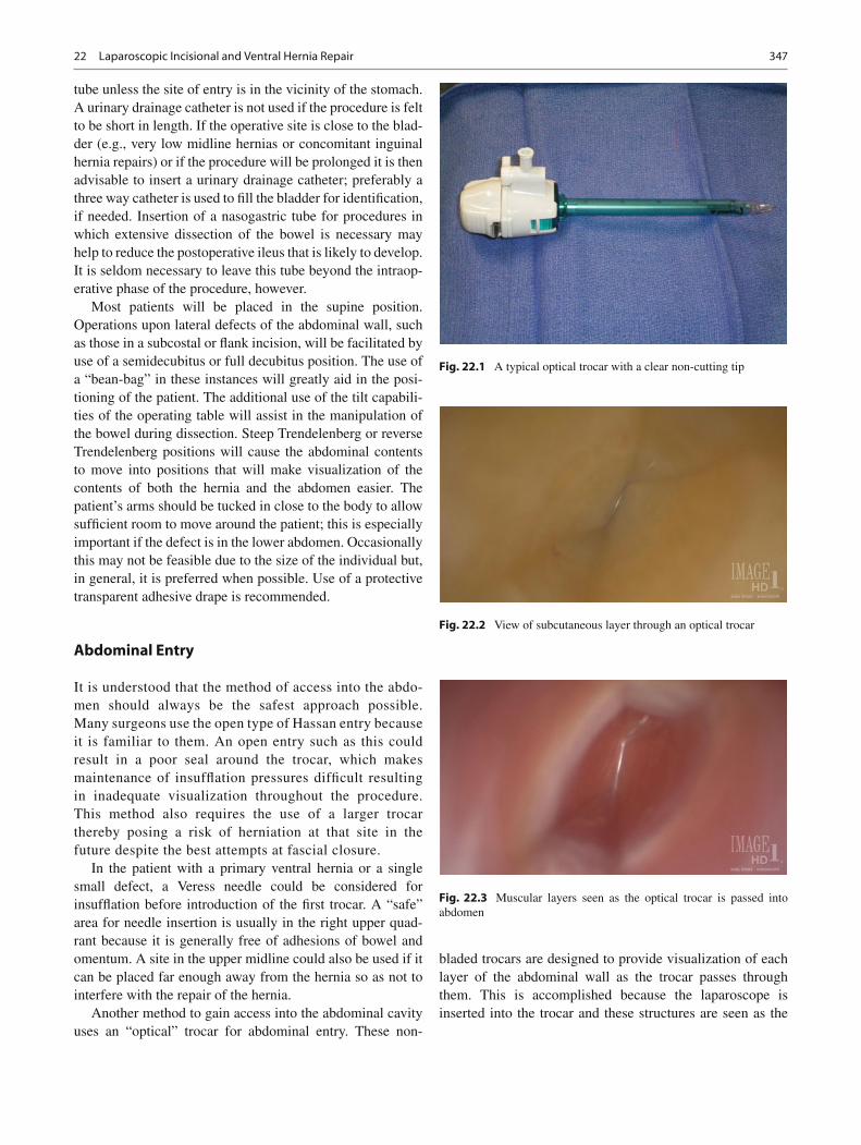
Fig. 22.1 A typical optical trocar with a clear non-cutting tip
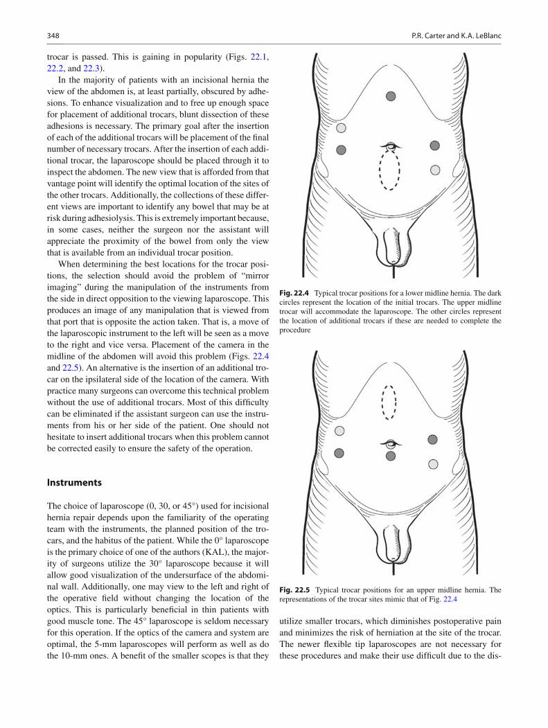
Fig. 22.2 View of subcutaneous layer through an optical trocar
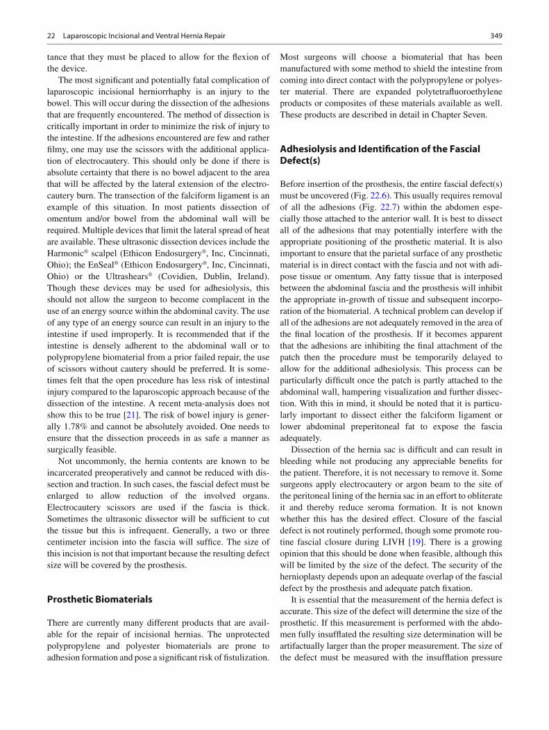
Fig. 22.3 Muscular layers seen as the optical trocar is passed into abdomen
348 P.R. Carter and K.A. LeBlanc
trocar is passed. This is gaining in popularity (Figs. 22.1 , 22.2 , and 22.3 ).
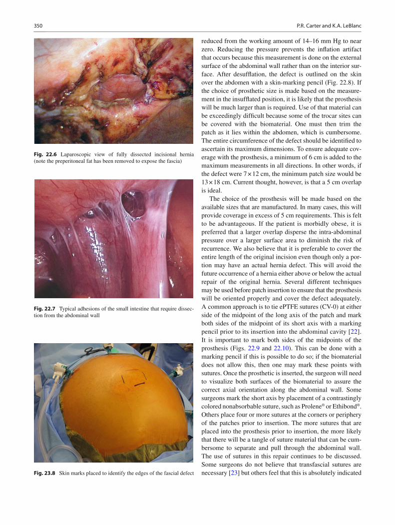
In the majority of patients with an incisional hernia the view of the abdomen is, at least partially, obscured by adhe-sions. To enhance visualization and to free up enough space for placement of additional trocars, blunt dissection of these adhesions is necessary. The primary goal after the insertion of each of the additional trocars will be placement of the fi nal number of necessary trocars. After the insertion of each addi-tional trocar, the laparoscope should be placed through it to inspect the abdomen. The new view that is afforded from that vantage point will identify the optimal location of the sites of the other trocars. Additionally, the collections of these differ-ent views are important to identify any bowel that may be at risk during adhesiolysis. This is extremely important because, in some cases, neither the surgeon nor the assistant will appreciate the proximity of the bowel from only the view that is available from an individual trocar position.
When determining the best locations for the trocar posi-tions, the selection should avoid the problem of “mirror imaging” during the manipulation of the instruments from the side in direct opposition to the viewing laparoscope. This produces an image of any manipulation that is viewed from that port that is opposite the action taken. That is, a move of the laparoscopic instrument to the left will be seen as a move to the right and vice versa. Placement of the camera in the midline of the abdomen will avoid this problem (Figs. 22.4 and 22.5 ). An alternative is the insertion of an additional tro-car on the ipsilateral side of the location of the camera. With practice many surgeons can overcome this technical problem without the use of additional trocars. Most of this dif fi culty can be eliminated if the assistant surgeon can use the instru-ments from his or her side of the patient. One should not hesitate to insert additional trocars when this problem cannot be corrected easily to ensure the safety of the operation.
Instruments
The choice of laparoscope (0, 30, or 45°) used for incisional hernia repair depends upon the familiarity of the operating team with the instruments, the planned position of the tro-cars, and the habitus of the patient. While the 0° laparoscope is the primary choice of one of the authors (KAL), the major-ity of surgeons utilize the 30° laparoscope because it will allow good visualization of the undersurface of the abdomi-nal wall. Additionally, one may view to the left and right of the operative fi eld without changing the location of the optics. This is particularly bene fi cial in thin patients with good muscle tone. The 45° laparoscope is seldom necessary for this operation. If the optics of the camera and system are optimal, the 5-mm laparoscopes will perform as well as do the 10-mm ones. A bene fi t of the smaller scopes is that they
utilize smaller trocars, which diminishes postoperative pain and minimizes the risk of herniation at the site of the trocar. The newer fl exible tip laparoscopes are not necessary for these procedures and make their use dif fi cult due to the dis-
Fig. 22.4 Typical trocar positions for a lower midline hernia. The dark circles represent the location of the initial trocars. The upper midline trocar will accommodate the laparoscope. The other circles represent the location of additional trocars if these are needed to complete the procedure
Fig. 22.5 Typical trocar positions for an upper midline hernia. The representations of the trocar sites mimic that of Fig. 22.4
34922 Laparoscopic Incisional and Ventral Hernia Repair
tance that they must be placed to allow for the fl exion of the device.
The most signi fi cant and potentially fatal complication of laparoscopic incisional herniorrhaphy is an injury to the bowel. This will occur during the dissection of the adhesions that are frequently encountered. The method of dissection is critically important in order to minimize the risk of injury to the intestine. If the adhesions encountered are few and rather fi lmy, one may use the scissors with the additional applica-tion of electrocautery. This should only be done if there is absolute certainty that there is no bowel adjacent to the area that will be affected by the lateral extension of the electro-cautery burn. The transection of the falciform ligament is an example of this situation. In most patients dissection of omentum and/or bowel from the abdominal wall will be required. Multiple devices that limit the lateral spread of heat are available. These ultrasonic dissection devices include the Harmonic ® scalpel (Ethicon Endosurgery ® , Inc, Cincinnati, Ohio); the EnSeal ® (Ethicon Endosurgery ® , Inc, Cincinnati, Ohio) or the Ultrashears ® (Covidien, Dublin, Ireland). Though these devices may be used for adhesiolysis, this should not allow the surgeon to become complacent in the use of an energy source within the abdominal cavity. The use of any type of an energy source can result in an injury to the intestine if used improperly. It is recommended that if the intestine is densely adherent to the abdominal wall or to polypropylene biomaterial from a prior failed repair, the use of scissors without cautery should be preferred. It is some-times felt that the open procedure has less risk of intestinal injury compared to the laparoscopic approach because of the dissection of the intestine. A recent meta-analysis does not show this to be true [ 21 ] . The risk of bowel injury is gener-ally 1.78% and cannot be absolutely avoided. One needs to ensure that the dissection proceeds in as safe a manner as surgically feasible.
Not uncommonly, the hernia contents are known to be incarcerated preoperatively and cannot be reduced with dis-section and traction. In such cases, the fascial defect must be enlarged to allow reduction of the involved organs. Electrocautery scissors are used if the fascia is thick. Sometimes the ultrasonic dissector will be suf fi cient to cut the tissue but this is infrequent. Generally, a two or three centimeter incision into the fascia will suf fi ce. The size of this incision is not that important because the resulting defect size will be covered by the prosthesis.
Prosthetic Biomaterials
There are currently many different products that are avail-able for the repair of incisional hernias. The unprotected polypropylene and polyester biomaterials are prone to adhesion formation and pose a signi fi cant risk of fi stulization.
Most surgeons will choose a biomaterial that has been manufactured with some method to shield the intestine from coming into direct contact with the polypropylene or polyes-ter material. There are expanded polytetra fl uoroethylene products or composites of these materials available as well. These products are described in detail in Chapter Seven.
Adhesiolysis and Identi fi cation of the Fascial Defect(s)
Before insertion of the prosthesis, the entire fascial defect(s) must be uncovered (Fig. 22.6 ). This usually requires removal of all the adhesions (Fig. 22.7 ) within the abdomen espe-cially those attached to the anterior wall. It is best to dissect all of the adhesions that may potentially interfere with the appropriate positioning of the prosthetic material. It is also important to ensure that the parietal surface of any prosthetic material is in direct contact with the fascia and not with adi-pose tissue or omentum. Any fatty tissue that is interposed between the abdominal fascia and the prosthesis will inhibit the appropriate in-growth of tissue and subsequent incorpo-ration of the biomaterial. A technical problem can develop if all of the adhesions are not adequately removed in the area of the fi nal location of the prosthesis. If it becomes apparent that the adhesions are inhibiting the fi nal attachment of the patch then the procedure must be temporarily delayed to allow for the additional adhesiolysis. This process can be particularly dif fi cult once the patch is partly attached to the abdominal wall, hampering visualization and further dissec-tion. With this in mind, it should be noted that it is particu-larly important to dissect either the falciform ligament or lower abdominal preperitoneal fat to expose the fascia adequately.
Dissection of the hernia sac is dif fi cult and can result in bleeding while not producing any appreciable bene fi ts for the patient. Therefore, it is not necessary to remove it. Some surgeons apply electrocautery or argon beam to the site of the peritoneal lining of the hernia sac in an effort to obliterate it and thereby reduce seroma formation. It is not known whether this has the desired effect. Closure of the fascial defect is not routinely performed, though some promote rou-tine fascial closure during LIVH [ 19 ] . There is a growing opinion that this should be done when feasible, although this will be limited by the size of the defect. The security of the hernioplasty depends upon an adequate overlap of the fascial defect by the prosthesis and adequate patch fi xation.
It is essential that the measurement of the hernia defect is accurate. This size of the defect will determine the size of the prosthetic. If this measurement is performed with the abdo-men fully insuf fl ated the resulting size determination will be artifactually larger than the proper measurement. The size of the defect must be measured with the insuf fl ation pressure
350 P.R. Carter and K.A. LeBlanc
reduced from the working amount of 14–16 mm Hg to near zero. Reducing the pressure prevents the in fl ation artifact that occurs because this measurement is done on the external surface of the abdominal wall rather than on the interior sur-face. After desuf fl ation, the defect is outlined on the skin over the abdomen with a skin-marking pencil (Fig. 22.8). If the choice of prosthetic size is made based on the measure-ment in the insuf fl ated position, it is likely that the prosthesis will be much larger than is required. Use of that material can be exceedingly dif fi cult because some of the trocar sites can be covered with the biomaterial. One must then trim the patch as it lies within the abdomen, which is cumbersome. The entire circumference of the defect should be identi fi ed to ascertain its maximum dimensions. To ensure adequate cov-erage with the prosthesis, a minimum of 6 cm is added to the maximum measurements in all directions. In other words, if the defect were 7 × 12 cm, the minimum patch size would be 13 × 18 cm. Current thought, however, is that a 5 cm overlap is ideal.
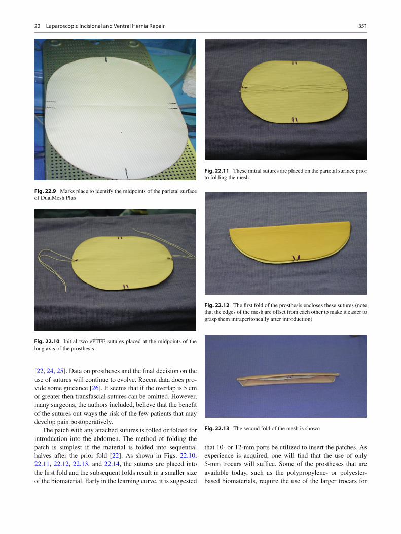
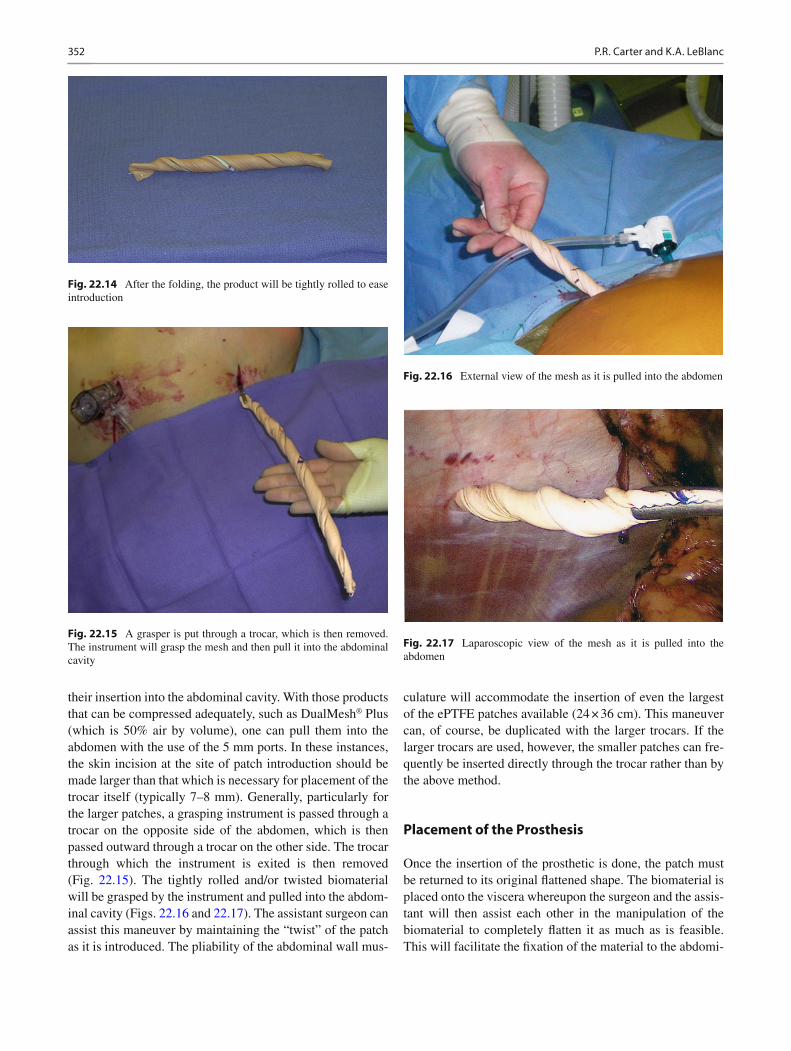
The choice of the prosthesis will be made based on the available sizes that are manufactured. In many cases, this will provide coverage in excess of 5 cm requirements. This is felt to be advantageous. If the patient is morbidly obese, it is preferred that a larger overlap disperse the intra-abdominal pressure over a larger surface area to diminish the risk of recurrence. We also believe that it is preferable to cover the entire length of the original incision even though only a por-tion may have an actual hernia defect. This will avoid the future occurrence of a hernia either above or below the actual repair of the original hernia. Several different techniques may be used before patch insertion to ensure that the prosthesis will be oriented properly and cover the defect adequately. A common approach is to tie ePTFE sutures (CV-0) at either side of the midpoint of the long axis of the patch and mark both sides of the midpoint of its short axis with a marking pencil prior to its insertion into the abdominal cavity [ 22 ] . It is important to mark both sides of the midpoints of the prosthesis (Figs. 22.9 and 22.10 ). This can be done with a marking pencil if this is possible to do so; if the biomaterial does not allow this, then one may mark these points with sutures. Once the prosthetic is inserted, the surgeon will need to visualize both surfaces of the biomaterial to assure the correct axial orientation along the abdominal wall. Some surgeons mark the short axis by placement of a contrastingly colored nonabsorbable suture, such as Prolene ® or Ethibond ® . Others place four or more sutures at the corners or periphery of the patches prior to insertion. The more sutures that are placed into the prosthesis prior to insertion, the more likely that there will be a tangle of suture material that can be cum-bersome to separate and pull through the abdominal wall. The use of sutures in this repair continues to be discussed. Some surgeons do not believe that transfascial sutures are necessary [ 23 ] but others feel that this is absolutely indicated
Fig. 22.7 Typical adhesions of the small intestine that require dissec-tion from the abdominal wall
Fig. 22.6 Laparoscopic view of fully dissected incisional hernia (note the preperitoneal fat has been removed to expose the fascia)
Fig. 23.8 Skin marks placed to identify the edges of the fascial defect
35122 Laparoscopic Incisional and Ventral Hernia Repair
[ 22, 24, 25 ] . Data on prostheses and the fi nal decision on the use of sutures will continue to evolve. Recent data does pro-vide some guidance [ 26 ] . It seems that if the overlap is 5 cm or greater then transfascial sutures can be omitted. However, many surgeons, the authors included, believe that the bene fi t of the sutures out ways the risk of the few patients that may develop pain postoperatively.
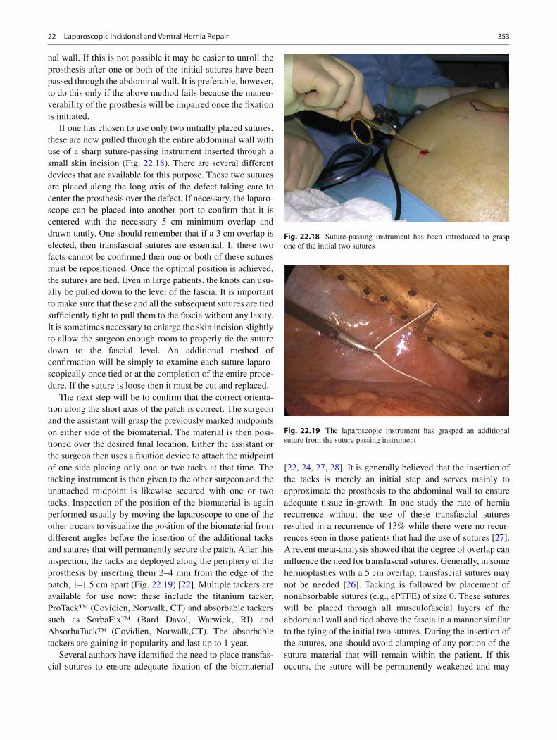
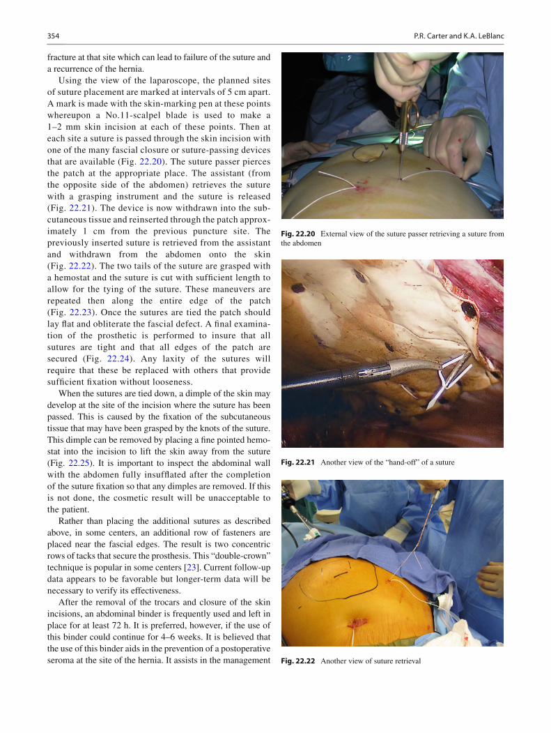
The patch with any attached sutures is rolled or folded for introduction into the abdomen. The method of folding the patch is simplest if the material is folded into sequential halves after the prior fold [ 22 ] . As shown in Figs. 22.10 , 22.11 , 22.12 , 22.13 , and 22.14 , the sutures are placed into the fi rst fold and the subsequent folds result in a smaller size of the biomaterial. Early in the learning curve, it is suggested
that 10- or 12-mm ports be utilized to insert the patches. As experience is acquired, one will fi nd that the use of only 5-mm trocars will suf fi ce. Some of the prostheses that are available today, such as the polypropylene- or polyester-based biomaterials, require the use of the larger trocars for
Fig. 22.9 Marks place to identify the midpoints of the parietal surface of DualMesh Plus
Fig. 22.10 Initial two ePTFE sutures placed at the midpoints of the long axis of the prosthesis
Fig. 22.11 These initial sutures are placed on the parietal surface prior to folding the mesh
Fig. 22.12 The fi rst fold of the prosthesis encloses these sutures (note that the edges of the mesh are offset from each other to make it easier to grasp them intraperitoneally after introduction)
Fig. 22.13 The second fold of the mesh is shown
352 P.R. Carter and K.A. LeBlanc
their insertion into the abdominal cavity. With those products that can be compressed adequately, such as DualMesh ® Plus (which is 50% air by volume), one can pull them into the abdomen with the use of the 5 mm ports. In these instances, the skin incision at the site of patch introduction should be made larger than that which is necessary for placement of the trocar itself (typically 7–8 mm). Generally, particularly for the larger patches, a grasping instrument is passed through a trocar on the opposite side of the abdomen, which is then passed outward through a trocar on the other side. The trocar through which the instrument is exited is then removed (Fig. 22.15 ). The tightly rolled and/or twisted biomaterial will be grasped by the instrument and pulled into the abdom-inal cavity (Figs. 22.16 and 22.17 ). The assistant surgeon can assist this maneuver by maintaining the “twist” of the patch as it is introduced. The pliability of the abdominal wall mus-
culature will accommodate the insertion of even the largest of the ePTFE patches available (24 × 36 cm). This maneuver can, of course, be duplicated with the larger trocars. If the larger trocars are used, however, the smaller patches can fre-quently be inserted directly through the trocar rather than by the above method.
Placement of the Prosthesis
Once the insertion of the prosthetic is done, the patch must be returned to its original fl attened shape. The biomaterial is placed onto the viscera whereupon the surgeon and the assis-tant will then assist each other in the manipulation of the biomaterial to completely fl atten it as much as is feasible. This will facilitate the fi xation of the material to the abdomi-
Fig. 22.14 After the folding, the product will be tightly rolled to ease introduction
Fig. 22.15 A grasper is put through a trocar, which is then removed. The instrument will grasp the mesh and then pull it into the abdominal cavity
Fig. 22.16 External view of the mesh as it is pulled into the abdomen
Fig. 22.17 Laparoscopic view of the mesh as it is pulled into the abdomen
35322 Laparoscopic Incisional and Ventral Hernia Repair
nal wall. If this is not possible it may be easier to unroll the prosthesis after one or both of the initial sutures have been passed through the abdominal wall. It is preferable, however, to do this only if the above method fails because the maneu-verability of the prosthesis will be impaired once the fi xation is initiated.
If one has chosen to use only two initially placed sutures, these are now pulled through the entire abdominal wall with use of a sharp suture-passing instrument inserted through a small skin incision (Fig. 22.18 ). There are several different devices that are available for this purpose. These two sutures are placed along the long axis of the defect taking care to center the prosthesis over the defect. If necessary, the laparo-scope can be placed into another port to con fi rm that it is centered with the necessary 5 cm minimum overlap and drawn tautly. One should remember that if a 3 cm overlap is elected, then transfascial sutures are essential. If these two facts cannot be con fi rmed then one or both of these sutures must be repositioned. Once the optimal position is achieved, the sutures are tied. Even in large patients, the knots can usu-ally be pulled down to the level of the fascia. It is important to make sure that these and all the subsequent sutures are tied suf fi ciently tight to pull them to the fascia without any laxity. It is sometimes necessary to enlarge the skin incision slightly to allow the surgeon enough room to properly tie the suture down to the fascial level. An additional method of con fi rmation will be simply to examine each suture laparo-scopically once tied or at the completion of the entire proce-dure. If the suture is loose then it must be cut and replaced.
The next step will be to con fi rm that the correct orienta-tion along the short axis of the patch is correct. The surgeon and the assistant will grasp the previously marked midpoints on either side of the biomaterial. The material is then posi-tioned over the desired fi nal location. Either the assistant or the surgeon then uses a fi xation device to attach the midpoint of one side placing only one or two tacks at that time. The tacking instrument is then given to the other surgeon and the unattached midpoint is likewise secured with one or two tacks. Inspection of the position of the biomaterial is again performed usually by moving the laparoscope to one of the other trocars to visualize the position of the biomaterial from different angles before the insertion of the additional tacks and sutures that will permanently secure the patch. After this inspection, the tacks are deployed along the periphery of the prosthesis by inserting them 2–4 mm from the edge of the patch, 1–1.5 cm apart (Fig. 22.19 ) [ 22 ] . Multiple tackers are available for use now: these include the titanium tacker, ProTack™ (Covidien, Norwalk, CT) and absorbable tackers such as SorbaFix™ (Bard Davol, Warwick, RI) and AbsorbaTack™ (Covidien, Norwalk,CT). The absorbable tackers are gaining in popularity and last up to 1 year.
Several authors have identi fi ed the need to place transfas-cial sutures to ensure adequate fi xation of the biomaterial
[ 22, 24, 27, 28 ] . It is generally believed that the insertion of the tacks is merely an initial step and serves mainly to approximate the prosthesis to the abdominal wall to ensure adequate tissue in-growth. In one study the rate of hernia recurrence without the use of these transfascial sutures resulted in a recurrence of 13% while there were no recur-rences seen in those patients that had the use of sutures [ 27 ] . A recent meta-analysis showed that the degree of overlap can in fl uence the need for transfascial sutures. Generally, in some hernioplasties with a 5 cm overlap, transfascial sutures may not be needed [ 26 ] . Tacking is followed by placement of nonabsorbable sutures (e.g., ePTFE) of size 0. These sutures will be placed through all musculofascial layers of the abdominal wall and tied above the fascia in a manner similar to the tying of the initial two sutures. During the insertion of the sutures, one should avoid clamping of any portion of the suture material that will remain within the patient. If this occurs, the suture will be permanently weakened and may
Fig. 22.18 Suture-passing instrument has been introduced to grasp one of the initial two sutures
Fig. 22.19 The laparoscopic instrument has grasped an additional suture from the suture passing instrument
354 P.R. Carter and K.A. LeBlanc
fracture at that site which can lead to failure of the suture and a recurrence of the hernia.
Using the view of the laparoscope, the planned sites of suture placement are marked at intervals of 5 cm apart. A mark is made with the skin-marking pen at these points whereupon a No.11-scalpel blade is used to make a 1–2 mm skin incision at each of these points. Then at each site a suture is passed through the skin incision with one of the many fascial closure or suture-passing devices that are available (Fig. 22.20 ). The suture passer pierces the patch at the appropriate place. The assistant (from the opposite side of the abdomen) retrieves the suture with a grasping instrument and the suture is released (Fig. 22.21 ). The device is now withdrawn into the sub-cutaneous tissue and reinserted through the patch approx-imately 1 cm from the previous puncture site. The previously inserted suture is retrieved from the assistant and withdrawn from the abdomen onto the skin (Fig. 22.22 ). The two tails of the suture are grasped with a hemostat and the suture is cut with suf fi cient length to allow for the tying of the suture. These maneuvers are repeated then along the entire edge of the patch (Fig. 22.23 ). Once the sutures are tied the patch should lay fl at and obliterate the fascial defect. A fi nal examina-tion of the prosthetic is performed to insure that all sutures are tight and that all edges of the patch are secured (Fig. 22.24 ). Any laxity of the sutures will require that these be replaced with others that provide suf fi cient fi xation without looseness.
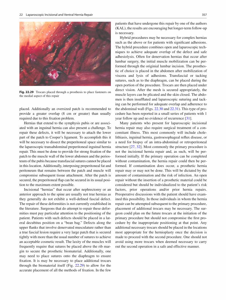
When the sutures are tied down, a dimple of the skin may develop at the site of the incision where the suture has been passed. This is caused by the fi xation of the subcutaneous tissue that may have been grasped by the knots of the suture. This dimple can be removed by placing a fi ne pointed hemo-stat into the incision to lift the skin away from the suture (Fig. 22.25 ). It is important to inspect the abdominal wall with the abdomen fully insuf fl ated after the completion of the suture fi xation so that any dimples are removed. If this is not done, the cosmetic result will be unacceptable to the patient.
Rather than placing the additional sutures as described above, in some centers, an additional row of fasteners are placed near the fascial edges. The result is two concentric rows of tacks that secure the prosthesis. This “double-crown” technique is popular in some centers [ 23 ] . Current follow-up data appears to be favorable but longer-term data will be necessary to verify its effectiveness.
After the removal of the trocars and closure of the skin incisions, an abdominal binder is frequently used and left in place for at least 72 h. It is preferred, however, if the use of this binder could continue for 4–6 weeks. It is believed that the use of this binder aids in the prevention of a postoperative seroma at the site of the hernia. It assists in the management
Fig. 22.20 External view of the suture passer retrieving a suture from the abdomen
Fig. 22.21 Another view of the “hand-off” of a suture
Fig. 22.22 Another view of suture retrieval
35522 Laparoscopic Incisional and Ventral Hernia Repair
of postoperative pain and does not appear to affect the respi-ratory effort of the patient.
Immediate Postoperative Considerations
Approximately 50% of these patients can be discharged on the same day of surgery. Generally this will be the patient that has a single defect, a hernia dimension of less than 25 cm [ 2 ] , few adhesions, and no incarcerated contents of the her-nia. The average length of stay is 1–2 days [ 7, 8, 12 ] . Patients can consume liquids the day of surgery and resume taking any regular medications immediately. Oral and parenteral sedatives are given as needed. Postoperatively, many patients will experience some degree of abdominal distension, which is usually proportional to the extent of adhesiolysis and the extent of bowel involvement. However, most patients can resume a regular diet the day after the operation. Occasionally, some patients will experience prolongation of the ileus. This should be managed by the usual methods; which would include a nasogastric tube when necessary.
Pain may be used as the guide to determine when patients can resume their normal activities. They are allowed to shower the next day. Patients may return to their daily activi-ties, including work, as soon as they can do so without marked pain. The majority of patients are able to drive within a week and resume job-related activities in 7–14 days. Most surgeons do not restrict the activities of these patients but allow the level of pain to dictate the increase in the level of activity.
After removal of the binder, many patients will note a fi rm bulge at the hernia site. The bulge may represent a seroma in the fi rst few weeks, but subsequently this area represents the cicatricial event that occurs in the majority of these patients. Seroma formation is a common occurrence after LIVH. However, it is rarely, if ever, necessary to aspirate these fl uid collections, as they will generally resolve without interven-tion. Aspiration will also expose the patient to a risk of the introduction of infection into the seroma.
Late Postoperative Considerations
In most patients with the cicatricial “bulge” and/or seroma at the hernia site, resolution will be noted within 2 months, depending on the size of the hernia and its contents. Occasionally the skin of the abdominal wall that overlaid the hernia will become erythematous within 4–6 days postopera-tively, usually in association with a distinct surface fi rmness but with little tenderness and without the presence of fever, chills, or leukocytosis (Fig. 22.26 ). This situation, which is seen in approximately 5–7% of patients, can persist for a few weeks and can be most unsettling. This is believed to be the
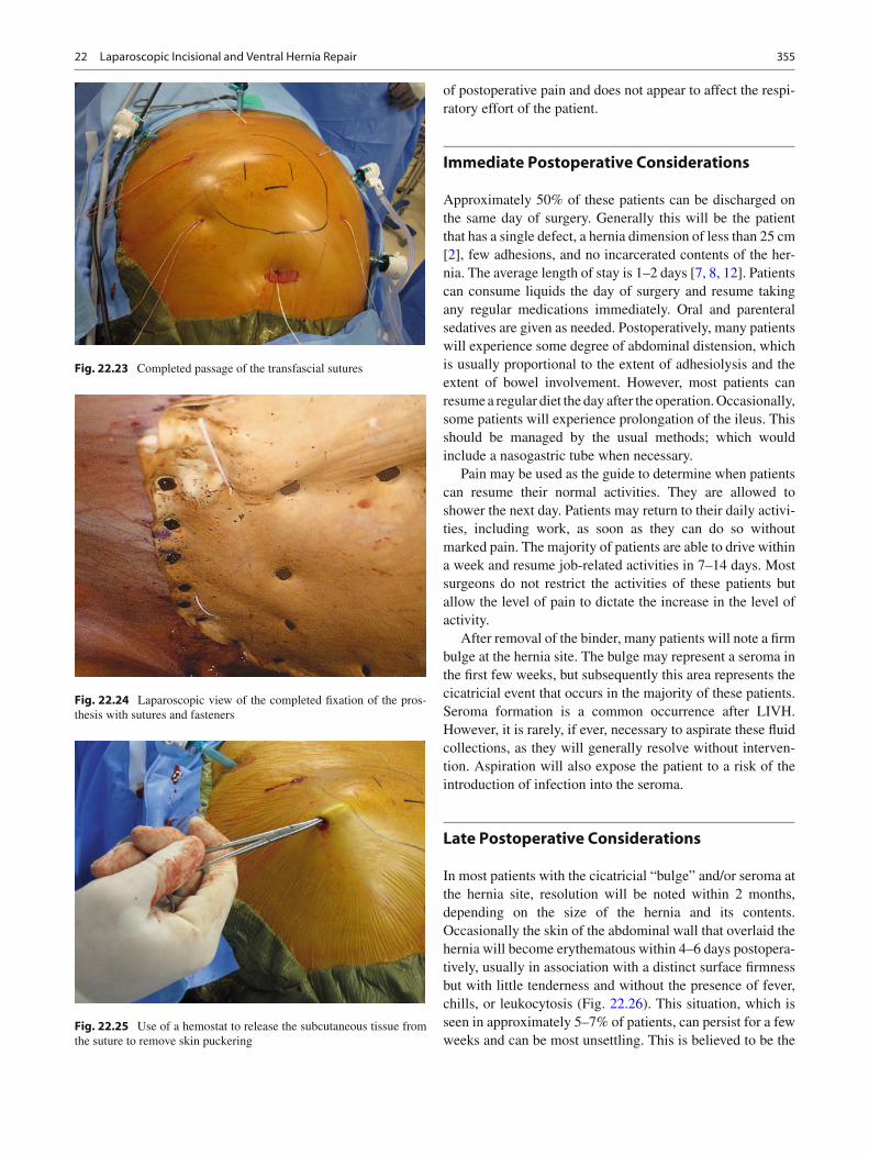
Fig. 22.23 Completed passage of the transfascial sutures
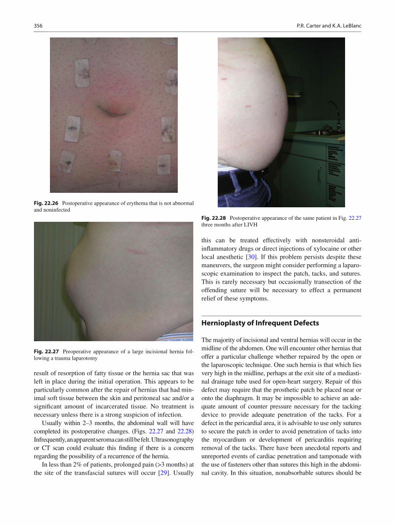
Fig. 22.24 Laparoscopic view of the completed fi xation of the pros-thesis with sutures and fasteners
Fig. 22.25 Use of a hemostat to release the subcutaneous tissue from the suture to remove skin puckering
356 P.R. Carter and K.A. LeBlanc
result of resorption of fatty tissue or the hernia sac that was left in place during the initial operation. This appears to be particularly common after the repair of hernias that had min-imal soft tissue between the skin and peritoneal sac and/or a signi fi cant amount of incarcerated tissue. No treatment is necessary unless there is a strong suspicion of infection.
Usually within 2–3 months, the abdominal wall will have completed its postoperative changes. (Figs. 22.27 and 22.28 ) Infrequently, an apparent seroma can still be felt. Ultrasonography or CT scan could evaluate this fi nding if there is a concern regarding the possibility of a recurrence of the hernia.
In less than 2% of patients, prolonged pain (>3 months) at the site of the transfascial sutures will occur [ 29 ] . Usually
this can be treated effectively with nonsteroidal anti-in fl ammatory drugs or direct injections of xylocaine or other local anesthetic [ 30 ] . If this problem persists despite these maneuvers, the surgeon might consider performing a laparo-scopic examination to inspect the patch, tacks, and sutures. This is rarely necessary but occasionally transection of the offending suture will be necessary to effect a permanent relief of these symptoms.
Hernioplasty of Infrequent Defects
The majority of incisional and ventral hernias will occur in the midline of the abdomen. One will encounter other hernias that offer a particular challenge whether repaired by the open or the laparoscopic technique. One such hernia is that which lies very high in the midline, perhaps at the exit site of a mediasti-nal drainage tube used for open-heart surgery. Repair of this defect may require that the prosthetic patch be placed near or onto the diaphragm. It may be impossible to achieve an ade-quate amount of counter pressure necessary for the tacking device to provide adequate penetration of the tacks. For a defect in the pericardial area, it is advisable to use only sutures to secure the patch in order to avoid penetration of tacks into the myocardium or development of pericarditis requiring removal of the tacks. There have been anecdotal reports and unreported events of cardiac penetration and tamponade with the use of fasteners other than sutures this high in the abdomi-nal cavity. In this situation, nonabsorbable sutures should be
Fig. 22.26 Postoperative appearance of erythema that is not abnormal and noninfected
Fig. 22.27 Preoperative appearance of a large incisional hernia fol-lowing a trauma laparotomy
Fig. 22.28 Postoperative appearance of the same patient in Fig. 22.27 three months after LIVH
35722 Laparoscopic Incisional and Ventral Hernia Repair
placed. Additionally an oversized patch is recommended to provide a greater overlap (8 cm or greater) than usually required due to this fi xation problem.
Hernias that extend to the symphysis pubis or are associ-ated with an inguinal hernia can also present a challenge. To repair these defects, it will be necessary to attach the lower part of the patch to Cooper’s ligament. To accomplish this it will be necessary to dissect the preperitoneal space similar to the laparoscopic transabdominal preperitoneal inguinal hernia repair. This must be done to provide for strong fi xation of the patch to the muscle wall of the lower abdomen and the perios-teum of the pubis because transfascial sutures cannot be placed in this location. Additionally, interposing preperitoneal fat and peritoneum that remains between the patch and muscle will compromise subsequent tissue attachment. After the patch is secured, the preperitoneal fl ap can be secured in its usual posi-tion to the maximum extent possible.
Incisional “hernias” that occur after nephrectomy or an anterior approach to the spine are usually not true hernias as they generally do not exhibit a well-de fi ned fascial defect. The repair of these deformities is not currently established in the literature. Surgeons that do attempt to repair these defor-mities must pay particular attention to the positioning of the patient. Patients with such defects should be placed in a lat-eral decubitus position on a “bean bag.” Defects along the upper fl anks that involve denervated musculature rather than a true fascial lesion require a very large patch that is secured tightly with more than the usual number of sutures to achieve an acceptable cosmetic result. The laxity of the muscles will frequently require that sutures be placed above the rib mar-gin to secure the prosthetic biomaterial. Additionally, one may need to place sutures onto the diaphragm to ensure fi xation. It is may be necessary to place additional trocars through the biomaterial itself (Fig. 22.29 ) to allow for the accurate placement of all the methods of fi xation. In the few
patients that have undergone this repair by one of the authors (KAL), the results are encouraging but longer-term follow-up is necessary.
Hybrid procedures may be necessary for complex hernias such as the above or for patients with signi fi cant adhesions. The hybrid procedure combines open and laparoscopic tech-niques to achieve adequate overlap of the defect and safe adhesiolysis. Often for denervation hernias that occur after lumbar surgery, the initial muscle mobilization can be per-formed through the originial lumbar incision. The prosthes-tic of choice is placed in the abdomen after mobilization of viscera and lysis of adhesions. Transfascial or tacking sutures, such as to the diaphragm, can be placed during the open portion of the procedure. Trocars are then placed under direct vision. After the mesh is secured appropriately, the muscle layers can be plicated and the skin closed. The abdo-men is then insuf fl ated and laparoscopic suturing and tack-ing can be performed for adequate overlap and adherence to the abdominal wall (Figs. 22.30 and 22.31 ). This type of pro-cedure has been reported in a small series of patients with 1 year follow up and no evidence of recurrence [ 31 ] .
Many patients who present for laparoscopic incisional hernia repair may also require surgical treatment of a con-comitant illness. This most commonly will include chole-lithiasis, inguinal hernia, gastroesophageal re fl ux disease, or a need for biopsy of an intra-abdominal or retroperitoneal structure [ 27, 32 ] . Most commonly the primary procedure is not the incisional hernia repair and, as such, will be per-formed initially. If the primary operation can be completed without contamination, the hernia repair could then be per-formed. If contamination does occur, a prosthetic hernia repair may or may not be done. This will be dictated by the amount of contamination and the risk of infection. An open repair without the insertion of a prosthetic material could be considered but should be individualized to the patient’s risk factors, prior operations and/or prior hernia repairs. Preoperative discussions with the patient should have exam-ined this possibility. In those individuals in whom the hernia repair can be attempted subsequent to the primary procedure, placement of additional trocars may be necessary. The sur-geon could plan on the future trocars at the initiation of the primary procedure but should not compromise the fi rst pro-cedure by the inappropriate positioning at that point. Any additional necessary trocars should be placed in the locations most appropriate for the hernioplasty once the decision is made to proceed with the second procedure. One should not avoid using more trocars when deemed necessary to carry out the second operation in a safe and effective manner.
Fig. 22.29 Trocars placed through a prosthesis to place fasteners on the medial aspect of this repair
358 P.R. Carter and K.A. LeBlanc
Results
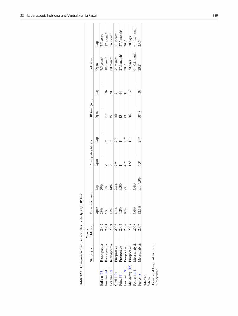
In the past decade, there has been a signi fi cant amount of literature comparing LIVH to open mesh repair. This new decade alone has yielded four prospective trials, three retro-spective trials and multiple meta-analysis and review papers (Table 22.1 ). Yet the literature fails to provide a standardiza-tion of technique in open mesh repairs. The Rives-Stoppa repair has a known recurrence rate ranging from 0 to 14% [ 1 ] ; however, Burger described a recurrence rate of 32% in open mesh repairs [ 6 ] . The majority of laparoscopic repairs described in comparative trials [ 7, 9, 10, 12, 33– 35 ] do adhere to the basic tenets of LIVH which include: 3 cm or greater mesh overlap and both transfascial sutures and tacks
for mesh fi xation as promoted by LeBlanc and colleagues [ 36 ] . This discordant approach to open mesh repair has chal-lenged a true comparison to LIVH in terms of overall recur-rence rates.
Pring and colleagues attempted to standardize their tech-nique by using ePTFE as an underlay with transfascial sutures in both open and laparoscopic repairs. Their results yielded a recurrence rate of 4.2% for open mesh repairs and 3.3% for laparoscopic repairs; this recurrence rate was not statistically different [ 7 ] . A meta-analysis performed by Forbes et al reviewed eight randomized controlled trials [ 11 ] . A similar study was done by Sajid et al on fi ve randomized controlled trials and Sains and colleagues reviewed fi ve com-parative trials [ 37, 38 ] ; all of these meta-analysis report no statistical difference in the recurrence rate between LIVH and open mesh repair. One of the largest meta-analysis was performed by Pierce and colleagues at Washington University. They reviewed 45 studies, of which 14 were paired studies and reported a recurrence rate of 3.1–4.3% for LIVH and 12.1% for open mesh repair [ 8 ] .
In a review of recent literature, the cumulative average of operating room time for LIVH was 87 and 91.5 min for open mesh repair, which supports a number of comparative stud-ies that report no statistical difference in OR time [ 9, 34, 37, 39 ] . However, other studies do show a statistical difference; LIVH has been shown in one meta-analysis to take 12 min longer than open mesh repair on average [ 35, 38 ] . This dis-crepancy is most likely secondary to the lack of standardiza-tion of open mesh repair and the learning curve for LIVH represented in earlier studies.
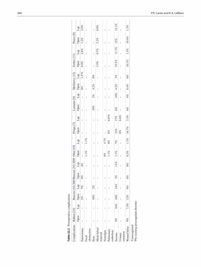
LIVH has been shown to have favorable results in shorter postoperative lengths of stay and overall decrease in wound infections and mesh removal [ 8, 11, 12, 37, 39, 40 ] . (Table 22.2 ) Pierce and colleagues found wound infections to be 4.6–8 fold higher in open mesh repairs when com-pared with LIVH [ 8 ] . A review of the National Surgical Quality Improvement Program (NSQIP) database, total complications were twice as high in open mesh repair in comparison to LIVH [ 39 ] . A common sequalae of LIVH is seroma formation. This complication is often underreported because it is routinely of no clinical signi fi cance. Very few studies document persistent seroma formation that required intervention.
LIVH is often accompanied with signi fi cant adhesiolyi-sis. A dreaded consequence of extensive adhesiolyis is injury to the intestine. Injury may be a result of direct laceration secondary to sharp or blunt dissection, but heightened vigi-lance is required for injuries caused by traction and remote serosal injuries that may go unrecognized. In a review of the literature by LeBlanc et al, the enterotomy rate for LIVH was 1.78% out of 3925 LIVH. According to this review, approxi-mately 18% of the time, an enterotomy is unrecognized which is associated with an increased mortality rate of 7.7%
Fig. 22.30 Use of laparoscopic fi xation device during the open portion of the hybrid procedure
Fig. 22.31 Completed open portion of hybrid procedure with laparo-scopic trocars in place
35922 Laparoscopic Incisional and Ventral Hernia Repair
Tab
le 2
2.1
C
ompa
riso
n of
rec
urre
nce
rate
s, p
ost-
Op
stay
, OR
tim
e
Stud
y ty
pe
Yea
r of
pu
blic
atio
n R
ecur
renc
e ra
tes
Post
-op
stay
(da
ys)
OR
tim
e (m
in)
Follo
w-u
p
Ope
n L
ap
Ope
n L
ap
Ope
n L
ap
Ope
n L
ap
Bal
lem
[ 33
] R
etro
spec
tive
2008
28
%
29%
–
– –
– 7.
5 ye
ars a
7.5
year
s B
enci
ni [
34 ]
Ret
rosp
ectiv
e 20
03
6%
0%
8 b 5 b
112
108
18 m
onth
b 17
mon
th b
Ben
cini
[ 35
] R
etro
spec
tive
2009
11
%
14%
2 a
3 a 35
70
60
mon
th a
56 m
onth
a O
lmi [
10 ]
Pros
pect
ive
2007
1.
1%
2.3%
9.
9 b 2.
7 b 15
1 61
24
mon
th a
24 m
onth
a Pr
ing
[ 7 ]
Pros
pect
ive
2008
4.
2%
3.3%
1 a
1 a 43
44
27
.5 m
onth
a 27
.5 m
onth
a L
oman
to [
9 ]
Pros
pect
ive
2006
10
%
2%
4.7 b
2.7 b
93
91
20.8
b 20
.8 b
McG
reev
y [ 1
2 ]
Pros
pect
ive
2003
–
– 1.
5 b
1.1 b
102
132
30 d
ays c
30 d
ays c
Forb
es [
11 ]
Met
a-an
alys
is
2009
3.
6%
3.4%
–
– –
– 6–
40.8
mon
th
6–40
.8 m
onth
Pi
erce
[ 8 ]
M
eta-
anal
ysis
20
07
12.1
%
3.1–
4.3%
4.
3 b 2.
4 b 10
4.5
103
20.2
d 25
.5 d
a Med
ian
b Mea
n c C
ompl
eted
leng
th o
f fo
llow
-up
d Uns
peci
fi ed
360 P.R. Carter and K.A. LeBlanc
Tab
le 2
2.2
Po
stop
erat
ive
com
plic
atio
ns
Com
plic
atio
ns
Bal
lem
[ 33
] B
enci
ni [
34 ]
2003
Ben
cini
[ 35
] 20
09 O
lmi [
10 ]
Prin
ge [
7 ]
Lom
anto
[ 9 ]
M
cGre
evy
[ 12 ]
Fo
rbes
[ 11
] Pi
erce
[ 8 ]
Ope
n L
ap
Ope
n L
ap
Ope
n L
ap
Ope
n L
ap
Ope
n L
ap
Ope
n L
ap
Ope
n L
ap
Ope
n L
ap
Ope
n L
ap
Ent
erot
omy
– –
2%
5%
0%
4%
– –
– –
– –
0%
1.5 a %
0.
9%
2.6%
1.
2%
2.9%
Feca
l ob
stru
ctio
n –
– –
– –
– 1.
1%
1.1%
–
– –
– –
– –
– –
–
Ileu
s –
– 10
%
2%
– –
– –
– –
10%
2%
4.
2%
0%
– –
– –
Mes
h In
fxn/
rem
oval
–
– –
– –
– –
– –
– –
– –
– 3.
5%
0.7%
3.
2%
0.9%
Neu
ralg
ia
– –
– –
– –
0%
4.7%
–
– –
– –
– –
– –
– Pu
lmon
ary
embo
lism
–
– –
– –
– 1.
1%
0%
0%
6.6 b %
–
– –
– –
– –
–
Sero
ma
9%
16%
10
%
14%
3%
11
%
1.1%
7%
33
%
17%
6%
10
%
4.2%
3%
15
.5%
11
.7%
12
%
12.1
%
Uri
nary
re
tent
ion
– –
– –
– –
– –
0%
6.6%
–
– –
– –
– –
–
Wou
nd I
nfxn
9%
7.
5%
12%
0%
8%
0%
8.
2%
1.1%
16
.7%
3.
3%
6%
4%
8.4%
0%
10
.1%
1.
5%
10.4
%
1.3%
a Unr
ecog
nize
d b P
re-e
xist
ing
proc
oagu
lant
dis
orde
r
36122 Laparoscopic Incisional and Ventral Hernia Repair
[ 21 ] . Should an enterotomy occur and is recognized, the injury should be repaired, of course. The next decision is whether or not to proceed with the repair of the hernia itself. The use of a prosthesis is to be avoided in conventional teaching but there is a growing opinion that the use of lower weight meshes might be considered in this situation as these seem to be less prone to infection. A primary repair of the hernia will be associated with a high risk of recurrence. Therefore, many experts recommend that the primary repair be avoided and the patient be returned to the operating room in several days [ 41 ] . With the introduction of biologic prod-ucts for the repair of the hernias in contaminated fi elds, per-haps these could be used in this situation. This has not been reported but it has been done in some cases.
The overall cost of LIVH has been shown to be equivalent with open mesh repair. A single institution prospectively col-lected data on 884 incisional hernias. There was no statistical difference in overall hospital cost for LIVH when compared to open mesh repair. LIVH was shown to have shorter length of stay, though operating time and cost of supplies were higher in LIVH. LIVH costs $6,725 compared with $7,445 for open mesh repair in total hospital costs and postoperative encounters [ 42 ] .
Obesity and LIVH
Obesity has been shown to be a major factor in hernia recur-rence. In a study of 160 patients, obesity was compared to other risk factors for hernia recurrence such as smoking, dia-betes, steroid use, and pulmonary disease. Obesity was the strongest predictor for hernia recurrence. Patients with a body mass index (BMI) of 38 were 4.2 times more likely to have a recurrent hernia in comparison to a patient with a BMI of 23 [ 18 ] . Congruent results were identi fi ed in a multi-institutional study of fi ve academic centers. This retrospective review found the recurrence rate to be signi fi cantly higher in mor-bidly obese patients with an odds ratio of 4.3 [ 17 ] .
Though some report a higher recurrence rate in obese patients, LIVH is safe and effective in this population of patients. LIVH has been shown to have less risk of wound complications, greater identi fi cation of multiple occult defects and wider mesh overlap. In a review of 168 patients at a single institution, perioperative complications after LIVH were not found to be statistically different from non-obese patients. Recurrence rates were related to defect size and size of mesh rather than obesity [ 43 ] . Ventral hernia repair is even promoted during laparoscopic bariatric surgery when concurrently identi fi ed. In patients who did not have their ventral hernia repaired during laparoscopic gastric bypass, there was an increased risk of intestinal incarceration during patient follow up [ 44 ] .
Conclusion
LIVH has a proven track record as an effective, safe, and durable option for ventral hernia repairs. There is general consensus that LIVH has comparable recurrence rates to open mesh repair, if not less risk of recurrence as seen in some prospective trials. Wound complications and mesh infections occur infrequently. Hospital stay is shortened and increasingly, LIVH is becoming the fi rst and only attempt at a disease that is commonly identi fi ed in 10–20% of postlapa-rotomy patients [ 1, 5, 19, 45 ] .
References
1. Jin J, Rosen M. Laparoscopic versus open ventral hernia repair. Surg Clin North Am. 2008;88:1083–100.
2. LeBlanc K, Booth W. Laparoscopic Repair of Incisional Abdominal Hernias using expanded polytetra fl uoroethylene: preliminary fi ndings. Surg Laparosc Endosc. 1993;3(1):39–41.
3. Stoppa R, Louis D, Verhaeghe P, et al. Current surgical treatment of post-operative eventrations. Int Surg. 1987;72(1):42–4.
4. Bauer J, Harris M, Gor fi ne S, et al. Rives-Stoppa procedure for repair of large incisional hernias: experience with 57 patients. Hernia. 2002;6:120–3.
5. den Hartog D, Dur A, Tuinebreijer W et al. (2008) Open surgical procedure for incisional hernias. The Cochrane Collaboration.
6. Burger J, Luijendijk R, Hop W, et al. Long-term follow-up of a randomized controlled trial of suture versus mesh repair of inci-sional hernia. Ann Surg. 2004;240(4):578–85.
7. Pring C, Tran V, O’Rourke N, et al. Laparoscopic versus open ven-tral hernia repair: a randomized controlled trial. ANZ J Surg. 2008;78:903–6.
8. Pierce R, Spitler J, Frisella M, et al. Pooled data analysis of laparo-scopic vs. open ventral hernia repair: 14 years of patient data accrual. Surg Endosc. 2007;21:378–86.
9. Lomanto D, Iyer S, Shabbir A, et al. Laparoscopic versus open ven-tral hernia mesh repair: a prospective study. Surg Endosc. 2006;20:1030–5.
10. Olmi S, Scaini A, Cesana G, et al. Laparoscopic versus open inci-sional hernia repair; an open randomized controlled trial. Surg Endosc. 2007;21:555–9.
11. Forbes S, Eskicioglu C, McLeod R, et al. Meta-analysis of random-ized controlled trials comparing open and laparoscopic ventral and incisional hernia repair with mesh. Br J Surg. 2009;96:851–8.
12. McGreevy J, Goodney P, Birkmeyer C, et al. A prospective study comparing the complication rates between laparoscopic and open ventral hernia repairs. Surg Endosc. 2003;17:1778–80.
13. Rosen M, Williams C, Jin J, et al. Laparoscopic versus open-com-ponent separation: a comparative analysis in a porcine model. Am J Surg. 2007;194:383–9.
14. Bachman S, Ramaswamy A, Ramshaw B. Early results of midline hernia repair using a minimally invasive component separation technique. Am Surg. 2009;75(7):572–7.
15. O’Malley C, Cunningham A. Physiologic changes during laparos-copy. Anesthesiol Clin North America. 2001;19(1):1–19.
16. Hesselink VJ, Luijendijk RW, de Wilt JH, et al. An evaluation of risk factors in incisional hernia recurrence. Surg Gynecol Obstet. 1993;176(3):228–34.
17. Tsereteli Z, Pryor B, Heniford B, et al. Laparoscopic ventral hernia repair (LIVH) in morbidly obese patients. Hernia. 2008;12:233–8.
362 P.R. Carter and K.A. LeBlanc
18. Sauerland S, Korenkov M, Kleinen T, et al. Obesity is a risk factor for recurrence after incisional hernia repair. Hernia. 2004;8:42–6.
19. Franklin M, Gonzalez J, Glass J, et al. Laparoscopic ventral and inci-sional hernia repair: an 11 year experience. Hernia. 2004;8:23–7.
20. Sarit C, Eliezer A, Mizrahi S. Minimally Invasive Repair of recur-rent strangulated umbilical hernia in cirrhotic patient with refrac-tory ascites. Liver Transpl. 2003;9:621–2.
21. LeBlanc K, Elieson M, III Corder J. Enterotomy and mortality rates of laparoscopic incisional and ventral hernia repair: a review of the literature. JSLS. 2007;11:408–14.
22. LeBlanc K. Current considerations in laparoscopic incisional and ventral herniorrhaphy. JSLS. 2004;4:131–9.
23. Carbajo M, Martin del Olmo J, Blanco J, et al. Laparoscopic treat-ment of ventral abdominal wall hernias: preliminary results in 100 Patients. JSLS. 2000;4:141–5.
24. Ramshaw BJ, Escartia P, Schwab J, et al. Comparison of laparo-scopic and open ventral herniorrhaphy. Am Surg. 1999;65:827–32.
25. Park A, Birch DW, Lovrics P. Laparoscopic and open incisional hernia repair: a comparison study. Surgery. 1998;124:816–22.
26. LeBlanc K. Laparoscopic incisional hernia repair: are Transfascial sutures necessary? A review of the literature. Surg Endosc. 2007;21:508–13.
27. LeBlanc K, Booth W, Whitaker J, et al. Laparoscopic incisional and ventral herniorrhaphy in 100 patients. Am J Surg. 2000;180(3):193–7.
28. DeMaria EJ, Moss JM, Sugerman HJ. Laparoscopic intraperito-neal polytetra fl uoroethylene (PTFE) prosthesic patch repair of ventral hernia. Prospective comparison to open prefascial polypro-pylene mesh repair. Surg Endosc. 2000;14(4):326–9.
29. Heniford B, Park A, Ramshaw B, et al. Laparoscopic repair of ven-tral hernias. Nine years’ experience with 850 consecutive hernias. Ann Surg. 2003;238:391–400.
30. Carbonell A, Harold K, Mahmutovic A, et al. Local injection for the treatment of suture site pain after laparoscopic ventral hernia repair. Am Surg. 2003;69:688–92.
31. Griniatsos J, Eugenia Y, Anastasios T, et al. A hybrid technique for recurrent incisional hernia repair. Surg Laparosc Endosc Percutan Tech. 2009;19:177–80.
32. Heniford B, Park A, Ramshaw B, et al. Laparoscopic ventral and inci-sional hernia repair in 407 patients. J Am Coll Surg. 2000;190:645–50.
33. Ballem N, Parikh R, Berber E. Laparoscopic versus open ventral her-nia repairs: 5 year recurrence rates. Surg Endosc. 2008;22:1935–40.
34. Bencini L, Sanchez L, Bof fi B, et al. Incisional hernia repair. Surg Endosc. 2003;17:1546–51.
35. Bencini L, Sanchez L, Bof fi B, et al. Comparison of laparoscopic and open repair for primary ventral hernias. Surg Laparosc Endosc Percutan Tech. 2009;19:341–4.
36. LeBlanc K, Whitaker J, Bellanger D, et al. Laparoscopic incisional and ventral hernioplasty: lessons learned from 200 patients. Hernia. 2003;7:118–24.
37. Sajid M, Bokhari S, Mallick A, et al. Laparoscopic versus open repair of incisional/ventral hernia: a meta-analysis. Am J Surg. 2009;197:64–72.
38. Sains P, Tilney H, Purkayastha S, et al. Outcomes following laparoscopic versus open repair of incisional hernia. World J Surg. 2006;30:2056–64.
39. Hwang C, Wichterman K, Alfrey E. Laparoscopic ventral hernia repair is safer than open repair: analysis of the NSQIP data. J Surg Res. 2009;156:213–6.
40. Misiakos E, Machairas A, Patapis P, et al. Laparoscopic ventral her-nia repair: pros and cons compared with open hernia repair. JSLS. 2008;12:117–25.
41. LeBlanc K. The Critical Technical Aspects of Laparoscopic Repair of Ventral and Incisional Hernias. Am Surg. 2001;67(8):809–12.
42. Earle D, Seymour N, Fellinger E, et al. Laparoscopic versus open incisional hernia repair. A single institution analysis of hospital resource utilization for 884 consecutive cases. Surg Endosc. 2006;20:71–5.
43. Ching S, Sarela A, Dexter S, et al. Comparison of early outcomes for laparoscopic ventral hernia repair between nonobese and mor-bidly obese patient populations. Surg Endosc. 2008;22:2244–50.
44. Eid G, Mattat S, Hamad G, et al. Repair of ventral hernias in mor-bidly obese patients undergoing laparoscopic gastric bypass should not be deferred. Surg Endosc. 2004;18:207–10.
45. Perrone J, Soper N, Eagon C, et al. Perioperative outcomes and complications of laparoscopic ventral hernia repair. Surgery. 2005;138:708–16.