Embed Size (px)
Citation preview
CASE REPORT Open Access
Management of delivery of a fetus withautosomal recessive polycystic kidneydisease: a case report of abdominaldystocia and review of the literatureSarah Belin1, Cristina Delco1, Paloma Parvex1, Sylviane Hanquinet2, Siv Fokstuen3,Begoña Martinez de Tejada1,4* and Isabelle Eperon1
Abstract
Background: Autosomal recessive renal polycystic kidney disease occurs in 1 in 20,000 live births. It is caused bymutations in both alleles of the PKHD1 gene. Management of delivery in cases of suspected autosomal recessiverenal polycystic kidney disease is rarely discussed, and literature concerning abdominal dystocia is extremely scarce.We present a case of a patient with autosomal recessive renal polycystic kidney disease whose delivery wascomplicated by abdominal dystocia, and we discuss the factors that determined the route and timing of delivery.
Case presentation: A 23-year-old Caucasian woman, G2 P0, with a prior unremarkable pregnancy was referred toour tertiary center at 31 weeks of gestation because of severe oligoamnios (amniotic fluid index = 2) andhyperechogenic, dedifferentiated, and enlarged fetal kidneys. She had no other genitourinary anomaly. Fetalmagnetic resonance imaging showed enlarged, hypersignal kidneys and severe pulmonary hypoplasia. We had ahigh suspicion of autosomal recessive renal polycystic kidney disease, and after discussion with our multidisciplinaryteam, the parents opted for conservative care. Ultrasound performed at 35 weeks of gestation showed a fetalestimated weight of 3550 g and an abdominal circumference of 377 mm, both above the 90th percentile. Becauseof the very rapid kidney growth and suspected risk of abdominal dystocia, we proposed induction of labor at 36weeks of gestation after corticosteroid administration for fetal lung maturation. Vaginal delivery was complicated byabdominal dystocia, which resolved by continuing expulsive efforts and gentle fetal traction. A 3300-g (P50–90)male infant was born with Apgar scores of 1-7-7 at 1, 5, and 10 minutes, respectively, and arterial and venousumbilical cord pH values of 7.23–7.33. Continuous peritoneal dialysis was started at day 2 of life because of anuria.Currently, the infant is 1 year old and is waiting for kidney transplant that should be performed once he reaches 10kg. Molecular analysis of PKHD1 performed on deoxyribonucleic acid (DNA) from the umbilical cord confirmedautosomal recessive renal polycystic kidney disease.
Conclusions: Management of delivery in cases of suspected autosomal recessive renal polycystic kidney diseaseneeds to be discussed because of the risk of abdominal dystocia. The route and timing of delivery depend on thesize of the fetal abdominal circumference and the gestational age. The rate of kidney growth must also be takeninto account.
Keywords: Abdominal dystocia, Autosomal recessive renal polycystic kidney disease, Delivery, Management
© The Author(s). 2019 Open Access This article is distributed under the terms of the Creative Commons Attribution 4.0International License (http://creativecommons.org/licenses/by/4.0/), which permits unrestricted use, distribution, andreproduction in any medium, provided you give appropriate credit to the original author(s) and the source, provide a link tothe Creative Commons license, and indicate if changes were made. The Creative Commons Public Domain Dedication waiver(http://creativecommons.org/publicdomain/zero/1.0/) applies to the data made available in this article, unless otherwise stated.
* Correspondence: [email protected] of Pediatrics, Gynecology and Obstetrics, University Hospitals ofGeneva, rue Gabriel-Perret-Gentil 14, 1205 Geneva, Switzerland4Faculty of Medicine, University of Geneva, University Hospitals of Geneva,rue Gabriel-Perret-Gentil 14, 1205 Geneva, SwitzerlandFull list of author information is available at the end of the article
Belin et al. Journal of Medical Case Reports (2019) 13:366 https://doi.org/10.1186/s13256-019-2293-3
BackgroundAutosomal recessive renal polycystic kidney disease(ARPKD) is a rare form of cystic kidney disease, occur-ring in approximately 1 in 20,000 live births [1]. It iscaused by mutations in the PKHD1 (polycystic kidneyand hepatic disease 1) gene, situated on chromosome6p12, which encodes for the protein fibrocystin [2]. Ithas an autosomal recessive mode of inheritance, andmost affected children are compound heterozygotes,inheriting a different ARPKD mutation from each parent[3]. Phenotype is variable, with up to 30% of cases dyingin the neonatal period or within the first year of life, pre-senting in utero with severe oligohydramnios leading toPotter’s sequence with pulmonary hypoplasia, character-istic facies, and limb deformations [4, 5]. In less severepresentations, prognosis is difficult to establish pre-natally and has been shown to correlate with the amountof amniotic fluid and lung volume calculated on thebasis of fetal magnetic resonance imaging (MRI) [6, 7].Prenatal counseling and clinical management are bestconducted by multidisciplinary care teams that includeobstetricians, neonatologists, pediatricians specialized innephrology, radiologists, and geneticists [3]. We presenta case of a pregnant woman with suspected fetal ARPKDdiagnosed at 31 weeks of gestation and discuss the ele-ments orienting pregnancy and delivery management.
Case presentationWe present a case of a spontaneous pregnancy in ahealthy nonconsanguineous couple with an unremark-able family history. The mother was 23-year-old and hada history of a voluntary pregnancy termination and ac-tive tobacco use (five to eight cigarettes per day). Sheunderwent regular pregnancy follow-up. First-trimesterscreening included an ultrasound (US) scan with a thinnuchal translucency (1.4 mm) and a very low risk for tri-somy 21 (1 in 19,000). Blood tests showed immunityagainst cytomegalovirus (immunoglobulin G [IgG]-posi-tive, IgM-negative), and a US scan at 20 weeks of gesta-tion showed no signs of fetal anomalies and a normalamount of amniotic fluid. A US scan at 28 weeks of ges-tation showed a slight reduction with an amniotic fluidindex (AFI) of 7 and a deepest pocket of 2.3 cm. At 31weeks of gestation, the patient was referred to our ter-tiary fetal medicine unit because of severe oligoamniosand enlarged kidneys. We confirmed the presence of se-vere oligoamnios (AFI = 2) as well as hyperechogenic,undifferentiated, and enlarged bilateral fetal kidneys(right side, 39 × 64 × 25 mm; left side, 33 × 63 × 39mm;>97th percentile [8]). There was no ectasia of the urinarytract. The bladder was visualized but only slightly filled,and no spontaneous micturition was seen. The malegenital organs of the fetus were without anomalies. Esti-mated fetal weight was above the 90th percentile at
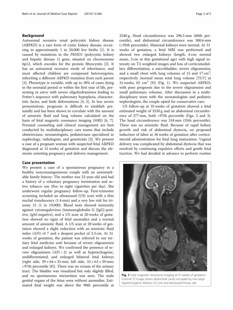
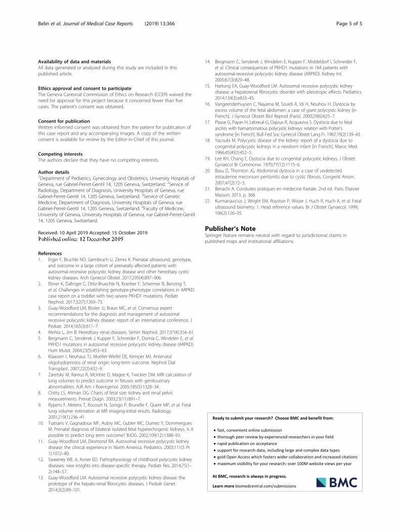
2240 g. Head circumference was 296.5 mm (60th per-centile), and abdominal circumference was 300.6 mm(>95th percentile). Maternal kidneys were normal. At 31weeks of gestation, a fetal MRI was performed andshowed two enlarged kidneys (length, 6 cm; normalmean, 3 cm at this gestational age) with high signal in-tensity on T2-weighted images and loss of corticomedul-lary differentiation, a microbladder, severe oligoamnios,and a small chest with lung volumes of 15 and 17 cm3,respectively (normal mean total lung volume [TLV] at31 weeks, 65 cm3 [9]) (Fig. 1). We suspected ARPKD,with poor prognosis due to the severe oligoamnios andsmall pulmonary volumes. After discussion in a multi-disciplinary team with the neonatologists and pediatricnephrologists, the couple opted for conservative care.US follow-up at 35 weeks of gestation showed a fetal
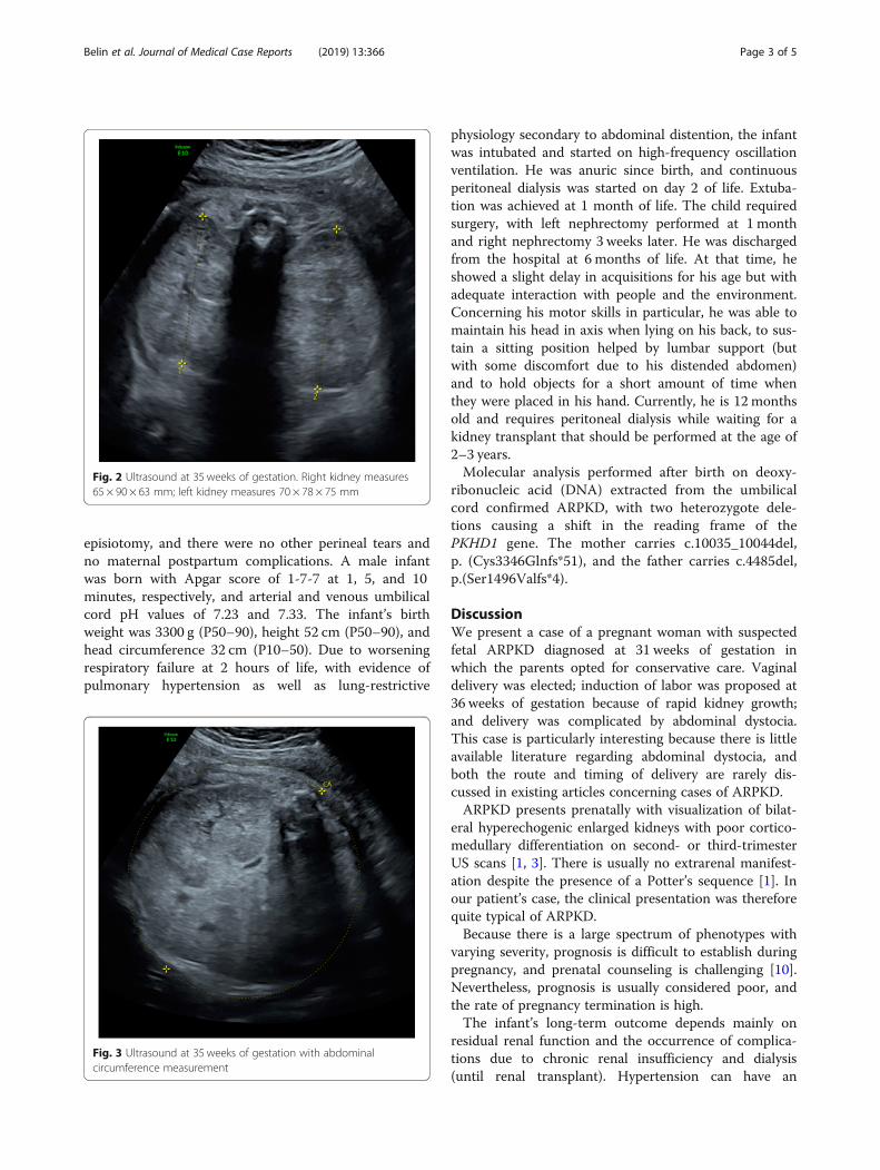
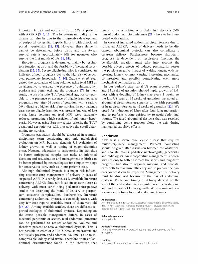
estimated weight of 3550 g and an abdominal circumfer-ence of 377 mm, both >97th percentile (Figs. 2 and 3).The head circumference was 318 mm (35th percentile).There was no amniotic fluid. Because of rapid kidneygrowth and risk of abdominal dystocia, we proposedinduction of labor at 36 weeks of gestation after cortico-steroid administration for fetal lung maturation. Vaginaldelivery was complicated by abdominal dystocia that wasresolved by continuing expulsive efforts and gentle fetaltraction. We had decided in advance to perform routine
Fig. 1 Fetal magnetic resonance imaging at 31 weeks of gestation.Coronal T2 image shows abdominal cavity occupied by two largehyperechogenic kidneys (5.5 cm) and decreased thorax size
Belin et al. Journal of Medical Case Reports (2019) 13:366 Page 2 of 5
episiotomy, and there were no other perineal tears andno maternal postpartum complications. A male infantwas born with Apgar score of 1-7-7 at 1, 5, and 10minutes, respectively, and arterial and venous umbilicalcord pH values of 7.23 and 7.33. The infant’s birthweight was 3300 g (P50–90), height 52 cm (P50–90), andhead circumference 32 cm (P10–50). Due to worseningrespiratory failure at 2 hours of life, with evidence ofpulmonary hypertension as well as lung-restrictive
physiology secondary to abdominal distention, the infantwas intubated and started on high-frequency oscillationventilation. He was anuric since birth, and continuousperitoneal dialysis was started on day 2 of life. Extuba-tion was achieved at 1 month of life. The child requiredsurgery, with left nephrectomy performed at 1 monthand right nephrectomy 3 weeks later. He was dischargedfrom the hospital at 6 months of life. At that time, heshowed a slight delay in acquisitions for his age but withadequate interaction with people and the environment.Concerning his motor skills in particular, he was able tomaintain his head in axis when lying on his back, to sus-tain a sitting position helped by lumbar support (butwith some discomfort due to his distended abdomen)and to hold objects for a short amount of time whenthey were placed in his hand. Currently, he is 12 monthsold and requires peritoneal dialysis while waiting for akidney transplant that should be performed at the age of2–3 years.Molecular analysis performed after birth on deoxy-
ribonucleic acid (DNA) extracted from the umbilicalcord confirmed ARPKD, with two heterozygote dele-tions causing a shift in the reading frame of thePKHD1 gene. The mother carries c.10035_10044del,p. (Cys3346Glnfs*51), and the father carries c.4485del,p.(Ser1496Valfs*4).
DiscussionWe present a case of a pregnant woman with suspectedfetal ARPKD diagnosed at 31 weeks of gestation inwhich the parents opted for conservative care. Vaginaldelivery was elected; induction of labor was proposed at36 weeks of gestation because of rapid kidney growth;and delivery was complicated by abdominal dystocia.This case is particularly interesting because there is littleavailable literature regarding abdominal dystocia, andboth the route and timing of delivery are rarely dis-cussed in existing articles concerning cases of ARPKD.ARPKD presents prenatally with visualization of bilat-
eral hyperechogenic enlarged kidneys with poor cortico-medullary differentiation on second- or third-trimesterUS scans [1, 3]. There is usually no extrarenal manifest-ation despite the presence of a Potter’s sequence [1]. Inour patient’s case, the clinical presentation was thereforequite typical of ARPKD.Because there is a large spectrum of phenotypes with
varying severity, prognosis is difficult to establish duringpregnancy, and prenatal counseling is challenging [10].Nevertheless, prognosis is usually considered poor, andthe rate of pregnancy termination is high.The infant’s long-term outcome depends mainly on
residual renal function and the occurrence of complica-tions due to chronic renal insufficiency and dialysis(until renal transplant). Hypertension can have an
Fig. 2 Ultrasound at 35 weeks of gestation. Right kidney measures65 × 90 × 63 mm; left kidney measures 70 × 78 × 75 mm
Fig. 3 Ultrasound at 35 weeks of gestation with abdominalcircumference measurement
Belin et al. Journal of Medical Case Reports (2019) 13:366 Page 3 of 5
important impact and occurs in up to 75% of patientswith ARPKD [3, 5, 11]. The long-term morbidity of thedisease can also be due to the progressive developmentof periportal congenital hepatic fibrosis, with associatedportal hypertension [12, 13]. However, these elementscannot be determined before birth, and the 5-yearsurvival rate is approximately 90% for neonates whosurvive the first month of life [11, 14].Short-term prognosis is determined mainly by respira-
tory function at birth and the feasibility of neonatal resus-citation [12, 15]. Severe oligohydramnios is considered anindicator of poor prognosis due to the high risk of associ-ated pulmonary hypoplasia [7, 10]. Zaretsky et al. sug-gested the calculation of lung volumes using fetal MRI asan alternative to evaluate the presence of pulmonary hy-poplasia and better estimate the prognosis [7]. In theirstudy, the use of a ratio, TLV/gestational age, was compar-able to the presence or absence of oligohydramnios as aprognostic tool after 26 weeks of gestation, with a ratio <0.9 indicating a higher risk of nonsurvival. In our patient’scase, severe oligohydramnios was present but was of lateonset. Lung volumes on fetal MRI were extremelyreduced, prompting a high suspicion of pulmonary hypo-plasia. However, using Zaretsky et al.’s criteria, the TLV/gestational age ratio was 1.03, thus above the cutoff deter-mining nonsurvival.Prognosis evaluation should be discussed in a multi-
disciplinary team considering not only radiologicalevaluation on MRI but also dynamic US evaluation ofkidney growth as well as timing of oligohydramniosonset. Neonatal adaptation to extrauterine life can thusbe better anticipated; couples can make an informeddecision; and resuscitation and management at birth canbe better planned by neonatologists for couples who optfor conservative care, such as in our patient’s case.Although abdominal dystocia is a major risk influen-
cing obstetric care, management of delivery in cases ofsuspected ARPKD is rarely discussed. Available literatureconcerning ARPKD does not focus on obstetric care atdelivery, with most series being pediatric retrospectivestudies not describing the mode of delivery or peripar-tum obstetric complications. Furthermore, literatureconcerning abdominal dystocia is extremely scarce, withvery few case reports available, most of them very old[16–20]. Among available articles, there are different re-ported etiologies of abdominal dystocia. Depending onthe cause, possible management differs. In cases ofmeconial peritonitis or ascites, fetal abdominal puncturecan be performed to reduce abdominal volume andtherefore prevent or resolve abdominal dystocia. This isnot possible in cases of ARPKD, because macrocysts arenot usually present, and abdominal volume is due to in-compressible kidney solid tissue. Therefore, values of ab-dominal circumference found in the literature that
seems to be associated with abdominal dystocia (400mm of abdominal circumference [21]) have to be inter-preted with caution.In cases of increased abdominal circumference due to
suspected ARPKD, mode of delivery needs to be dis-cussed. Abdominal dystocia can also complicate acesarean delivery. Furthermore, because short-termprognosis is dependent on respiratory function, thebenefit–risk equation must take into account thepossible adverse effects of induced prematurity versusthe possible negative impact of waiting longer, with in-creasing kidney volumes causing increasing mechanicalcompression and possibly complicating even moremechanical ventilation at birth.In our patient’s case, serial US scans repeated at 33
and 35 weeks of gestation showed rapid growth of kid-neys with a doubling of kidney size every 2 weeks. Atthe last US scan at 35 weeks of gestation, we noted anabdominal circumference superior to the 95th percentileof head circumference at 42 weeks of gestation [22]. Weopted for induction of labor after fetal lung maturationand to perform routine episiotomy to avoid abdominaltrauma. We faced abdominal dystocia that was resolvedby continuing gentle traction along the axis duringmaintained expulsive efforts.
ConclusionARPKD is a severe renal cystic disease that requiresmultidisciplinary management. Prenatal counselingshould be given after discussion between the obstetricaland neonatal teams, pediatric nephrologists, geneticists,and radiologists. An incorporative management is neces-sary not only to better estimate the short- and long-termprognosis but also to organize maternal and neonatalcare, both to maximize efficiency and to prepare the par-ents for what can be expected. Management of deliverymust be discussed because of the risk of abdominaldystocia. Route and timing of delivery depend on thesize of the fetal abdominal circumference, the gestationalage, and the rate of kidney growth. We recommend per-forming episiotomy to avoid abdominal trauma.
AbbreviationsAFI: Amniotic fluid index; ARPKD: Autosomal recessive renal polycystic kidneydisease; MRI: Magnetic resonance imaging; PKHD1: Polycystic kidney andhepatic disease 1 gene; TLV: Total lung volume; US: Ultrasound
AcknowledgementsNot applicable.
Authors’ contributionsSB and IE reviewed the literature. All authors read and approved the finalmanuscript.
FundingNot applicable; no funding was necessary for this article.
Belin et al. Journal of Medical Case Reports (2019) 13:366 Page 4 of 5
Availability of data and materialsAll data generated or analyzed during this study are included in thispublished article.
Ethics approval and consent to participateThe Geneva Cantonal Commission of Ethics on Research (CCER) waived theneed for approval for this project because it concerned fewer than fivecases. The patient’s consent was obtained.
Consent for publicationWritten informed consent was obtained from the patient for publication ofthis case report and any accompanying images. A copy of the writtenconsent is available for review by the Editor-in-Chief of this journal.
Competing interestsThe authors declare that they have no competing interests.
Author details1Department of Pediatrics, Gynecology and Obstetrics, University Hospitals ofGeneva, rue Gabriel-Perret-Gentil 14, 1205 Geneva, Switzerland. 2Service ofRadiology, Department of Diagnosis, University Hospitals of Geneva, rueGabriel-Perret-Gentil 14, 1205 Geneva, Switzerland. 3Service of GeneticMedicine, Department of Diagnosis, University Hospitals of Geneva, rueGabriel-Perret-Gentil 14, 1205 Geneva, Switzerland. 4Faculty of Medicine,University of Geneva, University Hospitals of Geneva, rue Gabriel-Perret-Gentil14, 1205 Geneva, Switzerland.
Received: 10 April 2019 Accepted: 15 October 2019
References1. Erger F, Bruchle NO, Gembruch U, Zerres K. Prenatal ultrasound, genotype,
and outcome in a large cohort of prenatally affected patients withautosomal-recessive polycystic kidney disease and other hereditary cystickidney diseases. Arch Gynecol Obstet. 2017;295(4):897–906.
2. Ebner K, Dafinger C, Ortiz-Bruechle N, Koerber F, Schermer B, Benzing T,et al. Challenges in establishing genotype-phenotype correlations in ARPKD:case report on a toddler with two severe PKHD1 mutations. PediatrNephrol. 2017;32(7):1269–73.
3. Guay-Woodford LM, Bissler JJ, Braun MC, et al. Consensus expertrecommendations for the diagnosis and management of autosomalrecessive polycystic kidney disease: report of an international conference. JPediatr. 2014;165(3):611–7.
4. Mehta L, Jim B. Hereditary renal diseases. Semin Nephrol. 2017;37(4):354–61.5. Bergmann C, Senderek J, Kupper F, Schneider F, Dornia C, Windelen E, et al.
PKHD1 mutations in autosomal recessive polycystic kidney disease (ARPKD).Hum Mutat. 2004;23(5):453–63.
6. Klaassen I, Neuhaus TJ, Mueller-Wiefel DE, Kemper MJ. Antenataloligohydramnios of renal origin: long-term outcome. Nephrol DialTransplant. 2007;22(2):432–9.
7. Zaretsky M, Ramus R, McIntire D, Magee K, Twickler DM. MRI calculation oflung volumes to predict outcome in fetuses with genitourinaryabnormalities. AJR Am J Roentgenol. 2005;185(5):1328–34.
8. Chitty LS, Altman DG. Charts of fetal size: kidney and renal pelvismeasurements. Prenat Diagn. 2003;23(11):891–7.
9. Rypens F, Metens T, Rocourt N, Sonigo P, Brunelle F, Quere MP, et al. Fetallung volume: estimation at MR imaging-initial results. Radiology.2001;219(1):236–41.
10. Tsatsaris V, Gagnadoux MF, Aubry MC, Gubler MC, Dumez Y, DommerguesM. Prenatal diagnosis of bilateral isolated fetal hyperechogenic kidneys. Is itpossible to predict long term outcome? BJOG. 2002;109(12):1388–93.
11. Guay-Woodford LM, Desmond RA. Autosomal recessive polycystic kidneydisease: the clinical experience in North America. Pediatrics. 2003;111(5 Pt1):1072–80.
12. Sweeney WE Jr, Avner ED. Pathophysiology of childhood polycystic kidneydiseases: new insights into disease-specific therapy. Pediatr Res. 2014;75(1–2):148–57.
13. Guay-Woodford LM. Autosomal recessive polycystic kidney disease: theprototype of the hepato-renal fibrocystic diseases. J Pediatr Genet.2014;3(2):89–101.
14. Bergmann C, Senderek J, Windelen E, Kupper F, Middeldorf I, Schneider F,et al. Clinical consequences of PKHD1 mutations in 164 patients withautosomal-recessive polycystic kidney disease (ARPKD). Kidney Int.2005;67(3):829–48.
15. Hartung EA, Guay-Woodford LM. Autosomal recessive polycystic kidneydisease: a hepatorenal fibrocystic disorder with pleiotropic effects. Pediatrics.2014;134(3):e833–45.
16. Vangeenderhuysen C, Nayama M, Souidi A, Idi H, Nouhou H. Dystocia byexcess volume of the fetal abdomen: a case of giant polycystic kidney [inFrench]. J Gynecol Obstet Biol Reprod (Paris). 2000;29(6):625–7.
17. Plasse G, Payan H, Lebreuil G, Dajoux R, Acquaviva S. Dystocia due to fetalascites with hamartomatous polycystic kidneys: relation with Potter'ssyndrome [in French]. Bull Fed Soc Gynecol Obstet Lang Fr. 1967;19(2):139–43.
18. Yacoubi M. Polycystic disease of the kidney: report of a dystocia due tocongenital polycystic kidneys in a newborn infant [in French]. Maroc Med.1966;45(492):452–5.
19. Lee KH, Chang E. Dystocia due to congenital polycystic kidneys. J ObstetGynaecol Br Commonw. 1970;77(12):1115–6.
20. Basu D, Thornton JG. Abdominal dystocia in a case of undetectedintrauterine meconium peritonitis due to cystic fibrosis. Congenit Anom.2007;47(2):72–3.
21. Benachi A. Conduites pratiques en médecine foetale. 2nd ed. Paris: ElsevierMasson; 2013. p. 368.
22. Kurmanavicius J, Wright EM, Royston P, Wisser J, Huch R, Huch A, et al. Fetalultrasound biometry: 1. Head reference values. Br J Obstet Gynaecol. 1999;106(2):126–35.
Publisher’s NoteSpringer Nature remains neutral with regard to jurisdictional claims inpublished maps and institutional affiliations.
Belin et al. Journal of Medical Case Reports (2019) 13:366 Page 5 of 5