Embed Size (px)
Citation preview
Abbreviations
and Acronyms
ADPKD ¼ autosomal dominantpolycystic kidney disease
LCD ¼ laparoscopic cystdecortication
SCS ¼ spinal cord stimulation
TAE ¼ transcatheter arterialembolization
Accepted for publication October 27, 2014.* Correspondence: Department of Urology,
Indiana University, 535 N. Barnhill Dr., Ste. 420,Indianapolis, Indiana 46202 (telephone: 317-948-3098; FAX: 317-944-0174; e-mail: [email protected]).
1470 j www.jurology.com
00
T
©
Review Article
Management of Pain in Autosomal Dominant PolycysticKidney Disease and Anatomy of Renal Innervation
Matthew W. Tellman, Clinton D. Bahler, Ashley M. Shumate,
Robert L. Bacallao and Chandru P. Sundaram*
From the Department of Urology (MWT, CDB, AMS, CPS) and Department of Nephrology (RLB),
Indiana University School of Medicine, Indianapolis, Indiana
Purpose: Chronic pain is a prominent feature of autosomal dominant polycystickidney disease that is difficult to treat and manage, often resulting in a decreasein quality of life. Understanding the underlying anatomy of renal innervationand the various etiologies of pain that occur in autosomal dominant polycystickidney disease can help guide proper treatments to manage pain. Reviewingpreviously studied treatments for pain in autosomal dominant polycystic kidneydisease can help characterize treatment in a stepwise fashion.
Materials and Methods: We performed a literature search of the etiology andmanagement of pain in autosomal dominant polycystic kidney disease andthe anatomy of renal innervation using PubMed� and Embase� from January1985 to April 2014 with limitations to human studies and English language.
Results: Pain occurs in the majority of patients with autosomal dominant poly-cystic kidney disease due to renal, hepatic and mechanical origins. Patientsmay experience different types of pain which can make it difficult to clinicallyconfirm its etiology. An anatomical and histological evaluation of the complexrenal innervation helps in understanding the mechanisms that can lead to renalpain. Understanding the complex nature of renal innervation is essential forsurgeons to perform renal denervation. The management of pain in autosomaldominant polycystic kidney disease should be approached in a stepwise fashion.Acute causes of renal pain must first be ruled out due to the high incidencein autosomal dominant polycystic kidney disease. For chronic pain, nonopioidanalgesics and conservative interventions can be used first, before opioid anal-gesics are considered. If pain continues there are surgical interventions such asrenal cyst decortication, renal denervation and nephrectomy that can targetpain produced by renal or hepatic cysts.
Conclusions: Chronic pain in patients with autosomal dominant polycystickidney disease is often refractory to conservative, medical and other noninvasivetreatments. There are effective surgical procedures that can be performed whenmore conservative treatments fail. Laparoscopic cyst decortication has been wellstudied and results in the relief of chronic renal pain in the majority of patients.In addition, renal denervation has been used successfully and could be performedconcurrently with cyst decortication. Nephrectomy should be reserved for pa-tients with intractable pain and renal failure when other modalities have failed.
Key Words: anatomy; innervation; kidney; pain management;
polycystic kidney, autosomal dominant
22-5347/15/1935-1470/0
HE JOURNAL OF UROLOGY®
2015 by AMERICAN UROLOGICAL ASSOCIATION EDUCATION AND RESEARCH, INC.
http://dx.doi.org/10.1016/j.juro.2014.10.124
Vol. 193, 1470-1478, May 2015
Printed in U.S.A.
PAIN MANAGEMENT IN AUTOSOMAL DOMINANT POLYCYSTIC KIDNEY DISEASE 1471
AUTOSOMAL dominant polycystic kidney disease isrelatively common, with a worldwide prevalenceof 1:400 to 1:1,000.1 Pain is a prominent feature ofall types of polycystic kidney disease, affectingmore than 60% of patients, and is most commonlylocated in the flank, followed by the back andabdomen.1,2 Pain is often present early in the dis-ease process and is the most common symptom thatleads to a diagnosis of the disease.3 The hardshipsof living with chronic pain can prevent patientswith ADPKD from performing physical and socialactivities, which detrimentally affects their qualityof life.4 The difficulty of pain management isdemonstrated by many patients, with up to 39%being somewhat or completely dissatisfied with paintreatment because they are physically unable to dowhat they would like.5 A better understanding ofthe etiology of pain that occurs in ADPKD in addi-tion to the underlying anatomy can help guidetreatment.
MATERIALS AND METHODSWe performed a literature search of the etiology and man-agement of pain in ADPKD using PubMed and Embasefrom January 1985 to April 2014 with limitations tohuman studies and English language. Search termsincluded pain management, chronic pain, treatment, ther-apy, polycystic kidney, autosomal dominant, cyst, hepatic,liver, kidney and renal. References of the studies foundwere reviewed. Further searches were performed usingMEDLINE� and Embase for each relevant treatment ofADPKD identified, with additional search terms includinganalgesics, Alexander technique, tolvaptan, opioids, aspi-ration, celiac plexus, splanchnic nerves, splanchnicectomy,block, ablation, spinal cord stimulation, sclerotherapy,decortication, laparoscopic, marsupialization, denervation,percutaneous, sympathectomy, nephrectomy, transplantand transcatheter arterial embolization.
A comprehensive review of the literature identified140 studies involving the presentation of pain andsymptomatic treatment in ADPKD. Of these studies30 were selected for this review based on appropriatestudy design, followup duration, number of patients andmethod of measuring pain control. The literature selectedconsisted of systematic reviews, randomized controlledtrials, cross-sectional studies, retrospective case seriesand case reports.
A literature search was also performed to betterdelineate the anatomy and histology of renal innervationusing PubMed and Embase with limitations to Englishlanguage. Search terms included anatomy, histology,renal, kidney, nerve, innervation, splanchnic, celiac,sympathetic, autonomic, sensory and afferent. Referencesof the studies found were reviewed and textbooks wereconsulted for additional information. Overall 54 articlesof anatomy and histology were reviewed, and 17 wereincluded based on the number of samples in the study,the method of histopathological sectioning and the pres-ence of afferent neural tissue.
RESULTS
Chronic Renal Pain
Renal cysts can lead to pain in the back, abdomenand flank region. Patients with ADPKD often expe-rience multiple types of pain, which can be describedas dull, an uncomfortable fullness, stabbing andcramping.3 Chronic pain due to cyst formationmay also present as persistent discomfort localizedto a small area that is aggravated by standing orwalking.6 Many patients also experience suddenonset of pain while performing physical activities.4
Renal mechanosensory nerves, which respond tochanges in pressure, and renal chemosensorynerves, which respond to ischemia or alterations ofthe renal interstitial fluid, have been identified inthe kidney.7 Cystic compression of the renal capsuleand parenchyma can lead to transmission of painthrough afferent sensory nerve fibers around therenal vasculature, in the corticomedullary connec-tive tissue and in the renal pelvic region.8 Pain is notrelated to kidney size early in the disease process(estimated glomerular filtration rate greater than60 ml/minute/1.73 m2) unless the kidneys areextremely large, with a height adjusted kidney vol-ume greater than 1,000 ml/m.5 Pain presentationbased on cyst size can be variable, as some patientswith smaller cysts can experience severe pain whileothers with larger cysts remain pain-free.6 Renalpain in ADPKD can present in various ways, makingit difficult to clinically confirm its etiology. Clinicalfindings often need to be correlated to diagnosticimages to confirm the source of the pain.
Mechanical Back Pain
Cystic enlargement of kidneys can lead to lumbarlordosis and an asymmetrical cystic enlargementof the kidneys can cause postural changes of thespine. These mechanisms can lead to stress anddegeneration of the spine, resulting in mechanicalback pain. Cystic enlargement of the liver can causemechanical back pain through this same mecha-nism. An observation was made that patientswith ADPKD have lumbodorsal muscle hypertro-phy, serving as further evidence of the mechanicalchanges that can occur in patients with ADPKD.6
Abdominal Fullness and Early Satiety
The feeling of abdominal fullness can occur due tocystic expansion of the kidney or liver and is presentin 20% of patients with ADPKD.5 Compression onthe stomach and duodenum can result in decreasedappetite and abdominal fullness, leading to a riskof malnutrition.5,6
Chronic Liver Pain
Hepatic cysts in ADPKD can be identified withmagnetic resonance imaging in up to 94% of
1472 PAIN MANAGEMENT IN AUTOSOMAL DOMINANT POLYCYSTIC KIDNEY DISEASE
patients 35 to 46 years old. Cyst volume and inci-dence increase with age as the disease progresses.9
Although most patients with hepatic cysts areasymptomatic, some experience abdominal fullnessand severe pain. Compression on the diaphragmcan also lead to shortness of breath. Compared topain from renal cysts, liver cysts can cause painthat is more severe, and resistant to conservative,medical and surgical intervention.6
RENAL NERVE ANATOMY AND HISTOLOGY
Extrinsic Renal Nerves
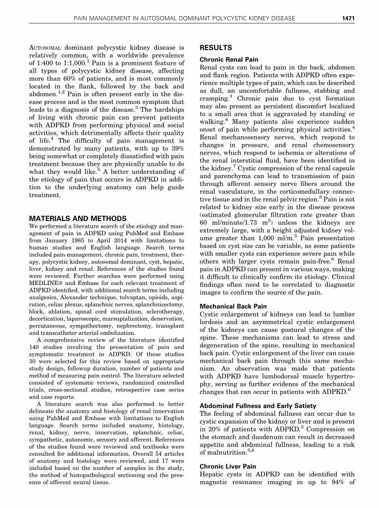
The renal plexus is a network of nerve filamentsand ganglia that are derived from direct branchesof the celiac plexus, celiac ganglia, aorticorenalganglia, thoracic splanchnic nerves, the upperlumbar splanchnic nerve and superior portions ofthe intermesenteric plexus (fig. 1). This has beendemonstrated in anatomical dissections of humancadavers.10,11 This pattern has been confirmedusing retrograde tracers in various animal models,although there is variation in the paravertebral anddorsal root ganglia of origin.7
The lesser thoracic splanchnic nerve, derivedfrom the 9th and 10th or 10th and 11th thoracicparavertebral ganglia, innervates the celiac andaorticorenal ganglia, which send branches to therenal plexus. The least thoracic splanchnic nerve,derived from the 12th thoracic paravertebralganglia, synapses directly on the renal plexus. Theupper lumbar splanchnic nerve, derived from the1st lumbar paravertebral ganglia, sends fibers tothe intermesenteric plexus as well as branches thatsynapse directly on the renal plexus. Fibers fromthe superior portion of the intermesenteric plexusalso run directly to the renal plexus.7,10e12
The greater splanchnic nerve may connect to therenal plexus through the aorticorenal or celiacganglia, which was seen in a minority of cadavers.Small connections to the renal plexus from the
Figure 1. Diagram of extrinsic renal innervation based on
human cadaveric dissections and animal models. Lighter text
and arrows represent nerve connections found in minority of
human cadavers.7,10e12
inferior portion of the intermesenteric plexus andsuperior portion of the hypogastric plexus werealso found in some cadavers.10 These connectionscould not be verified in other dissections.11
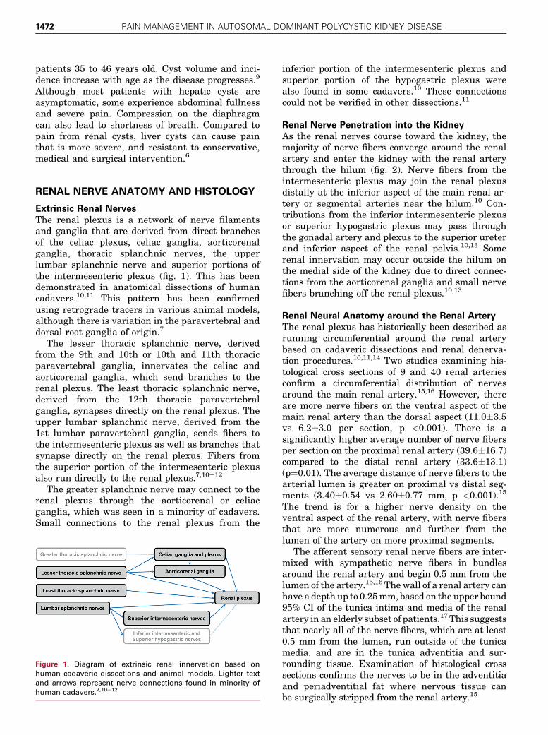
Renal Nerve Penetration into the Kidney
As the renal nerves course toward the kidney, themajority of nerve fibers converge around the renalartery and enter the kidney with the renal arterythrough the hilum (fig. 2). Nerve fibers from theintermesenteric plexus may join the renal plexusdistally at the inferior aspect of the main renal ar-tery or segmental arteries near the hilum.10 Con-tributions from the inferior intermesenteric plexusor superior hypogastric plexus may pass throughthe gonadal artery and plexus to the superior ureterand inferior aspect of the renal pelvis.10,13 Somerenal innervation may occur outside the hilum onthe medial side of the kidney due to direct connec-tions from the aorticorenal ganglia and small nervefibers branching off the renal plexus.10,13
Renal Neural Anatomy around the Renal Artery
The renal plexus has historically been described asrunning circumferential around the renal arterybased on cadaveric dissections and renal denerva-tion procedures.10,11,14 Two studies examining his-tological cross sections of 9 and 40 renal arteriesconfirm a circumferential distribution of nervesaround the main renal artery.15,16 However, thereare more nerve fibers on the ventral aspect of themain renal artery than the dorsal aspect (11.0�3.5vs 6.2�3.0 per section, p <0.001). There is asignificantly higher average number of nerve fibersper section on the proximal renal artery (39.6�16.7)compared to the distal renal artery (33.6�13.1)(p¼0.01). The average distance of nerve fibers to thearterial lumen is greater on proximal vs distal seg-ments (3.40�0.54 vs 2.60�0.77 mm, p <0.001).15
The trend is for a higher nerve density on theventral aspect of the renal artery, with nerve fibersthat are more numerous and further from thelumen of the artery on more proximal segments.
The afferent sensory renal nerve fibers are inter-mixed with sympathetic nerve fibers in bundlesaround the renal artery and begin 0.5 mm from thelumen of the artery.15,16 Thewall of a renal artery canhave a depthup to 0.25mm, based on the upper bound95% CI of the tunica intima and media of the renalartery in an elderly subset of patients.17 This suggeststhat nearly all of the nerve fibers, which are at least0.5 mm from the lumen, run outside of the tunicamedia, and are in the tunica adventitia and sur-rounding tissue. Examination of histological crosssections confirms the nerves to be in the adventitiaand periadventitial fat where nervous tissue canbe surgically stripped from the renal artery.15
Figure 2. Illustration of renal innervation based on dissections of Mitchell10
PAIN MANAGEMENT IN AUTOSOMAL DOMINANT POLYCYSTIC KIDNEY DISEASE 1473
Sympathetic Activity and ADPKD
Increased sympathetic nerve activity has beenimplicated in the pathophysiology of ADPKD.There is an increase in sympathetic nerve activityin hypertensive cases with ADPKD with normalrenal function and in those with impaired renalfunction.18 The renin-angiotensin-aldosterone sys-tem is activated to a greater extent in hypertensivecases with ADPKD than in those with essentialhypertension.19 Furthermore, angiotensin II canenhance epidermal growth factor, which promotescyst formation in the kidney.20
Early onset hypertension and increased sympa-thetic nervous activity could account for the highlevel of cardiovascular morbidity and mortalityin ADPKD, with up to 41% of patients havingleft ventricular hypertrophy in an initial de-mographic study.21 More recent evidence suggeststhe decreasing incidence of left ventricular hyper-trophy, now found in 3.9% of patients with ADPKD,could be due to improved hypertension controlearlier in the disease process, in addition to theuse of renin-angiotensin-aldosterone system antag-onists.22 The role of renin angiotensin blockadein ADPKD will be better understood when theHALT-PKD trial is completed.23 Renal denervationin the rat model of ADPKD causes a reduction in
cyst size, a decrease in blood pressure and im-provement in renal function,24 suggesting a furtherrole of sympathetic activity in the disease and po-tential benefit of renal denervation beyond paincontrol.
Initial Evaluation of Pain
Evaluation must start with a detailed history andphysical examination to determine if the pain ap-pears to be acute or chronic. Patients with ADPKDhave a higher incidence of acute causes of renalpain, including nephrolithiasis, cyst hemorrhage,pyelonephritis and cyst infection, which must beruled out.6
Physical examination may reveal palpableenlarged kidneys or liver. The presence of costo-vertebral angle tenderness upon percussion canresult from inflammation of the kidney andshould raise the suspicion for an acute cause ofrenal pain.
A ruptured or hemorrhagic cyst may be presentwhen there is an abrupt onset of sharp, localizedflank pain with associated gross hematuria. Conser-vative management can be started since the hema-turia normally resolves within 2 to 7 days.6 Aninfectious source of pain such as pyelonephritis orcyst infection should be suspected when a patient
1474 PAIN MANAGEMENT IN AUTOSOMAL DOMINANT POLYCYSTIC KIDNEY DISEASE
has a fever and leukocytosis in addition to in-creasing flank pain. Urine and blood cultures mustbe obtained, and proper antibiotics need to be initi-ated.1 Surgical intervention or fluid drainage maybe necessary if the infection does not respond toantibiotics.
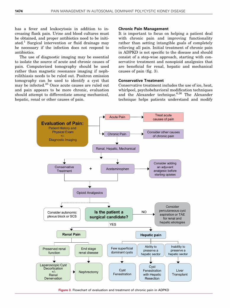
The use of diagnostic imaging may be essentialto isolate the source of acute and chronic causes ofpain. Computerized tomography should be usedrather than magnetic resonance imaging if neph-rolithiasis needs to be ruled out. Positron emissiontomography can be used to identify a cyst thatmay be infected.25 Once acute causes are ruled outand pain appears to be more chronic, evaluationshould attempt to differentiate among mechanical,hepatic, renal or other causes of pain.
Figure 3. Flowchart of evaluation and tr
Chronic Pain Management
It is important to focus on helping a patient dealwith chronic pain and improving functionalityrather than setting intangible goals of completelyrelieving all pain. Initial treatment of chronic painin ADPKD is not specific to the disease and shouldconsist of a step-wise approach, starting with con-servative treatment and nonopioid analgesics thatare beneficial for renal, hepatic and mechanicalcauses of pain (fig. 3).
Conservative Treatment
Conservative treatment includes the use of ice, heat,whirlpool, psychobehavioral modification techniquesand the Alexander technique.6,26 The Alexandertechnique helps patients understand and modify
eatment of chronic pain in ADPKD
PAIN MANAGEMENT IN AUTOSOMAL DOMINANT POLYCYSTIC KIDNEY DISEASE 1475
the postures and movements that result in pain. Ithas been shown to be beneficial in the treatment ofchronic back pain,27 and observations of its use bypatients with ADPKD have led to recommendationsof including it as a treatment option.6
Analgesics
The National Kidney Foundation recommends acet-aminophen as the preferred nonopioid analgesic forlong-term pain management in patients with under-lying renal diseases. Nonsteroidal anti-inflammatorydrugs can be used for acute episodes of pain. How-ever, long-term use is discouraged due to the poten-tial for renal toxicity and renal function should bemonitored with long-term use.28 These recommen-dations should be applied to patients with ADPKDsince 45% experience renal failure by age 60.1
Tramadol as well as other adjuvant analgesics,including clonidine, gabapentin, pregabalin, dulox-etine and amitriptyline, can be added to acetamin-ophen to help control pain before opioids areimplemented.6,26 If indicated, opioids should beinitiated as a trial to determine if they are appro-priate in treating pain. The initial opioid selected,dosing and titration should be based on the patient’spast analgesic use and response. Patients shouldbe evaluated periodically for effectiveness and sideeffects of the medication. An opioid rotation mayneed to be implemented if pain is refractory tohigher doses.29
Tolvaptan, a vasopressin V2 receptor antagonist,has been shown to cause a statistically significantdecrease in renal pain in patients with ADPKD,likely through a decrease in cystic pressure andfluid production. However, the small decrease inpain from 7 to 5 events per 100 person-years of fol-lowup (p¼0.007) in addition to the side effects ofpolyuria and polydipsia, limits the indication of itsuse for pain.30 While tolvaptan is not indicated totreat pain in ADPKD, it shows that future medicaltreatment that limits cyst growth could potentiallyimprove pain control in ADPKD.
Autonomic Nerve Block and SCS
Celiac plexus nerve block is used to control chronicvisceral pain and has been suggested to be of benefitin ADPKD.6,26 Anatomy previously discussed sug-gests that this procedure may only block a portionof renal sensory innervation since the least thoracicand lumbar splanchnic nerves do not relay throughthe celiac plexus. Targeting splanchnic nerveswould produce a wider blockade of renal sensoryoutflow compared to a blockade of the celiac plexus.Although it has not been documented in ADPKD,radio frequency ablation of the splanchnic nervesprovided pain relief in a patient with loin pain he-maturia syndrome.31 Celiac plexus nerve blocks
with radio frequency ablation of the intercostalnerves has been used to provide short-term painrelief in a patient with ADPKD, followed by theuse of SCS for longer term pain relief.32
Cyst Aspiration and Ablation
Cyst aspiration under ultrasound guidance is theleast invasive procedure used to reduce cyst size forrelief of pain. However, pain recurs in 67% of pa-tients after 18 months as the cysts reform.33 Inconjunction with cyst aspiration, sclerotherapy canbe added for further reduction in cyst size. Theseprocedures are most useful when there are only afew dominant cysts responsible for the patient’ssymptoms, which is uncommon in ADPKD. A vari-ety of sclerosing agents have been used, includingethanol, minocycline and n-butyl cyanoacrylatewith iodized oil. There is evidence of successfulreduction of clinical symptoms with sclerotherapyin 17 of 21 patients (81%) with ADPKD at a meanfollowup of 28.5 months.34 All patients had between3 and 6 cysts, which is a low number for ADPKD.
Cyst Decortication
Cyst decortication can relieve pain by releasingpressure on the renal capsule and parenchyma inaddition to reducing compression on the surround-ing tissue. Cyst decortication is the most extensivelystudied procedure for chronic pain relief in ADPKD,with a review showing successful pain relief inall 15 studies and evidence of sustained painrelief in the majority of patients 5 years out.35
Pain relief continued 1 year after the procedure in80% to 92% of patients, which decreased slightlywith time at longer followup visits. A long-termfollowup at a mean of 10.9 years showed that 8 of12 patients (67%) continued to have more than50% pain improvement.36 There does not appearto be any improvement in blood pressure control orrenal function.35 Caution is advised in performinglaparoscopic cyst decortication in patients withrenal impairment as a decreased preoperative esti-mated glomerular filtration rate is associatedwith progression to end stage renal disease afterLCD.36 However, there is no known cause for thisassociation.
LCD remains an option for pain management inpatients with decreased renal function to preserveremaining function by avoiding nephrectomy as analternative treatment for pain. For LCD to be mosteffective, almost all cysts that are accessible on allsurfaces of the kidney must be addressed. The kid-ney will need to be extensively mobilized andnephropexy may be required at the end of the pro-cedure. Laparoscopic ultrasound may be requiredto ensure that the collecting system is not enteredand smaller cysts are identified. Patients should be
1476 PAIN MANAGEMENT IN AUTOSOMAL DOMINANT POLYCYSTIC KIDNEY DISEASE
advised that they may experience a temporaryincrease in pain postoperatively due to irritatingfluid released from hemorrhagic cysts.
Renal Denervation
Laparoscopic renal denervation has been performedin an adolescent age group in a case series of 4 and12 patients with ADPKD (mean age 17 and12.4 years, respectively) who had chronic flank painrefractory to narcotics of 6 to 9 out of 10 on theBieri modified and Wong-Baker pain scales. Divi-sion of all nervous tissue near the hilum was per-formed, followed by circumferential dissection of thekidney to divide any nerves that did not coursethrough the hilum. All patients were pain-freewithout the need for analgesics at a mean followupof 11.5 and 25.5 months, respectively.37,38
A thoracoscopic approach of renal denervationhas been performed for pain refractory to narcoticsand repeated cyst aspirations. Sympathosplanch-nicectomy of the sympathetic chain and splanchnicnerves was performed, resulting in pain relief theday after the procedure and at a 2-year followup withno need for analgesics.39 This approach is beingusing in a clinical trial (NCT00571909) with initialreports of 6 of 9 patients being pain-free withoutanalgesics at 3-month followup.26 A thorough widedissection of the renal artery at the hilum may needto be performed during renal denervation to dissectnerves that may not have run the course of the renalartery. Alternatively it would be safer to performcircumferential renal denervation of the renal arterynear its origin with care to dissect nerve fibers on thelateral aspect of the aorta that may join the renalartery at more distal segments. Division of all tissueon the medial aspect of the kidney and along theproximal ureter can denervate remaining nerveconnections. Multiple renal arteries are present in28% of patients, based on a review of the anatomyof renal arteries.40 Renal innervation follows eachartery entering the kidney,10 implying that renaldenervation would need to be performed aroundeach artery.
Percutaneous transluminal renal denervationrelieved pain in a patient with ADPKD and a fol-lowup procedure on the contralateral side was per-formed based on this success.41 The failure ofpercutaneous transluminal renal denervation todecrease systolic blood pressure compared to a shamoperation raises some concern for the efficacy ofthis procedure to treat pain since the sensorynerves lie within the bundles of sympathetic fiberstargeted with this procedure.42 Renal denervationwould not likely be beneficial in patients who havemechanical associated pain or are experiencingearly satiety. However, it could be used in conjunc-tion with LCD or alone when renal cysts are too
small or deep for LCD to be beneficial. It is ourpractice to perform renal denervation along withextensive laparoscopic renal cyst decortication inpatients with refractory renal pain.
Nephrectomy
Nephrectomy is reserved for the treatment ofchronic renal pain in patients with end stage renaldisease when other modalities have failed. Thisprocedure can be performed unilaterally or bilater-ally, which necessitates the use of dialysis or renaltransplantation. Bilateral laparoscopic nephrec-tomy has been shown to decrease visual analog painscores from an average of 6.9 out of 10 preopera-tively to 0.5 at 3-month followup in 18 patientsstudied,43 with evidence of long-term pain reliefat 31 months in a separate study.44 A laparoscopicapproach is generally preferred compared to anopen procedure due to decreased blood loss,shorter recovery and decreased pain.44 Patientswith extremely large kidneys with a volume greaterthan 3,500 cc are at an increased risk of conversionto an open procedure and, thus, open nephrectomymay initially be considered in these patients.43
Unilateral nephrectomy with renal transplan-tation does not increase morbidity compared to renaltransplantation alone in patients with ADPKD.Renal transplantation with unilateral nephrectomyfollowed by laparoscopic unilateral nephrectomyhas a better perioperative outcome than renaltransplantation followed by bilateral laparoscopicnephrectomy.45 Unpublished data suggest thattransplantation with concurrent unilateral nephrec-tomy in patients with ADPKD results in a greaterdecrease in the number of antihypertensive medica-tions needed compared to transplantation alone.
Transcatheter Arterial Embolization
TAE of distal branches of the renal artery resultsin a reduction in renal volume by 47% 1 year aftertreatment.46 Reduction in renal volume decreasescompression on surrounding organs that can alle-viate feelings of abdominal fullness, discomfort andearly satiety. This procedure can be used in patientswith end stage renal disease in whom other treat-ment modalities have failed and in those who arepoor surgical candidates for nephrectomy.26 Symp-tomatic hepatic cysts can also be reduced by embo-lization of the hepatic segments involved.
Hepatic Cyst Treatment
A variety of surgical approaches can be performedfor pain associated with hepatic cysts, includingcyst fenestration, cyst fenestration with hepaticresection, liver transplantation and TAE. Cystfenestration alone can be used when there are afew superficial dominant cysts and the addition ofhepatic resection can be used in highly symptomatic
PAIN MANAGEMENT IN AUTOSOMAL DOMINANT POLYCYSTIC KIDNEY DISEASE 1477
patients if at least 1 hepatic sector can be preserved.Liver transplantation is indicated if a single hepaticsector cannot be preserved, but it may be difficultto obtain a liver for transplantation since the indi-cation in these patients is usually for symptomsand not for liver failure. While morbidity can be upto 63% using hepatic resection with cyst fenestra-tion, long-term success at a followup of 9 years hasbeen demonstrated, with 75% of patients showingnormalized or improved functionality measured byEastern Cooperative Oncology Group PerformanceStatus.47
CONCLUSIONSThe presentation of pain in patients with ADPKDcan vary due to the numerous mechanisms that can
generate pain in this patient population. Knowingthe etiology of pain can help a physician select anappropriate treatment. A variety of conservative,medical and noninvasive procedures can be used totreat chronic pain in ADPKD. However, pain isoften difficult to control and may be refractory toconservative measures. In this event there aresurgical procedures that can provide reliable relief.Laparoscopic cyst decortication has been exten-sively studied and proven to provide long-termpain relief in the majority of patients. Renaldenervation has been highly successful in the pe-diatric population and could be performed in addi-tion to laparoscopic cyst decortication. Finally,nephrectomy can be used in patients with end stagerenal disease when other treatment modalitieshave failed.
REFERENCES
1. Gabow PA: Autosomal dominant polycystickidney disease. N Engl J Med 1993; 329: 332.
2. Taylor M, Johnson AM, Tison M et al: Earlierdiagnosis of autosomal dominant polycystickidney disease: importance of family history andimplications for cardiovascular and renal com-plications. Am J Kidney Dis 2005; 46: 415.
3. Bajwa ZH, Sial KA, Malik AB et al: Pain patternsin patients with polycystic kidney disease.Kidney Int 2004; 66: 1561.
4. Heiwe S and Bjuke M: “An evil heritage”:interview study of pain and autosomal dominantpolycystic kidney disease. Pain Manag Nurs2009; 10: 134.
5. Miskulin DC, Abebe KZ, Chapman AB et al:Health-related quality of life in patients withautosomal dominant polycystic kidney diseaseand CKD stages 1-4: a cross-sectional study. AmJ Kidney Dis 2014; 63: 214.
6. Bajwa ZH, Gupta S, Warfield CA et al: Painmanagement in polycystic kidney disease. Kid-ney Int 2001; 60: 1631.
7. DiBona GF and Kopp UC: Neural control of renalfunction. Physiol Rev 1997; 77: 75.
8. Barajas L, Liu L and Powers K: Anatomy of therenal innervation: intrarenal aspects and gangliaof origin. Can J Physiol Pharmacol 1992; 70: 735.
9. Bae KT, Zhu F, Chapman AB et al: Magneticresonance imaging evaluation of hepatic cystsin early autosomal-dominant polycystic kidneydisease: the Consortium for Radiologic ImagingStudies of Polycystic Kidney Disease cohort. ClinJ Am Soc Nephrol 2006; 1: 64.
10. Mitchell GA: The nerve supply of the kidneys.Acta Anat (Basel) 1950; 10: 1.
11. Pick J: The innervation of abdominal viscera. In:The Autonomic Nervous System: Morphological,
Comparative, Clinical, and Surgical Aspects.Philadelphia: J. B. Lippincott Co. 1970; chapt 17,pp 313e319.
12. Loukas M, Klaassen Z, Merbs W et al: A reviewof the thoracic splanchnic nerves and celiacganglia. Clin Anat 2010; 23: 512.
13. Sripairojthikoon W and Wyss JM: Cells of originof the sympathetic renal innervation in rat. Am JPhysiol 1987; 252: F957.
14. Page IH and Heuer GJ: The effect of renaldenervation on patients suffering from nephritis.J Clin Invest 1935; 14: 443.
15. Sakakura K, Ladich E, Cheng Q et al: Anatomicassessment of sympathetic peri-arterial renalnerves in man. J Am Coll Cardiol 2014; 64: 635.
16. Atherton DS, Deep NL and Mendelsohn FO:Micro-anatomy of the renal sympathetic nervoussystem: a human postmortem histologic study.Clin Anat 2012; 25: 628.
17. Reddy S, Kumar P and Prasad K: Histomorpho-metric and sympathetic innervation of thehuman renal artery: a cadaveric study. Urol Ann2011; 3: 141.
18. Klein IH, Ligtenberg G, Oey PL et al: Sympatheticactivity is increased in polycystic kidney diseaseand is associated with hypertension. J Am SocNephrol 2001; 12: 2427.
19. Chapman AB, Johnson A, Gabow PA et al:The renin-angiotensin-aldosterone system andautosomal dominant polycystic kidney disease.N Engl J Med 1990; 323: 1091.
20. Torres VE, Sweeney WE Jr, Wang X et al:EGF receptor tyrosine kinase inhibition attenu-ates the development of PKD in Han:SPRD rats.Kidney Int 2003; 64: 1573.
21. Chapman AB, Johnson AM, Rainguet S et al: Leftventricular hypertrophy in autosomal dominant
polycystic kidney disease. J Am Soc Nephrol1997; 8: 1292.
22. Schrier RW: Renal volume, renin-angiotensin-aldosterone system, hypertension, and left ven-tricular hypertrophy in patients with autosomaldominant polycystic kidney disease. J Am SocNephrol 2009; 20: 1888.
23. Chapman AB: Approaches to testing new treat-ments in autosomal dominant polycystic kidneydisease: insights from the CRISP and HALT-PKDstudies. Clin J Am Soc Nephrol 2008; 3: 1197.
24. Gattone VH 2nd, Siqueira TM Jr, Powell CR et al:Contribution of renal innervation to hypertensionin rat autosomal dominant polycystic kidneydisease. Exp Biol Med (Maywood) 2008; 233:952.
25. Jouret F, Lhommel R, Devuyst O et al: Diagnosisof cyst infection in patients with autosomaldominant polycystic kidney disease: attributesand limitations of the current modalities. Ne-phrol Dial Transplant 2012; 27: 3746.
26. Hogan MC and Norby SM: Evaluation andmanagement of pain in autosomal dominantpolycystic kidney disease. Adv Chronic KidneyDis 2010; 17: e1.
27. Woodman JP and Moore NR: Evidence for theeffectiveness of Alexander Technique lessons inmedical and health-related conditions: a sys-tematic review. Int J Clin Pract 2012; 66: 98.
28. Henrich WL, Agodoa LE, Barrett B et al: Anal-gesics and the kidney: summary and recom-mendations to the Scientific Advisory Board ofthe National Kidney Foundation from an Ad HocCommittee of the National Kidney Foundation.Am J Kidney Dis 1996; 27: 162.
29. Chou R, Fanciullo GJ, Fine PG et al: Clinicalguidelines for the use of chronic opioid therapyin chronic noncancer pain. J Pain 2009; 10: 113.
1478 PAIN MANAGEMENT IN AUTOSOMAL DOMINANT POLYCYSTIC KIDNEY DISEASE
30. Torres VE, Chapman AB, Devuyst O et al: Tol-vaptan in patients with autosomal dominantpolycystic kidney disease. N Engl J Med 2012;367: 2407.
31. Moeschler SM, Hoelzer BC and Eldrige JS:A patient with loin hematuria syndrome andchronic flank pain treated with pulsed radio-frequency of the splanchnic nerves. Clin J Pain2013; 29: e26.
32. Walsh N and Sarria JE: Management of chronicpain in a patient with autosomal dominantpolycystic kidney disease by sequential celiacplexus blockade, radiofrequency ablation, andspinal cord stimulation. Am J Kidney Dis 2012;59: 858.
33. Bennett WM, Elzinga L, Golper TA et al:Reduction of cyst volume for symptomaticmanagement of autosomal dominant polycystickidney disease. J Urol 1987; 137: 620.
34. Kim SH, Kim SH and Cho JY: Cyst ablation usinga mixture of N-butyl cyanoacrylate and iodizedoil in patients with autosomal dominant poly-cystic kidney disease: the long-term results.Korean J Radiol 2009; 10: 377.
35. Millar M, Tanagho YS, Haseebuddin M et al:Surgical cyst decortication in autosomal domi-nant polycystic kidney disease. J Endourol 2013;27: 528.
36. Haseebuddin M, Tanagho YS, Millar M et al:Long-term impact of laparoscopic cyst decorti-cation on renal function, hypertension and paincontrol in patients with autosomal dominantpolycystic kidney disease. J Urol 2012; 188:1239.
37. Casale P, Meyers K and Kaplan B: Follow-upfor laparoscopic renal denervation and neph-ropexy for autosomal dominant polycystic kidneydisease-related pain in pediatrics. J Endourol2008; 22: 991.
38. Resnick M, Chang AY and Casale P: Laparoscopicrenal denervation and nephropexy for autosomaldominant polycystic kidney disease related painin adolescents. J Urol 2006; 175: 2274.
39. Chapuis O, Sockeel P, Pallas G et al: Thoraco-scopic renal denervation for intractable auto-somal dominant polycystic kidney disease-relatedpain. Am J Kidney Dis 2004; 43: 161.
40. Satyapal KS, Haffejee AA, Singh B et al: Addi-tional renal arteries: incidence and morphometry.Surg Radiol Anat 2001; 23: 33.
41. Casteleijn NF, de Jager RL, Neeleman MP et al:Chronic kidney pain in autosomal dominantpolycystic kidney disease: a case report of suc-cessful treatment by catheter-based renaldenervation. Am J Kidney Dis 2014; 63: 1019.
42. Bhatt DL, Kandzari DE, O’Neill WW et al: Acontrolled trial of renal denervation for resistanthypertension. N Engl J Med 2014; 370: 1393.
43. Lipke MC, Bargman V, Milgrom M et al: Limita-tions of laparoscopy for bilateral nephrectomyfor autosomal dominant polycystic kidney dis-ease. J Urol 2007; 177: 627.
44. Dunn MD, Portis AJ, Elbahnasy AM et al:Laparoscopic nephrectomy in patients with end-stage renal disease and autosomal dominantpolycystic kidney disease. Am J Kidney Dis 2000;35: 720.
45. Lucas SM, Mofunanya TC, Goggins WC et al:Staged nephrectomy versus bilateral laparo-scopic nephrectomy in patients with autosomaldominant polycystic kidney disease. J Urol 2010;184: 2054.
46. Ubara Y: New therapeutic option for autosomaldominant polycystic kidney disease patientswith enlarged kidney and liver. Ther Apher Dial2006; 10: 333.
47. Schnelldorfer T, Torres VE, Zakaria S et al:Polycystic liver disease: a critical appraisal ofhepatic resection, cyst fenestration, and livertransplantation. Ann Surg 2009; 250: 112.