Embed Size (px)
Citation preview
1
OTOLARYNGOLOGY ONLINE
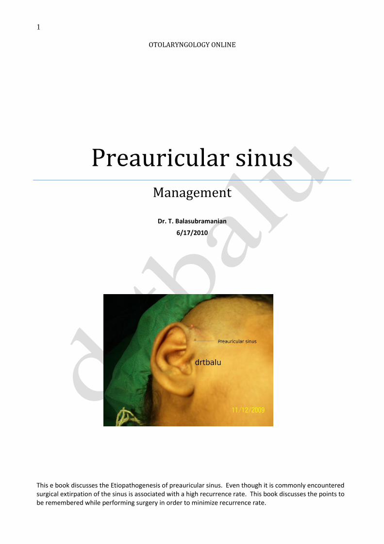
Preauricular sinus Management
Dr. T. Balasubramanian
6/17/2010
This e book discusses the Etiopathogenesis of preauricular sinus. Even though it is commonly encountered surgical extirpation of the sinus is associated with a high recurrence rate. This book discusses the points to be remembered while performing surgery in order to minimize recurrence rate.
Otolaryngology online
Preauricular sinus and its
management
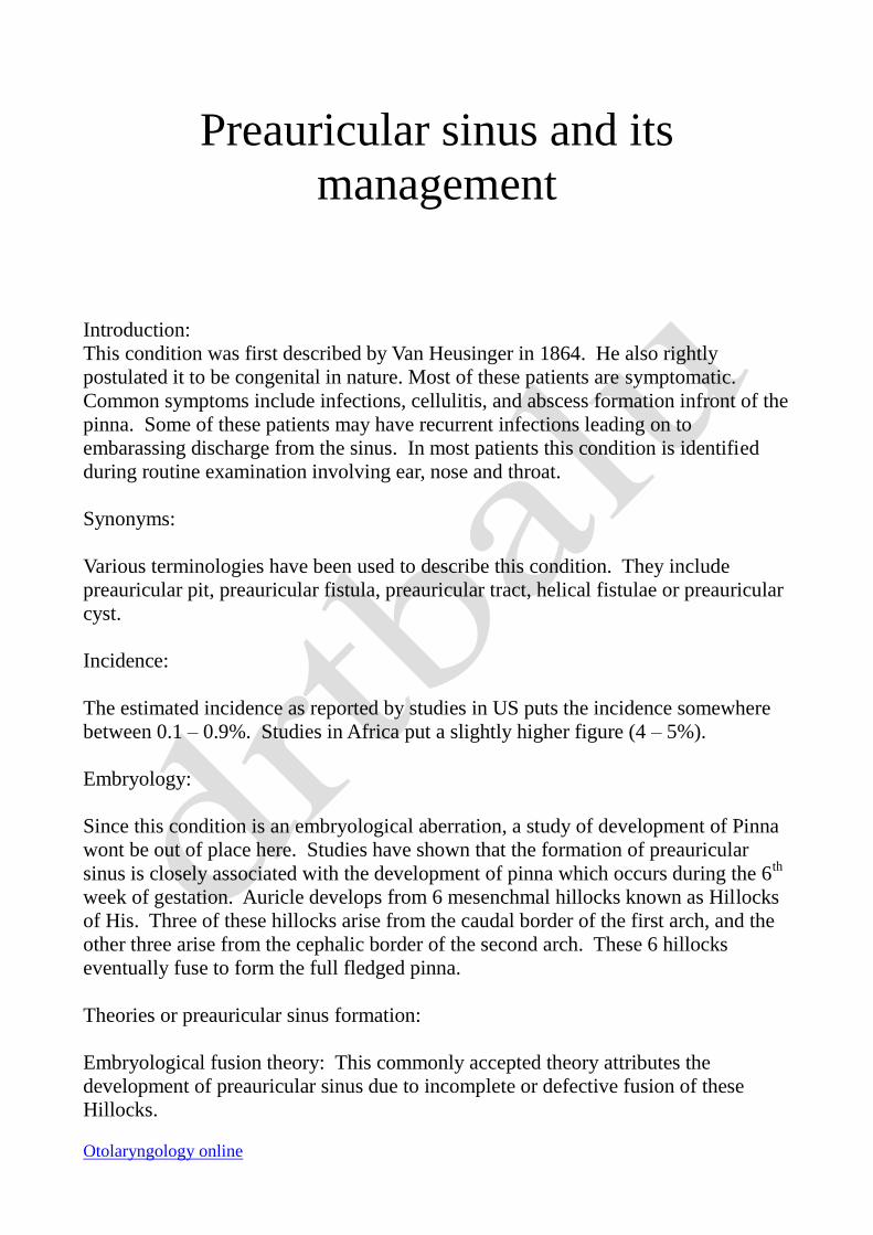
Introduction:
This condition was first described by Van Heusinger in 1864. He also rightly
postulated it to be congenital in nature. Most of these patients are symptomatic.
Common symptoms include infections, cellulitis, and abscess formation infront of the
pinna. Some of these patients may have recurrent infections leading on to
embarassing discharge from the sinus. In most patients this condition is identified
during routine examination involving ear, nose and throat.
Synonyms:
Various terminologies have been used to describe this condition. They include
preauricular pit, preauricular fistula, preauricular tract, helical fistulae or preauricular
cyst.
Incidence:
The estimated incidence as reported by studies in US puts the incidence somewhere
between 0.1 – 0.9%. Studies in Africa put a slightly higher figure (4 – 5%).
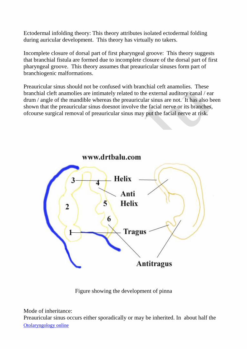
Embryology:
Since this condition is an embryological aberration, a study of development of Pinna
wont be out of place here. Studies have shown that the formation of preauricular
sinus is closely associated with the development of pinna which occurs during the 6th
week of gestation. Auricle develops from 6 mesenchmal hillocks known as Hillocks
of His. Three of these hillocks arise from the caudal border of the first arch, and the
other three arise from the cephalic border of the second arch. These 6 hillocks
eventually fuse to form the full fledged pinna.
Theories or preauricular sinus formation:
Embryological fusion theory: This commonly accepted theory attributes the
development of preauricular sinus due to incomplete or defective fusion of these
Hillocks.
Otolaryngology online
Ectodermal infolding theory: This theory attributes isolated ectodermal folding
during auricular development. This theory has virtually no takers.
Incomplete closure of dorsal part of first pharyngeal groove: This theory suggests
that branchial fistula are formed due to incomplete closure of the dorsal part of first
pharyngeal groove. This theory assumes that preauricular sinuses form part of
branchiogenic malformations.
Preauricular sinus should not be confused with branchial ceft anamolies. These
branchial cleft anamolies are intimately related to the external auditory canal / ear
drum / angle of the mandible whereas the preauricular sinus are not. It has also been
shown that the preauricular sinus doesnot involve the facial nerve or its branches,
ofcourse surgical removal of preauricular sinus may put the facial nerve at risk.
Figure showing the development of pinna
Mode of inheritance:
Preauricular sinus occurs either sporadically or may be inherited. In about half the
Otolaryngology online
number of patients it occurs in a sporadic manner and commonly on the right side.
Bilateral cases are commonly genetically inherited. Studies have shown that
inheritance is autosomal dominant with varying degrees of penetration (about 85%
penetration). Studies in China has shown chromosome 8q11 to be site of abnormal
gene which transmits preauricular sinus.
Preauricular sinus has been described as a part of number of syndromes. These
syndromes include:
1. BOR syndrome (Branchio oto renal syndrome) – defects in these patients
include outer, middle and inner ear deformities with conductive deafness. These
patients also have renal anamolies, lateral cervical fistulae, preauricular sinus, and
nasolacrimal duct stenosis and fistula.
2. Branchio oto urethral syndrome – These patients have sensorineural hearing
loss, preauricular sinus, renal anamolies like bifid ureters and bifid renal pelvis.
3. Branchio otic syndrome – This is a variant of BOR syndrome. These patients
have branchial anamolies, preauricular sinus, branchial fistula (unilateral) with no
renal dysplasia
4. Branchio oto costal syndrome – These patients have conductive deafness,
preauricular sinus, bilateral commissural lip, unilateral branchial fistula and rib
anamolies
5. Cat eye syndrome – Coloboma of iris, Preauricular sinus, imperforate anus and
down slanting of palpebral fissures
6. Trisomy 22 – These patients have bilateral preauricular sinus, antimongoloid
palpebral fissures, macroglossia, cleft palate, enlarged sublingual glands and short
lower limbs
Clinical features:
Preauricular sinus is seen as a small pit usually at the anterior margin of the
ascending limb of the helix. In some patients this opening may also be seen along the
postero superior margin of helix. Rarely it may be seen close to the tragus or lobule.
In almost all patients part of the tract blends with the perichondrium of the auricular
cartilage.
The sinus tract may follow a tortuous course. The sinus tract is usually superior and
lateral to the facial nerve and parotid gland. This feature differentiates it from
branchial cleft anamolies. Sometimes the preauricular sinus may lead to the
formation of subcutaneous cyst that is intimately related to the tragal cartilage and the
crus of helix.
Patients usually present with discharge from the preauricular sinus pit. Discharge
could be due to desquamating epithelial debris or infection. Studies have shown that
the common pathogens causing infection in the preauricular sinus include
staphylococcus, proteus, streptococcus and peptococcus.

Otolaryngology online
It is always better to rule out syndromes associated with preauricular sinus. Almost
majority of these syndromes involve kidney. There is intense debate raging whether
ultrasound examination should be performed as a routine in all patients with
preauricular sinus. Considering the commonality of the lesion and the cost and time
involved routine ultrasound in these patients are not indicated. Wang et al of
California came out with a set of indications when ultrasound abdomen should be
performed in these patients.
Wang's criteria in performing ultrasound examination in patients with preauricular
sinus:
1. Presence of another malformation / dysmorphic feature
2. Family history of deafness
3. Malformations involving pinna
4. Maternal history of gestational diabetes
Picture of a patient with infected preauricular sinus
Puretone audiometry:
This is another investigation that should routinely be performed in all patients with
preauricular sinuses.
Complications of preauricular sinus:
Otolaryngology online
Infection is the predominant complication. In the acute phase of infection (cellulitis
stage) management is by prescribing appropriate antibiotics in adequate doses. Since
the common infecting organism is staphylococcus aureus the drug of choice is a
combination of amoxycillin and clavulanic acid.
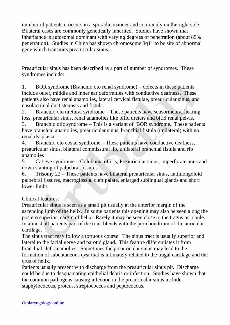
Abscess formation: Abscess in this area should always be drained. Incision and
drainage using a scalpel would cause extensive fibrosis causing difficulty in complete
surgical clearance of the area at a later date. Precisely for this reason Coatesworth et
al described a drainage procedure using lacrimal probe. This probe negates the need
for incision in this area and thus causes very little disturbance to the underlying
preauricular sinus tissue. In this technique of drainage the overlying skin is
anesthetized using 2% xylocaine infiltration. The blunt end of the lacrimal probe is
inserted into the sinus through the pit. This allows drainage to occur via the normal
opening which is usually present in front of the ascending limb of the helix. If
preauricular abscess does not drain when this technique is used then conventional
incision and drainage should be performed. Recurrent infections involving the
preauricular sinus should be managed by complete surgical resection of the sinus tract
completely during the stage of quiescence.
Image showing lacrimal probe which is used to drain preauricular abscess
Otolaryngology online
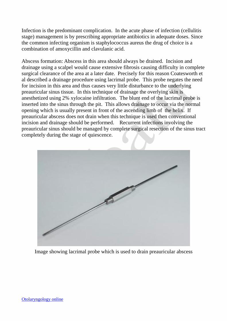
Figure showing the common sites of preauricular sinus involvement.
1. Anterior margin of ascending limb of helix (most common)
2. Superior to auricle
3. Along the posterior surface of cymba concha
4. Lobule
5. Posterior to auricle
Surgical excision of perauricular sinus:
While surgically excising the sinus tract care should be taken to completely remove
it. Incomplete removal of sinus tract is the commonest cause for recurrence. The
recurrence rate ranges between 1 – 45% depending on the procedure followed.
Simple sincectomy:
Otolaryngology online
This is the commonly used standard procedure for excising preauricular sinus. An
ellipse of skin surrounding the preauricular sinus tract is excised and dissected out
along with the tract. The tract can simply be identified by its glistening white color,
or methylene blue dye can be injected through the opening to faciliate easy
identification of the tract. Most of these fistulae follow the external auditory canal.
This procedure can be performed under local or general anesthesia. While operating
on children general anesthesia is preferred.
Jensma technique:
This technique was popularised by Jensma in 1970. It is actually a modification of
the classic sinusotomy procedure. This technique is also known as inside out
technique.
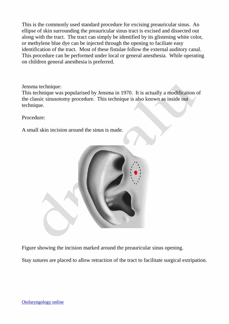
Procedure:
A small skin incision around the sinus is made.
Figure showing the incision marked around the preauricular sinus opening.
Stay sutures are placed to allow retraction of the tract to facilitate surgical extripation.
Otolaryngology online
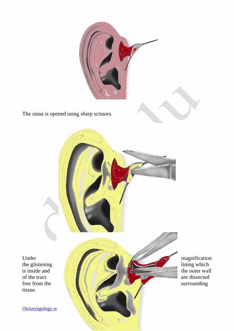
The sinus is opened using sharp scissors.
Under magnification
the glistening lining which
is inside and the outer wall
of the tract are dissected
free from the surrounding
tissue.
Otolaryngology online
The main advantage of this procedure is that the sinus can be viewed and followed
from both inside and outside. The classic procedure allows visualisation of the sinus
from only outside. All the tracts are opened and followed until the dead end is
reached. A lacrimal duct probe can be used to establish the direction of small tracts.
It should be borne in mind that one of the tracts could be closely adherent to the
perichondrium of the root of the helix / tragus. This piece of perichondrium along
with a small bit of underlying cartilage should be resected along with the specimen.
The medial limit of dissection is always the temporalis fascia. Before closure the
wound bed should be carefully examined for evidence of residual tracts.
Causes of recurrence:
1. Major cause of recurrence is inadequate removal of the mass.
2. Performing the surgery without magnification aids
3. Skill of the operating surgeon. This is rather important because surgeons
consider this case to be a minor procedure and hence pass it on either to a novice or
junior surgeon who may not be experienced enough in performing this type of
surgery.
Supra auricular approach:
This is a more radical approach. Major advantage of this approach is that it gives
excellent exposure and hence removal of the sinus tract is nearly complete. This
procedure has the lowest recurrence rate among all other surgical procedures for
Otolaryngology online
preauricular sinus remoaval.
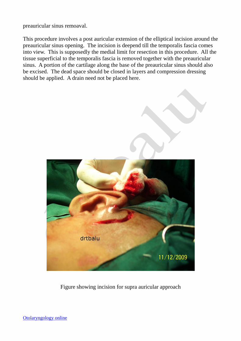
This procedure involves a post auricular extension of the elliptical incision around the
preauricular sinus opening. The incision is deepend till the temporalis fascia comes
into view. This is supposedly the medial limit for resection in this procedure. All the
tissue superficial to the temporalis fascia is removed together with the preauricular
sinus. A portion of the cartilage along the base of the preauricular sinus should also
be excised. The dead space should be closed in layers and compression dressing
should be applied. A drain need not be placed here.
Figure showing incision for supra auricular approach
Otolaryngology online
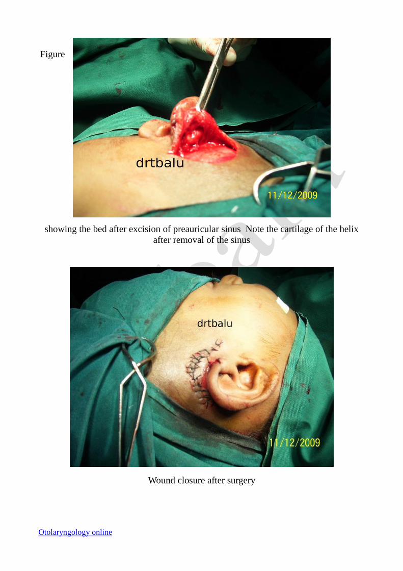
Figure
showing the bed after excision of preauricular sinus Note the cartilage of the helix
after removal of the sinus
Wound closure after surgery


![[OTOLARYNGOLOGY CLINICAL EXAMIANTION SERIES]otolaryngology.wdfiles.com/.../Clinical-Examination-Techniques-in... · Before proceeding with clinical examination a good history 1 taking](https://img.pdfslide.net/doc/110x75/5b543a147f8b9a27658ca338/otolaryngology-clinical-examiantion-series-before-proceeding-with-clinical.jpg)