Embed Size (px)
Citation preview
ORYCTOS vol. 7, 2008 61
Mandibular kinesis in Hesperornis
Larry D. Martin1 & Virginia L. Naples2
1Department of Ecology and Evolutionary Biology; Museum of Natural History and Biodiversity Center, University of Kan-sas, Lawrence, Kansas 66045, U. S. A. E-mail: [email protected] of Biological Sciences, Northern Illinois University, DeKalb, Illinois 60115-2861, U. S. A. E-mail: [email protected]
ABSTRACT - Some aspects of mandibular morphology are known for three hesperornithiform genera: Hesperornis, Para-hesperornis and Baptornis. All share a distinctive intramandibular joint between the angular and the splenial. A special pro-cess of the surangular extending between the splenial and the dentary bridges the joint. The symphysis appears to have been elongate and unfused, joining anteriorly with a short intersymphyseal bone. It appears that the mandibles spread posteriorly as the jaws opened, allowing the swallowing of larger prey. The closed mandible is very slender anteriorly, resembling some cetaceans, and seems highly adapted for the capture of fish. The discovery of fish remains in a preserved stomach cast of Baptornis gives direct support for this interpretation.
Key words: Hesperornis, mandibular kinesis, intraramal joint, intersymphyseal bone, intramandibular joint.
KINESIS MANDIBULAIRE DE HESPERORNIS – Quelques aspects de la morphologie mandibulaire sont connu pour les trois genres d’ hesperornithiformes: Hesperornis, Parahesperornis et Baptornis. Tous ont en commun une articulation intramandibulaire entre l’angulaire et le splénial. Un processus spécial du surangulaire s’étend entre le splénial et le dentaire à travers l’articulation. La symphyse semble avoir été allongée et sans fusion, rejoignant antérieurement un os intersym-physial court. Il apparaît que les mandibles s’écartaient postérieurement lors de l’ouverture des mâchoires, permettant ainsi d’avaler des proies plus grandes. La mâchoire est très étroite antérieurement, ce qui rappelle certains cétacés, et paraît très bien adaptée à la capture des poissons. La découverte de débris de poissons dans un contenu stomacal de Baptornis apporte un soutien direct à cette interprétation.
INTRODUCTION
The cranial kinesis of Hesperornis is described in detail by Buhler et al. (1998), who conclude that it is proki-netic, but do not discuss the mandible. Kinesis in the man-dible is discussed by Gregory (1951), who makes extensive comparisons with extinct giant lizards (mosasaurs), claiming that the mandibles of the two groups are similar in many respects, but especially in sharing an intramandibular joint between the splenial and the angular. This unusual joint was also found in jaws referred to Ichthyornis, and Gregory (1952) found the similarity between that genus and mosas-aurs so great that he suggested that the putative Ichthyornis jaws were really those of juvenile mosasaurs. Surprisingly, this bold suggestion was quickly accepted. Swinton (1975), in a book on fossil birds, even doubted that the Hesperornis jaws were avian. He did accept one mandible in the Univer-sity of Nebraska State Museum as evidence that Hesperornis was toothed and illustrated it (Swinton, 1975, Fig. 16, p. 37). Further study soon demonstrated that all of these sugges-tions were in error, and the toothed jaws were restored to
their avian skeletons (Gingerich, 1972; Martin and Stewart, 1977). Ironically, the one exception was the only specimen that had convinced Swinton, UNSM 5363. This turned out to be a genuine juvenile mosasaur. However, the question of mandibular kinesis remained, and Gingerich suggested that a somewhat similar kinesis in theropod dinosaurs provided further support of their affinity with birds. Most of this spec-ulation continued to be based on the original Marsh materials at Yale, and so the discovery of a more complete and better-preserved specimen of Hesperornis regalis (KUVP 71012) provides an opportunity to review the question of mandibu-lar kinesis in more detail.
THE MANDIBLE OF HESPERORNIS
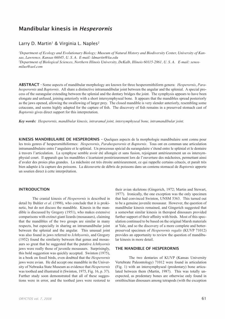
The two dentaries of KUVP (Kansas University Vertebrate Paleontology) 71012 were found in articulation (Fig. 1) with an intersymphyseal (predentary) bone articu-lated between them (Martin, 1987). This was totally un-expected, as predentary bones are otherwise only found in ornithischian dinosaurs among tetrapods (with the exception
ORYCTOS vol. 7, 2008 62
of the unusual bird, Teratornis). A quick search revealed a second hesperornithiform example with the type mandible of Parahesperornis alexi and the bone illustrated by Marsh (1880, Pl. II., fig. 12) as a basihyal provides an additional oc-currence in Hesperornis regalis. The unusual blunt anterior termination of the dentary in Hesperornis had been remarked upon (Gregory, 1951), and now could be explained, as the end bears a small oval facet for the articulation with the pre-dentary. The dorsal lateral surface of the dentary also bears a distinctive large pit that is matched by a similar depression in the predentary, presumably for a ligament that crossed the
joint and tied the jaw together (Fig. 2). The jaws are hinged anteriorly by this joint, while in modern birds the symphysis between the dentaries is fused. The predentary is triangular and fits inside the down turned tip of the premaxillaries. The premaxillaries are toothless, as is the predentary bone.
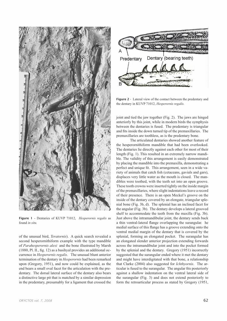
The articulated dentaries showed another feature of the hesperornithiform mandible that had been overlooked. The dentaries lie directly against each other for most of their length (Fig. 1). This resulted in an extremely narrow mandi-ble. The validity of this arrangement is easily demonstrated by placing the mandible into the premaxilla, demonstrating a perfect and unique fit. This arrangement, seen in a wide va-riety of animals that catch fish (cetaceans, gavials and gars), displaces very little water as the mouth is closed. The man-dibles were toothed, with the teeth set into an open groove. These tooth crowns were inserted tightly on the inside margin of the premaxillaries, where slight indentations leave a record of their presence. There is an open Meckel’s groove on the inside of the dentary covered by an elongate, triangular sple-nial bone (Fig. 3b, d). The splenial has an inclined facet for the angular (Fig. 3b). The dentary develops a lateral grooved shelf to accommodate the teeth from the maxilla (Fig. 3b). Just above the intramandibular joint, the dentary sends back a thin ventral-lateral flange overlapping the surangular. he medial surface of this flange has a groove extending onto the ventral medial margin of the dentary that is covered by the splenial, forming an elongated pocket. The surangular has an elongated slender anterior projection extending forwards across the intramandibular joint and into the pocket formed by the splenial and the dentary. Gregory (1951) incorrectly suggested that the surangular ended where it met the dentary and might have interdigitated with that bone, a relationship that Clarke (2004) also suggested for Ichthyornis. The ar-ticular is fused to the surangular. The angular fits posteriorly against a shallow indentation on the ventral lateral side of the surangular (Fig. 3) and does not extend posteriorly to form the retroarticular process as stated by Gregory (1951,
Figure 1 - Dentaries of KUVP 71012, Hesperornis regalis as found in situ.
Figure 2 - Lateral view of the contact between the predentary and the dentary in KUVP 71012, Hesperornis regalis.
ORYCTOS vol. 7, 2008 63
Figure 3 - Mandible of KUVP 71012, Hesperornis regalis: A. medial view of the left surangular with the articular and prearticular fused to it; B. medial view of the left mandible; C. lateral view of the left mandible; D. medial view of the left splenial; E. medial view of the right angular; F. dorsal view of the mandible when the jaws are closed; G. dorsal view of the mandible when the jaws are open, showing separation at the interdentary joint and bending at the intramandibular joints; H. ventral view of the right quadrate showing the articular surfaces.
ORYCTOS vol. 7, 2008 64
p. 348). The surangular runs forward above the angular’s dorsal medial surface extending to where the angular articu-lates with the splenial. At that junction, it fits into a shallow groove on the dorsal surface of the angular that locks the angular to the surangular. The angular is distinctly curved (convex labially), as compared to the surangular, and this curvature may help guide the bending of the surangular. The surangular is thin, flat, and presumably, bendable above the joint between the angular and the splenial. The thinner the bone, the greater the flexibility should be, but thinner bones are less strong; therefore three thin bones, each of which can flex independently, participating in the intraramal joint, allow this jaw region to bow laterally, while maintaining strength.
The surangular, articular and prearticular are tightly fused and hard to distinguish. The prearticular is not fused to the angular, a possibility suggested by Gregory (1951). It also does not extend across the intraramal joint, as suggested by Gregory (1951), as it does in mosasaurs. Because the coronoid and surangular end at the intramandibular joint and the prearticular crosses it in mosasaurs, Gregory’s 1951 mis-interpretation of the Yale Hesperornis material might have resulted from his use of mosasaurs to interpret it.
The quadrate has an exceptionally large and up-wardly turned orbital process. The ventral articulation has a large inwardly inclined medial trochlea and a small lateral one (Fig. 3h). This causes the jaws to spread as they open, bending them at the intramandibular joint. The dentaries then spread at the intersymphyseal joint, causing a signifi-cant increase in gape (fig. 3g). The posterior flange of the dentary and the surangular form an overlapping structure that facilitates bending at the interramal joint. As Gregory (1951) suggested, there is a slight rotation of the dentaries outward as the jaws open. This would dislodge the maxil-lary teeth and permit the captured prey to be rotated into the headfirst swallowing position favored by piscivores. The loss of teeth in the premaxilla may have facilitated this rotation, while the maxillary and dentary teeth could assist with hold-ing the prey for manipulation. Because of the incorporation of the arm into a wing (extremely reduced in Hesperornis), birds cannot use their manus to assist with the manipulation of food held in the mouth. This is not much of a problem when the food is small, as is the case with most insects, but larger prey may be caught sideways and have to be rotated for swallowing. A system of rotation not involving release of the prey is provided by movement (kinesis) within the jaw and may have been a major impetus for the evolution of the jaw kinesis characteristic of birds.
COMPARISONS
In modern birds the symphysis is fused, but there may be a bending zone just behind it that acts in a similar way to the joint between the dentaries and the intersymphy-seal bone. Such a joint would be difficult to develop within the relatively thick and inflexible dentaries of the toothed birds, and this may have promoted the development of a
synovial joint in the same position, resulting in a separate predentary ossification. The intraramal joint in Hesperornis is a combination of a synovial joint between the surangu-lar and the splenial and a bending zone across the posterior flange of the dentary and the surangular. In either case, ex-tinct toothed and modern piscivorous birds use intraramal bending to facilitate gape. Intraramal bending is also char-acteristic of the extinct bony-toothed birds, Odontopterygia (Zusi and Warheit, 1992). An intersymphyseal bone also occurs in the giant vulture-like Teratornis (Campbell and Tonni, 1982), representing independent evolution of a simi-lar gape mechanism.
The intraramal joint of mosasaurs and some dino-saurs is unlike that of Hesperornis in that they lack the poste-rior dentary flange and the anterior process of the surangular. Hesperornis also lacks a coronoid bone, a prominent feature in mosasaurs and most dinosaurs. In many animals the coro-noid would lie across and interfere with an intraramal joint (it lies behind the joint in mosasaurs). The supposed similar-ity between mosasaur and bird mandibles is overdrawn and the two are easily separated. These differences extend to the pleurodont implantation of the teeth in mosasaurs and the thecodont implantation in birds.
Parahesperornis has the same mandibular kinesis as Hesperornis, and an anterior part of a Baptornis angular shows the characteristic intraramal joint, so we might sup-pose that it occurs throughout Hesperornithiformes. The intraramal joint also occurs in Ichthyornis, and examination of the anterior tip of the dentary in a number of specimens of that genus reveals the characteristic facet for the preden-tary bone. This combination of features is unique enough to raise the possibility that it arose before foot-propelled div-ing developed in Hesperornithiformes and represents a com-plex synapomorphy uniting an early ornithurine clade, the Odontornithes of Marsh (1880). In contrast, Clarke’s (2004) description of Ichthyornis would suggest a very isolated po-sition for that genus.
Gregory (1952) described a large coronoid bone in Ichthyornis, and Clarke (2004) a smaller one. Gingerich (1972) was unable to find a coronoid. All of the birds, liv-ing and fossil that we are aware of, lack the coronoid, so its presence would be of considerable interest. According to Clarke (2004, Fig. 30), the surangular in Ichthyornis turns labially and inserts into a slot on the medial side of the den-tary, while the coronoid is a small sliver inserted between it and the prearticular. The prearticular extends across the joint between the angular and the splenial and inserts between the splenial and the dentary, the same position occupied by the surangular in Hesperornis (Clarke, 2004). The prearticular ends behind the intraramal joint in Hesperornis; there is no coronoid, and the surangular turns medially when it reaches the dentary rather than laterally. The dentary in Hesperornis extends across the intraramal joint rather than ending at the joint as described for Ichthyornis by Clarke (2004). This would indicate very different intraramal joints in hesperor-nithiforms and Ichthyornis. However, the Ichthyornis mate-
ORYCTOS vol. 7, 2008 65
rial is so broken and crushed that it could easily be misin-terpreted, and probably a re-examination in light of the new hesperornithiform material is warranted.
Hesperornis has a mandible that is not similar in de-tail to either mosasaurs or dinosaurs. Nor is it close to mod-ern birds, which have rhamphotheca-covered dentaries that permit different opportunities for bending. The predentary bone occupies the same position as a bending zone in mod-ern birds with intraramal joints, and the resistance to bending of the thicker tooth-bearing dentary may have forced the de-velopment of a synovial joint. A similar joint and predentary bone is suggested for Ichthyornis on the basis of a similar articular facet on the tip of the dentary to that for the preden-tary in Hesperornis.
CONCLUSIONS
We do not presently know the distribution of pre-dentary bones among early birds. They are absent from Archaeopteryx and other known members of the Sauriurae. Presumably, predentaries define a clade within the early Orni-thurae. It seems certain, on the basis of anterior dentary mor-phology, that they occur in the Ichthyornithiformes, as well as the Hesperornithiformes, thus uniting these two groups into what may be considered a superorder (Odontornithes of Marsh, 1880). They occur at the same position as a well-recognized bending zone in the dentary of modern birds that have an intramandibular joint, and this provides an analogue for their origin. Almost all known Mesozoic ornithurines are aquatic or water marginal, and many undoubtedly ate fish. The combination of a narrow mandible with a wide posterior gape has obvious advantages for a piscivore. The kinetic system in Hesperornis is unique and not really comparable to that of either mosasaurs or theropod dinosaurs.
ACKNOWLEDGEMENTS
We thank K. Campbell and D. Burnham for help-ful comments. The figures are by B. Platt, E. Dickey and J. Chorn. J. Ostrom and M. Turner made the Yale hesperorni-thiform material available for comparison.
REFERENCES
Buhler, P. 1992. Light bones in Birds; pp. 385-393. In Campbell, Jr., K. E. (ed.). Papers in Paleontology Honoring Pierce Brodkorb. Natural History Museum of Los Angeles County, Los Angeles.
Buhler, P., Martin, L. D., & Witmer, L. M. 1988. Cranial kinesis in the Late Cretaceous birds Hesperornis and Parahesperornis. Auk, 105: 111-122.
Campbell, K. C. & Tonni, E. P. 1982. Preliminary Observations on the Paleobiology and Evolution of Teratorns (Aves: Teratornithidae). J. Vert. Paleont. 1: 265-273.
Clarke, J. A. 2004. Morphology, phylogenetic taxonomy, and systematics of Ichthyornis and Apatornis (Avialae: Ornithurae), Bulletin of the American Museum of Natural History, 286: 1-179.
Gingerich, P. D. 1972. A new partial mandible of Ichthyornis. Condor, 74: 471-473.
Gregory, J. T. 1951. Convergent evolution: the jaws of Hesperornis and the Mosasaurs. Evolution, 5: 345-354.
Gregory, J. T. 1952. The jaws of the Cretaceous toothed birds, Ichthyornis and Hesperornis. Condor, 54: 73-88.
Marsh, O. C. 1880. Odontornithes: A monograph on the Extinct Toothed Birds of North America. Reports on Geological Exploration of the Fortieth Parallel, Vol. II, Government Printing, Washington.
Martin, L. D. 1983. The origin and early radiation of birds, pp. 291-338. In Bush, A. H. and Clark, Jr., J. A. (eds). Perspectives in Ornithology. Cambridge University Press, Cambridge.
Martin, L. D. 1987. The beginning of the modern avian radiation; pp. 9-19. In Mourer-Chauviré, C. (ed.). L’évolution des oiseaux d’après le témoignage des fossiles. Documents des Laboratoires de Géologie de Lyon, 99, Lyon.
Martin, L. D. & Bonner, O. 1977. An immature specimen of Baptornis advenus from the Cretaceous of Kansas. Auk, 94: 787-789.
Martin, L. D. & Stewart, J. D. 1977. Teeth in Ichthyornis (Class: Aves). Science, 195: 1331-1332.
Swinton, W. E. 1975. Fossil Birds. British Museum (Natural History). London. Publication 397, Burgess and Son (Abington), Ltd. Abington, Oxfordshire. Pp. 1-81.
Zusi, R. L. & Warheit, K. I. 1992. On the Evolution of Intraramal Mandibular Joints in Pseudodontorns (Aves: Odontopterygia); pp. 351-360. In Campbell, Jr., K. E., (ed.). Papers in Paleontology Honoring Pierce Brodkorb. Natural History Museum of Los Angeles County, Los Angeles.