Embed Size (px)
Citation preview
Mandibular third molars and anterior
crowding in the lower jaw. A
longitudinal study from 15 to 21 years
JESSIE POON
Faculty of Dentistry
University of Oslo, Norway
2009
Supervisor: Lisen Espeland

2
2
CONTENTS
CONTENTS .......................................................................................................................................... 2
SUMMARY .......................................................................................................................................... 3
AIM:................................................................................................................................................... 3
MATERIAL: ........................................................................................................................................ 3
METHODS: ......................................................................................................................................... 4
RESULTS: ........................................................................................................................................... 4
CONCLUSION: .................................................................................................................................... 5
INTRODUCTION ................................................................................................................................ 6
AIM ....................................................................................................................................................... 8
MATERIAL AND METHODS ........................................................................................................... 9
SAMPLES ............................................................................................................................................ 9
METHODS ........................................................................................................................................ 10
STATISTICAL ANALYSIS .................................................................................................................... 14
RESULTS AND DISCUSSION ......................................................................................................... 16
ERROR OF THE METHOD ................................................................................................................... 16
RESULTS .......................................................................................................................................... 16
DISCUSSION ..................................................................................................................................... 17
CONCLUSIONS ................................................................................................................................ 21
REFERENCES ................................................................................................................................... 22
APPENDIX ......................................................................................................................................... 27
3
3
SUMMARY
Aim:
The aim of the study is to examine whether presence of third molars contribute to changes in
anterior dental alignment in the lower jaw in individuals from adolescence to young
adulthood, when compared to the development in individuals with congenitally missing third
molars.
Material:
The present study was based on 69 individuals selected from the Oslo Craniofacial Growth
Archives. The study included patient with longitudinal series of orthopantomograms and
plaster models at age 15 years (T1) and at 21 years (T2), and presence of bilateral mandibular
third molars (M1) or congenitally missing third molars (M2) at each age stage (table 8).
Congenital absence of third molars was determined using panoramic radiographs. Third
molars were considered to be congenitally missing when no mineralization of the crown
could be seen.
4
4
Methods:
Standardized measurements were carried out on the plaster models at time-points T1 and T2
for all the 69 patients. Contact point displacement and intercanine width in the lower jaw
were measured applying the computer program Facad (Ilexis AB, Linkøping, Sweden). All
the statistical analyses were done in SPSS (SPSS Inc, Chicago, US).
Results:
The results of this study:
60 patients with bilateral mandibular third molars from showed no significant
changes from 15 to 21 years of age in the lower anterior alignment, or intercanine
width.
9 patients with bilateral agenesis mandibular third molars show no significant
changes in the lower anterior alignment, but a change in intercanine width.
There were small decreases in intercanine width, with the most significant change
occurring in patients with agenesis of both mandibular third molars from 15 to 21
years (p=0,036).

5
5
Conclusion:
Our study suggests that changes in alignment of lower incisors are not related to the presence
of third mandibular molars.
6
6
INTRODUCTION
Late mandibular incisor crowding is a well-recognized clinical problem (Kaplan 1974,
Lindqvist and Thilander 1982, Espeland and Aasen 2005, Tüfekçi et al., 2009), and may be
due to increased concern of dental appearances in today’s society. Anterior dental crowding
is perhaps the most frequently occurring malocclusion trait (Rao 2009). It is common to see
crowding in the incisor area, particularly in the lower jaw after puberty (Lindqvist 1982).
Many factors may influence development or changes in the anterior alignment during
growth. Factors may include: growth (Bishara), erupting third molars (Lindqvist 1982,
Richardson 1989), anterior component of force during third molar eruption (Niedzielska
2005), presence or congenitally missing third molars (Sidlauskas and Trakiniene 2006),
muscle forces (Björk 47, Sillman 64, and Siatkowski 74), space conditions (Richardson
1983, Quinn 1985), changing facial morphology and growth of anatomical structures
(Bondevik 2002), and relapse after orthodontic treatment (Little 83, Sinclair 83, Bondevik
98, Espeland and Aasen 2005) etc.
The relationship between changes in anterior alignment and third molars has been of interest
for many years. Third molars may have an impact on dental arch crowding, and impacted
third molars, especially in the mandible, are of concern in management of orthodontic
patients (Bishara 1999, Pham et al. 2006). However several studies have reported that there

7
7
is no relationship between erupting third molars and late anterior crowding, while others
state that there is a definite association.
The aim of this study was to identify factors associated with changes in alignment of the
lower anterior dental arch in young Norwegian adults.

8
8
AIM
The aim of this study is to examine if there is any correlation between the presence of
bilateral lower third molars and changes in lower incisor alignment by using a longitudinal
study design. The objectives are:
To examine changes in lower arch from 15 to 21 years.
To examine if these changes are affected by whether patients have bilateral
mandibular third molars or congenitally missing third molar.
9
9
MATERIAL AND METHODS
Samples
The sample consisted of 69 patients who were selected from the archives at the Department
of Orthodontics, University of Oslo. These archives originally established as a project of
longitudinal growth including individuals born 1958-1972. They were all living in Nittedal, a
community of about 16 000 inhabitants near Oslo. The archive includes plaster models,
lateral cephalograms, panoramic radiographs, and facial photographs, collected every third
year from the age of 6 until 21 years. Accordingly, between 9 and 12 years of age The
University of Oslo Growth Archives represents a normal population, and after 12 years of
age the material is selected as most individuals have acceptable occlusal conditions.
Individuals who received orthodontic treatments were excluded, and in most cases, not
further examined.
Criteria for inclusion in the sample used in the present study:
1) Individuals with longitudinal series of orthopantomograms and plaster models from
15 until 21 years. Two age stages were analyzed: 15 (T1) and 21 (T2) years.
2) A: Presence of both lower third molars on the orthopantomogram taken at each of the
2 stages (M1).
10
10
2) B: Congenitally missing mandibular third molars on the orthopantomogram taken at
each of the 2 stages (M2).
A previously selected sample of 92 individuals was available from an earlier research project
that used the same archives. 13 individuals who did not fulfill the present selection criteria
were excluded from the study. The final sample was reduced to 69 patients, 60 with presence
of bilateral lower third molars (M1) and 9 with bilateral congenitally missing third
mandibular molars (M2).
Methods
A total of 138 plaster models from 69 individuals at T1 and T2 were first scanned, and then
transferred to Facad, a software program used for cephalometric analysis (Facad, Ilexix AB,
Linkøping, Sweden).
Millimetre scale (ABFO nr.2), an internationally recognized measuring scale available from
Section for Forensic odontology, UiO, was placed next to the plaster models while scanning.
The plaster models were marked with patient’s identification number and age (15 and 21
years) before scanning. Both upper and lower jaw appears on the photo taken at these two
stages.
We used the same method as Camilla Rao, Department of Orthodontics in her project for
measurement of irregularity and intercanine width (Rao 2009).
11
11
Computer generated measurements
The software Facad was used for the measurements obtained from the photos. The
photographs were calibrated before measurements were recorded in Facad. All the
measurements were done in the same order each time, as precribed in the programme Facad.
Contact point displacement for the incisors, inter-canine width in mandibular arch was
measured.
Measurement method
1. Contact point displacement
Recordings were made on plaster models. Mandibular anterior alignment was measured
according to Camilla Rao's definition of landmarks for contact point displacement. Contact
point displacement was measured for all lower incisors.
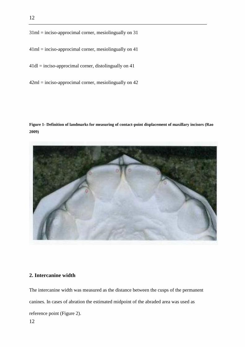
Break of contact is defined as the labio-lingual distance between two neighboring incisors`
inciso-approximal corners, measured between the most lingually positioned corners along the
occlusal plane (Rao 2009) (Figure 1).
Labeling of the landmarks (Rao 2009):
32ml = inciso-approximal corner, mesiolingually on 32
31dl = inciso-approcimal corner, distolingually on 31
12
12
31ml = inciso-approcimal corner, mesiolingually on 31
41ml = inciso-approcimal corner, mesiolingually on 41
41dl = inciso-approcimal corner, distolingually on 41
42ml = inciso-approcimal corner, mesiolingually on 42
Figure 1- Definition of landmarks for measuring of contact-point displacement of maxillary incisors (Rao
2009)
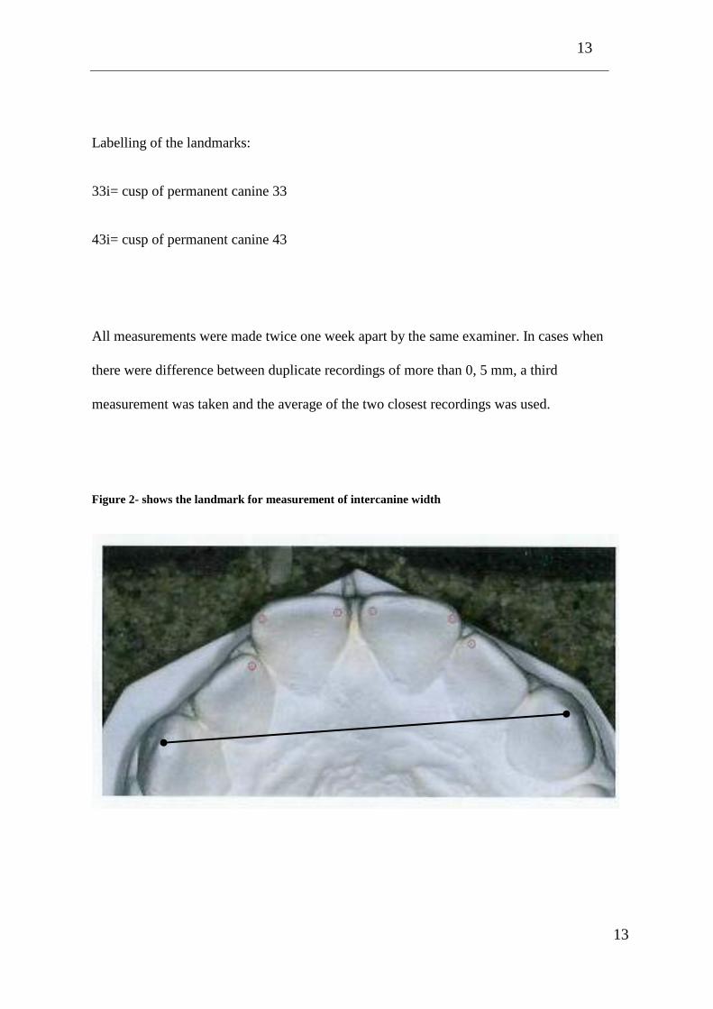
2. Intercanine width
The intercanine width was measured as the distance between the cusps of the permanent
canines. In cases of abration the estimated midpoint of the abraded area was used as
reference point (Figure 2).
13
13
Labelling of the landmarks:
33i= cusp of permanent canine 33
43i= cusp of permanent canine 43
All measurements were made twice one week apart by the same examiner. In cases when
there were difference between duplicate recordings of more than 0, 5 mm, a third
measurement was taken and the average of the two closest recordings was used.
Figure 2- shows the landmark for measurement of intercanine width
14
14
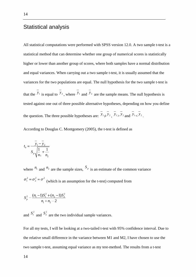
Statistical analysis
All statistical computations were performed with SPSS version 12.0. A two sample t-test is a
statistical method that can determine whether one group of numerical scores is statistically
higher or lower than another group of scores, where both samples have a normal distribution
and equal variances. When carrying out a two sample t-test, it is usually assumed that the
variances for the two populations are equal. The null hypothesis for the two sample t-test is
that the 1y
is equal to 2y
, where 1y
and 2y
are the sample means. The null hypothesis is
tested against one out of three possible alternative hypotheses, depending on how you define
the question. The three possible hypotheses are: 1y
≠ 2y
, 1y
> 2y
and 1y
< 2y
.
According to Douglas C. Montgomery (2005), the t-test is defined as
1 20
1 2
1 1p
y yt
Sn n
where 1n and 2n
are the sample sizes, pS is an estimate of the common variance
2 2 2
1 2 (which is an assumption for the t-test) computed from
2 22 1 1 2 2
1 2
( 1) ( 1)
2p
n S n SS
n n
and 2
1S and
2
2S are the two individual sample variances.
For all my tests, I will be looking at a two-tailed t-test with 95% confidence interval. Due to
the relative small difference in the variance between M1 and M2, I have chosen to use the
two sample t-test, assuming equal variance as my test-method. The results from a t-test

15
15
assuming unequal variance gives approximately the same results. (Refeance Montgomery,
Douglas C. 2005)
The following significance test were used:
Analyzing the difference between M1 and M2
Analyzing the differences between T1 and T2.
16
16
RESULTS AND DISCUSSION
In this chapter the results of the statistical analysis will be presented and seen in relation to
previous studies.
Error of the method
The reproducibility of the measurements was assessed by statistically analyzing the
difference between double measurements taken one week apart. In cases when there were
difference between duplicate recordings of more than 0, 5 mm, a third measurement was
taken and the average of the two closest recordings was used.
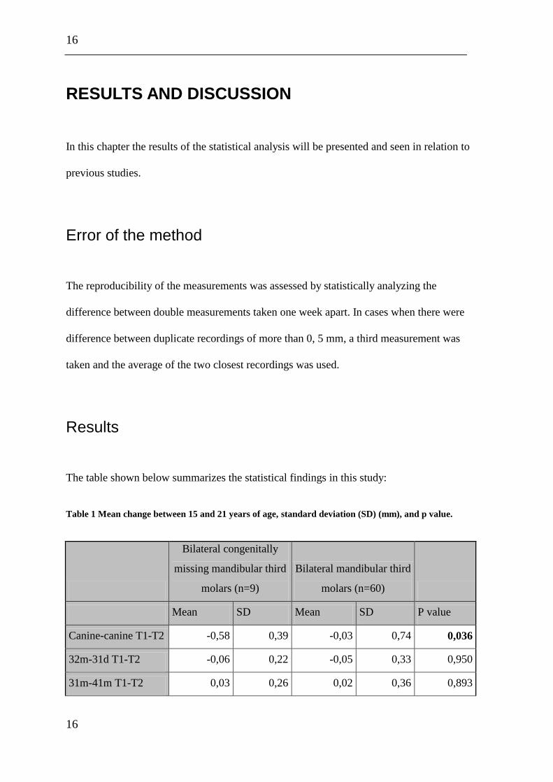
Results
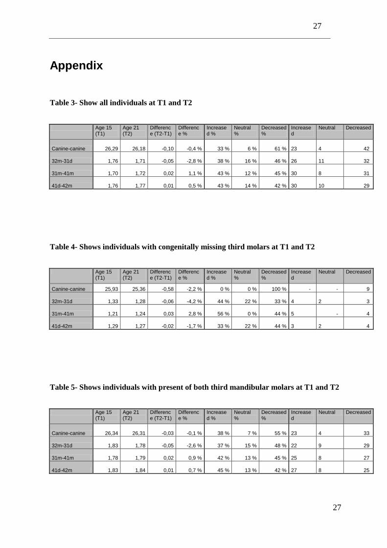
The table shown below summarizes the statistical findings in this study:
Table 1 Mean change between 15 and 21 years of age, standard deviation (SD) (mm), and p value.
Bilateral congenitally
missing mandibular third
molars (n=9)
Bilateral mandibular third
molars (n=60)
Mean SD Mean SD P value
Canine-canine T1-T2 -0,58 0,39 -0,03 0,74 0,036
32m-31d T1-T2 -0,06 0,22 -0,05 0,33 0,950
31m-41m T1-T2 0,03 0,26 0,02 0,36 0,893
17
17
41d-42m T1-T2 -0,02 0,20 0,01 0,30 0,733
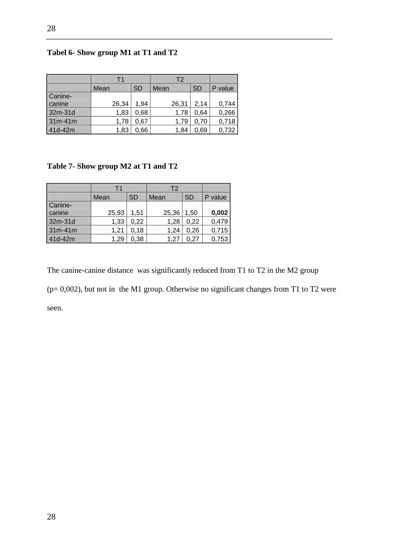
Intercanine width change (T1-T2):
Mean intercanine width was significantly more reduced in the M2 group than in the M1
group (p=0.036). As can be seen from the table above, the mean value of this change in
width was -0,58 mm in the M2 group, and only -0,03 mm in the M1 group.
Changes from T1 to T2 in the other three distances measured did not differ significantly
between the groups.
In addition, we analysed group M1 and M2 at T1 and T2 to examine if there were significant
values that support our findings in Table1. These Tables are presented in the Appendix
( page 27 and 28).
Discussion
This study included 69 individuals with plaster models and panoramic radiographs at age 15
and 21 years. All individuals were selected from the Oslo Craniofacial Growth Archive. In
60 (87 %) individuals bilateral lower third molars (M1), were present compared to 9 (13 %)
who had congenitally missing mandibular third molars (M2).
Sinclair and Little studied a group of individuals from 13 to 20 years who had not undergone
orthodontic treatment and they observed a decrease in arch length and intercanine width, and
that lower incisor irregularity increased during young adulthood (Sinclair and Little 1983). In
another study, Årtun also demonstrated an increase of incisor irregularity and reduction of
18
18
intercanine width and arch length long-term following orthodontic treatment and retention.
The degree of relapse was associated with narrow pretreatment intercanine width and incisor
irregularity (Årtun et al. 1996).
The influence of third molars on the alignment of the anterior dentition is controversial
(Kaplan 1974, Lindqvist 1982, Tüfekçi et al. 2009). In a study Richardson found that anterior
crowding is present more often in patients with third molars than in subjects with these teeth
absent (Richardson 1989). This is supported with the findings in Lindqvist’s (1982) study.
She claimed that in cases with severe crowding removal of the third molars could be
recommended (Lindqvist 1982). Sidlauskas and Trakiniene also confirm these theories in
their study where they found greater tendency for crowding of the mandibular anterior teeth
expressed in groups with third molars present than in groups with these teeth missing
(Sidlauskas and Trakiniene 2006).
To the contrary Ades reported in her study no differences in dental arch length and
irrespective crowding of presence or absence of third molars in orthodontic patients 10 years
post-retention. Therefore, it was concluded that removal of third molars to alleviate anterior
crowding may not be justified (Ades 1990). Bishara also concludet in his study that third
molars do not play a signicicant role in mandibular anterior crowding (Bishara 1999)
Congenitally missing third molars is frequently observed, and although the frequency ranges
widely, varying from 0 % among an unspecified sample of skulls from Tasmania to 49 % in
an unspecified sample of Hungarian skulls. Other radiographic studies of Caucasian
populations observe prevalences between 7% and 26% (Banks 1934, Hellman 1936). In our
study we found 9 individuals with congenitally missing third molars, compared to 60
individuals with presence of both mandibular third molars. The sample in group M2 may be
19
19
considered small as it represents only 13 % of the whole study sample. However, if we did
not exclude individuals with presence of congenitally missing third molar, which did not
fulfil out criteria for inclusion, the rate would be higher.
In the present study we found no significant differences in anterior alignment and
irrespective of presence mandibular third molars or congenitally missing third molars.
However, the findings indicate that there is a significantly higher risk for intercanine width
change from T1-T2 among patients in group M2 compared to group M1.
Earlier studies have shown a small but significant reduction in the lower intercanine width
(Sillman 1964, Knott 1972, Blake and Bibby 1998). These findings are supported by Sinclair
and Little (1983) who found significant change (mean 0,75mm reduction) in the intercanine
distance especially in women, between 13 to 20 years of age. Bishara et al. did also observe a
reduction of 0, 4 and 0, 6 mm in the lower intercanine width, but only after 25 years (Bishara
et al. 1996).
We cannot exlude the possibility that some of the results that were non-significant in our
statistical analysis would have been significant if a more advanced statistical ananysis had
been performed.
Table 2- Overview of some studies that have examined changes in anterior alignment
STUDY NO
PATIENTS AGE TREATMENT INDEX
REMARKS
(CONCLUSION))
Ade
1990 97
This finding suggests
that the
recommendation
for mandibular third
molar removal with
the objective of
alleviating or
20
20
preventing long-term
mandibular incisor
irregularity may not be
justified.
Aasen,
Espeland
2005
56
Stripping
0,5-5mm
(gj.snitt 1.9)
Little`s
index
Lomg term changes in
lower incisor, 3 years
after debonding
Keene
1964 195 17-25
Agnesi of thirs molars
give more frequently
space in both arches
and less frequently
crowding. Following
also smaller
mesiodistal crown
diameter of the lower
right first molar.
Margrethe
Richardsson
1983
10, 4male and
6 female.
Ex. 3rd
molar
unilateral
Ex. Of a lower second
molar can reduce the
possibility or severity
of late lower arch
crowding.
Margrethe
Richardsson
1986
48 Ex. 1.premolars
A greater increase in
molar space occurs in
mandibular first
premolar extraction
compared with non-
extraction cases.
Ross g.
Kaplan
1971
75
The presence of third
molars does not
appear to produce a
greater degree of
lower anterior
crowding and
rotational relapse after
the cessation of
retention than that
which occurs in
patients with third
molar agnesis.
Berit
Lindqvist
52
( 23 boys, 29
girls)
13-19
( gj.snitt
15 ½)
Ex. third molars
Ex. Second
molars
Extraction can be
recommended in
severe crowding.
Robert M.
Little
1988
31 Ex. of 4
premolar Little`s
Relapse after
orthodontic treatment
Robert M.
Little
2002
26 6-23
Without lifetime
retention, the strategy
of arch development
will yield
unacceptable results.
(The degree of
relapse are significant
and alarming)
21
21
CONCLUSIONS
Our study suggests that mandibular third molar is not a contributing factor to crowding in
mandibular alignment. A greater reduction in intercanine width occurred when mandibular
third molars were missing when compared to cases where third molars were present.
22
22
REFERENCES
1. Aasen T., Espeland L. An approach to maintain orthodontic alignment of lower
incisors without the use of retainers. European Journal of Orthodontics 27: 209-214,
2005
2. Ades AG., Joondeph DR., Little RM., Chapko MK. A long-term study of the
relationship of third molars to changes in the mandibular dental arch. American
Journal of Orthodontics and Dentofacial Orthopedics. 97: 323-335, 1990
3. Banks HV. Incidence of third molar development. Angle Orthodontist. 4: 223–233,
1934
4. Bishara SE. Third molars: a dilemma! Or is it? American Journal of Orthodontics and
Dentofacial Orthopedics. Stomatologija. 115: 628–633, 1999
5. Bishara SE., Jakobsen JR. Arch width changes from 6 weeks to 45 years of age.
American Journal of Orthodontics and Dentofacial Orthopedics. 111: 401-409, 1997
6. Bishara SE., Peterson LC. Changes in facial dimensions and relationships between
the ages of 5 and 25 years. American Journal of Orthodontics and Dentofacial
Orthopedics. 85: 238-52, 1984
7. Bishara SE., Treder JE. , Damon P. and Olsen M. Changes in the dental arches
dentition between 25 and 45 years of age. Angle Orthodontist. 66: 417–422, 1996
8. Björk, A. The face in profile: An anthropological x-ray investigation on Swedish
children and conscripts. American Journal of Orthodontics. 34: 691-699, 1948
23
23
9. Blake M., Bibby K. Retention and stability: A review of the literature. American
Journal of Orthodontics and Dentofacial Orthopedics. 114: 299-306, 1998
10. Bolton A. Disharmony in tooth size and its relation to the analysis and treatment of
malocclusion. Angle Orthodontist. 28: 113-130, 1958
11. Bondevik O. Changes in occlusion between 23-34 years. Angle Orthodontist. 68: 75-
80, 1998
12. Burleigh T., Surbeck BT., Årtun J. Associations between initial, posttreatment, and
postretention alignment of maxillary anterior teeth. American Journal of Orthodontics
and Dentofacial Orthopedics. 113: 186-195, 1998
13. Crosby DR., Alexander CG. The occurrence of tooth size discrepancies among
different malocclusion groups. American Journal of Orthodontics and Dentofacial
Orthopedics. 95: 457-461, 1989
14. Dahlberg G. Statistical method for medical and biological students. London: George
Allen and Unwin Ltd. 122-132, 1940
15. De La Cruz A., Sampson P., Little RM., Årtun J., Shapiro PA. Long-term changes in
arch form after orthodontic treatment. American Journal of Orthodontics and
Dentofacial Orthopedics. 107: 518-530, 1995
16. Hellman M. Our third molar teeth, their eruption, presence and absence. Dental
Cosmos. 78:750-62, 1936
17. Kaplan RG. Mandibular third molars and postretention crowding. American Journal
of Orthodontics and Dentofacial Orthopedics. 66: 411-430, 1974
24
24
18. Knott VB. Longitudinal study of dental arch widths at four stages of dentition. Angle
Orthodontist. 42: 387–394, 1972
19. Lindqvist B., Thilander B. Extraction of third molars in cases of anticipated crowding
in the lower jaw. American Journal of Orthodontics and Dentofacial Orthopedics. 81:
130-139, 1982
20. Little RM. Stability and relapse: Early treatment of arch length deficiency. American
Journal of Orthodontics and Dentofacial Orthopedics. 121: 578-81, 2002
21. Little RM. The irregularity index: A quantitative score of mandibular anterior
alignment. American Journal of Orthodontics. 68: 554-563, 1975
22. Montgomery, Douglas C. Design and Analysis of Experiments. Hoboken, NJ: John
Wiley & Sons, Inc. 6 edition, 2005
23. Nanda RS. Agenesis of the third molar in man. American Journal of Orthodontics.
40: 698-706, 1954
24. Niedzielska I. Third molar influence on dental arch crowding. European Journal of
Orthodontics. 27: 518–523, 2005
25. Norderval K., Wisth PJ., Böe OE. Mandibular anterior crowding in relation to tooth
size and craniofacial morphology. Scandinavian Journal of Dental Research. 83: 267-
273, 1975
26. Pham L., Fang W. Change in inclination and eruption of mandibular third molars: a
longitudinal radiographic study among 12 to 21-year-olds. Thesis, Master of
Dentistry, University of Oslo 2006
25
25
27. Quinn GW. Extraction of four second molars. Angle Orthodontist. 55: 58-69, 1985
28. Rao C. Factors influencing the stability of maxillary incisors after orthodontic
treatment. Thesis, University of Oslo 2009
29. Richardson ME. Lower arch crowding in the third decade. European Journal of
Orthodontics. 20: 597-607, 1998
30. Richardson ME. The effect of lower second molar extraction on late lower arch
crowding. Angle Orthodontist. 53: 25-28, 1983
31. Richardson ME. The effect of mandibular first premolar extraction on third molar
space. Angle Orthodontist. 59: 291-294, 1989
32. Richardson ME. The role of the third molar in the cause of late lower arch crowding:
a review. American Journal Orthodontics and Dentofacial Orthodpedics. 95: 79-83,
1989
33. Schütz-Fransson U., Bjerklin K., Kurol J. Long-term development in the mandible
and incisor crowding with and without an orthodontic stabilizing appliance. Journal
of Orofacial Orthopedics 59: 63-72, 1998
34. Siatkowski RE. Incisor uprighting: Mechanism for late secondary crowding in the
anterior segments of the dental arches. American Journal of Orthodontics. 66: 398-
410, 1974
35. Sidlauskas A, Trakiniene G. Effect of the lower third molars on the lower dental arch
crowding. Stomatologija. 8: 80-84, 2006
36. Sillman JH. Dimensional changes of the dental arches: Longitudinal study from birth
to 25 years. American Journal of Orthodontics. 50: 824-842, 1964
26
26
37. Sinclair PM., Little RM. Maturation of untreated normal occlusions. American
Journal of Orthodontics and Dentofacial Orthopedics. 83: 114-123, 1983
38. Tüfekçi E. ̧Kallunki J., Huggare J. Opinions of American and Swedish Orthodontists
about the Role of Erupting Third Molars as a Cause of Dental Crowding. Angle
Orthodontist. 79: 1139-1142, 2009
39. Årtun J., Garol JD., Little RM. Long-term stability of mandibular incisors following
successful treatment of Class II, Division 1, malocclusions. Angle Orthodontist. 66:
229-238, 1996
27
27
Appendix
Table 3- Show all individuals at T1 and T2
Age 15 (T1)
Age 21 (T2)
Difference (T2-T1)
Difference %
Increased %
Neutral %
Decreased %
Increased
Neutral Decreased
Canine-canine 26,29 26,18 -0,10 -0,4 % 33 % 6 % 61 % 23
4 42
32m-31d 1,76 1,71 -0,05 -2,8 % 38 % 16 % 46 % 26
11 32
31m-41m 1,70 1,72 0,02 1,1 % 43 % 12 % 45 % 30
8 31
41d-42m 1,76 1,77 0,01 0,5 % 43 % 14 % 42 % 30
10 29
Table 4- Shows individuals with congenitally missing third molars at T1 and T2
Age 15 (T1)
Age 21 (T2)
Difference (T2-T1)
Difference %
Increased %
Neutral %
Decreased %
Increased
Neutral Decreased
Canine-canine 25,93 25,36 -0,58 -2,2 % 0 % 0 % 100 % - - 9
32m-31d 1,33 1,28 -0,06 -4,2 % 44 % 22 % 33 % 4
2 3
31m-41m 1,21 1,24 0,03 2,8 % 56 % 0 % 44 % 5 - 4
41d-42m 1,29 1,27 -0,02 -1,7 % 33 % 22 % 44 % 3
2 4
Table 5- Shows individuals with present of both third mandibular molars at T1 and T2
Age 15 (T1)
Age 21 (T2)
Difference (T2-T1)
Difference %
Increased %
Neutral %
Decreased %
Increased
Neutral Decreased
Canine-canine 26,34 26,31 -0,03 -0,1 % 38 % 7 % 55 % 23
4 33
32m-31d 1,83 1,78 -0,05 -2,6 % 37 % 15 % 48 % 22
9 29
31m-41m 1,78 1,79 0,02 0,9 % 42 % 13 % 45 % 25
8 27
41d-42m 1,83 1,84 0,01 0,7 % 45 % 13 % 42 % 27
8 25
28
28
Tabel 6- Show group M1 at T1 and T2
T1 T2
Mean SD Mean SD P value
Canine-canine 26,34 1,94 26,31 2,14 0,744
32m-31d 1,83 0,68 1,78 0,64 0,266
31m-41m 1,78 0,67 1,79 0,70 0,718
41d-42m 1,83 0,66 1,84 0,69 0,732
Table 7- Show group M2 at T1 and T2
T1 T2
Mean SD Mean SD P value
Canine-canine 25,93 1,51 25,36 1,50 0,002
32m-31d 1,33 0,22 1,28 0,22 0,479
31m-41m 1,21 0,18 1,24 0,26 0,715
41d-42m 1,29 0,38 1,27 0,27 0,753
The canine-canine distance was significantly reduced from T1 to T2 in the M2 group
(p= 0,002), but not in the M1 group. Otherwise no significant changes from T1 to T2 were
seen.
29
29
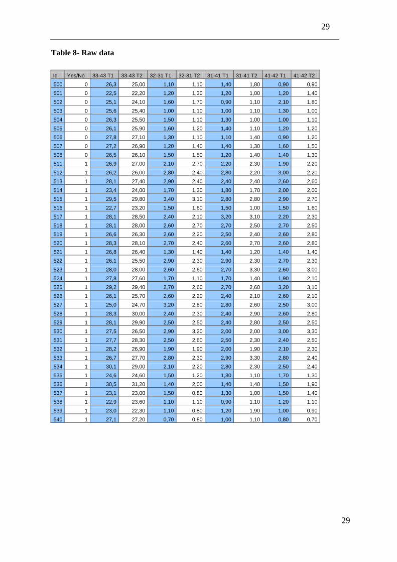
Table 8- Raw data
Id Yes/No 33-43 T1 33-43 T2 32-31 T1 32-31 T2 31-41 T1 31-41 T2 41-42 T1 41-42 T2
500 0 26,3 25,00 1,10 1,10 1,40 1,80 0,90 0,90
501 0 22,5 22,20 1,20 1,30 1,20 1,00 1,20 1,40
502 0 25,1 24,10 1,60 1,70 0,90 1,10 2,10 1,80
503 0 25,6 25,40 1,00 1,10 1,00 1,10 1,30 1,00
504 0 26,3 25,50 1,50 1,10 1,30 1,00 1,00 1,10
505 0 26,1 25,90 1,60 1,20 1,40 1,10 1,20 1,20
506 0 27,8 27,10 1,30 1,10 1,10 1,40 0,90 1,20
507 0 27,2 26,90 1,20 1,40 1,40 1,30 1,60 1,50
508 0 26,5 26,10 1,50 1,50 1,20 1,40 1,40 1,30
511 1 26,9 27,00 2,10 2,70 2,20 2,30 1,90 2,20
512 1 26,2 26,00 2,80 2,40 2,80 2,20 3,00 2,20
513 1 28,1 27,40 2,90 2,40 2,40 2,40 2,60 2,60
514 1 23,4 24,00 1,70 1,30 1,80 1,70 2,00 2,00
515 1 29,5 29,80 3,40 3,10 2,80 2,80 2,90 2,70
516 1 22,7 23,20 1,50 1,60 1,50 1,00 1,50 1,60
517 1 28,1 28,50 2,40 2,10 3,20 3,10 2,20 2,30
518 1 28,1 28,00 2,60 2,70 2,70 2,50 2,70 2,50
519 1 26,6 26,30 2,60 2,20 2,50 2,40 2,60 2,80
520 1 28,3 28,10 2,70 2,40 2,60 2,70 2,60 2,80
521 1 26,8 26,40 1,30 1,40 1,40 1,20 1,40 1,40
522 1 26,1 25,50 2,90 2,30 2,90 2,30 2,70 2,30
523 1 28,0 28,00 2,60 2,60 2,70 3,30 2,60 3,00
524 1 27,8 27,60 1,70 1,10 1,70 1,40 1,90 2,10
525 1 29,2 29,40 2,70 2,60 2,70 2,60 3,20 3,10
526 1 26,1 25,70 2,60 2,20 2,40 2,10 2,60 2,10
527 1 25,0 24,70 3,20 2,80 2,80 2,60 2,50 3,00
528 1 28,3 30,00 2,40 2,30 2,40 2,90 2,60 2,80
529 1 28,1 29,90 2,50 2,50 2,40 2,80 2,50 2,50
530 1 27,5 26,50 2,90 3,20 2,00 2,00 3,00 3,30
531 1 27,7 28,30 2,50 2,60 2,50 2,30 2,40 2,50
532 1 28,2 26,90 1,90 1,90 2,00 1,90 2,10 2,30
533 1 26,7 27,70 2,80 2,30 2,90 3,30 2,80 2,40
534 1 30,1 29,00 2,10 2,20 2,80 2,30 2,50 2,40
535 1 24,6 24,60 1,50 1,20 1,30 1,10 1,70 1,30
536 1 30,5 31,20 1,40 2,00 1,40 1,40 1,50 1,90
537 1 23,1 23,00 1,50 0,80 1,30 1,00 1,50 1,40
538 1 22,9 23,60 1,10 1,10 0,90 1,10 1,20 1,10
539 1 23,0 22,30 1,10 0,80 1,20 1,90 1,00 0,90
540 1 27,1 27,20 0,70 0,80 1,00 1,10 0,80 0,70
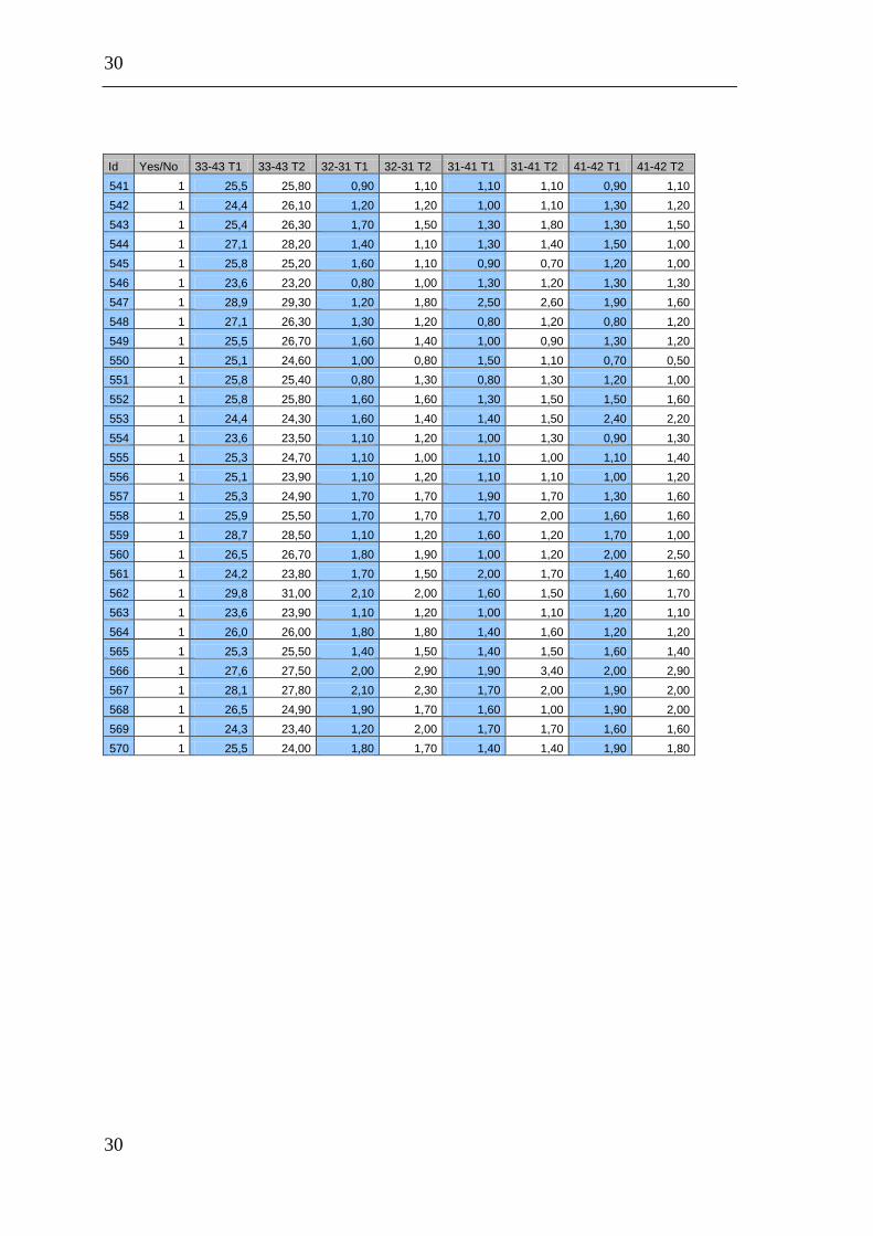
30
30
Id Yes/No 33-43 T1 33-43 T2 32-31 T1 32-31 T2 31-41 T1 31-41 T2 41-42 T1 41-42 T2
541 1 25,5 25,80 0,90 1,10 1,10 1,10 0,90 1,10
542 1 24,4 26,10 1,20 1,20 1,00 1,10 1,30 1,20
543 1 25,4 26,30 1,70 1,50 1,30 1,80 1,30 1,50
544 1 27,1 28,20 1,40 1,10 1,30 1,40 1,50 1,00
545 1 25,8 25,20 1,60 1,10 0,90 0,70 1,20 1,00
546 1 23,6 23,20 0,80 1,00 1,30 1,20 1,30 1,30
547 1 28,9 29,30 1,20 1,80 2,50 2,60 1,90 1,60
548 1 27,1 26,30 1,30 1,20 0,80 1,20 0,80 1,20
549 1 25,5 26,70 1,60 1,40 1,00 0,90 1,30 1,20
550 1 25,1 24,60 1,00 0,80 1,50 1,10 0,70 0,50
551 1 25,8 25,40 0,80 1,30 0,80 1,30 1,20 1,00
552 1 25,8 25,80 1,60 1,60 1,30 1,50 1,50 1,60
553 1 24,4 24,30 1,60 1,40 1,40 1,50 2,40 2,20
554 1 23,6 23,50 1,10 1,20 1,00 1,30 0,90 1,30
555 1 25,3 24,70 1,10 1,00 1,10 1,00 1,10 1,40
556 1 25,1 23,90 1,10 1,20 1,10 1,10 1,00 1,20
557 1 25,3 24,90 1,70 1,70 1,90 1,70 1,30 1,60
558 1 25,9 25,50 1,70 1,70 1,70 2,00 1,60 1,60
559 1 28,7 28,50 1,10 1,20 1,60 1,20 1,70 1,00
560 1 26,5 26,70 1,80 1,90 1,00 1,20 2,00 2,50
561 1 24,2 23,80 1,70 1,50 2,00 1,70 1,40 1,60
562 1 29,8 31,00 2,10 2,00 1,60 1,50 1,60 1,70
563 1 23,6 23,90 1,10 1,20 1,00 1,10 1,20 1,10
564 1 26,0 26,00 1,80 1,80 1,40 1,60 1,20 1,20
565 1 25,3 25,50 1,40 1,50 1,40 1,50 1,60 1,40
566 1 27,6 27,50 2,00 2,90 1,90 3,40 2,00 2,90
567 1 28,1 27,80 2,10 2,30 1,70 2,00 1,90 2,00
568 1 26,5 24,90 1,90 1,70 1,60 1,00 1,90 2,00
569 1 24,3 23,40 1,20 2,00 1,70 1,70 1,60 1,60
570 1 25,5 24,00 1,80 1,70 1,40 1,40 1,90 1,80

![LONGITUDINAL CHANGES OF DENTAL ARCHES IN GROWING … · eruption meaning that a small amount of crowding in the mandibular arch at this time is normal [11]. The growth process continues](https://img.pdfslide.net/doc/110x75/5e984c43943b7133f670e1a4/longitudinal-changes-of-dental-arches-in-growing-eruption-meaning-that-a-small-amount.jpg)