Embed Size (px)
Citation preview
International Journal of Engineering Research & Science (IJOER) ISSN: [2395-6992] [Vol-5, Issue-7, July- 2019]
Page | 7
Manual Correction of Myopia, Hypermetropia, Astigmatism,
Presbyopia & Keratoconus. 1Prof. Bijay Kumar Parida, MS, FRCS
2Ms. Anannya Anupurva
1Muthusamy Virtual University of Post Graduate Ophthalmology
2MVJ Medical College.
Abstract— A new method has been designed to correct myopia, hypermetropia, astigmatism and presbyopia of very high
power. If 1mm of
cornea is tucked, then it corrects 3.174 D & If 1mm of horizontal diameter of cornea is reduced, then it
corrects +4.1D. This procedure is reliable, safe, cheapest and simplest. Because of the use of simple procedure to split the
cornea, adjustable nonabsorbable sutures, correction of very high myopia and hypermetropia along with astigmatism and
presbyopia, the procedure is expected to be accepted.
Keywords— Arc of cornea, chord, angle of arc, myopia, hypermetropia, astigmatism, formula.
I. INTRODUCTION
LASIK (laser-assisted in situ keratomileusis) is a type of refractive surgery for the correction of myopia, hypermetropia and
astigmatism. LASIK is most similar to another surgical corrective procedure, photorefractive keratectomy (PRK), and
LASEK. All represent advances over radial keratectomy in the surgical treatment of refractive errors of vision. For patients
with moderate to high myopia or thin corneas which cannot be treated with LASIK and PRK, the phakic intraocular lens is
an alternative.1,2
Some people with poor outcomes from LASIK surgical procedures report a significantly reduced quality of life because of
vision problems or physical pain associated with surgery. However, 30% of post-LASIK referrals to tertiary ophthalmology
care centers have been shown to be due to chronic dry eye.3,4
.
A type of LASIK, known as presbyLASIK, may be used in presbyopia.5
Flap complications,6,7,8,9
slipped flap and flap interface particles are other complications.
Together with his colleagues, Charles Munnerlyn founded VISX USA inc.
SMILE is an acronym that stands for “Small Incision Lenticule Extraction” and is the latest advancement in laser vision
correction. During a SMILE procedure, the femtosecond laser is used to cut a small corneal lenticule (precise lens shaped
disc within cornea) and then it is removed via a small keyhole incision. The biggest difference and advancement between the
two is the fact that SMILE laser eye surgery is a flapless procedure.
II. PRINCIPLE
If the curvature of cornea is responsible for myopia or hypermetropia, it can either be flattened or steepened to change the
power of cornea. For example, if the cornea is made flatter, then the myopia is reduced. Whereas, if the cornea is made
steeper, then the power of the cornea is increased, refractive index of the cornea and other factors responsible for ocular
refractive power remaining unchanged. Assuming the refractive power of anterior surface of cornea is +49D, after deducting
-6D power of posterior surface of cornea, the net power of cornea is +43D.
International Journal of Engineering Research & Science (IJOER) ISSN: [2395-6992] [Vol-5, Issue-7, July- 2019]
Page | 8
FIGURE 1
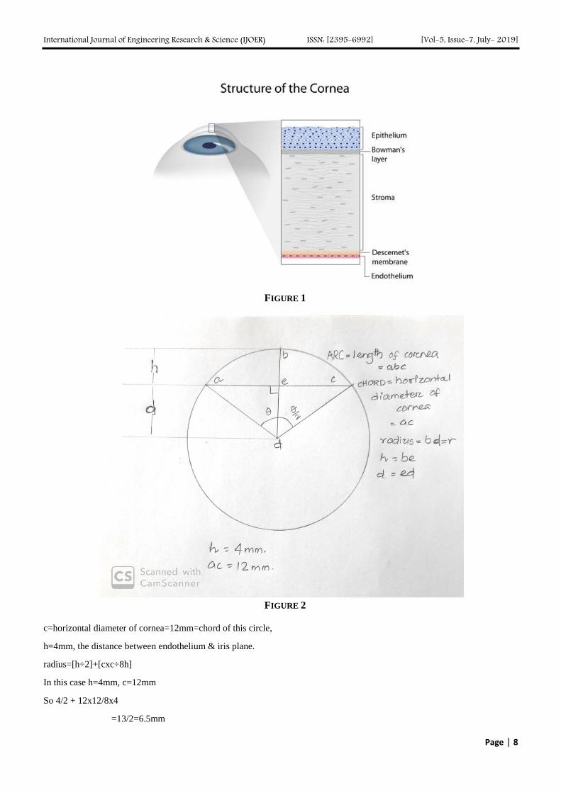
FIGURE 2
c=horizontal diameter of cornea=12mm=chord of this circle,
h=4mm, the distance between endothelium & iris plane.
radius=[h÷2]+[cxc÷8h]
In this case h=4mm, c=12mm
So 4/2 + 12x12/8x4
=13/2=6.5mm
International Journal of Engineering Research & Science (IJOER) ISSN: [2395-6992] [Vol-5, Issue-7, July- 2019]
Page | 9
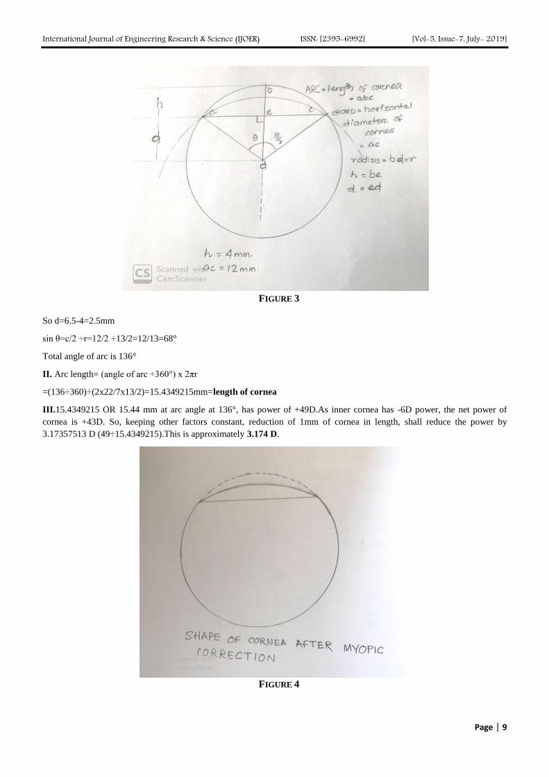
FIGURE 3
So d=6.5-4=2.5mm
sin θ=c/2 ÷r=12/2 ÷13/2=12/13=68°
Total angle of arc is 136°
II. Arc length= (angle of arc ÷360°) x 2πr
=(136÷360)÷(2x22/7x13/2)=15.4349215mm=length of cornea
III.15.4349215 OR 15.44 mm at arc angle at 136°, has power of +49D.As inner cornea has -6D power, the net power of
cornea is +43D. So, keeping other factors constant, reduction of 1mm of cornea in length, shall reduce the power by
3.17357513 D (49÷15.4349215).This is approximately 3.174 D.
FIGURE 4

International Journal of Engineering Research & Science (IJOER) ISSN: [2395-6992] [Vol-5, Issue-7, July- 2019]
Page | 10
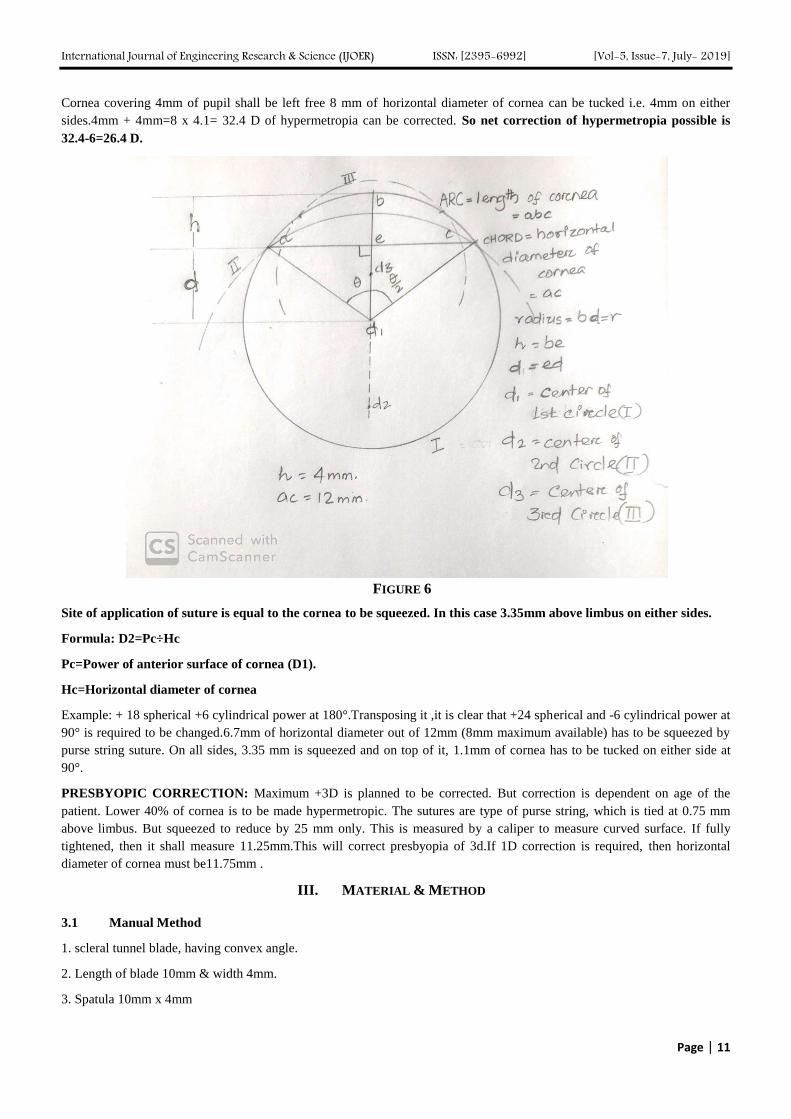
If 1mm of cornea is tucked on either sides that will flatten the cornea and correct 2 x 3.174 =6.348D of myopia. If 0.5 mm of
cornea is tucked, then correction of 3.174 ÷ 2=1.587D of myopia.
2mm of cornea on either side (4mm) is tucked; 11 mm of cornea is left. If 3mm on either sides of cornea is tucked(=6mm in
total), then 9mm is left to be flattened. If 4 mm on either sides is tucked (8mm), then 7 mm is left. If 5mm on either sides is
tucked (10mm), then 5mm of clear cornea is left. Central cornea of 4 mm is essential to be clear.
Thus (10 x 3.174=) -31.74D can be corrected, which is equal to (31.74-6=) 25.74D.
FORMULA; D2=[(Pc÷Lc)x(Lt)]-6
Pc=power of anterior surface of cornea.
Lc =length of cornea=(θ÷360°)x2πr
Lt=length of cornea tucked.
Sin θ = perpendicular÷hypotenuse=p/h
Example: myopia of -20 D spherical & -6 cylindrical error at 90 °.
*7.179000858=7.18mm of cornea has to be replicated. On either sides 3.59mm to be tucked.
*to correct -6 cylindrical, 2.15370257 mm (=2.154mm) has to be plicated.
So 1.07685129 (= 1.1mm) on either sides of cornea at 180° to be tucked more at 180°.So at 180°,3.6+1.1=4.7mm has to be
tucked.
Except 1mm of peripheral cornea at periphery and 4 mm at center, rest of the cornea is available for the procedure.
VII. Similarly, 12mm of horizontal diameter of cornea, other conditions remaining constant is associated with +49D. So
1mm of horizontal diameter is associated with +4.08333333(=+4.1D)
FIGURE 5
International Journal of Engineering Research & Science (IJOER) ISSN: [2395-6992] [Vol-5, Issue-7, July- 2019]
Page | 11
Cornea covering 4mm of pupil shall be left free 8 mm of horizontal diameter of cornea can be tucked i.e. 4mm on either
sides.4mm + 4mm=8 x 4.1= 32.4 D of hypermetropia can be corrected. So net correction of hypermetropia possible is
32.4-6=26.4 D.
FIGURE 6
Site of application of suture is equal to the cornea to be squeezed. In this case 3.35mm above limbus on either sides.
Formula: D2=Pc÷Hc
Pc=Power of anterior surface of cornea (D1).
Hc=Horizontal diameter of cornea
Example: + 18 spherical +6 cylindrical power at 180°.Transposing it ,it is clear that +24 spherical and -6 cylindrical power at
90° is required to be changed.6.7mm of horizontal diameter out of 12mm (8mm maximum available) has to be squeezed by
purse string suture. On all sides, 3.35 mm is squeezed and on top of it, 1.1mm of cornea has to be tucked on either side at
90°.
PRESBYOPIC CORRECTION: Maximum +3D is planned to be corrected. But correction is dependent on age of the
patient. Lower 40% of cornea is to be made hypermetropic. The sutures are type of purse string, which is tied at 0.75 mm
above limbus. But squeezed to reduce by 25 mm only. This is measured by a caliper to measure curved surface. If fully
tightened, then it shall measure 11.25mm.This will correct presbyopia of 3d.If 1D correction is required, then horizontal
diameter of cornea must be11.75mm .
III. MATERIAL & METHOD
3.1 Manual Method
1. scleral tunnel blade, having convex angle.
2. Length of blade 10mm & width 4mm.
3. Spatula 10mm x 4mm

International Journal of Engineering Research & Science (IJOER) ISSN: [2395-6992] [Vol-5, Issue-7, July- 2019]
Page | 12
4. 2 types of caliper specially designed of measurement of curvature.
5. Fine tip marker pen
6. Gentian violet
7. 8-0 prolene sutures
8. Fine tooth forceps
9. Needle holder
10. Tie forceps
3.2 Procedure:
FIGURE 7
Myopia; firstly, 1mm away from limbus ,a 4mm wide incisions are made to a depth of 0.15mm.On this plane, a scleral
tunnel blade can split cornea stroma into 2 parts,and the splitting is extended upto centre of cornea. Similarly, the cornea is

International Journal of Engineering Research & Science (IJOER) ISSN: [2395-6992] [Vol-5, Issue-7, July- 2019]
Page | 13
split from diagonally opposite site(if 1st one at 0° axis, then the 2nd one on 180° axis.) similarly, incision and splitting is
done form 90° and 270°.The splitting of the cornea is completed in 360° to free the epithelial layer from stroma.
Method to plicate part of cornea is by running a sure through cornea at 3/4th depth & come out at desired end of the length.
Then similarly run it from opposite side and exit near the point of entry, preferably 4 mm wide, to take adequate bite and tuck
it as well as prevent cheese cutting.
FIGURE 8
FIGURE 9
International Journal of Engineering Research & Science (IJOER) ISSN: [2395-6992] [Vol-5, Issue-7, July- 2019]
Page | 14
Hypermetropia:
FIGURE 10
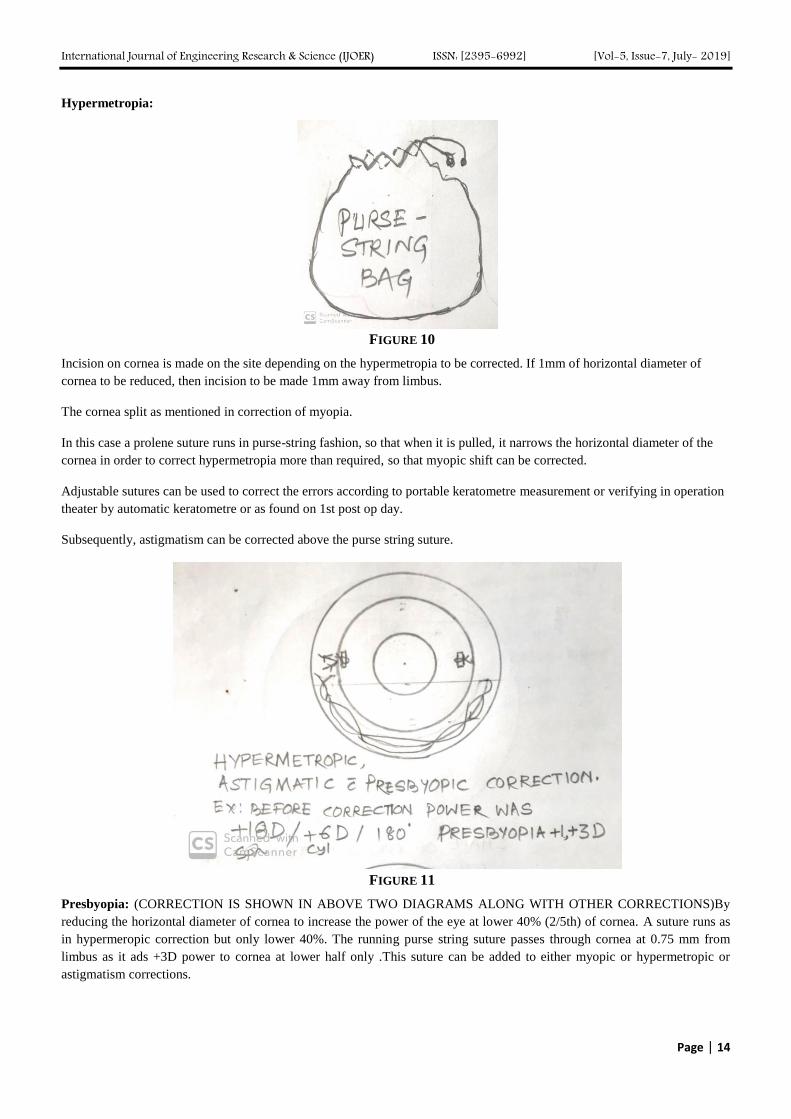
Incision on cornea is made on the site depending on the hypermetropia to be corrected. If 1mm of horizontal diameter of
cornea to be reduced, then incision to be made 1mm away from limbus.
The cornea split as mentioned in correction of myopia.
In this case a prolene suture runs in purse-string fashion, so that when it is pulled, it narrows the horizontal diameter of the
cornea in order to correct hypermetropia more than required, so that myopic shift can be corrected.
Adjustable sutures can be used to correct the errors according to portable keratometre measurement or verifying in operation
theater by automatic keratometre or as found on 1st post op day.
Subsequently, astigmatism can be corrected above the purse string suture.
FIGURE 11
Presbyopia: (CORRECTION IS SHOWN IN ABOVE TWO DIAGRAMS ALONG WITH OTHER CORRECTIONS)By
reducing the horizontal diameter of cornea to increase the power of the eye at lower 40% (2/5th) of cornea. A suture runs as
in hypermeropic correction but only lower 40%. The running purse string suture passes through cornea at 0.75 mm from
limbus as it ads +3D power to cornea at lower half only .This suture can be added to either myopic or hypermetropic or
astigmatism corrections.
International Journal of Engineering Research & Science (IJOER) ISSN: [2395-6992] [Vol-5, Issue-7, July- 2019]
Page | 15
IV. ADVANTAGES
1. It does not require femtosecond or excimer laser which is exuberantly costly.
2. It does not require raising IOP to 60mm Hg for microkeratome.
3. This delicate procedure shall be cheapest and can be used by an expert hand.
4. Very high myopia, hypermetropia, astigmatism and presbyopia can be corrected.
5. It does not require a corneal flap to be raised.
V. DISADVANTAGES
1. It is time consuming.
2. Learning curve much higher that LASIK or SMILE procedure.
VI. CONCLUSION
Manual correction of myopia, hypermetropia, astigmatism and presbyopia is simplest, cheapest and reliable. If 1 mm of
cornea is tucked, then it corrects 3.174 D & If 1 mm of horizontal diameter of cornea is reduced, then it corrects +4.1D.
Because of use of simple procedure to split the cornea, tie the sutures either to tuck it or use of purse string suture to squeeze
the horizontal length of cornea, correction of very high myopia, hyprmetropia, astigmatism and presbyopia, this procedure
has lots of promises in future.
REFERENCES
[1] Lovisolo,CF, Reinstein DZ; Reinstein(Nov-Dec 2005)”Phakic intraocular Lenses.”Survey of Ophthalmology.50(6):549-87.
[2] Sanders DR, Vukich JA; Vukich (May 200K),”Comparison of Implantable Contact Lens and Laser Assisted In Situ keratomileusis
for moderate to High Myopia.”Cornea 22(4):324-334.
[3] Le vinson,B.A;Rapuano,CJ;Cohen EJ:Hammer Smith K.M.;Ayres B.D; Laibson, PR(2008).”Referrals to the Wills Eye Institute
Cornea Service after laser in situ keratomileusis: reasons for patient dissatisfaction.”Journal of Cataract & Refractive Surgery 34(1).
[4] Jabbur,N.S;Saktani,K;O’Brien,T.P.(2004).”Survey of Complications and recommendations for management in dissatisfied patients
seeking consultation after refractive surgery.”Journal of Catarcat and Refractive surgery.30(9):1867-74.
[5] Carrillo C,Chayet AS,Dougherty PJ,Montes M,Magallanes R,Najman J,Fleitman J,Morales A(2005).”Incidence of complications
during flap creation in LASIK using the NIDEK MK-2000 microkeratome in 26,000 cases.” J Refract Surg.21(5 Suppli):S 655-7.
[6] Eye surgery Education Council.”Lasikinstitute.org.Archived from the Original on 2011-09-28.Rerived 2011-12-10.
[7] ”Eye Surgery Education Council.”Lasikinstitute.org.Archived from the Original on 2011-09-28.Retrieved 2011-12-10.
[8] Tham VM,Maloney RK(May 2000).”Microkeratome complications of laser in situ keratomileusis”Ophthalmology,107(5):920-4.
[9] Vesauloma M,Perez-Santoria J,Petroll WM,Linna T,Alio J,Terro T(1 February 2000).”Corneal Stroma changes included by myopic
LASIK.”Invest.Ohthalmol.Vis.Sci.41(2)369-76.


![Anannya [July-2011]](https://img.pdfslide.net/doc/110x75/577d275c1a28ab4e1ea3b91f/anannya-july-2011.jpg)