Embed Size (px)
Citation preview
1 Folio 1
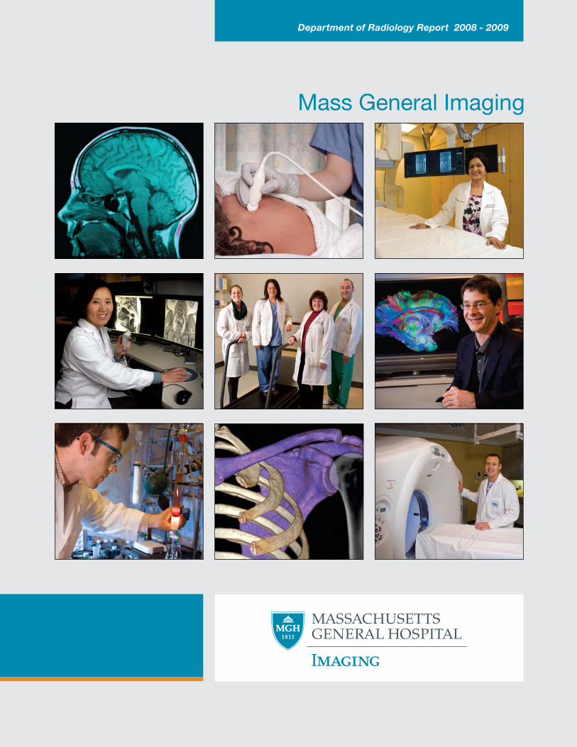
Mass General Imaging
Department of Radiology Report 2008 - 2009
Table of Contents Department Overview and Major Initiatives
1 Introduction from the Chairman 3 Quality and Safety 6 CT Dose Reduction 8 Strategic Initiatives 9 Clinical Facilities and Equipment 12 Community Radiology and Teleradiology 13 Customer Outreach and Marketing 14 3-D Imaging Service and Computer Aided Diagnostics Laboratory 15 Information Technology 18 Education
19 In Memoriam: Gordon Brownell, PhD, 1922 - 2008
Imaging from Primary Care to Complex Treatments
21 Abdominal Imaging and Intervention 24 Breast Imaging 26 Cardiovascular Imaging and Intervention 29 Emergency Radiology 31 Musculoskeletal Imaging & Intervention 33 Neuroradiology 36 Nuclear Medicine and Molecular Imaging 38 Pediatric Imaging 40 Thoracic Imaging
42 Staff at Work & New Cyclotron Installation
Research
45 Overview of Research Centers 48 Center for Translational Nuclear Medicine and Molecular Imaging 50 Athinoula A. Martinos Center for Biomedical Imaging Hybrid PET/MR Imaging Advances in MR Technology & Image-Guided Molecular Therapy 56 Center for Systems Biology: In Vivo Imaging of Immune Cell Activity Statistics
58 Impact of Radiology Order Entry and Decision Support on Outpatient CT Utilization 58 Research Funding Directory
59 Senior Administration & Directors
1
Message from the Chairman
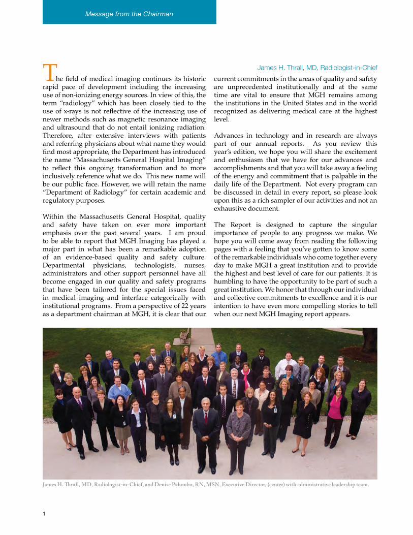
James H. Thrall, MD, Radiologist-in-ChiefThe field of medical imaging continues its historic rapid pace of development including the increasing use of non-ionizing energy sources. In view of this, the term “radiology” which has been closely tied to the use of x-rays is not reflective of the increasing use of newer methods such as magnetic resonance imaging and ultrasound that do not entail ionizing radiation. Therefore, after extensive interviews with patients and referring physicians about what name they would find most appropriate, the Department has introduced the name “Massachusetts General Hospital Imaging” to reflect this ongoing transformation and to more inclusively reference what we do. This new name will be our public face. However, we will retain the name “Department of Radiology” for certain academic and regulatory purposes.
Within the Massachusetts General Hospital, quality and safety have taken on ever more important emphasis over the past several years. I am proud to be able to report that MGH Imaging has played a major part in what has been a remarkable adoption of an evidence-based quality and safety culture. Departmental physicians, technologists, nurses, administrators and other support personnel have all become engaged in our quality and safety programs that have been tailored for the special issues faced in medical imaging and interface categorically with institutional programs. From a perspective of 22 years as a department chairman at MGH, it is clear that our
current commitments in the areas of quality and safety are unprecedented institutionally and at the same time are vital to ensure that MGH remains among the institutions in the United States and in the world recognized as delivering medical care at the highest level.
Advances in technology and in research are always part of our annual reports. As you review this year’s edition, we hope you will share the excitement and enthusiasm that we have for our advances and accomplishments and that you will take away a feeling of the energy and commitment that is palpable in the daily life of the Department. Not every program can be discussed in detail in every report, so please look upon this as a rich sampler of our activities and not an exhaustive document.
The Report is designed to capture the singular importance of people to any progress we make. We hope you will come away from reading the following pages with a feeling that you’ve gotten to know some of the remarkable individuals who come together every day to make MGH a great institution and to provide the highest and best level of care for our patients. It is humbling to have the opportunity to be part of such a great institution. We honor that through our individual and collective commitments to excellence and it is our intention to have even more compelling stories to tell when our next MGH Imaging report appears.
James H. Thrall, MD, Radiologist-in-Chief, and Denise Palumbo, RN, MSN, Executive Director, (center) with administrative leadership team.
5
3
Quality and Safety
Over the past few years there has been a renewed focus on patient safety and the quality of care. The Joint Commission has named several National Patient Safety Goals, Partners Healthcare has set new standards, and Mass General Imaging has initiated its own strategies to improve safety. In 2008, the Department appointed a Patient Safety Officer, Karen Miguel, RN, to work with Hani Abujudeh, MD, the Director of Quality Assurance. They work with the Quality and Safety Strategic Council, which has adopted the STEEEP (Safe, Timely, Effective, Efficient, Equitable, and Patient-centered) guidelines of the Institute of Medicine – to devise ways to meet mandated goals as well as to develop and apply additional processes and procedures to improve patient safety and minimize errors. As a result of these initiatives, the Department now operates at a new level of excellence.
Universal Protocol
In order to minimize errors during surgery or other interventional procedural errors, all those involved must now carry out a set of steps, known as the Universal Protocol, in which everyone involved in a procedure, including the patient, agrees that the correct procedure will be performed on the correct patient on the correct side. That they have the correct patient is confirmed by pre-procedure verification using two means of identification. They must also confirm that all relevant documents match the patient, all consent forms have been signed, and all the necessary supplies for the procedure are ready for use. Next, a licensed practitioner who will be involved in the procedure must mark the correct site of the procedure on the skin. Finally, in the procedure room immediately before the start of the procedure, everybody who is involved must participate in a Time Out, following a checklist that confirms the pre-procedure verification process and reviews need for antibiotics, medications, and safety precautions specific to that patient.
In collaboration with the hospital’s Center for Quality and Safety Office, the Radiology Safety Office conducted Department-wide efforts to educate all the Imaging staff (radiologists, technologists, and nurses) on the Universal Protocol through seminars as well as an online education module.
National Patient Safety Goals
The Department has also addressed the 2009 National Patient Safety Goals of correct patient identification, improved staff communication, safe management of medication, and the prevention of infection, falls, and surgical errors. The Joint Commission can now come at any time to evaluate how well its standards are being met.
While few medications are used in radiology, vigilance is still required to make sure that they are all handled, stored, labeled, and administered correctly. Although adverse events such as extravasations and reactions are rare, they need to be recorded consistently and promptly. For some years, Partners has collected data on reactions to iodinated contrast agents used in CT. It is now requiring that all the radiology departments in the Partners HealthCare system report allergic reactions to gadolinium contrast agents.
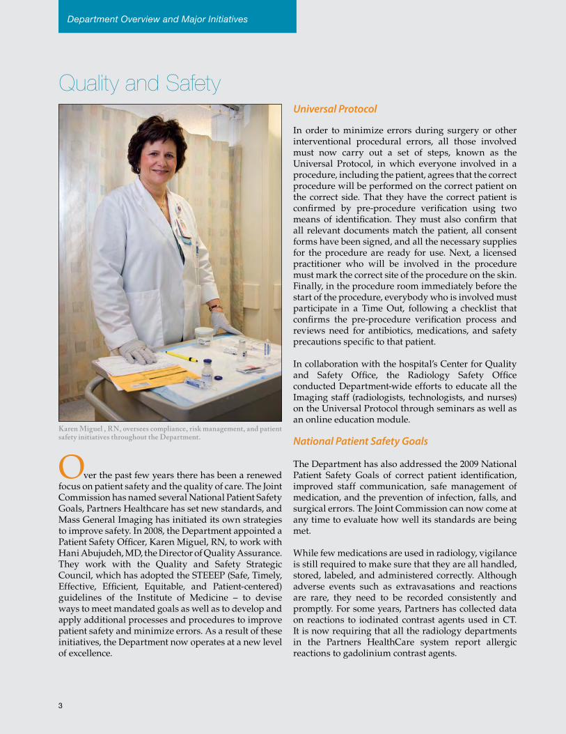
Karen Miguel , RN, oversees compliance, risk management, and patient safety initiatives throughout the Department.
Department Overview and Major Initiatives
In a busy Department that is rid-dled with laborious processes, it is tempting to find shortcuts that in-crease risk. In order to make pro-cesses as efficient as possible, there is an ongoing quality management group working on establishing best practices. In addition, a behavior au-diting program is in place to ensure that best practices are followed. For example, auditors regularly moni-tor hand washing before and af-ter patient contact, proper patient identification, transfer of pre- and post-procedure transfer notes, and universal protocol. The results of these audits are recorded, provid-ing feedback on performance and raising standards.
Safety Champions
In another effort to promote safety and high-quality care, Mass Gen-eral Imaging has initiated a Safety Champion program. The Safety Champions are front line indi-viduals from each work area, who, along with their managers, serve as local resources to support their col-leagues in understanding the Na-tional Patient Safety Goals and in navigating the complexities of the regulatory requirements governing the practice of radiology.
The Champions instill the importance of continuously maintaining high standards and moving away from sporadically “getting ready” for regulatory visits from the Joint Commission.
The Safety Champions held their first retreat in June 2009, with over 50 attendees representing 17 areas across the Department. Besides celebrat-ing the Champions, the day’s program focused on the their influential position in educating, communicating and motivating staff across all areas, and their importance in reinforcing the Department’s motto, “Every Patient, Every Process: Quality and Safety, Built In.”
Physician Evaluations
The Joint Commission has also mandated physician evaluations, with a focused physician performance evaluation on recruitment and ongoing physician performance evaluations at regular intervals thereafter. Each division in the Department has devised its own specific evaluation criteria, such as compliance with the standard report structure, timeliness of completion of the final report, peer review of major findings in randomly selected studies, and the rate of common complications associated with interventional procedures.
Effective Communication
Highly Effective Team Training, a method first developed in the aviation industry, has been introduced successfully in a pilot program to improve communication and teamwork in vascular and neuro-interventional radiology and will soon be launched in other areas. In this program, the radiology teams have been trained in SBAR (Situation, Background, Assessment, and Recommendation), closed loop communication, assertion techniques, and briefing and debriefing skills.
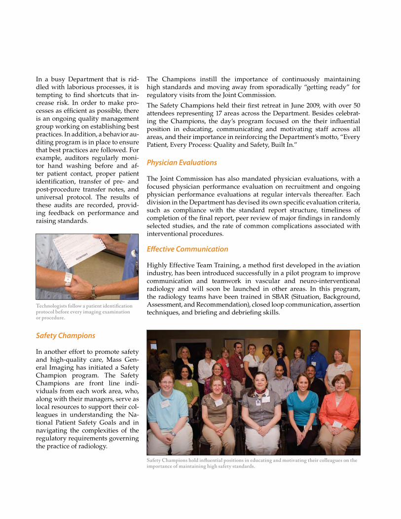
Safety Champions hold influential positions in educating and motivating their colleagues on the importance of maintaining high safety standards.
Technologists follow a patient identification protocol before every imaging examination or procedure.
Department Overview and Major Initiatives
Quality and SafetyIn SBAR, caregivers use concise and clear language, starting with the current situation that needs attention, background information about the patient, an assessment of what may be needed, and a recommendation as to what needs to be done. In closed loop communication, the listener repeats back what has been said to confirm that the message has been received correctly. Team members also learned how to speak up and share their concerns using critical language and common phrases that drive home the urgency of the situation and the clear request for action while striking a balance between persistence and politeness.
They also learned pre-procedure briefing and post-procedure debriefing techniques. Briefing is intended to anticipate possible problems and special needs of the patient while debriefing is intended to discuss the completed procedure and consider whether and how the process might be improved.
Electronic Incident Reporting
The Department is now participating in a Partners-wide program of electronic incident reporting of adverse events and near misses. This program is intended to foster an open, non-punitive environment in which people are encouraged to talk about errors and to investigate reasons why they happened and to seek ways in which such errors might be avoided in the future. Consequently, there has been an increase in the number of reported safety events. The new openness is being encouraged through meetings in which safety issues and failures are presented and people have an opportunity to express their own experiences and fears in regard to patient safety. At the same time, staff actions that averted errors are celebrated.
Automated Retrieval of Patient Information
Electronic medical records (EMRs) contain a wealth of data that could be used to improve patient care. In 2005, Michael E. Zalis, MD, and Mitchell A. Harris, PhD, began work on an ontological search system, Queriable Patient Inference Dossier (QPID), that could tap into EMRs to retrieve information relevant to the clinical encounter at hand. In the Department, QPID is currently used for finding duplicate orders in the computerized radiology order entry (ROE) system. In this way, QPID has substantially reduced the number of unnecessary examinations.
Policy, Guideline, and Procedure ManualRevision of the Policy, Guideline, and Procedure Manual is another big undertaking in the Department. The new Web-based manual will have a consistent template-based structure for policies and guidelines, and a new standardized review process has been established for all new policies and guidelines. It will make use of links via keywords to other related policies and guidelines to make it much easier for caregivers to find the information that they are looking for. In addition, the new manual will be easier to update, with an in-built mechanism to ensure that each policy and guideline will be reviewed and updated, if necessary, at regular intervals.
Employee Safety
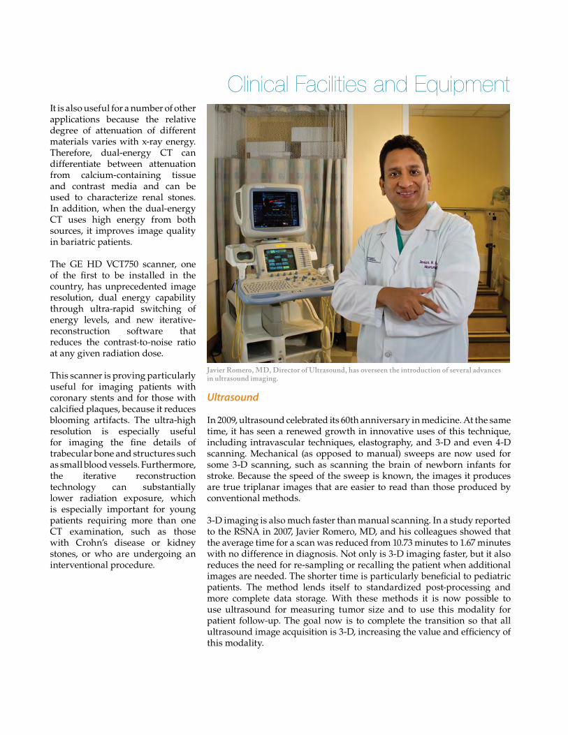
High employee safety standards are also a concern of the Department, and steps have been taken to minimize workplace injuries. For example, in the past few years it was noted that the number of repetitive stress injuries to sonographers was unacceptably high. Nationally, more than 80% report pain or injury due to the unnatural grip used when holding transducers, the repetitive pressing required to image patients, and sonographer posturing required to obtain images. In order to reduce the rate of injuries, Janice Wright, Operations Manager for Ultrasound, and Javier Romero, MD, Director of Ultrasound, have brought in new ergonomically improved equipment with lighter transducers and connecting cables that are easier to use, adjustable monitors and keyboards on the imaging equipment, and examination beds that are adjustable to a variety of positions.
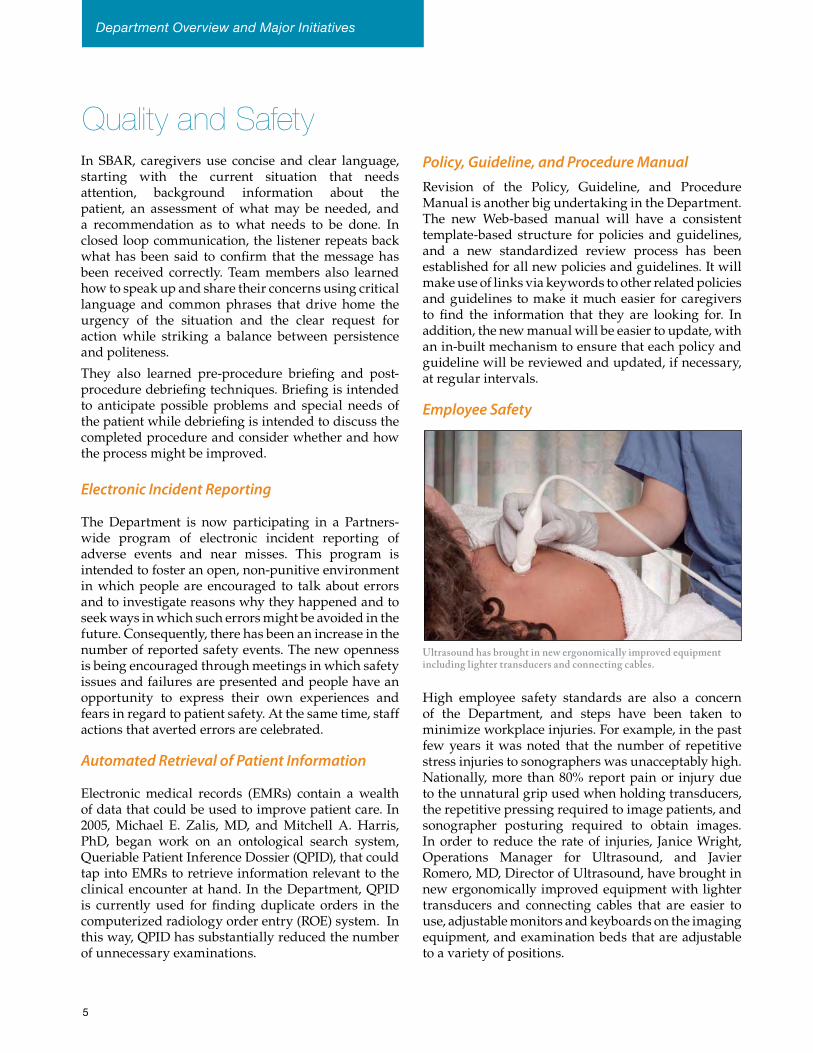
Ultrasound has brought in new ergonomically improved equipment including lighter transducers and connecting cables.
5
In addition, ergonomics specialists were consulted to observe and then train sonographers, showing them how to adjust tables and chairs for better positioning and teaching them stretching exercises. The instruction is reinforced with training videos, and sonographers are observed for compliance. Jennifer McDowell, Technical Manager of Ultrasound, has been instrumental in training and ensuring that sonographers are using proper ergonomics. As a result of this effort, the number of injuries has been reduced by more than 50%.
CT Dose Reduction
The diagnostic value of CT scanning has led to huge increases in its utilization. As a result, it is estimated that CT scans now deliver almost half of the estimated collective dose of medically related radiation exposure in the United States. Although there is no doubt that the benefits of CT outweigh the risks, there is increasing recognition of the possibility of harm. Recognizing this concern, Mass General Imaging has been in the forefront in developing and applying dose reduction strategies for CT imaging while maintaining high diagnostic image quality.
CT Dose Reduction: Pediatric Protocols
Pediatric patients are of special concern when considering radiation exposure. Scanning protocols for children, developed at Mass General, adjust exposure to the weight of the patient, the image contrast demands of the particular study, and whether it is an initial diagnostic image or a follow-up image, for which a noisier image may be sufficient. These protocols are displayed as a color-coded chart to aid the technologists in selecting the most appropriate CT protocols for pediatric patients.
Implementation of these new protocols required training more than 70 technologists, who work on different shifts and at three separate locations, on the application of these protocols on scanners with various detector geometries (eight-, 16-, and 64-section multidetector CT scanners). Despite these challenges, compliance was high. In a large study, Sarabjeet Singh, MBBS, MMST, Mannudeep K. Kalra, MD, and Sjirk J. Westra, MD, have demonstrated that since the adoption of these protocols, the radiation dose to pediatric patients has been reduced by as much as 80%.
These protocols have now been introduced in all Partners HealthCare hospitals, including the Newton Wellesley Hospital and North Shore Children’s Hospital. The introduction of these protocols has resulted in a reduction of undesirable intra- and inter-hospital variability and CT dosing, which is an important outcome measure in quality improvement. Through Partners Radiology, these quality improvement efforts are currently being expanded to adult CT imaging across the Partners network, with the aim of ensuring a seamless transition between pediatric and adult CT protocols at age 18 years.
GE has recently published a technical bulletin, Featherlight Imaging, CT Procedure-Based Protocols for pediatric patients and small adults, based on the protocols developed at Mass General. In addition, GE has invited Dr. Kalra to act as a consultant in its CT 4Kids program and to be a consultant and educator in a course that will be presented across the country to educate technologists and radiologists in reduced-dose CT protocols.
In addition, Dr. Westra has been active in the education of radiologists and trainees about optimal CT techniques to reduce dose in children, and has given lectures for referring clinicians (pediatricians and surgeons) about the appropriateness of pediatric CT imaging in light of its expanding utilization.
CT Dose Reduction: Adult Imaging
All the other clinical divisions have worked on developing reduced-dose protocols while maintaining high diagnostic image quality. For example, in Thoracic Radiology, Matthew W. Gilman, MD, has worked closely with Dr. Kalra to develop new protocols. Because smaller bodies attenuate less and require less
Our staff have been in the forefront in developing and applying dose reduction
strategies for CT.
Department Overview and Major Initiatives
Quality and Safety
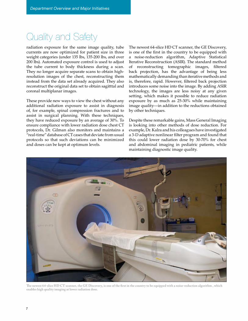
The newest 64-slice HD CT scanner, the GE Discovery, is one of the first in the country to be equipped with a noise-reduction algorithm , which enables high quality imaging at lower radiation dose.
radiation exposure for the same image quality, tube currents are now optimized for patient size in three weight categories (under 135 lbs, 135-200 lbs, and over 200 lbs). Automated exposure control is used to adjust the tube current to body thickness during a scan. They no longer acquire separate scans to obtain high-resolution images of the chest, reconstructing them instead from the data set already acquired. They also reconstruct the original data set to obtain sagitttal and coronal multiplanar images.
These provide new ways to view the chest without any additional radiation exposure to assist in diagnosis of, for example, spinal compression fractures and to assist in surgical planning. With these techniques, they have reduced exposure by an average of 30%. To ensure compliance with lower radiation dose chest CT protocols, Dr. Gilman also monitors and maintains a “real-time” database of CT cases that deviate from usual protocols so that such deviations can be minimized and doses can be kept at optimum levels.
The newest 64-slice HD CT scanner, the GE Discovery, is one of the first in the country to be equipped with a noise-reduction algorithm, Adaptive Statistical Iterative Reconstruction (ASIR). The standard method of reconstructing tomographic images, filtered back projection, has the advantage of being less mathematically demanding than iterative methods and is, therefore, rapid. However, filtered back projection introduces some noise into the image. By adding ASIR technology, the images are less noisy at any given setting, which makes it possible to reduce radiation exposure by as much as 25-30% while maintaining image quality—in addition to the reductions obtained by other techniques.
Despite these remarkable gains, Mass General Imaging is looking into other methods of dose reduction. For example, Dr. Kalra and his colleagues have investigated a 3-D adaptive nonlinear filter program and found that this could lower radiation dose by 30-70% for chest and abdominal imaging in pediatric patients, while maintaining diagnostic image quality.
7
Strategic Initiatives
Each year during the fall, the Department holds a Strategic Retreat to celebrate its accomplishments and assess ongoing and future challenges. The Department is proud that the Press Ganey Employee Survey showed that employee satisfaction had improved for the fourth year in a row and was higher than 86% of organizations of a similar size. The second year of the Department’s safety office had seen the implementation of several new initiatives. New state-of-the-art imaging equipment had been installed and facilities expanded. The marketing team had conducted an extensive analysis that yielded a new logo that clarifies and reinforces the values of the Department. At the same time, the Department recognized that there is a continuing need to refine operations and improve patient care; a task that is made more challenging by difficulties presented by the current fiscal environment and the anticipation of slower growth than in the past.
The outcome of the Strategic Retreat included seven new initiatives. The first is to update the new employee orientation process with a new focus on the mission, vision, and values of the Department, which are encapsulated in a new tagline, Every Patient, Every Process, Quality and Safety Built-in. A new peer-review program for technologists, based on one that already exists for radiologists, was envisaged as an opportunity to provide constructive and non-punitive feedback that would enable them to improve their practice. Explorations are underway for the development and implementation of safety champions. The Quality Management and Education (QME) team, directed by Max Gomez, MPH., is supporting the implementation of these initiatives.
In order to facilitate the smooth adoption of the new version of IDXrad, scheduled for 2009, the Department
has contracted with a consulting firm, the Breakaway Group, which will provide role-based simulation modules in order to speed the learning process and ease the transition. In order to share knowledge and improve operational management, more formal meetings will be held that include both radiologists and technologists. And a new pilot program is underway that is intended to reduce the logistical problems of fitting in ad hoc requests for inpatient examinations by designing a new scheduling template in ROE that solicits needed information from inpatient units in a timely and proactive way.
Strategic Councils
There is also a renewed focus on the Strategic Councils, and QME team members are providing assistance and supplying resources to each of them in order to help them achieve their goals. Each of the Councils now has a well-publicized mission statement, charter, and accountability/responsibility statement, each of which have been validated and confirmed by the Executive Council. The Innovation Council has been restructured with the goal of providing a forum for staff, especially front-line staff, to propose innovative ideas that could improve safety or efficiency in their practice. In addition, the Innovation Council will provide staff with the opportunity to learn, and put into practice, the
• QualityandSafetyCouncil• CustomerServiceCouncil• TheAccessGroup• ClinicalInformaticsAdvisoryGroup• InnovationCouncil• EducationAdvisoryBoard
Strategic Initiatives needed tools to flesh out and test their ideas, present them to senior leadership and, if accepted, supply the resources to implement them.
There is one new Strategic Council, the Clinical Informatics Advisory Group, whose role is to fill a gap in the decision-making process. This will be a proactive group that will anticipate resource requirements, constraints, and obstacles before engaging in a project. It will gain consensus on the reasoning behind information technology (IT) decisions, make those decisions clear to the Department as a whole, and provide a forum for and opportunity to reprioritize IT projects to meet immediate or critical needs.
Lean Training
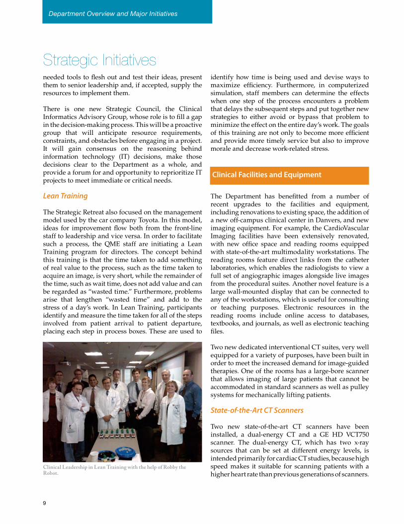
The Strategic Retreat also focused on the management model used by the car company Toyota. In this model, ideas for improvement flow both from the front-line staff to leadership and vice versa. In order to facilitate such a process, the QME staff are initiating a Lean Training program for directors. The concept behind this training is that the time taken to add something of real value to the process, such as the time taken to acquire an image, is very short, while the remainder of the time, such as wait time, does not add value and can be regarded as “wasted time.” Furthermore, problems arise that lengthen “wasted time” and add to the stress of a day’s work. In Lean Training, participants identify and measure the time taken for all of the steps involved from patient arrival to patient departure, placing each step in process boxes. These are used to
identify how time is being used and devise ways to maximize efficiency. Furthermore, in computerized simulation, staff members can determine the effects when one step of the process encounters a problem that delays the subsequent steps and put together new strategies to either avoid or bypass that problem to minimize the effect on the entire day’s work. The goals of this training are not only to become more efficient and provide more timely service but also to improve morale and decrease work-related stress.
Clinical Facilities and Equipment
The Department has benefitted from a number of recent upgrades to the facilities and equipment, including renovations to existing space, the addition of a new off-campus clinical center in Danvers, and new imaging equipment. For example, the CardioVascular Imaging facilities have been extensively renovated, with new office space and reading rooms equipped with state-of-the-art multimodality workstations. The reading rooms feature direct links from the catheter laboratories, which enables the radiologists to view a full set of angiographic images alongside live images from the procedural suites. Another novel feature is a large wall-mounted display that can be connected to any of the workstations, which is useful for consulting or teaching purposes. Electronic resources in the reading rooms include online access to databases, textbooks, and journals, as well as electronic teaching files.
Two new dedicated interventional CT suites, very well equipped for a variety of purposes, have been built in order to meet the increased demand for image-guided therapies. One of the rooms has a large-bore scanner that allows imaging of large patients that cannot be accommodated in standard scanners as well as pulley systems for mechanically lifting patients.
State-of-the-Art CT Scanners
Two new state-of-the-art CT scanners have been installed, a dual-energy CT and a GE HD VCT750 scanner. The dual-energy CT, which has two x-ray sources that can be set at different energy levels, is intended primarily for cardiac CT studies, because high speed makes it suitable for scanning patients with a higher heart rate than previous generations of scanners.
Clinical Leadership in Lean Training with the help of Robby the Robot.
Department Overview and Major Initiatives
9
It is also useful for a number of other applications because the relative degree of attenuation of different materials varies with x-ray energy. Therefore, dual-energy CT can differentiate between attenuation from calcium-containing tissue and contrast media and can be used to characterize renal stones. In addition, when the dual-energy CT uses high energy from both sources, it improves image quality in bariatric patients.
The GE HD VCT750 scanner, one of the first to be installed in the country, has unprecedented image resolution, dual energy capability through ultra-rapid switching of energy levels, and new iterative-reconstruction software that reduces the contrast-to-noise ratio at any given radiation dose.
This scanner is proving particularly useful for imaging patients with coronary stents and for those with calcified plaques, because it reduces blooming artifacts. The ultra-high resolution is especially useful for imaging the fine details of trabecular bone and structures such as small blood vessels. Furthermore, the iterative reconstruction technology can substantially lower radiation exposure, which is especially important for young patients requiring more than one CT examination, such as those with Crohn’s disease or kidney stones, or who are undergoing an interventional procedure.
Ultrasound
In 2009, ultrasound celebrated its 60th anniversary in medicine. At the same time, it has seen a renewed growth in innovative uses of this technique, including intravascular techniques, elastography, and 3-D and even 4-D scanning. Mechanical (as opposed to manual) sweeps are now used for some 3-D scanning, such as scanning the brain of newborn infants for stroke. Because the speed of the sweep is known, the images it produces are true triplanar images that are easier to read than those produced by conventional methods.
3-D imaging is also much faster than manual scanning. In a study reported to the RSNA in 2007, Javier Romero, MD, and his colleagues showed that the average time for a scan was reduced from 10.73 minutes to 1.67 minutes with no difference in diagnosis. Not only is 3-D imaging faster, but it also reduces the need for re-sampling or recalling the patient when additional images are needed. The shorter time is particularly beneficial to pediatric patients. The method lends itself to standardized post-processing and more complete data storage. With these methods it is now possible to use ultrasound for measuring tumor size and to use this modality for patient follow-up. The goal now is to complete the transition so that all ultrasound image acquisition is 3-D, increasing the value and efficiency of this modality.
Javier Romero, MD, Director of Ultrasound, has overseen the introduction of several advances in ultrasound imaging.
Clinical Facilities and EquipmentStrategic Initiatives
11
Clinical Facilities and Equipment
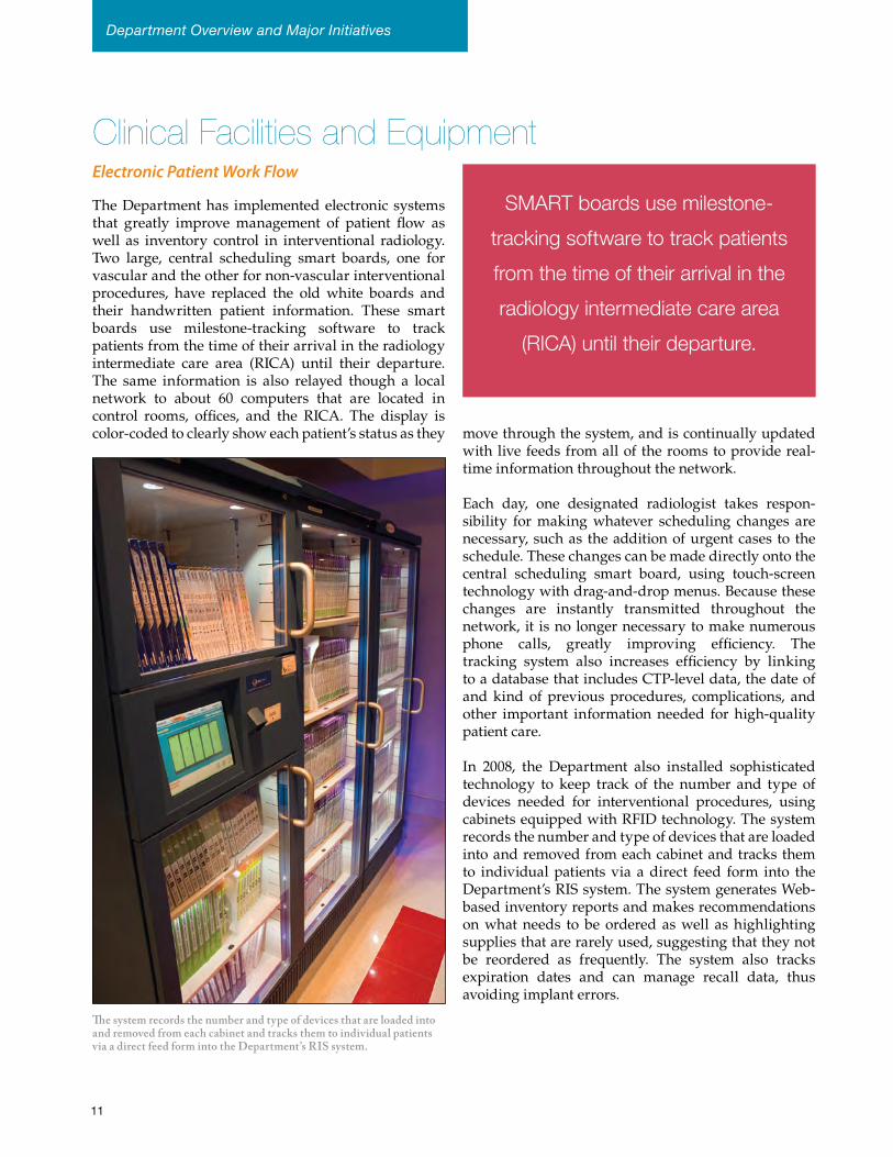
The system records the number and type of devices that are loaded into and removed from each cabinet and tracks them to individual patients via a direct feed form into the Department’s RIS system.
Electronic Patient Work Flow
The Department has implemented electronic systems that greatly improve management of patient flow as well as inventory control in interventional radiology. Two large, central scheduling smart boards, one for vascular and the other for non-vascular interventional procedures, have replaced the old white boards and their handwritten patient information. These smart boards use milestone-tracking software to track patients from the time of their arrival in the radiology intermediate care area (RICA) until their departure. The same information is also relayed though a local network to about 60 computers that are located in control rooms, offices, and the RICA. The display is color-coded to clearly show each patient’s status as they move through the system, and is continually updated
with live feeds from all of the rooms to provide real-time information throughout the network.
Each day, one designated radiologist takes respon-sibility for making whatever scheduling changes are necessary, such as the addition of urgent cases to the schedule. These changes can be made directly onto the central scheduling smart board, using touch-screen technology with drag-and-drop menus. Because these changes are instantly transmitted throughout the network, it is no longer necessary to make numerous phone calls, greatly improving efficiency. The tracking system also increases efficiency by linking to a database that includes CTP-level data, the date of and kind of previous procedures, complications, and other important information needed for high-quality patient care.
In 2008, the Department also installed sophisticated technology to keep track of the number and type of devices needed for interventional procedures, using cabinets equipped with RFID technology. The system records the number and type of devices that are loaded into and removed from each cabinet and tracks them to individual patients via a direct feed form into the Department’s RIS system. The system generates Web-based inventory reports and makes recommendations on what needs to be ordered as well as highlighting supplies that are rarely used, suggesting that they not be reordered as frequently. The system also tracks expiration dates and can manage recall data, thus avoiding implant errors.
SMART boards use milestone-
tracking software to track patients
from the time of their arrival in the
radiology intermediate care area
(RICA) until their departure.
Department Overview and Major Initiatives
Community-Based Imaging Centers
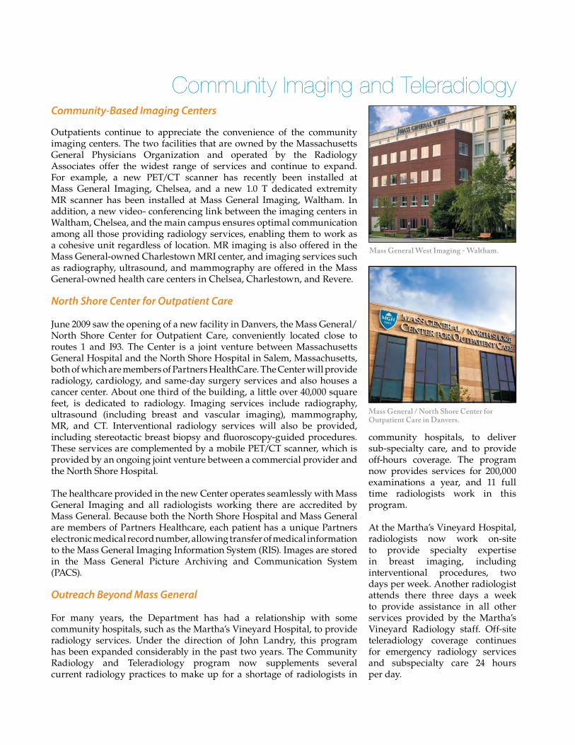
Outpatients continue to appreciate the convenience of the community imaging centers. The two facilities that are owned by the Massachusetts General Physicians Organization and operated by the Radiology Associates offer the widest range of services and continue to expand. For example, a new PET/CT scanner has recently been installed at Mass General Imaging, Chelsea, and a new 1.0 T dedicated extremity MR scanner has been installed at Mass General Imaging, Waltham. In addition, a new video- conferencing link between the imaging centers in Waltham, Chelsea, and the main campus ensures optimal communication among all those providing radiology services, enabling them to work as a cohesive unit regardless of location. MR imaging is also offered in the Mass General-owned Charlestown MRI center, and imaging services such as radiography, ultrasound, and mammography are offered in the Mass General-owned health care centers in Chelsea, Charlestown, and Revere.
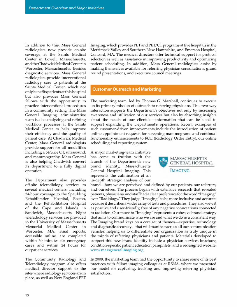
North Shore Center for Outpatient Care
June 2009 saw the opening of a new facility in Danvers, the Mass General/ North Shore Center for Outpatient Care, conveniently located close to routes 1 and I93. The Center is a joint venture between Massachusetts General Hospital and the North Shore Hospital in Salem, Massachusetts, both of which are members of Partners HealthCare. The Center will provide radiology, cardiology, and same-day surgery services and also houses a cancer center. About one third of the building, a little over 40,000 square feet, is dedicated to radiology. Imaging services include radiography, ultrasound (including breast and vascular imaging), mammography, MR, and CT. Interventional radiology services will also be provided, including stereotactic breast biopsy and fluoroscopy-guided procedures. These services are complemented by a mobile PET/CT scanner, which is provided by an ongoing joint venture between a commercial provider and the North Shore Hospital.
The healthcare provided in the new Center operates seamlessly with Mass General Imaging and all radiologists working there are accredited by Mass General. Because both the North Shore Hospital and Mass General are members of Partners Healthcare, each patient has a unique Partners electronic medical record number, allowing transfer of medical information to the Mass General Imaging Information System (RIS). Images are stored in the Mass General Picture Archiving and Communication System (PACS).
Outreach Beyond Mass General
For many years, the Department has had a relationship with some community hospitals, such as the Martha’s Vineyard Hospital, to provide radiology services. Under the direction of John Landry, this program has been expanded considerably in the past two years. The Community Radiology and Teleradiology program now supplements several current radiology practices to make up for a shortage of radiologists in
community hospitals, to deliver sub-specialty care, and to provide off-hours coverage. The program now provides services for 200,000 examinations a year, and 11 full time radiologists work in this program.
At the Martha’s Vineyard Hospital, radiologists now work on-site to provide specialty expertise in breast imaging, including interventional procedures, two days per week. Another radiologist attends there three days a week to provide assistance in all other services provided by the Martha’s Vineyard Radiology staff. Off-site teleradiology coverage continues for emergency radiology services and subspecialty care 24 hours per day.
Mass General / North Shore Center for Outpatient Care in Danvers.
Community Imaging and Teleradiology
Mass General West Imaging - Waltham.
In addition to this, Mass General radiologists now provide on-site coverage at the Saints Medical Center in Lowell, Massachusetts, and the Chadwick Medical Center in Worcester, Massachusetts. Besides diagnostic services, Mass General radiologists provide interventional radiology care to patients at the Saints Medical Center, which not only benefits patients at this hospital but also provides Mass General fellows with the opportunity to practice interventional procedures in a community setting. The Mass General Imaging administrative team is also analyzing and refining workflow processes at the Saints Medical Center to help improve their efficiency and the quality of patient care. At Chadwick Medical Center, Mass General radiologists provide support for all modalities including a 64 Slice CT, ultrasound, and mammography. Mass General is also helping Chadwick convert its department to a fully digital operation.
The Department also provides off-site teleradiology services to several medical centers, including 24-hour coverage to the Spaulding Rehabilitation Hospital, Boston, and the Rehabilitation Hospital of the Cape and Islands in Sandwich, Massachusetts. Night teleradiology services are provided to the University of Massachusetts Memorial Medical Center in Worcester, MA. Final reports, accessible online, are completed within 30 minutes for emergency cases and within 24 hours for outpatient services.
The Community Radiology and Teleradiology program also offers medical director support to the sites where radiology services are in place, as well as New England PET
Imaging, which provides PET and PET/CT programs at five hospitals in the Merrimack Valley and Southern New Hampshire, and Emerson Hospital, Concord, MA. The medical directors offer technical support for protocol selection as well as assistance in improving productivity and optimizing patient scheduling. In addition, Mass General radiologists assist by making themselves available for referring physician consultations, grand round presentations, and executive council meetings.
Customer Outreach and Marketing
The marketing team, led by Thomas G. Marshall, continues to execute on its primary mission of outreach to referring physicians. This two-way interaction supports the Department’s objectives not only by increasing awareness and utilization of our services but also by absorbing insights about the needs of our clientele—information that can be used to support expanding the Department’s operations. Recent examples of such customer-driven improvements include the introduction of patient online appointment requests for screening mammograms and continual functionality enhancements to ROE (Radiology Order Entry), our online scheduling and reporting system.
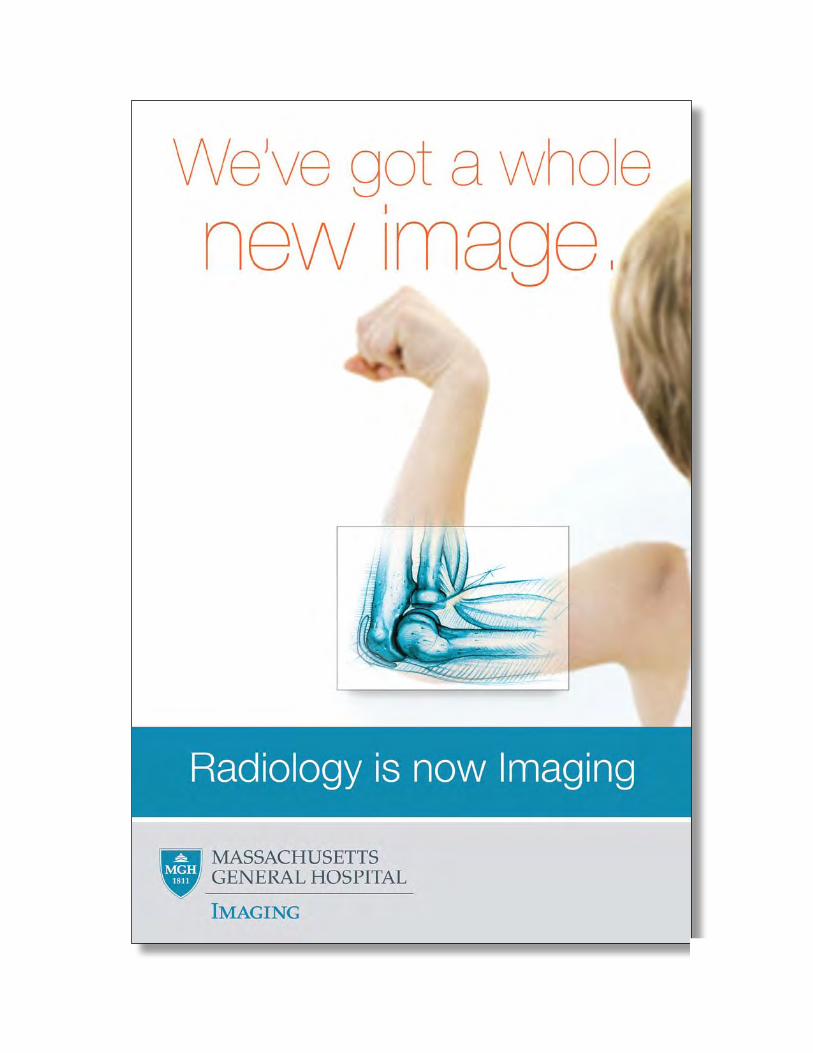
A major marketing-team initiative has come to fruition with the launch of the Department’s new brand identity, Massachusetts General Hospital Imaging. This represents the culmination of an in-depth strategic analysis of our brand—how we are perceived and defined by our patients, our referrers, and ourselves. The process began with extensive research that revealed patients, physicians, and staff had a clear preference for the word “Imaging” over “Radiology.” They judge “Imaging” to be more inclusive and accurate because it describes a wider array of tests and procedures. They also view it as positive and user-friendly, free of any negative connotations connected to radiation. Our move to “Imaging” represents a cohesive brand strategy that aims to communicate who we are and what we do in a consistent way. The Imaging brand keys on a core set of themes—expertise, technology, and diagnostic accuracy—that will manifest across all our communication vehicles, helping us to differentiate our organization as truly unique in the minds of referring physicians and patients. Materials developed to support this new brand identity include a physician services brochure, condition-specific patient education pamphlets, and a redesigned website, www.massgeneralimaging.org.
In 2008, the marketing team had the opportunity to share some of its best practices with fellow imaging colleagues at RSNA, where we presented our model for capturing, tracking and improving referring physician satisfaction.
Department Overview and Major Initiatives
13
3D imaging is now a vital component of healthcare as a diagnostic aid for radiologists as well as other physicians who benefit from precise anatomical visualization as an aid to treatment planning. 3D visualization is also helpful to patients because they can understand their condition and actively participate in their treatment. Over the past few years, the 3D Imaging and Computer-Aided Diagnostics Laboratory has developed a wide range of clinically validated 3D reconstructions for all branches of radiology. In addition to serving the Massachusetts General community, the 3D Imaging Laboratory now offers its services to other institutions through Tele3D Advantage (http://tele3dadvantage.net). In this way, many small hospitals and imaging centers can benefit from these advanced imaging techniques without having to overcome the challenges and costs associated with providing and operating their own 3D service.
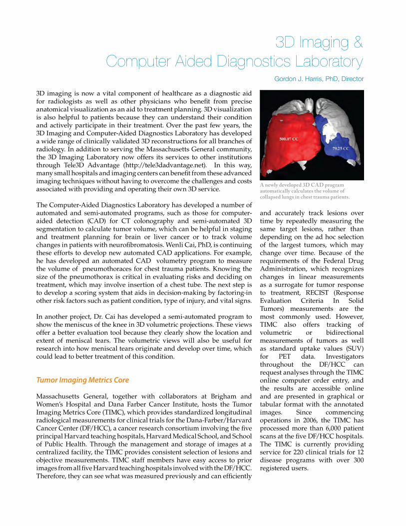
The Computer-Aided Diagnostics Laboratory has developed a number of automated and semi-automated programs, such as those for computer-aided detection (CAD) for CT colonography and semi-automated 3D segmentation to calculate tumor volume, which can be helpful in staging and treatment planning for brain or liver cancer or to track volume changes in patients with neurofibromatosis. Wenli Cai, PhD, is continuing these efforts to develop new automated CAD applications. For example, he has developed an automated CAD volumetry program to measure the volume of pneumothoraces for chest trauma patients. Knowing the size of the pneumothorax is critical in evaluating risks and deciding on treatment, which may involve insertion of a chest tube. The next step is to develop a scoring system that aids in decision-making by factoring-in other risk factors such as patient condition, type of injury, and vital signs.
In another project, Dr. Cai has developed a semi-automated program to show the meniscus of the knee in 3D volumetric projections. These views offer a better evaluation tool because they clearly show the location and extent of meniscal tears. The volumetric views will also be useful for research into how meniscal tears originate and develop over time, which could lead to better treatment of this condition.
Tumor Imaging Metrics Core
Massachusetts General, together with collaborators at Brigham and Women’s Hospital and Dana Farber Cancer Institute, hosts the Tumor Imaging Metrics Core (TIMC), which provides standardized longitudinal radiological measurements for clinical trials for the Dana-Farber/Harvard Cancer Center (DF/HCC), a cancer research consortium involving the five principal Harvard teaching hospitals, Harvard Medical School, and School of Public Health. Through the management and storage of images at a centralized facility, the TIMC provides consistent selection of lesions and objective measurements. TIMC staff members have easy access to prior images from all five Harvard teaching hospitals involved with the DF/HCC. Therefore, they can see what was measured previously and can efficiently
and accurately track lesions over time by repeatedly measuring the same target lesions, rather than depending on the ad hoc selection of the largest tumors, which may change over time. Because of the requirements of the Federal Drug Administration, which recognizes changes in linear measurements as a surrogate for tumor response to treatment, RECIST (Response Evaluation Criteria In Solid Tumors) measurements are the most commonly used. However, TIMC also offers tracking of volumetric or bidirectional measurements of tumors as well as standard uptake values (SUV) for PET data. Investigators throughout the DF/HCC can request analyses through the TIMC online computer order entry, and the results are accessible online and are presented in graphical or tabular format with the annotated images. Since commencing operations in 2006, the TIMC has processed more than 6,000 patient scans at the five DF/HCC hospitals. The TIMC is currently providing service for 220 clinical trials for 12 disease programs with over 300 registered users.
3D Imaging & Computer Aided Diagnostics Laboratory
Gordon J. Harris, PhD, Director
A newly developed 3D CAD program automatically calculates the volume of collapsed lungs in chest trauma patients.
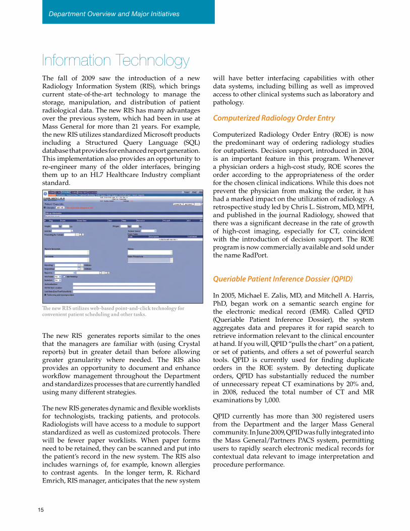
Information TechnologyThe fall of 2009 saw the introduction of a new Radiology Information System (RIS), which brings current state-of-the-art technology to manage the storage, manipulation, and distribution of patient radiological data. The new RIS has many advantages over the previous system, which had been in use at Mass General for more than 21 years. For example, the new RIS utilizes standardized Microsoft products including a Structured Query Language (SQL) database that provides for enhanced report generation. This implementation also provides an opportunity to re-engineer many of the older interfaces, bringing them up to an HL7 Healthcare Industry compliant standard.
The new RIS generates reports similar to the ones that the managers are familiar with (using Crystal reports) but in greater detail than before allowing greater granularity where needed. The RIS also provides an opportunity to document and enhance workflow management throughout the Department and standardizes processes that are currently handled using many different strategies.
The new RIS generates dynamic and flexible worklists for technologists, tracking patients, and protocols. Radiologists will have access to a module to support standardized as well as customized protocols. There will be fewer paper worklists. When paper forms need to be retained, they can be scanned and put into the patient’s record in the new system. The RIS also includes warnings of, for example, known allergies to contrast agents. In the longer term, R. Richard Emrich, RIS manager, anticipates that the new system
will have better interfacing capabilities with other data systems, including billing as well as improved access to other clinical systems such as laboratory and pathology.
Computerized Radiology Order Entry
Computerized Radiology Order Entry (ROE) is now the predominant way of ordering radiology studies for outpatients. Decision support, introduced in 2004, is an important feature in this program. Whenever a physician orders a high-cost study, ROE scores the order according to the appropriateness of the order for the chosen clinical indications. While this does not prevent the physician from making the order, it has had a marked impact on the utilization of radiology. A retrospective study led by Chris L. Sistrom, MD, MPH, and published in the journal Radiology, showed that there was a significant decrease in the rate of growth of high-cost imaging, especially for CT, coincident with the introduction of decision support. The ROE program is now commercially available and sold under the name RadPort.
Queriable Patient Inference Dossier (QPID)
In 2005, Michael E. Zalis, MD, and Mitchell A. Harris, PhD, began work on a semantic search engine for the electronic medical record (EMR). Called QPID (Queriable Patient Inference Dossier), the system aggregates data and prepares it for rapid search to retrieve information relevant to the clinical encounter at hand. If you will, QPID “pulls the chart” on a patient, or set of patients, and offers a set of powerful search tools. QPID is currently used for finding duplicate orders in the ROE system. By detecting duplicate orders, QPID has substantially reduced the number of unnecessary repeat CT examinations by 20% and, in 2008, reduced the total number of CT and MR examinations by 1,000.
QPID currently has more than 300 registered users from the Department and the larger Mass General community. In June 2009, QPID was fully integrated into the Mass General/Partners PACS system, permitting users to rapidly search electronic medical records for contextual data relevant to image interpretation and procedure performance.
The new RIS utilizes web-based point-and-click technology for convenient patient scheduling and other tasks.
Department Overview and Major Initiatives
15
administration, and automated patient and procedure summaries tied to diagnostic and interventional schedules. QPID can also be used for online search of medical records for data that might be useful for imaging interpretation and interventional procedures. These are expected to improve time efficiency for a diverse set of Radiology operations.
Drs. Zalis and Harris are also developing a customizable search function, in which an individual radiologist or administrator can edit, save, and share queries with other users, which will permit them to leverage and learn from each others’ experience and insight. QPID applications have been successfully applied to non-Radiology applications in Gastroenterology, Palliative Care, and the Mass General Office of Quality and Safety; others will soon follow.
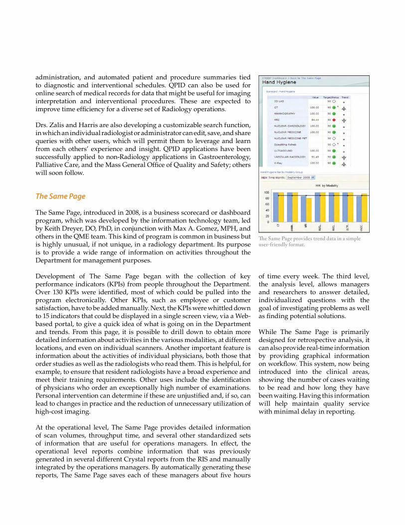
The Same Page
The Same Page, introduced in 2008, is a business scorecard or dashboard program, which was developed by the information technology team, led by Keith Dreyer, DO, PhD, in conjunction with Max A. Gomez, MPH, and others in the QME team. This kind of program is common in business but is highly unusual, if not unique, in a radiology department. Its purpose is to provide a wide range of information on activities throughout the Department for management purposes.
Development of The Same Page began with the collection of key performance indicators (KPIs) from people throughout the Department. Over 130 KPIs were identified, most of which could be pulled into the program electronically. Other KPIs, such as employee or customer satisfaction, have to be added manually. Next, the KPIs were whittled down to 15 indicators that could be displayed in a single screen view, via a Web-based portal, to give a quick idea of what is going on in the Department and trends. From this page, it is possible to drill down to obtain more detailed information about activities in the various modalities, at different locations, and even on individual scanners. Another important feature is information about the activities of individual physicians, both those that order studies as well as the radiologists who read them. This is helpful, for example, to ensure that resident radiologists have a broad experience and meet their training requirements. Other uses include the identification of physicians who order an exceptionally high number of examinations. Personal intervention can determine if these are unjustified and, if so, can lead to changes in practice and the reduction of unnecessary utilization of high-cost imaging.
At the operational level, The Same Page provides detailed information of scan volumes, throughput time, and several other standardized sets of information that are useful for operations managers. In effect, the operational level reports combine information that was previously generated in several different Crystal reports from the RIS and manually integrated by the operations managers. By automatically generating these reports, The Same Page saves each of these managers about five hours
The Same Page provides trend data in a simple user-friendly format.
of time every week. The third level, the analysis level, allows managers and researchers to answer detailed, individualized questions with the goal of investigating problems as well as finding potential solutions.
While The Same Page is primarily designed for retrospective analysis, it can also provide real-time information by providing graphical information on workflow. This system, now being introduced into the clinical areas, showing the number of cases waiting to be read and how long they have been waiting. Having this information will help maintain quality service with minimal delay in reporting.
Information TechnologyRENDER: An On-Line Searchable Radiology Study Repository
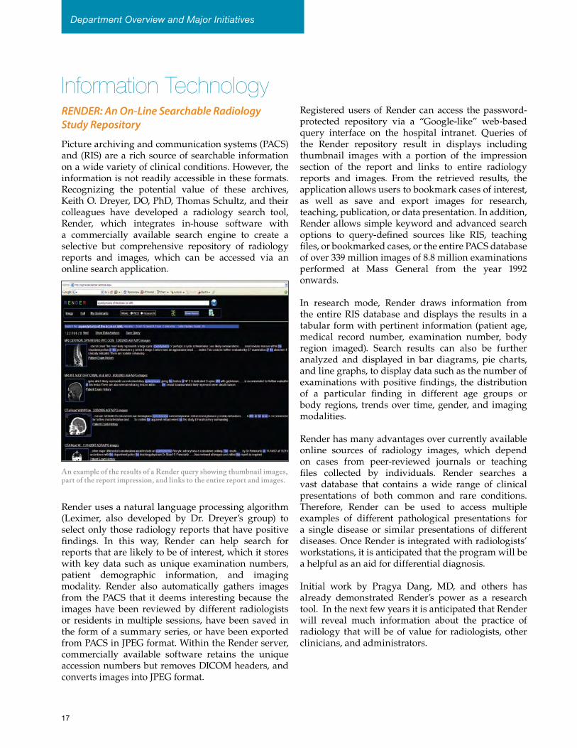
Picture archiving and communication systems (PACS) and (RIS) are a rich source of searchable information on a wide variety of clinical conditions. However, the information is not readily accessible in these formats. Recognizing the potential value of these archives, Keith O. Dreyer, DO, PhD, Thomas Schultz, and their colleagues have developed a radiology search tool, Render, which integrates in-house software with a commercially available search engine to create a selective but comprehensive repository of radiology reports and images, which can be accessed via an online search application.
Render uses a natural language processing algorithm (Leximer, also developed by Dr. Dreyer’s group) to select only those radiology reports that have positive findings. In this way, Render can help search for reports that are likely to be of interest, which it stores with key data such as unique examination numbers, patient demographic information, and imaging modality. Render also automatically gathers images from the PACS that it deems interesting because the images have been reviewed by different radiologists or residents in multiple sessions, have been saved in the form of a summary series, or have been exported from PACS in JPEG format. Within the Render server, commercially available software retains the unique accession numbers but removes DICOM headers, and converts images into JPEG format.
Registered users of Render can access the password-protected repository via a “Google-like” web-based query interface on the hospital intranet. Queries of the Render repository result in displays including thumbnail images with a portion of the impression section of the report and links to entire radiology reports and images. From the retrieved results, the application allows users to bookmark cases of interest, as well as save and export images for research, teaching, publication, or data presentation. In addition, Render allows simple keyword and advanced search options to query-defined sources like RIS, teaching files, or bookmarked cases, or the entire PACS database of over 339 million images of 8.8 million examinations performed at Mass General from the year 1992 onwards.
In research mode, Render draws information from the entire RIS database and displays the results in a tabular form with pertinent information (patient age, medical record number, examination number, body region imaged). Search results can also be further analyzed and displayed in bar diagrams, pie charts, and line graphs, to display data such as the number of examinations with positive findings, the distribution of a particular finding in different age groups or body regions, trends over time, gender, and imaging modalities.
Render has many advantages over currently available online sources of radiology images, which depend on cases from peer-reviewed journals or teaching files collected by individuals. Render searches a vast database that contains a wide range of clinical presentations of both common and rare conditions. Therefore, Render can be used to access multiple examples of different pathological presentations for a single disease or similar presentations of different diseases. Once Render is integrated with radiologists’ workstations, it is anticipated that the program will be a helpful as an aid for differential diagnosis.
Initial work by Pragya Dang, MD, and others has already demonstrated Render’s power as a research tool. In the next few years it is anticipated that Render will reveal much information about the practice of radiology that will be of value for radiologists, other clinicians, and administrators.
An example of the results of a Render query showing thumbnail images, part of the report impression, and links to the entire report and images.
Department Overview and Major Initiatives
17
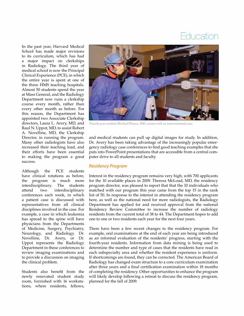
Fourth year resident Michael Preece, MD, assists with an interventional case.
In the past year, Harvard Medical School has made major revisions to its curriculum, which has had a major impact on clerkships in Radiology. The third year of medical school is now the Principal Clinical Experience (PCE), in which the entire year is spent at one of the three HMS teaching hospitals. Almost 50 students spend the year at Mass General, and the Radiology Department now runs a clerkship course every month, rather than every other month as before. For this reason, the Department has appointed two Associate Clerkship directors, Laura L. Avery, MD, and Raul N. Uppot, MD, to assist Robert A. Novelline, MD, the Clerkship Director, in running the program. Many other radiologists have also increased their teaching load, and their efforts have been essential to making the program a great success.
Although the PCE students have clinical rotations as before, the program is much more interdisciplinary. The students attend two interdisciplinary conferences each week, in which a patient case is discussed with representatives from all clinical disciplines involved in the case. For example, a case in which leukemia has spread to the spine will have physicians from the Departments of Medicine, Surgery, Psychiatry, Neurology, and Radiology. Dr. Novelline, Dr. Avery, or Dr. Uppot represents the Radiology Department in these conferences to review imaging examinations and to provide a discussion on imaging the clinical problem.
Students also benefit from the newly renovated student study room, furnished with 16 worksta-tions, where residents, fellows,
and medical students can pull up digital images for study. In addition, Dr. Avery has been taking advantage of the increasingly popular emer-gency radiology case conferences to find good teaching examples that she puts into PowerPoint presentations that are accessible from a central com-puter drive to all students and faculty.
Residency Program
Interest in the residency program remains very high, with 700 applicants for the 10 available places in 2009. Theresa McLoud, MD, the residency program director, was pleased to report that that the 10 individuals who matched with our program this year came from the top 15 in the rank list of 50. In response to the interest in attending the residency program here, as well as the national need for more radiologists, the Radiology Department has applied for and received approval from the national Residency Review Committee to increase the number of radiology residents from the current total of 38 to 44. The Department hopes to add one to one or two residents each year for the next four years.
There have been a few recent changes to the residency program. For example, oral examinations at the end of each year are being introduced as an informal evaluation of the residents’ progress, starting with the fourth-year residents. Information from data mining is being used to determine the number and type of cases that the residents have read in each subspecialty area and whether the resident experience is uniform. If shortcomings are found, they can be corrected. The American Board of Radiology has changed exam structure to a core curriculum examination after three years and a final certification examination within 18 months of completing the residency. Other opportunities to enhance the program will likely develop following a retreat to discuss the residency program, planned for the fall of 2009.
Education
19
Gordon Brownell, PhD, 1922-2008
Gordon Brownell was a pioneer in the field of nuclear medicine whose achievements included the development of radioactive tracer kinetics, radiation dosimetry methods, and positron imaging. Brownell first came to Mass General in 1949 as a physicist in the Thyroid Unit, where he aided in investigations into the use of radioactive iodine for the diagnosis and treatment of thyroid disease. In 1951, he was a member of a team that traveled to Mendoza, Argentina, for a landmark controlled
clinical research study. By applying mathematics to study the kinetics of radioactive iodine uptake in patients with endemic goiter, the team shed light on how the thyroid gland adapted to lack of iodine.
In 1950, Brownell founded the Physics Research Laboratory at Mass General and, in that year, built his first positron imaging device. He was one of the first in the world to recognize the potential of positron emitters to provide greater sensitivity and superior image resolution compared with gamma emitters. He realized that positrons only travel a few millimeters before interacting with an electron, resulting in the production of two high-energy photons that travel in diametrically opposite directions. Therefore, an instrument that could detect the simultaneous arrival of pairs of photons could be used to calculate the source of the emissions without the need for collimation.
In 1952, Brownell built the first positron imaging scanner designed for clinical use, which used a pair of detectors mounted on a moving platform to diagnose brain tumors and determine their approximate location. The first patient to benefit from this device was a 7-year-old girl, Holly Jane Hunter, whose tumor was diagnosed and located with positron scanning, allowing William H. Sweet, MD, of the Neurosurgical Service to successfully remove the tumor. By 1959, positron scanning had been used for diagnostic purposes on more than 3,500 patients at Mass General.
In 1962, Brownell built his first multiple-detector positron imaging device, which was specifically designed for brain imaging and
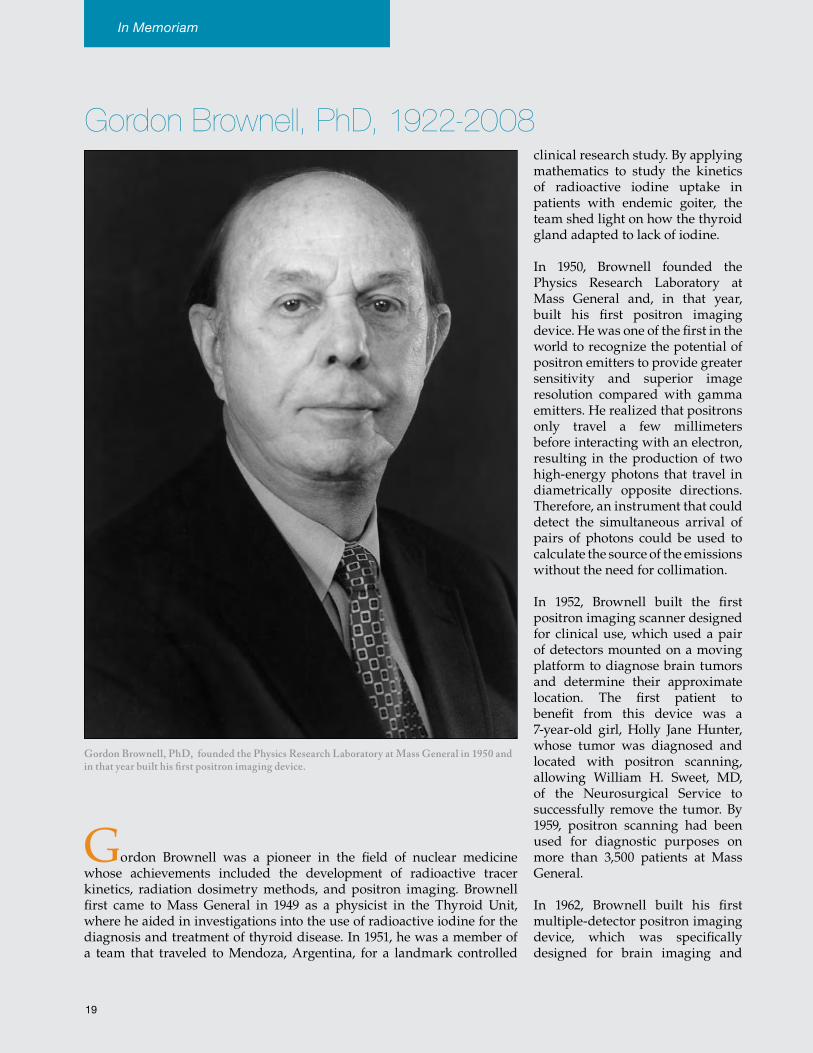
Gordon Brownell, PhD, founded the Physics Research Laboratory at Mass General in 1950 and in that year built his first positron imaging device.
In Memoriam
was used clinically for nearly a decade. This machine was designed to produce two-dimensional images but could provide some three-dimensional information by focusing on planes parallel to the detector arrays and selecting the plane with the sharpest image. In 1968, Brownell designed and built the first tomographic device, the PC-1, which used three-dimensional arrays of detectors.
In 1970, David Chesler, an MIT-trained information theorist and a colleague of Brownell’s at Mass General, devised and tested a computer simulation method, filtered back projection, to produce three-dimensional images. He presented his data at the Meeting on Tomographic Imaging in Nuclear Medicine in 1972. Positron emission tomography (PET) had been born. It is interesting to note that Chesler’s analytic technique did not require iteration, as did the method developed by Godfrey Hounsfield for CT. Before long, the advantages of filtered back projection were recognized and adopted for CT scanning, becoming the standard technique until recently, when more advanced methods requiring more computation became available.
The next in the series of PET scanners, PC-II, which was developed and built between 1971 and 1976, became the basis for a commercial version developed by the Cyclotron Corporation. The next logical step was the development of a circular or cylindrical array of detectors, which were first built in Berkeley, CA. However, Charles Burnham in the Physics Laboratory at Mass General devised a method that used analog coding, which permitted the use of multiple small detectors identified by a smaller number of phototubes. This concept was used to build the first high-resolution PET scanner without moving detectors, the PCR-I, in 1983. A computer controlled table, operated in step and shoot mode, was used to obtain multiple image slices. This device remained in continuous use for 20 years, until 2003, for animal studies. The PCR-II, which had a cylindrical array of detectors, was a massive volumetric multi-slice device built between 1988 and 1995.
Dr. Brownell’s other contributions included the development of the concept of internal radiation dose. Together with Gerald Hine, Brownell published a book, Radiation Dosimetry, in 1956, which remained the definitive work on this topic for several decades. Indeed, the parameters the duo conceived still find use in recent books on determining radiation dose.
Brownell won numerous awards, including election to the Institute of Medicine (2002); the Coolidge Award from the American Association of Physics (1987); and three awards from the Society of Nuclear Medicine: the Paul C. Abersold Award (1975), the Georg von Hevesy Memorial Award (1979), and the Loevinger-Berman Award for Excellence in Internal Dosimetry (2006). Dr. Brownell served as honorary physicist in the Mass General Department of Radiology and professor emeritus in the Department of Nuclear Science and Engineering at MIT until his death in 2008.
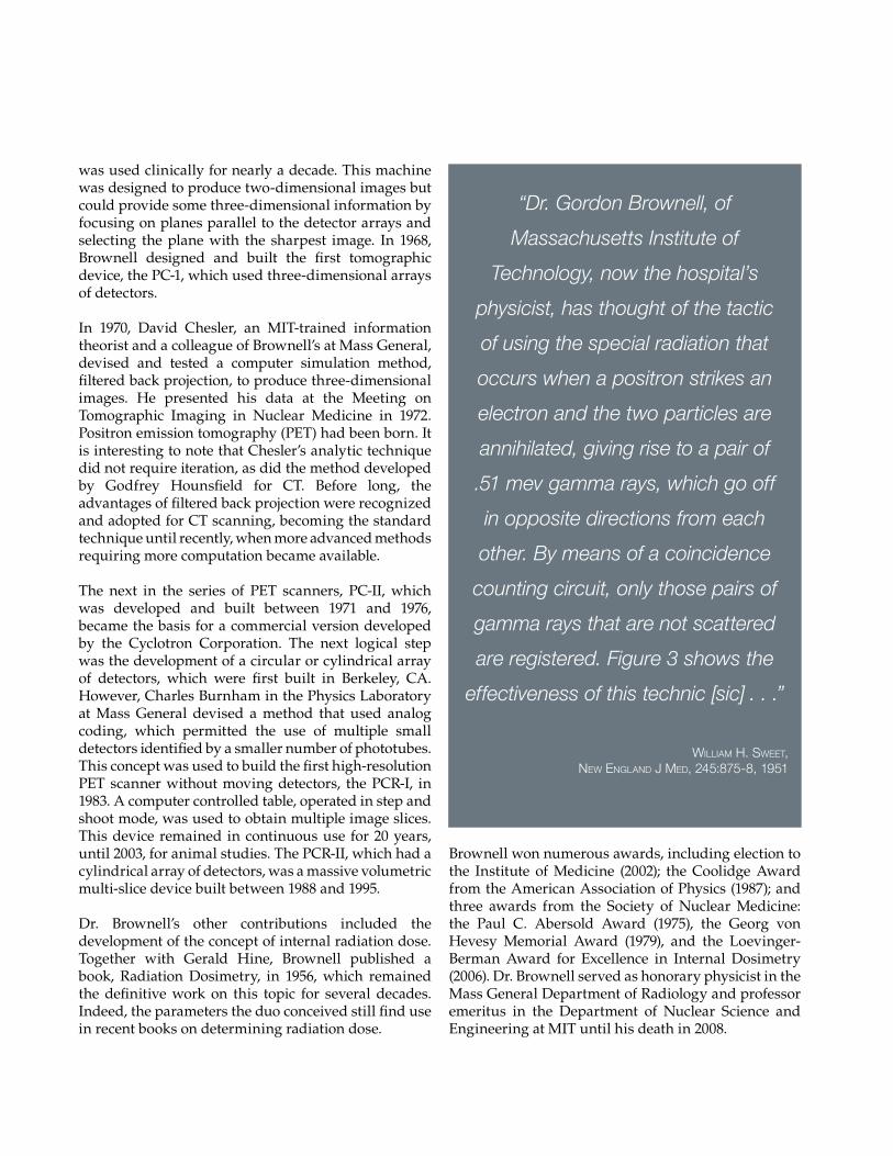
“Dr. Gordon Brownell, of
Massachusetts Institute of
Technology, now the hospital’s
physicist, has thought of the tactic
of using the special radiation that
occurs when a positron strikes an
electron and the two particles are
annihilated, giving rise to a pair of
.51 mev gamma rays, which go off
in opposite directions from each
other. By means of a coincidence
counting circuit, only those pairs of
gamma rays that are not scattered
are registered. Figure 3 shows the
effectiveness of this technic [sic] . . .”
William H. SWeet, NeW eNglaNd J med, 245:875-8, 1951
24
Abdominal Imaging & Intervention
The Division of Abdominal Imaging and Intervention takes pride in applying current imaging technologies and interventions to the fullest extent possible as well as striving to overcome current limitations in the ability to diagnose and treat disease. In addition to introducing advanced imaging methods into routine care, members of the Division are exploring novel MR pulse sequences and time-efficient protocols together with new hardware and software in order to shorten scan time for MR imaging. They have also been working on the clinical introduction of new contrast agents for both MR and PET/CT imaging.
The volume of work continues to grow. This is especially apparent for PET/CT imaging because research has demonstrated the value of this imaging technique for the diagnosis, staging, and follow-up of numerous cancers of the abdomen, pelvis, and, as demonstrated in recent research by research fellow Johannes B. Roedl, MD, for esophageal cancer. In response to the increased demand and to increase accessibility, the Department is now offering appointments on Saturday on the new outpatient PET/CT scanner in Chelsea.
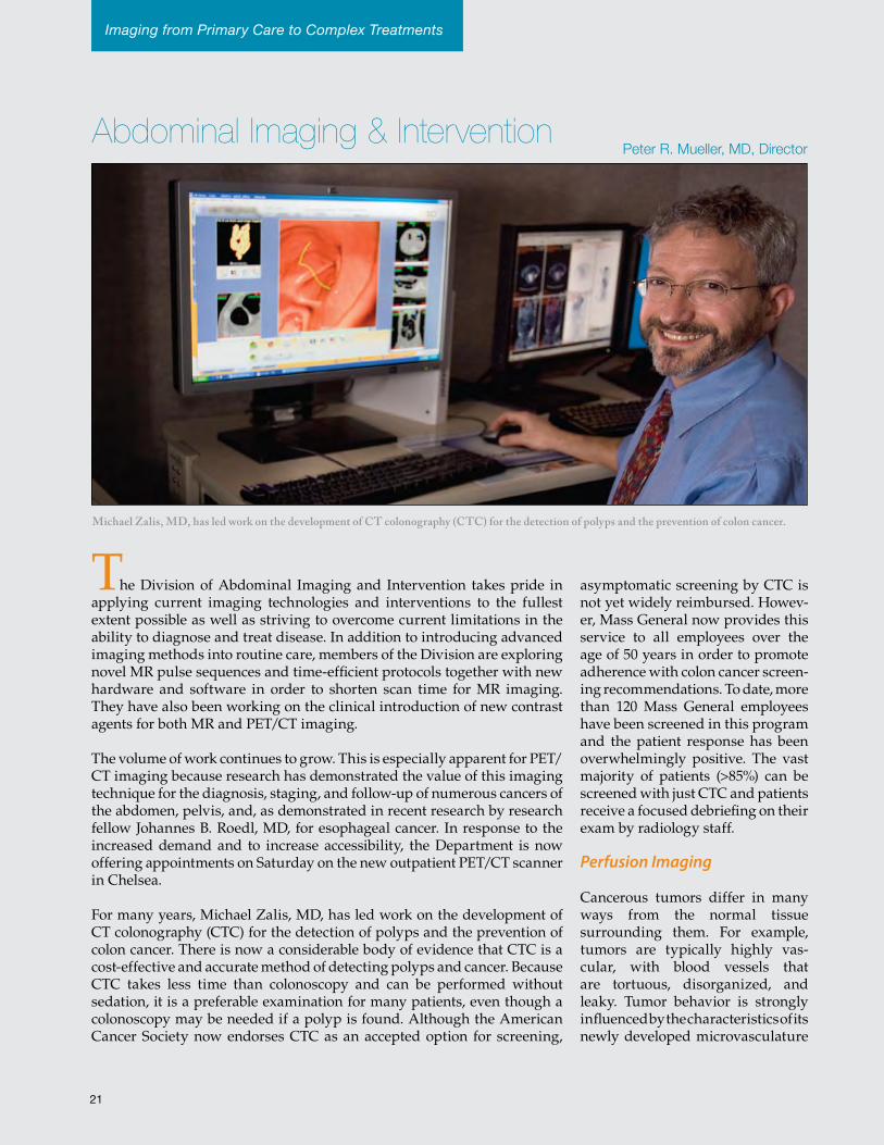
For many years, Michael Zalis, MD, has led work on the development of CT colonography (CTC) for the detection of polyps and the prevention of colon cancer. There is now a considerable body of evidence that CTC is a cost-effective and accurate method of detecting polyps and cancer. Because CTC takes less time than colonoscopy and can be performed without sedation, it is a preferable examination for many patients, even though a colonoscopy may be needed if a polyp is found. Although the American Cancer Society now endorses CTC as an accepted option for screening,
asymptomatic screening by CTC is not yet widely reimbursed. Howev-er, Mass General now provides this service to all employees over the age of 50 years in order to promote adherence with colon cancer screen-ing recommendations. To date, more than 120 Mass General employees have been screened in this program and the patient response has been overwhelmingly positive. The vast majority of patients (>85%) can be screened with just CTC and patients receive a focused debriefing on their exam by radiology staff.
Perfusion Imaging
Cancerous tumors differ in many ways from the normal tissue surrounding them. For example, tumors are typically highly vas-cular, with blood vessels that are tortuous, disorganized, and leaky. Tumor behavior is strongly influenced by the characteristics of its newly developed microvasculature
Michael Zalis, MD, has led work on the development of CT colonography (CTC) for the detection of polyps and the prevention of colon cancer.
Imaging from Primary Care to Complex Treatments
Peter R. Mueller, MD, Director
21
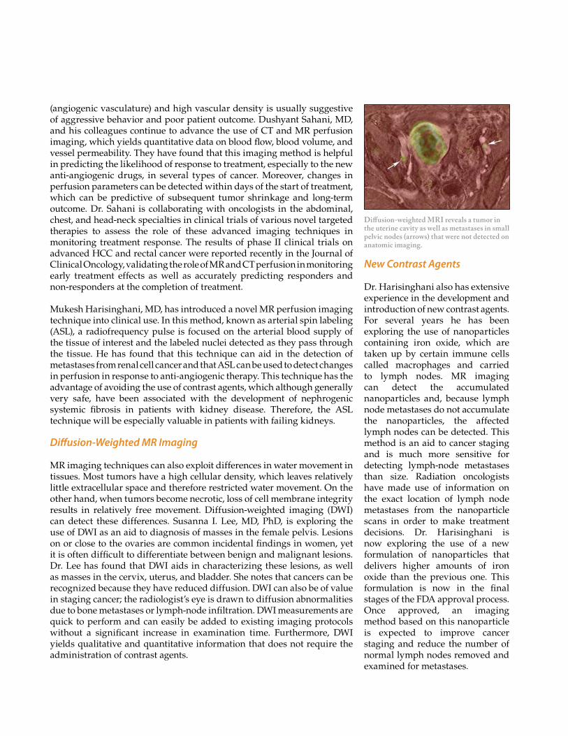
Diffusion-weighted MRI reveals a tumor in the uterine cavity as well as metastases in small pelvic nodes (arrows) that were not detected on anatomic imaging.
(angiogenic vasculature) and high vascular density is usually suggestive of aggressive behavior and poor patient outcome. Dushyant Sahani, MD, and his colleagues continue to advance the use of CT and MR perfusion imaging, which yields quantitative data on blood flow, blood volume, and vessel permeability. They have found that this imaging method is helpful in predicting the likelihood of response to treatment, especially to the new anti-angiogenic drugs, in several types of cancer. Moreover, changes in perfusion parameters can be detected within days of the start of treatment, which can be predictive of subsequent tumor shrinkage and long-term outcome. Dr. Sahani is collaborating with oncologists in the abdominal, chest, and head-neck specialties in clinical trials of various novel targeted therapies to assess the role of these advanced imaging techniques in monitoring treatment response. The results of phase II clinical trials on advanced HCC and rectal cancer were reported recently in the Journal of Clinical Oncology, validating the role of MR and CT perfusion in monitoring early treatment effects as well as accurately predicting responders and non-responders at the completion of treatment.
Mukesh Harisinghani, MD, has introduced a novel MR perfusion imaging technique into clinical use. In this method, known as arterial spin labeling (ASL), a radiofrequency pulse is focused on the arterial blood supply of the tissue of interest and the labeled nuclei detected as they pass through the tissue. He has found that this technique can aid in the detection of metastases from renal cell cancer and that ASL can be used to detect changes in perfusion in response to anti-angiogenic therapy. This technique has the advantage of avoiding the use of contrast agents, which although generally very safe, have been associated with the development of nephrogenic systemic fibrosis in patients with kidney disease. Therefore, the ASL technique will be especially valuable in patients with failing kidneys.
Diffusion-Weighted MR Imaging
MR imaging techniques can also exploit differences in water movement in tissues. Most tumors have a high cellular density, which leaves relatively little extracellular space and therefore restricted water movement. On the other hand, when tumors become necrotic, loss of cell membrane integrity results in relatively free movement. Diffusion-weighted imaging (DWI) can detect these differences. Susanna I. Lee, MD, PhD, is exploring the use of DWI as an aid to diagnosis of masses in the female pelvis. Lesions on or close to the ovaries are common incidental findings in women, yet it is often difficult to differentiate between benign and malignant lesions. Dr. Lee has found that DWI aids in characterizing these lesions, as well as masses in the cervix, uterus, and bladder. She notes that cancers can be recognized because they have reduced diffusion. DWI can also be of value in staging cancer; the radiologist’s eye is drawn to diffusion abnormalities due to bone metastases or lymph-node infiltration. DWI measurements are quick to perform and can easily be added to existing imaging protocols without a significant increase in examination time. Furthermore, DWI yields qualitative and quantitative information that does not require the administration of contrast agents.
New Contrast Agents
Dr. Harisinghani also has extensive experience in the development and introduction of new contrast agents. For several years he has been exploring the use of nanoparticles containing iron oxide, which are taken up by certain immune cells called macrophages and carried to lymph nodes. MR imaging can detect the accumulated nanoparticles and, because lymph node metastases do not accumulate the nanoparticles, the affected lymph nodes can be detected. This method is an aid to cancer staging and is much more sensitive for detecting lymph-node metastases than size. Radiation oncologists have made use of information on the exact location of lymph node metastases from the nanoparticle scans in order to make treatment decisions. Dr. Harisinghani is now exploring the use of a new formulation of nanoparticles that delivers higher amounts of iron oxide than the previous one. This formulation is now in the final stages of the FDA approval process. Once approved, an imaging method based on this nanoparticle is expected to improve cancer staging and reduce the number of normal lymph nodes removed and examined for metastases.
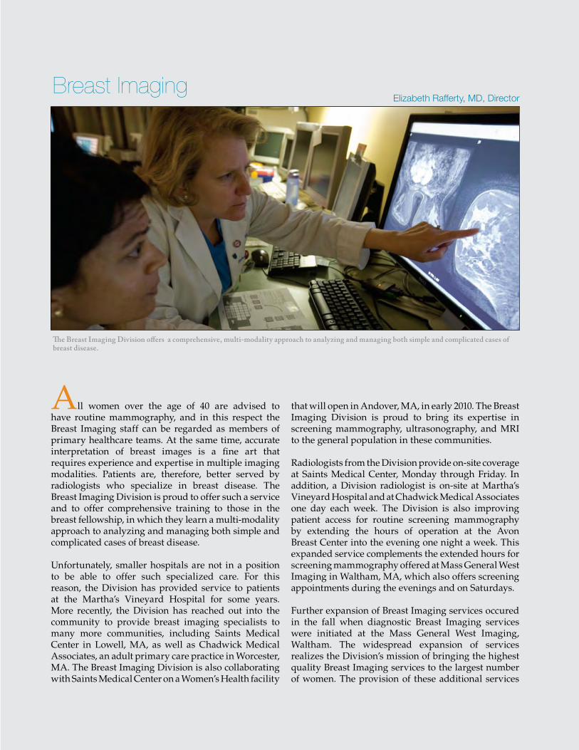
Nanoparticle structure with iron oxide core and dextran coating.
Abdominal Imaging & InterventionAlexander Guimaraes, MD, PhD, has been awarded an RSNA scholar grant to explore another use of the magnetic nanoparticles: detecting early stage Type I diabetes. In this disease, an autoimmune reaction attacks the pancreatic islets, which results in the accumulation of macrophages and inflammation in the islets, leading to their destruction. Because macrophages pick up the nanoparticles, Dr. Guimaraes hypothesized that insulitis, which precedes full-blown diabetes, could be detected using this agent. Dr. Guimaraes, under the direction of Ralph Weissleder, MD, PhD, in a collaborative effort with the Joslin Diabetes Center, has now worked out an effective three-stage MR imaging protocol. The protocol includes an initial non-contrast image, an image acquired soon after nanoparticle administration, and a third image 48 hours later. In a pilot study of patients with suspected early-onset diabetes, the investigators have found that inflammation can be detected in the first image taken after nanoparticle administration due to the high vascular permeability found in inflamed tissue, while the delayed image shows inflammation due to the accumulation of nanoparticle-laden macrophages.
Because the nanoparticle contrast agent has a prolonged intravascular half-life, Dr. Guimaraes is now investigating its use as an MR perfusion imaging agent in mice. His preliminary results demonstrate that this method is sensitive to changes in the microvasculature in response to anti-angiogenic therapy and often predicts response to therapy prior to tumor volumetric change. He hopes to soon translate these efforts to clinical studies of the response to cancer therapy in patients.
Thermal Ablation
The interventional radiologists have expanded their use of thermal ablation methods for treating small tumors. Procedures now include cryoablation and microwave ablation as well as radiofrequency ablation. These treatments slow disease progression in hepatocellular cancer patients who are waiting for a liver transplant or have other unresectable liver tumors. Thermal ablation therapy is also used in patients with renal tumors who are poor candidates for surgery or when there is a clinical need to preserve renal function. The treatments have a high technical success rate and deliver good outcomes with little post-procedural pain.
Volumetric pseudocolorized MR maps depicting the uptake of magnetic nanoparticles in the pancreas highlight the promise of this technique for detecting inflammation associated with early-onset type I diabetes (TID).
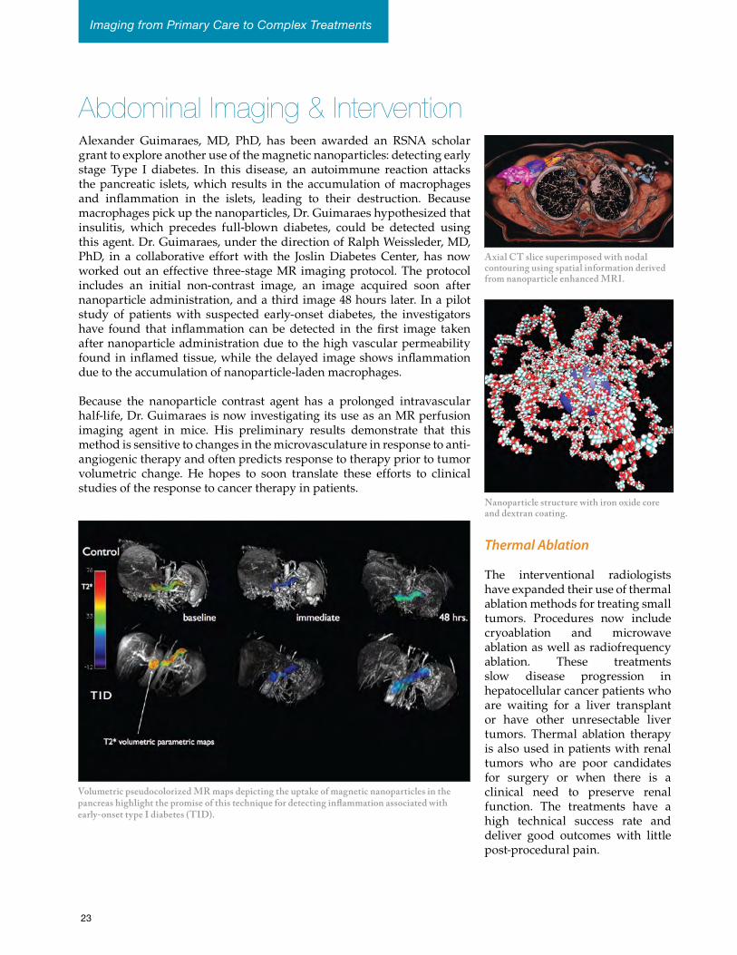
Axial CT slice superimposed with nodal contouring using spatial information derived from nanoparticle enhanced MRI.
23
Imaging from Primary Care to Complex Treatments
27
Elizabeth Rafferty, MD, Director
The Breast Imaging Division offers a comprehensive, multi-modality approach to analyzing and managing both simple and complicated cases of breast disease.
All women over the age of 40 are advised to have routine mammography, and in this respect the Breast Imaging staff can be regarded as members of primary healthcare teams. At the same time, accurate interpretation of breast images is a fine art that requires experience and expertise in multiple imaging modalities. Patients are, therefore, better served by radiologists who specialize in breast disease. The Breast Imaging Division is proud to offer such a service and to offer comprehensive training to those in the breast fellowship, in which they learn a multi-modality approach to analyzing and managing both simple and complicated cases of breast disease.
Unfortunately, smaller hospitals are not in a position to be able to offer such specialized care. For this reason, the Division has provided service to patients at the Martha’s Vineyard Hospital for some years. More recently, the Division has reached out into the community to provide breast imaging specialists to many more communities, including Saints Medical Center in Lowell, MA, as well as Chadwick Medical Associates, an adult primary care practice in Worcester, MA. The Breast Imaging Division is also collaborating with Saints Medical Center on a Women’s Health facility
that will open in Andover, MA, in early 2010. The Breast Imaging Division is proud to bring its expertise in screening mammography, ultrasonography, and MRI to the general population in these communities.
Radiologists from the Division provide on-site coverage at Saints Medical Center, Monday through Friday. In addition, a Division radiologist is on-site at Martha’s Vineyard Hospital and at Chadwick Medical Associates one day each week. The Division is also improving patient access for routine screening mammography by extending the hours of operation at the Avon Breast Center into the evening one night a week. This expanded service complements the extended hours for screening mammography offered at Mass General West Imaging in Waltham, MA, which also offers screening appointments during the evenings and on Saturdays.
Further expansion of Breast Imaging services occured in the fall when diagnostic Breast Imaging services were initiated at the Mass General West Imaging, Waltham. The widespread expansion of services realizes the Division’s mission of bringing the highest quality Breast Imaging services to the largest number of women. The provision of these additional services
Breast Imaging
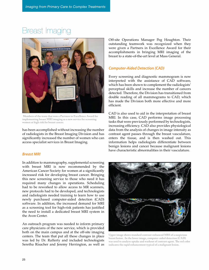
Members of the team that won a Partners in Excellence Award for implementing breast MRI imaging as a new service for screening women at high risk for breast cancer.
Breast Imaging
has been accomplished without increasing the number of radiologists in the Breast Imaging Division and has significantly increased the number of women who can access specialist services in Breast Imaging.
Breast MRI
In addition to mammography, supplemental screening with breast MRI is now recommended by the American Cancer Society for women at a significantly increased risk for developing breast cancer. Bringing this new screening service to those who need it has required many changes in operations. Scheduling had to be reworked to allow access to MR scanners, new protocols had to be developed, and technologists and radiologists needed training to learn how to use newly purchased computer-aided detection (CAD) software. In addition, the increased demand for MRI as a screening tool for high-risk patients has justified the need to install a dedicated breast MRI system in the Avon Center.
An outreach program was needed to inform primary care physicians of the new service, which is provided both on the main campus and at the off-site imaging centers. The team that put all these changes in place was led by Dr. Rafferty and included technologists Seretha Risacher and Jeremy Herrington, as well as
Off-site Operations Manager Peg Houghton. Their outstanding teamwork was recognized when they were given a Partners in Excellence Award for their accomplishments in bringing MRI imaging of the breast to a state-of-the-art level at Mass General.
Computer-Aided Detection (CAD)
Every screening and diagnostic mammogram is now interpreted with the assistance of CAD software, which has been shown to complement the radiologists’ perceptual skills and increase the number of cancers detected. Therefore, the Division has transitioned from double reading of all mammograms to CAD, which has made the Division both more effective and more efficient.
CAD is also used to aid in the interpretation of breast MRI. In this case, CAD performs image processing tasks that were previously performed by technologists, increasing efficiency. CAD also provides physiological data from the analysis of changes in image intensity as contrast agent passes through the breast vasculature, enters the tissue, and is then washed out. This information helps radiologists differentiate between benign lesions and cancer because malignant lesions have characteristic abnormalities in their vasculature.
Upper image shows standard contrast-enhanced MRI of a suspicious breast lesion . In the lower image, computer-aided detection (CAD) was used to analyze uptake and washout of contrast agent. The red color indicates the rapid enhancement typical of a malignant lesion.
25
Imaging from Primary Care to Complex Treatments
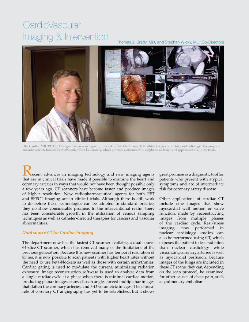
29
The Cardiac MR/PET/CT Program is a research group, directed by Udo Hoffmann, MD, which bridges cardiology and radiology. The program includes a newly funded CardioVascular Core Laboratory, which provides assistance with all phases of design and application of clinical trials.
Thomas J. Brady, MD, and Stephan Wicky, MD, Co-Directors
Recent advances in imaging technology and new imaging agents that are in clinical trials have made it possible to examine the heart and coronary arteries in ways that would not have been thought possible only a few years ago. CT scanners have become faster and produce images of higher resolution. New radiopharmaceutical agents for both PET and SPECT imaging are in clinical trials. Although there is still work to do before these technologies can be adopted in standard practice, they do show considerable promise. In the interventional realm, there has been considerable growth in the utilization of venous sampling techniques as well as catheter-directed therapies for cancers and vascular abnormalities.
Dual source CT for Cardiac Imaging
The department now has the fastest CT scanner available, a dual-source 64-slice CT scanner, which has removed many of the limitations of the previous generation. Because this new scanner has temporal resolution of 83 ms, it is now possible to scan patients with higher heart rates without the need to use beta-blockers as well as those with certain arrhythmias. Cardiac gating is used to modulate the current, minimizing radiation exposure. Image reconstruction software is used to analyze data from a single cardiac cycle at a phase when there is minimal cardiac motion, producing planar images at any chosen angle, curved multiplanar images that flatten the coronary arteries, and 3-D volumetric images. The clinical role of coronary CT angiography has yet to be established, but it shows
great promise as a diagnostic tool for patients who present with atypical symptoms and are of intermediate risk for coronary artery disease.
Other applications of cardiac CT include cine images that show myocardial wall motion or valve function, made by reconstructing images from multiple phases of the cardiac cycle. Rest/stress imaging, now performed in nuclear cardiology studies, can also be performed using CT, which exposes the patient to less radiation than nuclear cardiology while visualizing coronary arteries as well as myocardial perfusion. Because images of the lungs are included in these CT scans, they can, depending on the scan protocol, be examined for other causes of chest pain, such as pulmonary embolism.
CardioVascular Imaging & Intervention
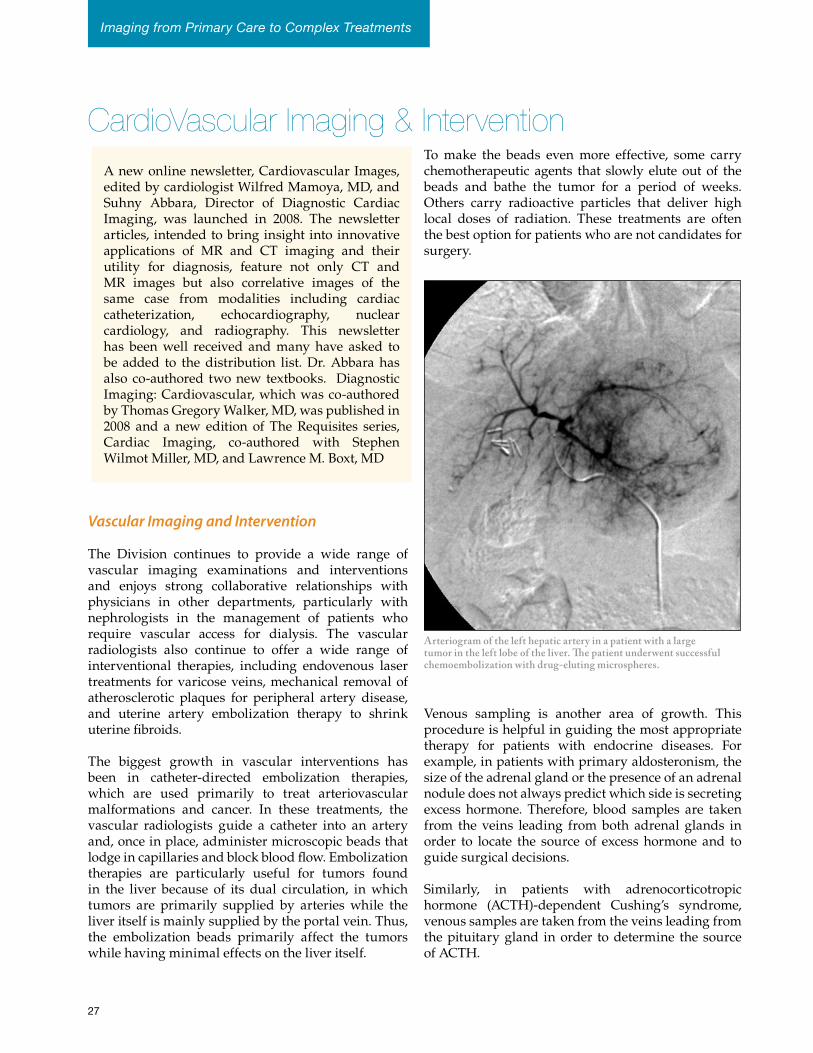
CardioVascular Imaging & InterventionTo make the beads even more effective, some carry chemotherapeutic agents that slowly elute out of the beads and bathe the tumor for a period of weeks. Others carry radioactive particles that deliver high local doses of radiation. These treatments are often the best option for patients who are not candidates for surgery.
Venous sampling is another area of growth. This procedure is helpful in guiding the most appropriate therapy for patients with endocrine diseases. For example, in patients with primary aldosteronism, the size of the adrenal gland or the presence of an adrenal nodule does not always predict which side is secreting excess hormone. Therefore, blood samples are taken from the veins leading from both adrenal glands in order to locate the source of excess hormone and to guide surgical decisions.
Similarly, in patients with adrenocorticotropic hormone (ACTH)-dependent Cushing’s syndrome, venous samples are taken from the veins leading from the pituitary gland in order to determine the source of ACTH.
Vascular Imaging and Intervention
The Division continues to provide a wide range of vascular imaging examinations and interventions and enjoys strong collaborative relationships with physicians in other departments, particularly with nephrologists in the management of patients who require vascular access for dialysis. The vascular radiologists also continue to offer a wide range of interventional therapies, including endovenous laser treatments for varicose veins, mechanical removal of atherosclerotic plaques for peripheral artery disease, and uterine artery embolization therapy to shrink uterine fibroids.
The biggest growth in vascular interventions has been in catheter-directed embolization therapies, which are used primarily to treat arteriovascular malformations and cancer. In these treatments, the vascular radiologists guide a catheter into an artery and, once in place, administer microscopic beads that lodge in capillaries and block blood flow. Embolization therapies are particularly useful for tumors found in the liver because of its dual circulation, in which tumors are primarily supplied by arteries while the liver itself is mainly supplied by the portal vein. Thus, the embolization beads primarily affect the tumors while having minimal effects on the liver itself.
Arteriogram of the left hepatic artery in a patient with a large tumor in the left lobe of the liver. The patient underwent successful chemoembolization with drug-eluting microspheres.
A new online newsletter, Cardiovascular Images, edited by cardiologist Wilfred Mamoya, MD, and Suhny Abbara, Director of Diagnostic Cardiac Imaging, was launched in 2008. The newsletter articles, intended to bring insight into innovative applications of MR and CT imaging and their utility for diagnosis, feature not only CT and MR images but also correlative images of the same case from modalities including cardiac catheterization, echocardiography, nuclear cardiology, and radiography. This newsletter has been well received and many have asked to be added to the distribution list. Dr. Abbara has also co-authored two new textbooks. Diagnostic Imaging: Cardiovascular, which was co-authored by Thomas Gregory Walker, MD, was published in 2008 and a new edition of The Requisites series, Cardiac Imaging, co-authored with Stephen Wilmot Miller, MD, and Lawrence M. Boxt, MD
Imaging from Primary Care to Complex Treatments
27
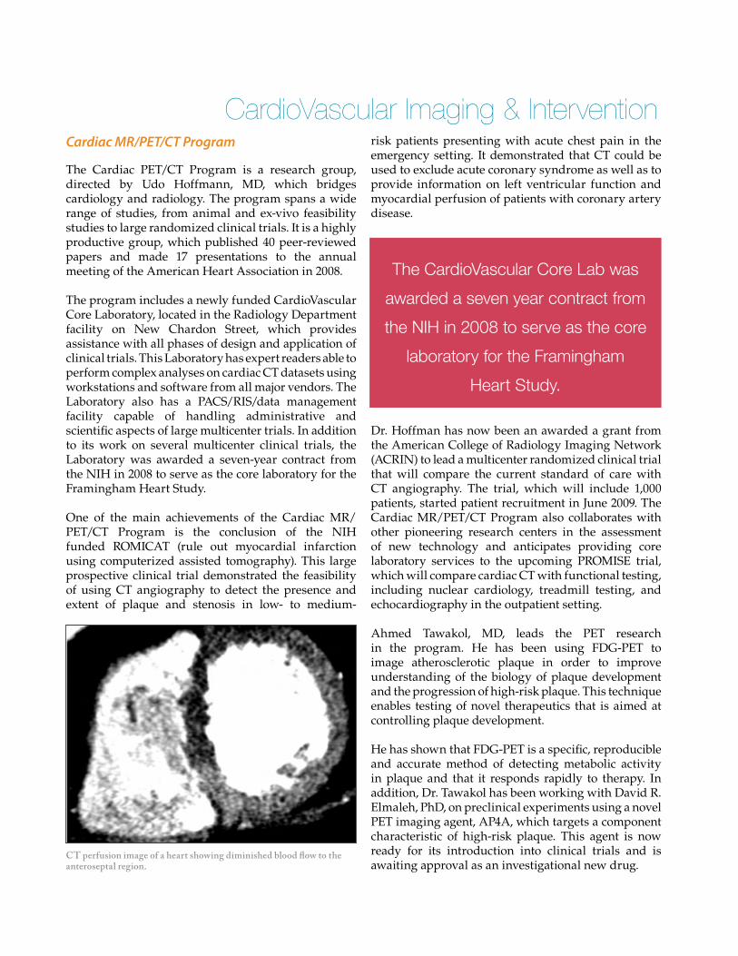
CardioVascular Imaging & Interventionrisk patients presenting with acute chest pain in the emergency setting. It demonstrated that CT could be used to exclude acute coronary syndrome as well as to provide information on left ventricular function and myocardial perfusion of patients with coronary artery disease.
Dr. Hoffman has now been an awarded a grant from the American College of Radiology Imaging Network (ACRIN) to lead a multicenter randomized clinical trial that will compare the current standard of care with CT angiography. The trial, which will include 1,000 patients, started patient recruitment in June 2009. The Cardiac MR/PET/CT Program also collaborates with other pioneering research centers in the assessment of new technology and anticipates providing core laboratory services to the upcoming PROMISE trial, which will compare cardiac CT with functional testing, including nuclear cardiology, treadmill testing, and echocardiography in the outpatient setting.
Ahmed Tawakol, MD, leads the PET research in the program. He has been using FDG-PET to image atherosclerotic plaque in order to improve understanding of the biology of plaque development and the progression of high-risk plaque. This technique enables testing of novel therapeutics that is aimed at controlling plaque development.
He has shown that FDG-PET is a specific, reproducible and accurate method of detecting metabolic activity in plaque and that it responds rapidly to therapy. In addition, Dr. Tawakol has been working with David R. Elmaleh, PhD, on preclinical experiments using a novel PET imaging agent, AP4A, which targets a component characteristic of high-risk plaque. This agent is now ready for its introduction into clinical trials and is awaiting approval as an investigational new drug.
Cardiac MR/PET/CT Program
The Cardiac PET/CT Program is a research group, directed by Udo Hoffmann, MD, which bridges cardiology and radiology. The program spans a wide range of studies, from animal and ex-vivo feasibility studies to large randomized clinical trials. It is a highly productive group, which published 40 peer-reviewed papers and made 17 presentations to the annual meeting of the American Heart Association in 2008.
The program includes a newly funded CardioVascular Core Laboratory, located in the Radiology Department facility on New Chardon Street, which provides assistance with all phases of design and application of clinical trials. This Laboratory has expert readers able to perform complex analyses on cardiac CT datasets using workstations and software from all major vendors. The Laboratory also has a PACS/RIS/data management facility capable of handling administrative and scientific aspects of large multicenter trials. In addition to its work on several multicenter clinical trials, the Laboratory was awarded a seven-year contract from the NIH in 2008 to serve as the core laboratory for the Framingham Heart Study.
One of the main achievements of the Cardiac MR/PET/CT Program is the conclusion of the NIH funded ROMICAT (rule out myocardial infarction using computerized assisted tomography). This large prospective clinical trial demonstrated the feasibility of using CT angiography to detect the presence and extent of plaque and stenosis in low- to medium-
CT perfusion image of a heart showing diminished blood flow to the anteroseptal region.
The CardioVascular Core Lab was
awarded a seven year contract from
the NIH in 2008 to serve as the core
laboratory for the Framingham
Heart Study.
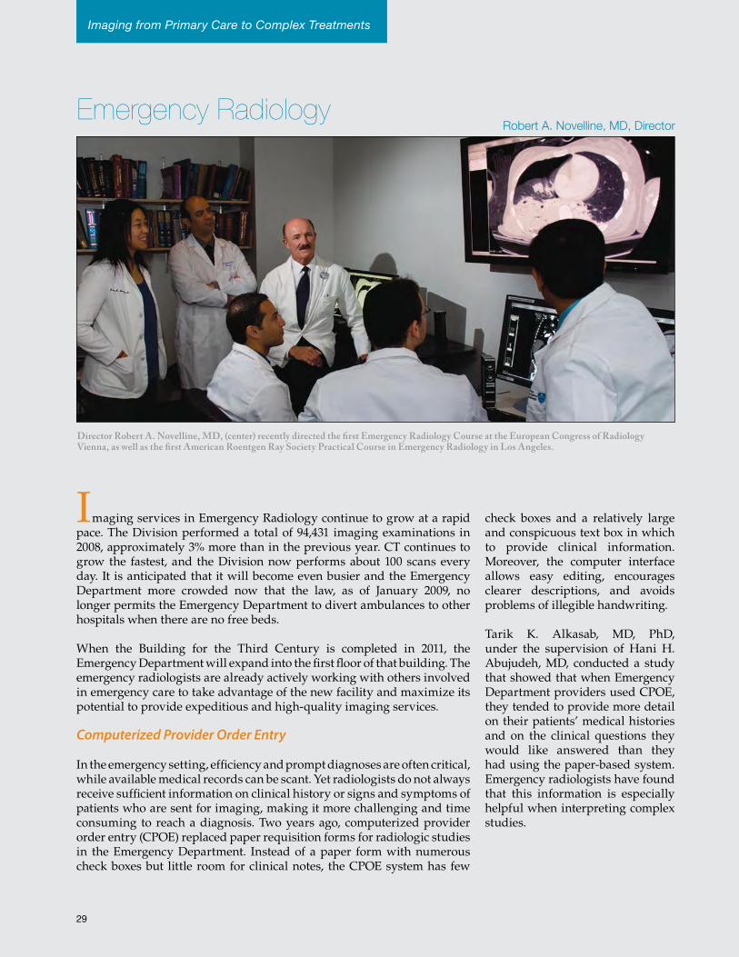
32
Director Robert A. Novelline, MD, (center) recently directed the first Emergency Radiology Course at the European Congress of Radiology Vienna, as well as the first American Roentgen Ray Society Practical Course in Emergency Radiology in Los Angeles.
Robert A. Novelline, MD, DirectorEmergency Radiology
Imaging services in Emergency Radiology continue to grow at a rapid pace. The Division performed a total of 94,431 imaging examinations in 2008, approximately 3% more than in the previous year. CT continues to grow the fastest, and the Division now performs about 100 scans every day. It is anticipated that it will become even busier and the Emergency Department more crowded now that the law, as of January 2009, no longer permits the Emergency Department to divert ambulances to other hospitals when there are no free beds.
When the Building for the Third Century is completed in 2011, the Emergency Department will expand into the first floor of that building. The emergency radiologists are already actively working with others involved in emergency care to take advantage of the new facility and maximize its potential to provide expeditious and high-quality imaging services.
Computerized Provider Order Entry
In the emergency setting, efficiency and prompt diagnoses are often critical, while available medical records can be scant. Yet radiologists do not always receive sufficient information on clinical history or signs and symptoms of patients who are sent for imaging, making it more challenging and time consuming to reach a diagnosis. Two years ago, computerized provider order entry (CPOE) replaced paper requisition forms for radiologic studies in the Emergency Department. Instead of a paper form with numerous check boxes but little room for clinical notes, the CPOE system has few
check boxes and a relatively large and conspicuous text box in which to provide clinical information. Moreover, the computer interface allows easy editing, encourages clearer descriptions, and avoids problems of illegible handwriting.
Tarik K. Alkasab, MD, PhD, under the supervision of Hani H. Abujudeh, MD, conducted a study that showed that when Emergency Department providers used CPOE, they tended to provide more detail on their patients’ medical histories and on the clinical questions they would like answered than they had using the paper-based system. Emergency radiologists have found that this information is especially helpful when interpreting complex studies.
Imaging from Primary Care to Complex Treatments
29
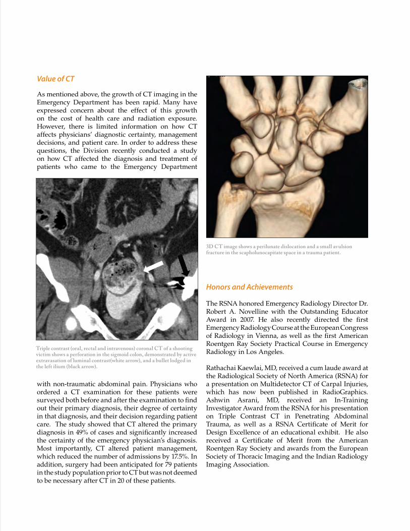
Triple contrast (oral, rectal and intravenous) coronal CT of a shooting victim shows a perforation in the sigmoid colon, demonstrated by active extravasation of luminal contrast(white arrow), and a bullet lodged in the left ilium (black arrow).
Value of CT
As mentioned above, the growth of CT imaging in the Emergency Department has been rapid. Many have expressed concern about the effect of this growth on the cost of health care and radiation exposure. However, there is limited information on how CT affects physicians’ diagnostic certainty, management decisions, and patient care. In order to address these questions, the Division recently conducted a study on how CT affected the diagnosis and treatment of patients who came to the Emergency Department
with non-traumatic abdominal pain. Physicians who ordered a CT examination for these patients were surveyed both before and after the examination to find out their primary diagnosis, their degree of certainty in that diagnosis, and their decision regarding patient care. The study showed that CT altered the primary diagnosis in 49% of cases and significantly increased the certainty of the emergency physician’s diagnosis. Most importantly, CT altered patient management, which reduced the number of admissions by 17.5%. In addition, surgery had been anticipated for 79 patients in the study population prior to CT but was not deemed to be necessary after CT in 20 of these patients.
Honors and Achievements
The RSNA honored Emergency Radiology Director Dr. Robert A. Novelline with the Outstanding Educator Award in 2007. He also recently directed the first Emergency Radiology Course at the European Congress of Radiology in Vienna, as well as the first American Roentgen Ray Society Practical Course in Emergency Radiology in Los Angeles.
Rathachai Kaewlai, MD, received a cum laude award at the Radiological Society of North America (RSNA) for a presentation on Multidetector CT of Carpal Injuries, which has now been published in RadioGraphics. Ashwin Asrani, MD, received an In-Training Investigator Award from the RSNA for his presentation on Triple Contrast CT in Penetrating Abdominal Trauma, as well as a RSNA Certificate of Merit for Design Excellence of an educational exhibit. He also received a Certificate of Merit from the American Roentgen Ray Society and awards from the European Society of Thoracic Imaging and the Indian Radiology Imaging Association.
3D CT image shows a perilunate dislocation and a small avulsion fracture in the scapholunocapitate space in a trauma patient.
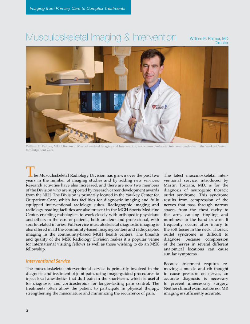
34
William E. Palmer, MD, Director of Musculoskeletal Imaging and Intervention, in the musculoskeletal interventional suite in the Yawkey Center for Outpatient Care.
William E. Palmer, MD Director
Musculoskeletal Imaging & Intervention
The Musculoskeletal Radiology Division has grown over the past two years in the number of imaging studies and by adding new services. Research activities have also increased, and there are now two members of the Division who are supported by research career development awards from the NIH. The Division is primarily located in the Yawkey Center for Outpatient Care, which has facilities for diagnostic imaging and fully equipped interventional radiology suites. Radiographic imaging and radiology reading facilities are also present in the MGH Sports Medicine Center, enabling radiologists to work closely with orthopedic physicians and others in the care of patients, both amateur and professional, with sports-related injuries. Full-service musculoskeletal diagnostic imaging is also offered in all the community-based imaging centers and radiographic imaging in the community-based MGH health centers. The breadth and quality of the MSK Radiology Division makes it a popular venue for international visiting fellows as well as those wishing to do an MSK fellowship.
Interventional ServiceThe musculoskeletal interventional service is primarily involved in the diagnosis and treatment of joint pain, using image-guided procedures to inject local anesthetics that dull pain in the short-term, which is useful for diagnosis, and corticosteroids for longer-lasting pain control. The treatments often allow the patient to participate in physical therapy, strengthening the musculature and minimizing the recurrence of pain.
The latest musculoskeletal inter-ventional service, introduced by Martin Torriani, MD, is for the diagnosis of neurogenic thoracic outlet syndrome. This syndrome results from compression of the nerves that pass through narrow spaces from the chest cavity to the arm, causing tingling and numbness in the hand or arm. It frequently occurs after injury to the soft tissue in the neck. Thoracic outlet syndrome is difficult to diagnose because compression of the nerves in several different anatomical locations can cause similar symptoms.
Because treatment requires re-moving a muscle and rib thought to cause pressure on nerves, an accurate diagnosis is necessary to prevent unnecessary surgery. Neither clinical examination nor MR imaging is sufficiently accurate.
Imaging from Primary Care to Complex Treatments
31
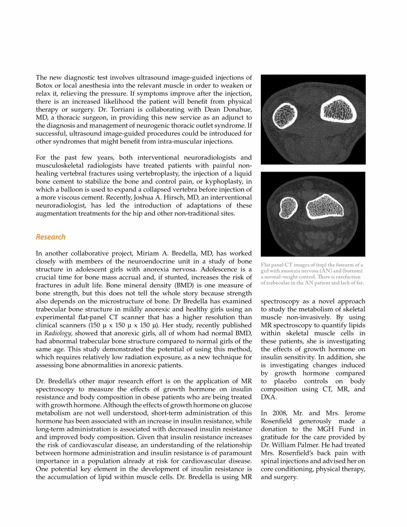
Flat panel CT images of (top) the forearm of a girl with anorexia nervosa (AN) and (bottom) a normal-weight control. There is rarefaction of trabeculae in the AN patient and lack of fat.
The new diagnostic test involves ultrasound image-guided injections of Botox or local anesthesia into the relevant muscle in order to weaken or relax it, relieving the pressure. If symptoms improve after the injection, there is an increased likelihood the patient will benefit from physical therapy or surgery. Dr. Torriani is collaborating with Dean Donahue, MD, a thoracic surgeon, in providing this new service as an adjunct to the diagnosis and management of neurogenic thoracic outlet syndrome. If successful, ultrasound image-guided procedures could be introduced for other syndromes that might benefit from intra-muscular injections.
For the past few years, both interventional neuroradiologists and musculoskeletal radiologists have treated patients with painful non-healing vertebral fractures using vertebroplasty, the injection of a liquid bone cement to stabilize the bone and control pain, or kyphoplasty, in which a balloon is used to expand a collapsed vertebra before injection of a more viscous cement. Recently, Joshua A. Hirsch, MD, an interventional neuroradiologist, has led the introduction of adaptations of these augmentation treatments for the hip and other non-traditional sites.
Research
In another collaborative project, Miriam A. Bredella, MD, has worked closely with members of the neuroendocrine unit in a study of bone structure in adolescent girls with anorexia nervosa. Adolescence is a crucial time for bone mass accrual and, if stunted, increases the risk of fractures in adult life. Bone mineral density (BMD) is one measure of bone strength, but this does not tell the whole story because strength also depends on the microstructure of bone. Dr Bredella has examined trabecular bone structure in mildly anorexic and healthy girls using an experimental flat-panel CT scanner that has a higher resolution than clinical scanners (150 μ x 150 μ x 150 μ). Her study, recently published in Radiology, showed that anorexic girls, all of whom had normal BMD, had abnormal trabecular bone structure compared to normal girls of the same age. This study demonstrated the potential of using this method, which requires relatively low radiation exposure, as a new technique for assessing bone abnormalities in anorexic patients.
Dr. Bredella’s other major research effort is on the application of MR spectroscopy to measure the effects of growth hormone on insulin resistance and body composition in obese patients who are being treated with growth hormone. Although the effects of growth hormone on glucose metabolism are not well understood, short-term administration of this hormone has been associated with an increase in insulin resistance, while long-term administration is associated with decreased insulin resistance and improved body composition. Given that insulin resistance increases the risk of cardiovascular disease, an understanding of the relationship between hormone administration and insulin resistance is of paramount importance in a population already at risk for cardiovascular disease. One potential key element in the development of insulin resistance is the accumulation of lipid within muscle cells. Dr. Bredella is using MR
spectroscopy as a novel approach to study the metabolism of skeletal muscle non-invasively. By using MR spectroscopy to quantify lipids within skeletal muscle cells in these patients, she is investigating the effects of growth hormone on insulin sensitivity. In addition, she is investigating changes induced by growth hormone compared to placebo controls on body composition using CT, MR, and DXA.
In 2008, Mr. and Mrs. Jerome Rosenfield generously made a donation to the MGH Fund in gratitude for the care provided by Dr. William Palmer. He had treated Mrs. Rosenfield’s back pain with spinal injections and advised her on core conditioning, physical therapy, and surgery.
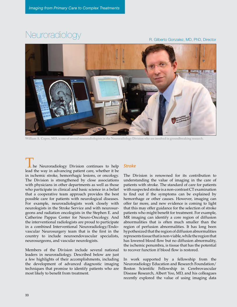
36
Imaging from Primary Care to Complex Treatments
William A. Copen, MD, is one of several neuroradiologists in the Neuroradiology Division who are involved in groundbreaking research.
R. Gilberto Gonzalez, MD, PhD, Director
The Neuroradiology Division continues to help lead the way in advancing patient care, whether it be in ischemic stroke, hemorrhagic lesions, or oncology. The Division is strengthened by close associations with physicians in other departments as well as those who participate in clinical and basic science in a belief that a cooperative team approach provides the best possible care for patients with neurological diseases. For example, neuroradiologists work closely with neurologists in the Stroke Service and with neurosur-geons and radiation oncologists in the Stephen E. and Catherine Pappas Center for Neuro-Oncology. And the interventional radiologists are proud to participate in a combined Interventional Neuroradiology/Endo- vascular Neurosurgery team that is the first in the country to include neuroendovascular specialists, neurosurgeons, and vascular neurologists.
Members of the Division include several national leaders in neuroradiology. Described below are just a few highlights of their accomplishments, including the development of advanced diagnostic imaging techniques that promise to identify patients who are most likely to benefit from treatment.
Stroke
The Division is renowned for its contribution to understanding the value of imaging in the care of patients with stroke. The standard of care for patients with suspected stroke is a non-contrast CT examination to find out if the symptoms can be explained by hemorrhage or other causes. However, imaging can offer far more, and new evidence is coming to light that this may offer guidance for the selection of stroke patients who might benefit for treatment. For example, MR imaging can identify a core region of diffusion abnormalities that is often much smaller than the region of perfusion abnormalities. It has long been hypothesized that the region of diffusion abnormalities represents tissue that is non-viable, while the region that has lowered blood flow but no diffusion abnormality, the ischemic penumbra, is tissue that has the potential to recover function if blood flow is restored.
In work supported by a fellowship from the Neuroradiology Education and Research Foundation/Boston Scientific Fellowship in Cerebrovascular Disease Research, Albert Yoo, MD, and his colleagues recently explored the value of using imaging data
Neuroradiology
33
Together, these studies provide strong evidence of the value of perfusion and diffusion imaging and may lead not only to a paradigm shift in the way that stroke patients are treated but also hope that there will be a reduction in the number of people who are severely disabled by strokes. Furthermore, although many institutions do not have the facilities to perform diffusion-weighted or perfusion-weighted MR imaging of acute stroke patients, they can identify patients with proximal arterial occlusions using CT angiography. Because time to treatment appears less critical than in the past, these images could be used to select patients who may benefit from intra-arterial interventions, such as clot retrieval or intra-arterial thrombolysis. These patients could then be transferred to a stroke center where advanced imaging and interventional facilities are available.
Cerebral Hemorrhage
Approximately 10-15% of strokes are caused by hemorrhage within the brain (intraparenchymal hemorrhage). A number of underlying pathologies cause these strokes, and it is important to determine the cause in order to manage the patient correctly and avoid further bleeding whenever possible. Although ruptured vascular lesions are a relatively uncommon cause of these strokes, it is especially important to identify them because they are potentially treatable. Javier M. Romero, MD, led the largest study to date to assess the diagnostic accuracy of multidetector CT angiography for detecting vascular lesions in cases of intraparenchymal hemorrhage: a retrospective review of 623 consecutive cases over a period of nine years.
to select patients for intra-arterial therapy. They discovered that patients whose diffusion abnormalities were larger than 70 ml fared poorly even if blood flow was restored. On the other hand, if the lesion was smaller than this and reperfusion was successful within 3.5 hours after imaging, patients recovered well. This ground-breaking research is the first real evidence of the benefits of imaging data for the selection of patients for treatment rather than time from symptom onset.
Current guidelines recommend clot-busting therapy only if the patient arrives for treatment within three hours of symptom onset. However, William A. Copen, MD, and his colleagues found, unexpectedly, that the presence of an ischemic penumbra is common after nine hours, especially in patients with a proximal arterial occlusion, the same group of patients as in Dr. Yoo’s study. This finding has the potential to expand the accepted window of time in which patients will benefit from intra-arterial therapy.
In a pooled analysis of the MERCI and Multi MERCI Trials of arterial interventional therapies, Raul G. Nogueira, MD, and his colleagues, also obtained evidence that successful intervention can be beneficial many hours after symptom onset. In this cohort of patients, who were selected without benefit of advanced imaging techniques and treated within eight hours from stroke onset, Dr. Nogueira demonstrated that vessel recanalization was one of the most important predictors of good outcome, while time from symptom onset to reperfusion failed to predict outcomes. In 2009, Dr. Nogueira received the Robert G. Siekert New Investigator Award in Stroke from the American Heart Association for this work.
Dr. Nogueira is also the Principal Investigator for the DAWN trial, which is using diffusion and perfusion imaging to triage patients with wake-up strokes or who arrive too late to qualify for standard treatment. This trial will test the hypothesis that imaging-based endovascular therapy is safe and improves outcomes in stroke patients with proximal anterior circulation occlusions who present beyond the typical therapeutic window. The preliminary data of the DAWN trial were recently presented at the American Academy of Neurology meeting, and Dr. Nogueira’s abstract was one of two from the neurovascular field that was included in the Highlight Session of that meeting.
Evidence of the value of perfusion
and diffusion imaging may lead to
a paradigm shift in the way that
stroke patients are treated.
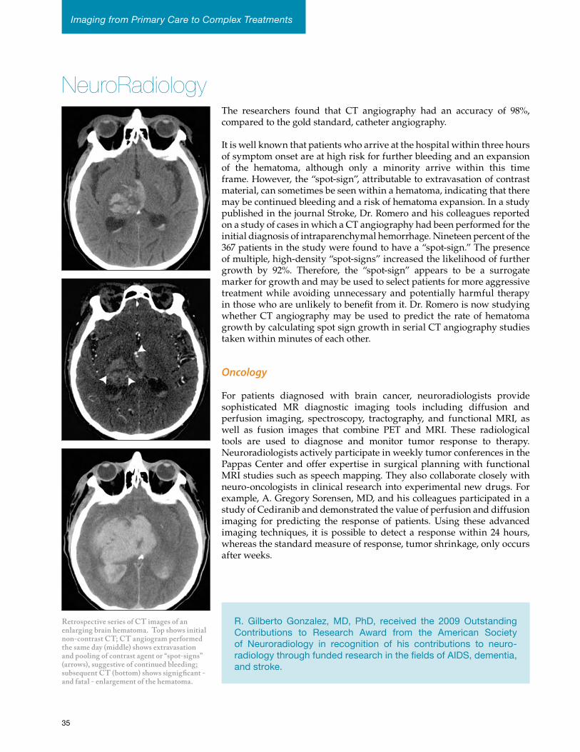
Retrospective series of CT images of an enlarging brain hematoma. Top shows initial non-contrast CT; CT angiogram performed the same day (middle) shows extravasation and pooling of contrast agent or “spot-signs” (arrows), suggestive of continued bleeding; subsequent CT (bottom) shows signigficant - and fatal - enlargement of the hematoma.
NeuroRadiologyThe researchers found that CT angiography had an accuracy of 98%, compared to the gold standard, catheter angiography.
It is well known that patients who arrive at the hospital within three hours of symptom onset are at high risk for further bleeding and an expansion of the hematoma, although only a minority arrive within this time frame. However, the “spot-sign”, attributable to extravasation of contrast material, can sometimes be seen within a hematoma, indicating that there may be continued bleeding and a risk of hematoma expansion. In a study published in the journal Stroke, Dr. Romero and his colleagues reported on a study of cases in which a CT angiography had been performed for the initial diagnosis of intraparenchymal hemorrhage. Nineteen percent of the 367 patients in the study were found to have a “spot-sign.” The presence of multiple, high-density “spot-signs” increased the likelihood of further growth by 92%. Therefore, the “spot-sign” appears to be a surrogate marker for growth and may be used to select patients for more aggressive treatment while avoiding unnecessary and potentially harmful therapy in those who are unlikely to benefit from it. Dr. Romero is now studying whether CT angiography may be used to predict the rate of hematoma growth by calculating spot sign growth in serial CT angiography studies taken within minutes of each other.
Oncology
For patients diagnosed with brain cancer, neuroradiologists provide sophisticated MR diagnostic imaging tools including diffusion and perfusion imaging, spectroscopy, tractography, and functional MRI, as well as fusion images that combine PET and MRI. These radiological tools are used to diagnose and monitor tumor response to therapy. Neuroradiologists actively participate in weekly tumor conferences in the Pappas Center and offer expertise in surgical planning with functional MRI studies such as speech mapping. They also collaborate closely with neuro-oncologists in clinical research into experimental new drugs. For example, A. Gregory Sorensen, MD, and his colleagues participated in a study of Cediranib and demonstrated the value of perfusion and diffusion imaging for predicting the response of patients. Using these advanced imaging techniques, it is possible to detect a response within 24 hours, whereas the standard measure of response, tumor shrinkage, only occurs after weeks.
R. Gilberto Gonzalez,MD, PhD, received the 2009 OutstandingContributions to Research Award from the American Societyof Neuroradiology in recognition of his contributions to neuro-radiologythroughfundedresearchinthefieldsofAIDS,dementia,andstroke.
35
Imaging from Primary Care to Complex Treatments
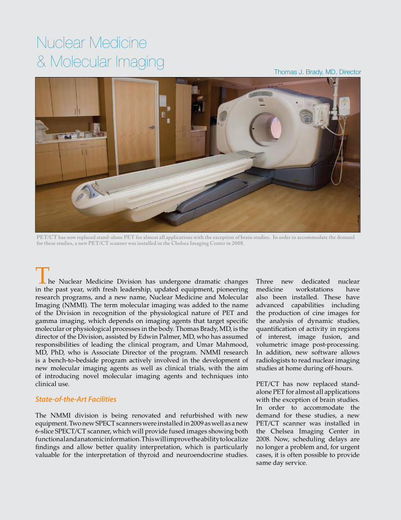
39
PET/CT has now replaced stand-alone PET for almost all applications with the exception of brain studies. In order to accommodate the demand for these studies, a new PET/CT scanner was installed in the Chelsea Imaging Center in 2008.
The Nuclear Medicine Division has undergone dramatic changes in the past year, with fresh leadership, updated equipment, pioneering research programs, and a new name, Nuclear Medicine and Molecular Imaging (NMMI). The term molecular imaging was added to the name of the Division in recognition of the physiological nature of PET and gamma imaging, which depends on imaging agents that target specific molecular or physiological processes in the body. Thomas Brady, MD, is the director of the Division, assisted by Edwin Palmer, MD, who has assumed responsibilities of leading the clinical program, and Umar Mahmood, MD, PhD, who is Associate Director of the program. NMMI research is a bench-to-bedside program actively involved in the development of new molecular imaging agents as well as clinical trials, with the aim of introducing novel molecular imaging agents and techniques into clinical use.
State-of-the-Art Facilities
The NMMI division is being renovated and refurbished with new equipment. Two new SPECT scanners were installed in 2009 as well as a new 6-slice SPECT/CT scanner, which will provide fused images showing both functional and anatomic information. This will improve the ability to localize findings and allow better quality interpretation, which is particularly valuable for the interpretation of thyroid and neuroendocrine studies.
Three new dedicated nuclear medicine workstations have also been installed. These have advanced capabilities including the production of cine images for the analysis of dynamic studies, quantification of activity in regions of interest, image fusion, and volumetric image post-processing. In addition, new software allows radiologists to read nuclear imaging studies at home during off-hours.
PET/CT has now replaced stand-alone PET for almost all applications with the exception of brain studies. In order to accommodate the demand for these studies, a new PET/CT scanner was installed in the Chelsea Imaging Center in 2008. Now, scheduling delays are no longer a problem and, for urgent cases, it is often possible to provide same day service.
Thomas J. Brady, MD, Director
Nuclear Medicine & Molecular Imaging
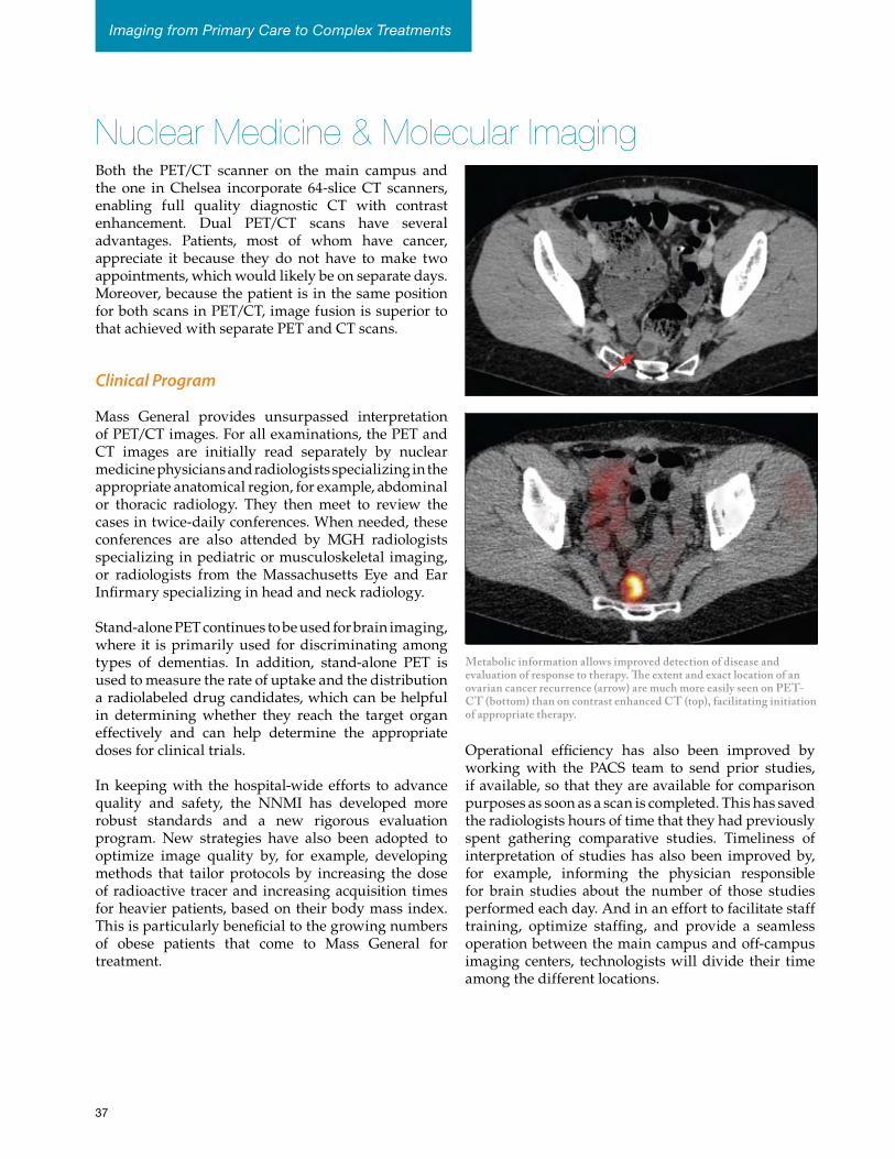
Nuclear Medicine & Molecular ImagingBoth the PET/CT scanner on the main campus and the one in Chelsea incorporate 64-slice CT scanners, enabling full quality diagnostic CT with contrast enhancement. Dual PET/CT scans have several advantages. Patients, most of whom have cancer, appreciate it because they do not have to make two appointments, which would likely be on separate days. Moreover, because the patient is in the same position for both scans in PET/CT, image fusion is superior to that achieved with separate PET and CT scans.
Clinical Program
Mass General provides unsurpassed interpretation of PET/CT images. For all examinations, the PET and CT images are initially read separately by nuclear medicine physicians and radiologists specializing in the appropriate anatomical region, for example, abdominal or thoracic radiology. They then meet to review the cases in twice-daily conferences. When needed, these conferences are also attended by MGH radiologists specializing in pediatric or musculoskeletal imaging, or radiologists from the Massachusetts Eye and Ear Infirmary specializing in head and neck radiology.
Stand-alone PET continues to be used for brain imaging, where it is primarily used for discriminating among types of dementias. In addition, stand-alone PET is used to measure the rate of uptake and the distribution a radiolabeled drug candidates, which can be helpful in determining whether they reach the target organ effectively and can help determine the appropriate doses for clinical trials.
In keeping with the hospital-wide efforts to advance quality and safety, the NNMI has developed more robust standards and a new rigorous evaluation program. New strategies have also been adopted to optimize image quality by, for example, developing methods that tailor protocols by increasing the dose of radioactive tracer and increasing acquisition times for heavier patients, based on their body mass index. This is particularly beneficial to the growing numbers of obese patients that come to Mass General for treatment.
Operational efficiency has also been improved by working with the PACS team to send prior studies, if available, so that they are available for comparison purposes as soon as a scan is completed. This has saved the radiologists hours of time that they had previously spent gathering comparative studies. Timeliness of interpretation of studies has also been improved by, for example, informing the physician responsible for brain studies about the number of those studies performed each day. And in an effort to facilitate staff training, optimize staffing, and provide a seamless operation between the main campus and off-campus imaging centers, technologists will divide their time among the different locations.
Metabolic information allows improved detection of disease and evaluation of response to therapy. The extent and exact location of an ovarian cancer recurrence (arrow) are much more easily seen on PET-CT (bottom) than on contrast enhanced CT (top), facilitating initiation of appropriate therapy.
37
Imaging from Primary Care to Complex Treatments
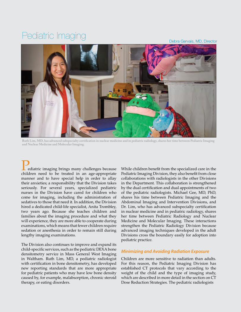
41
Ruth Lim, MD, has advanced subspecialty certification in nuclear medicine and in pediatric radiology, shares her time between Pediatric Imaging and Nuclear Medicine and Molecular Imaging.
Pediatric imaging brings many challenges because children need to be treated in an age-appropriate manner and to have special help in order to allay their anxieties; a responsibility that the Division takes seriously. For several years, specialized pediatric nurses in the Division have cared for children who come for imaging, including the administration of sedatives to those that need it. In addition, the Division hired a dedicated child-life specialist, Anita Trombley, two years ago. Because she teaches children and families about the imaging procedure and what they will experience, they are more able to cooperate during examinations, which means that fewer children require sedation or anesthesia in order to remain still during lengthy imaging examinations.
The Division also continues to improve and expand its child-specific services, such as the pediatric DEXA bone densitometry service in Mass General West Imaging in Waltham. Ruth Lim, MD, a pediatric radiologist with certification in bone densitometry, has developed new reporting standards that are more appropriate for pediatric patients who may have low bone density caused by, for example, malabsorption, chronic steroid therapy, or eating disorders.
While children benefit from the specialized care in the Pediatric Imaging Division, they also benefit from close collaborations with radiologists in the other Divisions in the Department. This collaboration is strengthened by the dual certification and dual appointments of two of the pediatric radiologists. Michael Gee, MD, PhD, shares his time between Pediatric Imaging and the Abdominal Imaging and Intervention Divisions, and Dr. Lim, who has advanced subspecialty certification in nuclear medicine and in pediatric radiology, shares her time between Pediatric Radiology and Nuclear Medicine and Molecular Imaging. These interactions strengthen the Pediatric Radiology Division because advanced imaging techniques developed in the adult Divisions cross the boundary easily for adoption into pediatric practice.
Minimizing and Avoiding Radiation Exposure Children are more sensitive to radiation than adults. For this reason, the Pediatric Imaging Division has established CT protocols that vary according to the weight of the child and the type of imaging study, which are described in more detail in the section on CT Dose Reduction Strategies. The pediatric radiologists
Debra Gervais, MD, DirectorPediatric Imaging
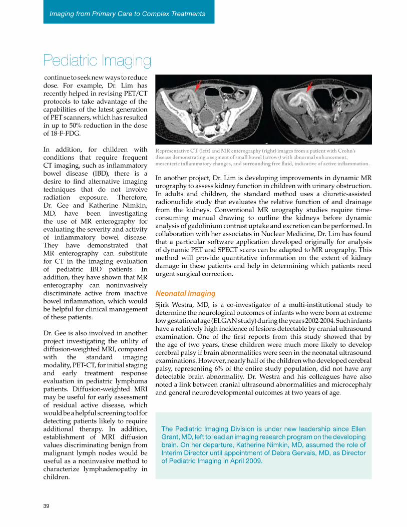
Pediatric Imaging continue to seek new ways to reduce dose. For example, Dr. Lim has recently helped in revising PET/CT protocols to take advantage of the capabilities of the latest generation of PET scanners, which has resulted in up to 50% reduction in the dose of 18-F-FDG.
In addition, for children with conditions that require frequent CT imaging, such as inflammatory bowel disease (IBD), there is a desire to find alternative imaging techniques that do not involve radiation exposure. Therefore, Dr. Gee and Katherine Nimkin, MD, have been investigating the use of MR enterography for evaluating the severity and activity of inflammatory bowel disease. They have demonstrated that MR enterography can substitute for CT in the imaging evaluation of pediatric IBD patients. In addition, they have shown that MR enterography can noninvasively discriminate active from inactive bowel inflammation, which would be helpful for clinical management of these patients.
Dr. Gee is also involved in another project investigating the utility of diffusion-weighted MRI, compared with the standard imaging modality, PET-CT, for initial staging and early treatment response evaluation in pediatric lymphoma patients. Diffusion-weighted MRI may be useful for early assessment of residual active disease, which would be a helpful screening tool for detecting patients likely to require additional therapy. In addition, establishment of MRI diffusion values discriminating benign from malignant lymph nodes would be useful as a noninvasive method to characterize lymphadenopathy in children.
In another project, Dr. Lim is developing improvements in dynamic MR urography to assess kidney function in children with urinary obstruction. In adults and children, the standard method uses a diuretic-assisted radionuclide study that evaluates the relative function of and drainage from the kidneys. Conventional MR urography studies require time-consuming manual drawing to outline the kidneys before dynamic analysis of gadolinium contrast uptake and excretion can be performed. In collaboration with her associates in Nuclear Medicine, Dr. Lim has found that a particular software application developed originally for analysis of dynamic PET and SPECT scans can be adapted to MR urography. This method will provide quantitative information on the extent of kidney damage in these patients and help in determining which patients need urgent surgical correction.
Neonatal ImagingSjirk Westra, MD, is a co-investigator of a multi-institutional study to determine the neurological outcomes of infants who were born at extreme low gestational age (ELGAN study) during the years 2002-2004. Such infants have a relatively high incidence of lesions detectable by cranial ultrasound examination. One of the first reports from this study showed that by the age of two years, these children were much more likely to develop cerebral palsy if brain abnormalities were seen in the neonatal ultrasound examinations. However, nearly half of the children who developed cerebral palsy, representing 6% of the entire study population, did not have any detectable brain abnormality. Dr. Westra and his colleagues have also noted a link between cranial ultrasound abnormalities and microcephaly and general neurodevelopmental outcomes at two years of age.
Representative CT (left) and MR enterography (right) images from a patient with Crohn’s disease demonstrating a segment of small bowel (arrows) with abnormal enhancement, mesenteric inflammatory changes, and surrounding free fluid, indicative of active inflammation.
ThePediatric ImagingDivision isundernewleadershipsinceEllenGrant,MD,lefttoleadanimagingresearchprogramonthedevelopingbrain.Onherdeparture,KatherineNimkin,MD,assumedtheroleofInterimDirectoruntilappointmentofDebraGervais,MD,asDirectorofPediatricImaginginApril2009.
39
Imaging from Primary Care to Complex Treatments
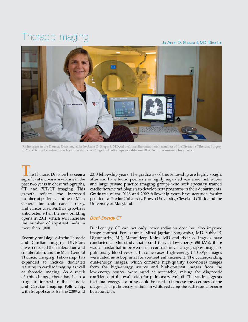
43
Radiologists in the Thoracic Division, led by Jo-Anne O. Shepard, MD, (above), in collaboration with members of the Division of Thoracic Surgery at Mass General, continue to be leaders in the use of CT-guided radiofrequency ablation (RFA) in the treatment of lung cancer.
The Thoracic Division has seen a significant increase in volume in the past two years in chest radiographs, CT, and PET/CT imaging. This growth reflects the increased number of patients coming to Mass General for acute care, surgery, and cancer care. Further growth is anticipated when the new building opens in 2011, which will increase the number of inpatient beds to more than 1,000.
Recently radiologists in the Thoracic and Cardiac Imaging Divisions have increased their interaction and collaboration, and the Mass General Thoracic Imaging Fellowship has expanded to include dedicated training in cardiac imaging as well as thoracic imaging. As a result of this change, there has been a surge in interest in the Thoracic and Cardiac Imaging Fellowship, with 64 applicants for the 2009 and
2010 fellowship years. The graduates of this fellowship are highly sought after and have found positions in highly regarded academic institutions and large private practice imaging groups who seek specialty trained cardiothoracic radiologists to develop new programs in their departments. Graduates of the 2008 and 2009 fellowship years have accepted faculty positions at Baylor University, Brown University, Cleveland Clinic, and the University of Maryland.
Dual-Energy CT
Dual-energy CT can not only lower radiation dose but also improve image contrast. For example, Minal Jagtiani Sangwaiya, MD, Subba R. Digumarthy, MD, Mannudeep Kalra, MD and their colleagues have conducted a pilot study that found that, at low-energy (80 kVp), there was a substantial improvement in contrast in CT angiography images of pulmonary blood vessels. In some cases, high-energy (140 kVp) images were rated as suboptimal for contrast enhancement. The corresponding dual-energy images, which combine high-quality (low-noise) images from the high-energy source and high-contrast images from the low-energy source, were rated as acceptable, raising the diagnostic confidence of the evaluation for pulmonary emboli. The study suggests that dual-energy scanning could be used to increase the accuracy of the diagnosis of pulmonary embolism while reducing the radiation exposure by about 28%.
Jo-Anne O. Shepard, MD, DirectorThoracic Imaging
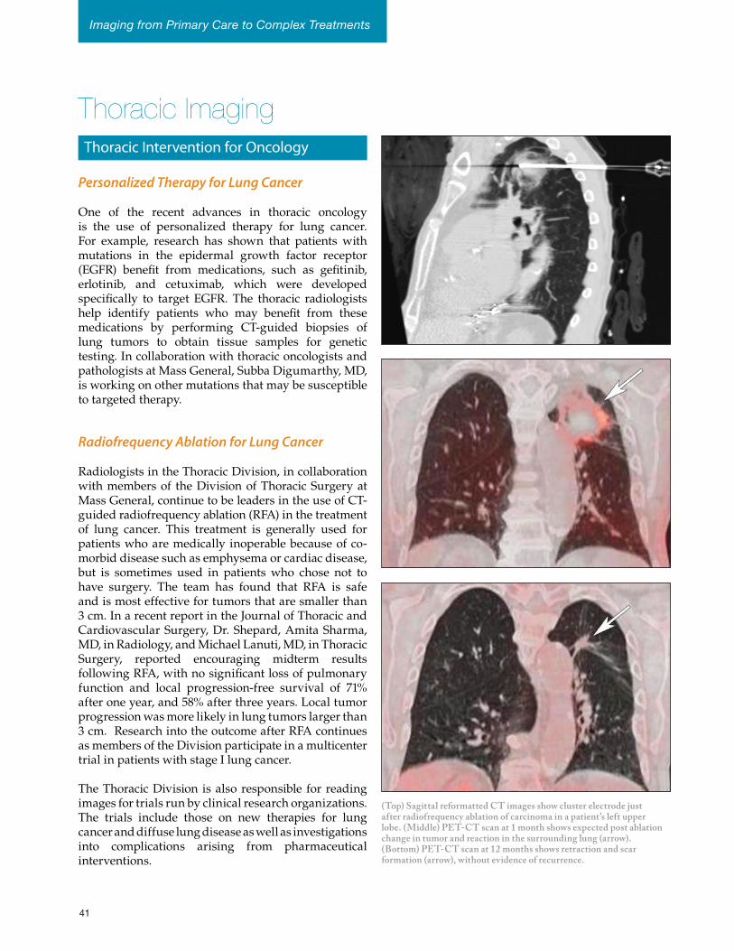
Thoracic Imaging
(Top) Sagittal reformatted CT images show cluster electrode just after radiofrequency ablation of carcinoma in a patient’s left upper lobe. (Middle) PET-CT scan at 1 month shows expected post ablation change in tumor and reaction in the surrounding lung (arrow). (Bottom) PET-CT scan at 12 months shows retraction and scar formation (arrow), without evidence of recurrence.
Personalized Therapy for Lung Cancer
One of the recent advances in thoracic oncology is the use of personalized therapy for lung cancer. For example, research has shown that patients with mutations in the epidermal growth factor receptor (EGFR) benefit from medications, such as gefitinib, erlotinib, and cetuximab, which were developed specifically to target EGFR. The thoracic radiologists help identify patients who may benefit from these medications by performing CT-guided biopsies of lung tumors to obtain tissue samples for genetic testing. In collaboration with thoracic oncologists and pathologists at Mass General, Subba Digumarthy, MD, is working on other mutations that may be susceptible to targeted therapy.
Radiofrequency Ablation for Lung Cancer
Radiologists in the Thoracic Division, in collaboration with members of the Division of Thoracic Surgery at Mass General, continue to be leaders in the use of CT-guided radiofrequency ablation (RFA) in the treatment of lung cancer. This treatment is generally used for patients who are medically inoperable because of co-morbid disease such as emphysema or cardiac disease, but is sometimes used in patients who chose not to have surgery. The team has found that RFA is safe and is most effective for tumors that are smaller than 3 cm. In a recent report in the Journal of Thoracic and Cardiovascular Surgery, Dr. Shepard, Amita Sharma, MD, in Radiology, and Michael Lanuti, MD, in Thoracic Surgery, reported encouraging midterm results following RFA, with no significant loss of pulmonary function and local progression-free survival of 71% after one year, and 58% after three years. Local tumor progression was more likely in lung tumors larger than 3 cm. Research into the outcome after RFA continues as members of the Division participate in a multicenter trial in patients with stage I lung cancer.
The Thoracic Division is also responsible for reading images for trials run by clinical research organizations. The trials include those on new therapies for lung cancer and diffuse lung disease as well as investigations into complications arising from pharmaceutical interventions.
Thoracic Intervention for Oncology
41
Imaging from Primary Care to Complex Treatments
Staff at Work
Michael Van Nostrand, RT(R), Ellison 2 Imaging
Chuck Huberdeau, RT(R)(MR), Mass General / North Shore Center for Outpatient Care
Julie Henry, RTR(M) Mammographer & Technical Manager CT Staff
MGH Chelsea HealthCare Staff & Student
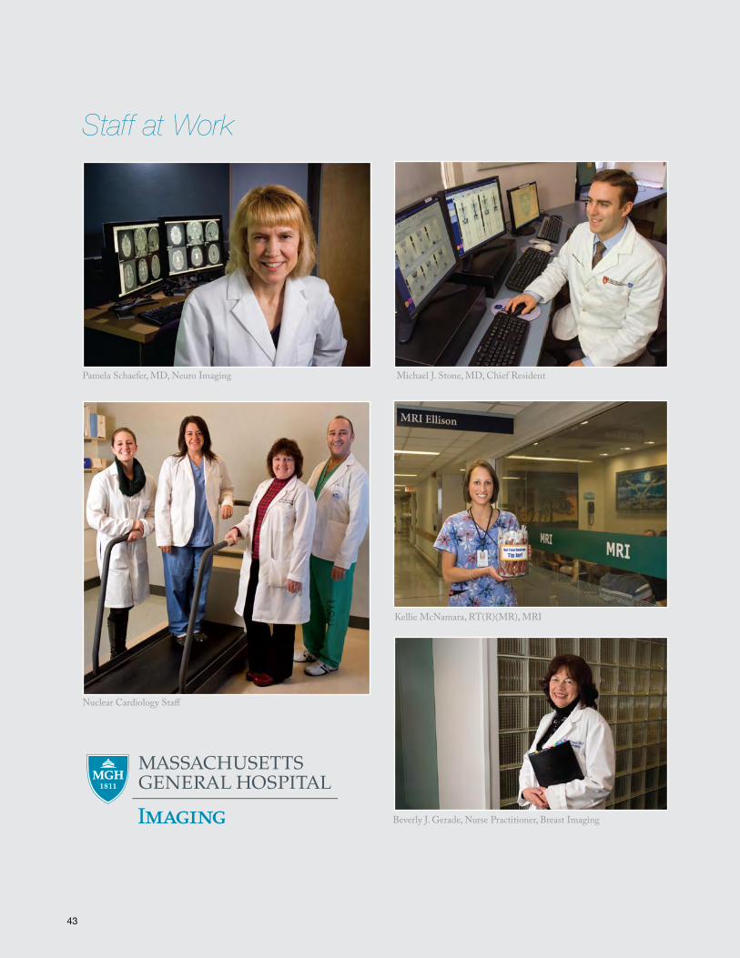
46
Staff at Work
Nuclear Cardiology Staff
Michael J. Stone, MD, Chief Resident
Beverly J. Gerade, Nurse Practitioner, Breast Imaging
Pamela Schaefer, MD, Neuro Imaging
Kellie McNamara, RT(R)(MR), MRI
43
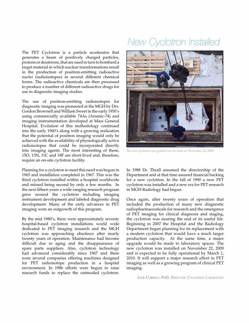
47
New Cyclotron InstalledThe PET Cyclotron is a particle accelerator that generates a beam of positively charged particles, protons or deuterons, that are used in turn to bombard a target material in which nuclear transformations result in the production of positron-emitting radioactive nuclei (radioisotopes) in several different chemical forms. The radioactive chemicals are then processed to produce a number of different radioactive drugs for use in diagnostic imaging studies.
The use of positron-emitting radioisotopes for diagnostic imaging was pioneered at the MGH by Drs. Gordon Brownell and William Sweet in the early 1950’s using commercially available 74As (Arsenic-74) and imaging instrumentation developed at Mass General Hospital. Evolution of this methodology continued into the early 1960’s along with a growing realization that the potential of positron imaging would only be achieved with the availability of physiologically active radioisotopes that could be incorporated directly into imaging agents. The most interesting of these, 15O, 13N, 11C and 18F are short-lived and, therefore, require an on-site cyclotron facility.
Planning for a cyclotron to meet this need was begun in 1965 and installation completed in 1967. This was the third cyclotron installed within a hospital worldwide and missed being second by only a few months. In the next fifteen years a wide ranging research program grew around the cyclotron including imaging instrument development and labeled diagnostic drug development. Many of the early advances in PET imaging were an outgrowth of this program.
By the mid 1980’s, there were approximately seventy hospital-based cyclotron installations world wide dedicated to PET imaging research and the MGH cyclotron was approaching obsolesce after nearly twenty years of operation. Maintenance had become difficult due to aging and the disappearance of spare parts suppliers. Also, cyclotron technology had advanced considerably since 1967 and there were several companies offering machines designed for PET radioisotope production in a hospital environment. In 1986 efforts were begun to raise research funds to replace the outmoded cyclotron.
In 1988 Dr. Thrall assumed the directorship of the Department and at that time assured financial backing for a new cyclotron. In the fall of 1990 a new PET cyclotron was installed and a new era for PET research in MGH Radiology had begun.
Once again, after twenty years of operation that included the production of many new diagnostic radiopharmaceuticals for research and the emergence of PET imaging for clinical diagnosis and staging, the cyclotron was nearing the end of its useful life. Beginning in 2007 the Hospital and the Radiology Department began planning for its replacement with a modern cyclotron that would have a much larger production capacity. At the same time, a major upgrade would be made to laboratory spaces. The new cyclotron was installed on November 22, 2009 and is expected to be fully operational by March 1, 2010. It will support a major research effort in PET imaging as well as a growing program of clinical PET imaging.
The new cyclotron was guided into its vault on November 22, 2009.
Jack correia, PhD, Director, cyclotron laboratory
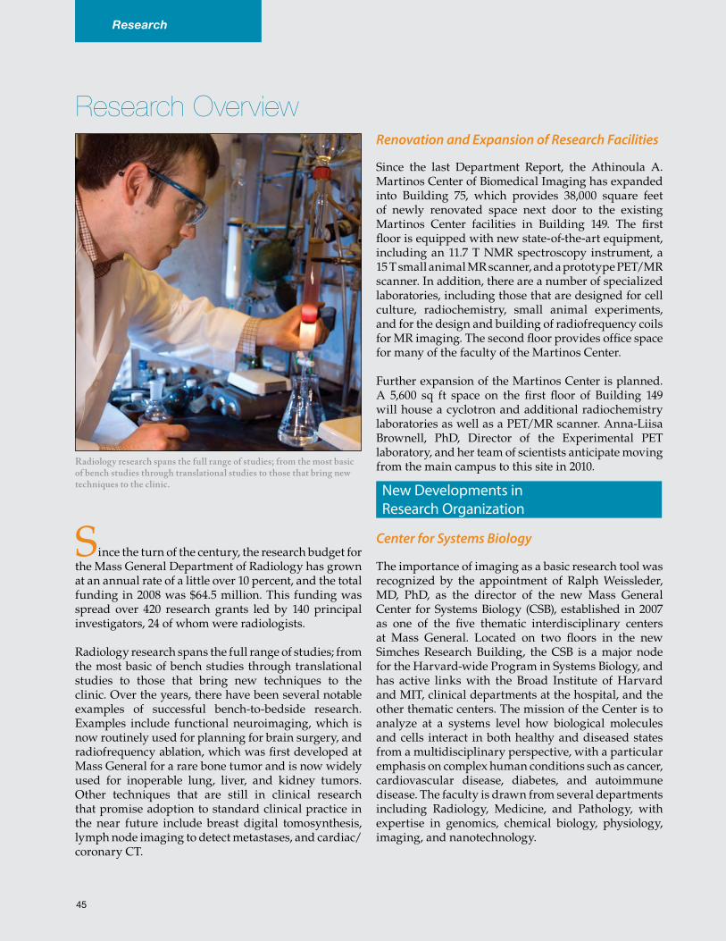
48
Research Overview
Radiology research spans the full range of studies; from the most basic of bench studies through translational studies to those that bring new techniques to the clinic.
Research
Since the turn of the century, the research budget for the Mass General Department of Radiology has grown at an annual rate of a little over 10 percent, and the total funding in 2008 was $64.5 million. This funding was spread over 420 research grants led by 140 principal investigators, 24 of whom were radiologists.
Radiology research spans the full range of studies; from the most basic of bench studies through translational studies to those that bring new techniques to the clinic. Over the years, there have been several notable examples of successful bench-to-bedside research. Examples include functional neuroimaging, which is now routinely used for planning for brain surgery, and radiofrequency ablation, which was first developed at Mass General for a rare bone tumor and is now widely used for inoperable lung, liver, and kidney tumors. Other techniques that are still in clinical research that promise adoption to standard clinical practice in the near future include breast digital tomosynthesis, lymph node imaging to detect metastases, and cardiac/coronary CT.
Renovation and Expansion of Research Facilities
Since the last Department Report, the Athinoula A. Martinos Center of Biomedical Imaging has expanded into Building 75, which provides 38,000 square feet of newly renovated space next door to the existing Martinos Center facilities in Building 149. The first floor is equipped with new state-of-the-art equipment, including an 11.7 T NMR spectroscopy instrument, a 15 T small animal MR scanner, and a prototype PET/MR scanner. In addition, there are a number of specialized laboratories, including those that are designed for cell culture, radiochemistry, small animal experiments, and for the design and building of radiofrequency coils for MR imaging. The second floor provides office space for many of the faculty of the Martinos Center.
Further expansion of the Martinos Center is planned. A 5,600 sq ft space on the first floor of Building 149 will house a cyclotron and additional radiochemistry laboratories as well as a PET/MR scanner. Anna-Liisa Brownell, PhD, Director of the Experimental PET laboratory, and her team of scientists anticipate moving from the main campus to this site in 2010.
Center for Systems Biology
The importance of imaging as a basic research tool was recognized by the appointment of Ralph Weissleder, MD, PhD, as the director of the new Mass General Center for Systems Biology (CSB), established in 2007 as one of the five thematic interdisciplinary centers at Mass General. Located on two floors in the new Simches Research Building, the CSB is a major node for the Harvard-wide Program in Systems Biology, and has active links with the Broad Institute of Harvard and MIT, clinical departments at the hospital, and the other thematic centers. The mission of the Center is to analyze at a systems level how biological molecules and cells interact in both healthy and diseased states from a multidisciplinary perspective, with a particular emphasis on complex human conditions such as cancer, cardiovascular disease, diabetes, and autoimmune disease. The faculty is drawn from several departments including Radiology, Medicine, and Pathology, with expertise in genomics, chemical biology, physiology, imaging, and nanotechnology.
New Developments in Research Organization
45
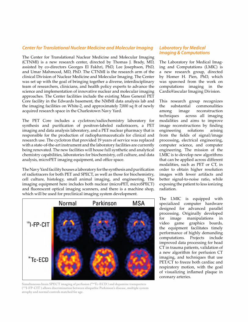
Laboratory for Medical Imaging & Computations
The Laboratory for Medical Imag-ing and Computations (LMIC) is a new research group, directed by Homer H. Pien, PhD, which was spawned from the work on computations imaging in the CardioVascular Imaging Division.
This research group recognizes the substantial commonalities among image reconstruction techniques across all imaging modalities and aims to improve image reconstructions by finding engineering solutions arising from the fields of signal/image processing, electrical engineering, computer science, and computer engineering. The mission of the LMIC is to develop new algorithms that can be applied across different modalities, such as PET or CT, in order to obtain higher resolution images with fewer artifacts and better signal-to-noise ratio, while exposing the patient to less ionizing radiation.
The LMIC is equipped with specialized computer hardware designed for advanced parallel processing. Originally developed for image manipulations in video game graphics boards, the equipment facilitates timely performance of highly demanding computations. Projects include improved data processing for head CT in trauma patients, validation of a new algorithm for perfusion CT imaging, and techniques that use PET/CT to freeze both cardiac and respiratory motion, with the goal of visualizing inflamed plaque in coronary arteries.
Center for Translational Nuclear Medicine and Molecular Imaging
The Center for Translational Nuclear Medicine and Molecular Imaging (CTNMI) is a new research center, directed by Thomas J. Brady, MD, assisted by co-directors Georges El Fakhri, PhD, Lee Josephson, PhD, and Umar Mahmood, MD, PhD. The CTNMI is the research arm of the clinical Division of Nuclear Medicine and Molecular Imaging. The Center was set up with the goal of bringing together a diverse, interdisciplinary team of researchers, clinicians, and health policy experts to advance the science and implementation of innovative nuclear and molecular imaging approaches. The Center facilities include the existing Mass General PET Core facility in the Edwards basement, the NMMI data analysis lab and the imaging facilities on White-2, and approximately 7,000 sq ft of newly acquired research space in the Charlestown Navy Yard.
The PET Core includes a cyclotron/radiochemistry laboratory for synthesis and purification of positron-labeled radiotracers, a PET imaging and data analysis laboratory, and a PET nuclear pharmacy that is responsible for the production of radiopharmaceuticals for clinical and research use. The cyclotron that provided 19 years of service was replaced with a state-of-the-art instrument and the laboratory facilities are currently being renovated. The new facilities will house full synthetic and analytical chemistry capabilities, laboratories for biochemistry, cell culture, and data analysis, microPET imaging equipment, and office space.
The Navy Yard facility houses a laboratory for the synthesis and purification of radiotracers for both PET and SPECT, as well as those for biochemistry, cell culture, histology, small animal imaging, and engineering. The imaging equipment here includes both nuclear (microPET, microSPECT) and fluorescent optical imaging scanners, and there is a machine shop, which will be used for preclinical imaging system development.
Simultaneous brain SPECT imaging of perfusion (99mTc-ECD ) and dopamine transporters (123I-FP-CIT ) allows discrimination between idiopathic Parkinson’s disease, multiple system atrophy and normal controls matched for age.
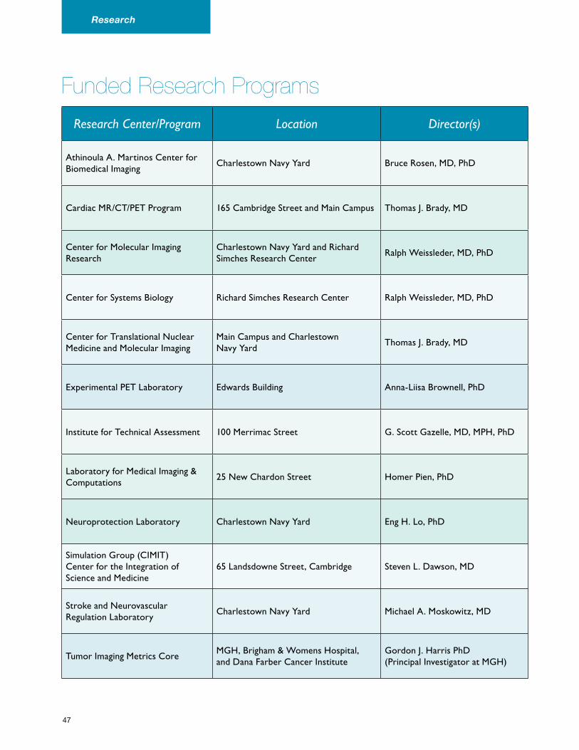
Funded Research Programs
Research Center/Program Location Director(s)
Athinoula A. Martinos Center for Biomedical Imaging
Charlestown Navy Yard Bruce Rosen, MD, PhD
Cardiac MR/CT/PET Program 165 Cambridge Street and Main Campus Thomas J. Brady, MD
Center for Molecular Imaging Research
Charlestown Navy Yard and Richard Simches Research Center
Ralph Weissleder, MD, PhD
Center for Systems Biology Richard Simches Research Center Ralph Weissleder, MD, PhD
Center for Translational Nuclear Medicine and Molecular Imaging
Main Campus and Charlestown Navy Yard
Thomas J. Brady, MD
Experimental PET Laboratory Edwards Building Anna-Liisa Brownell, PhD
Institute for Technical Assessment 100 Merrimac Street G. Scott Gazelle, MD, MPH, PhD
Laboratory for Medical Imaging & Computations
25 New Chardon Street Homer Pien, PhD
Neuroprotection Laboratory Charlestown Navy Yard Eng H. Lo, PhD
Simulation Group (CIMIT) Center for the Integration of Science and Medicine
65 Landsdowne Street, Cambridge Steven L. Dawson, MD
Stroke and Neurovascular Regulation Laboratory
Charlestown Navy Yard Michael A. Moskowitz, MD
Tumor Imaging Metrics CoreMGH, Brigham & Womens Hospital, and Dana Farber Cancer Institute
Gordon J. Harris PhD (Principal Investigator at MGH)
47
Research
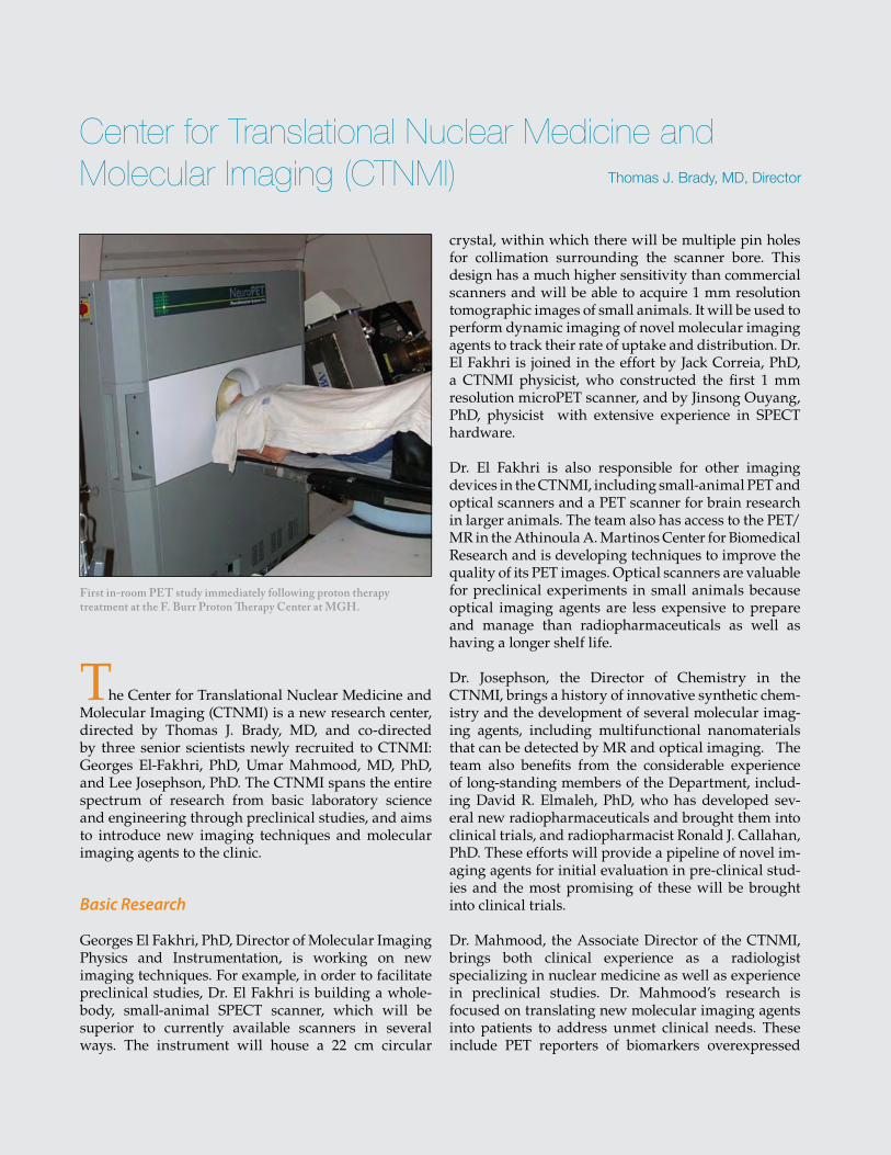
51
Thomas J. Brady, MD, Director
Center for Translational Nuclear Medicine and Molecular Imaging (CTNMI)
First in-room PET study immediately following proton therapy treatment at the F. Burr Proton Therapy Center at MGH.
The Center for Translational Nuclear Medicine and Molecular Imaging (CTNMI) is a new research center, directed by Thomas J. Brady, MD, and co-directed by three senior scientists newly recruited to CTNMI: Georges El-Fakhri, PhD, Umar Mahmood, MD, PhD, and Lee Josephson, PhD. The CTNMI spans the entire spectrum of research from basic laboratory science and engineering through preclinical studies, and aims to introduce new imaging techniques and molecular imaging agents to the clinic.
Basic Research
Georges El Fakhri, PhD, Director of Molecular Imaging Physics and Instrumentation, is working on new imaging techniques. For example, in order to facilitate preclinical studies, Dr. El Fakhri is building a whole-body, small-animal SPECT scanner, which will be superior to currently available scanners in several ways. The instrument will house a 22 cm circular
crystal, within which there will be multiple pin holes for collimation surrounding the scanner bore. This design has a much higher sensitivity than commercial scanners and will be able to acquire 1 mm resolution tomographic images of small animals. It will be used to perform dynamic imaging of novel molecular imaging agents to track their rate of uptake and distribution. Dr. El Fakhri is joined in the effort by Jack Correia, PhD, a CTNMI physicist, who constructed the first 1 mm resolution microPET scanner, and by Jinsong Ouyang, PhD, physicist with extensive experience in SPECT hardware.
Dr. El Fakhri is also responsible for other imaging devices in the CTNMI, including small-animal PET and optical scanners and a PET scanner for brain research in larger animals. The team also has access to the PET/MR in the Athinoula A. Martinos Center for Biomedical Research and is developing techniques to improve the quality of its PET images. Optical scanners are valuable for preclinical experiments in small animals because optical imaging agents are less expensive to prepare and manage than radiopharmaceuticals as well as having a longer shelf life.
Dr. Josephson, the Director of Chemistry in the CTNMI, brings a history of innovative synthetic chem-istry and the development of several molecular imag-ing agents, including multifunctional nanomaterials that can be detected by MR and optical imaging. The team also benefits from the considerable experience of long-standing members of the Department, includ-ing David R. Elmaleh, PhD, who has developed sev-eral new radiopharmaceuticals and brought them into clinical trials, and radiopharmacist Ronald J. Callahan, PhD. These efforts will provide a pipeline of novel im-aging agents for initial evaluation in pre-clinical stud-ies and the most promising of these will be brought into clinical trials.
Dr. Mahmood, the Associate Director of the CTNMI, brings both clinical experience as a radiologist specializing in nuclear medicine as well as experience in preclinical studies. Dr. Mahmood’s research is focused on translating new molecular imaging agents into patients to address unmet clinical needs. These include PET reporters of biomarkers overexpressed
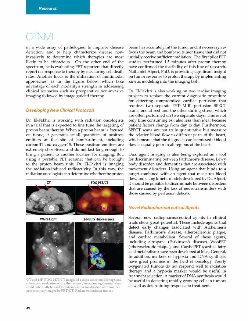
CTNMIin a wide array of pathologies, to improve disease detection, and to help characterize disease non-invasively to determine which therapies are most likely to be efficacious. On the other end of the spectrum, he is evaluating PET reporters that directly report on response to therapy by measuring cell death rates. Another focus is the utilization of multimodal approaches, as in the figure below, which take advantage of each modality’s strength in addressing clinical scenarios such as preoperative non-invasive imaging followed by image guided therapy.
Developing New Clinical Protocols
Dr. El-Fakhri is working with radiation oncologists in a trial that is expected to fine tune the targeting of proton beam therapy. When a proton beam is focused on tissue, it generates small quantities of positron emitters at the site of bombardment, including carbon-11 and oxygen-15. These positron emitters are extremely short-lived and do not last long enough to bring a patient to another location for imaging. But, using a portable PET scanner that can be brought to the proton beam unit, Dr. El-Fakhri is imaging the radiation-induced radioactivity. In this way, the radiation oncologists can determine whether the proton
beam has accurately hit the tumor and, if necessary, re-focus the beam and bombard tumor tissue that did not initially receive sufficient radiation. The first pilot PET studies performed 1.5 minutes after proton therapy have confirmed the feasibility of this line of research. Nathaniel Alpert, PhD, is providing significant insight on tumor response to proton therapy by implementing kinetic modeling into the imaging task.
Dr. El-Fakhri is also working on two cardiac imaging projects to replace the current diagnostic procedure for detecting compromised cardiac perfusion that requires two separate 99mTc-MIBI perfusion SPECT scans, one at rest and the other during stress, which are often performed on two separate days. This is not only time consuming but also less than ideal because patient factors change from day to day. Furthermore, SPECT scans are not truly quantitative but measure the relative blood flow to different parts of the heart, which means that the diagnosis can be missed if blood flow is equally poor in all regions of the heart.
Dual agent imaging is also being explored as a tool for discriminating between Parkinson’s disease, Lewy body disorder, and dementias that are associated with movement disorders. Using an agent that binds to a target combined with an agent that measures blood flow, and using kinetic models developed by Dr. Alpert, it should be possible to discriminate between disorders that are caused by the loss of neurotransmitters with those caused by perfusion deficits.
Novel Radiopharmaceutical Agents
Several new radiopharmaceutical agents in clinical trials show great potential. These include agents that detect early changes associated with Alzheimer’s disease, Parkinson’s disease, atherosclerotic plaque, and cardiac metabolism. Several of these agents, including altropane (Parkinson’s disease), VasoPET (atherosclerotic plaque), and CardioPET (cardiac fatty acid metabolism) have been developed at Mass General. In addition, markers of hypoxia and DNA synthesis have great promise in the field of oncology. Poorly oxygenated tumors do not respond well to radiation therapy and a hypoxia marker would be useful in treatment selection. A marker of DNA synthesis would be useful in detecting rapidly growing cells in tumors as well as determining response to treatment.
CT and 18F-FDG PET/CT images of a colon cancer model (top), and subsequent evaluation with a fluorescent glucose analog (bottom) that could potentially be used for intraoperative localization of tumor foci preoperatively imaged by PET/CT. Red arrows indicate tumors.
49
Research
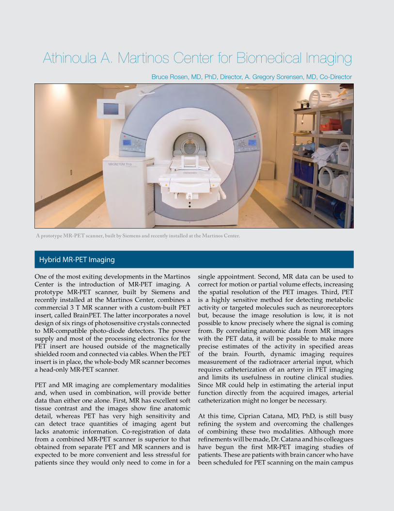
53
A prototype MR-PET scanner, built by Siemens and recently installed at the Martinos Center.
Athinoula A. Martinos Center for Biomedical Imaging
One of the most exiting developments in the Martinos Center is the introduction of MR-PET imaging. A prototype MR-PET scanner, built by Siemens and recently installed at the Martinos Center, combines a commercial 3 T MR scanner with a custom-built PET insert, called BrainPET. The latter incorporates a novel design of six rings of photosensitive crystals connected to MR-compatible photo-diode detectors. The power supply and most of the processing electronics for the PET insert are housed outside of the magnetically shielded room and connected via cables. When the PET insert is in place, the whole-body MR scanner becomes a head-only MR-PET scanner.
PET and MR imaging are complementary modalities and, when used in combination, will provide better data than either one alone. First, MR has excellent soft tissue contrast and the images show fine anatomic detail, whereas PET has very high sensitivity and can detect trace quantities of imaging agent but lacks anatomic information. Co-registration of data from a combined MR-PET scanner is superior to that obtained from separate PET and MR scanners and is expected to be more convenient and less stressful for patients since they would only need to come in for a
single appointment. Second, MR data can be used to correct for motion or partial volume effects, increasing the spatial resolution of the PET images. Third, PET is a highly sensitive method for detecting metabolic activity or targeted molecules such as neuroreceptors but, because the image resolution is low, it is not possible to know precisely where the signal is coming from. By correlating anatomic data from MR images with the PET data, it will be possible to make more precise estimates of the activity in specified areas of the brain. Fourth, dynamic imaging requires measurement of the radiotracer arterial input, which requires catheterization of an artery in PET imaging and limits its usefulness in routine clinical studies. Since MR could help in estimating the arterial input function directly from the acquired images, arterial catheterization might no longer be necessary.
At this time, Ciprian Catana, MD, PhD, is still busy refining the system and overcoming the challenges of combining these two modalities. Although more refinements will be made, Dr. Catana and his colleagues have begun the first MR-PET imaging studies of patients. These are patients with brain cancer who have been scheduled for PET scanning on the main campus
Bruce Rosen, MD, PhD, Director, A. Gregory Sorensen, MD, Co-Director
Hybrid MR-PET Imaging
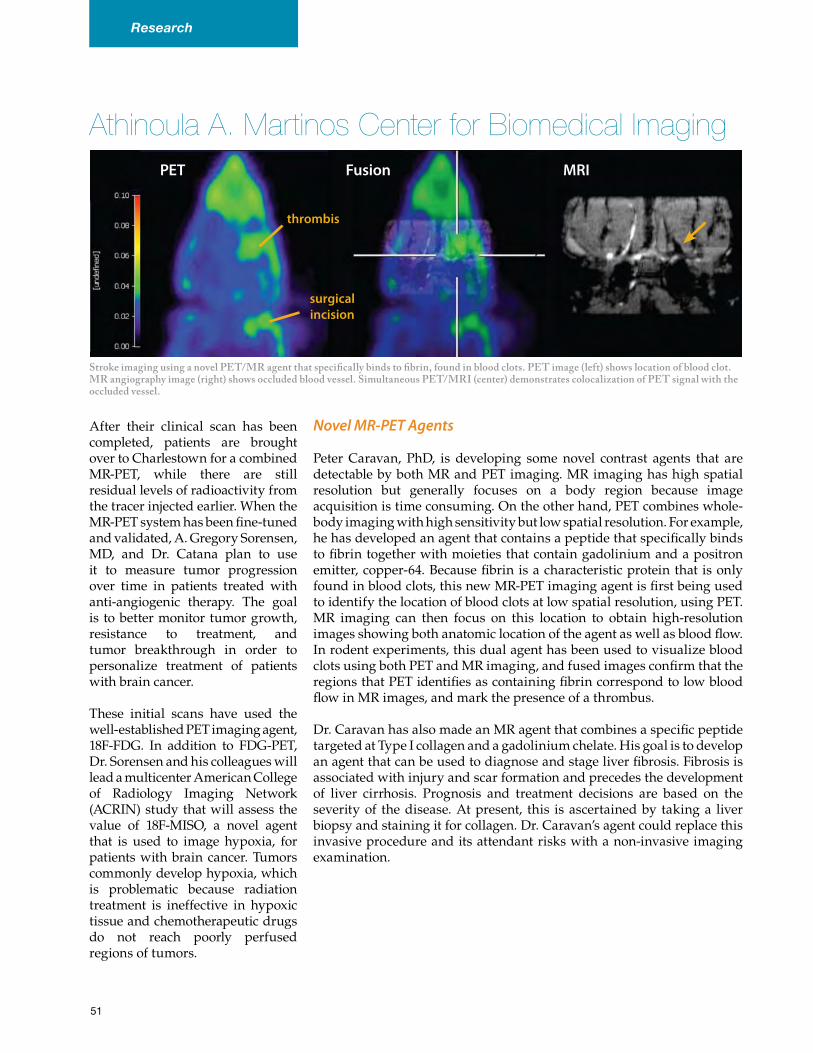
MRI
Stroke imaging using a novel PET/MR agent that specifically binds to fibrin, found in blood clots. PET image (left) shows location of blood clot. MR angiography image (right) shows occluded blood vessel. Simultaneous PET/MRI (center) demonstrates colocalization of PET signal with the occluded vessel.
Athinoula A. Martinos Center for Biomedical Imaging
After their clinical scan has been completed, patients are brought over to Charlestown for a combined MR-PET, while there are still residual levels of radioactivity from the tracer injected earlier. When the MR-PET system has been fine-tuned and validated, A. Gregory Sorensen, MD, and Dr. Catana plan to use it to measure tumor progression over time in patients treated with anti-angiogenic therapy. The goal is to better monitor tumor growth, resistance to treatment, and tumor breakthrough in order to personalize treatment of patients with brain cancer.
These initial scans have used the well-established PET imaging agent, 18F-FDG. In addition to FDG-PET, Dr. Sorensen and his colleagues will lead a multicenter American College of Radiology Imaging Network (ACRIN) study that will assess the value of 18F-MISO, a novel agent that is used to image hypoxia, for patients with brain cancer. Tumors commonly develop hypoxia, which is problematic because radiation treatment is ineffective in hypoxic tissue and chemotherapeutic drugs do not reach poorly perfused regions of tumors.
Novel MR-PET Agents
Peter Caravan, PhD, is developing some novel contrast agents that are detectable by both MR and PET imaging. MR imaging has high spatial resolution but generally focuses on a body region because image acquisition is time consuming. On the other hand, PET combines whole-body imaging with high sensitivity but low spatial resolution. For example, he has developed an agent that contains a peptide that specifically binds to fibrin together with moieties that contain gadolinium and a positron emitter, copper-64. Because fibrin is a characteristic protein that is only found in blood clots, this new MR-PET imaging agent is first being used to identify the location of blood clots at low spatial resolution, using PET. MR imaging can then focus on this location to obtain high-resolution images showing both anatomic location of the agent as well as blood flow. In rodent experiments, this dual agent has been used to visualize blood clots using both PET and MR imaging, and fused images confirm that the regions that PET identifies as containing fibrin correspond to low blood flow in MR images, and mark the presence of a thrombus.
Dr. Caravan has also made an MR agent that combines a specific peptide targeted at Type I collagen and a gadolinium chelate. His goal is to develop an agent that can be used to diagnose and stage liver fibrosis. Fibrosis is associated with injury and scar formation and precedes the development of liver cirrhosis. Prognosis and treatment decisions are based on the severity of the disease. At present, this is ascertained by taking a liver biopsy and staining it for collagen. Dr. Caravan’s agent could replace this invasive procedure and its attendant risks with a non-invasive imaging examination.
PET Fusion
thrombis
surgical incision
51
Research
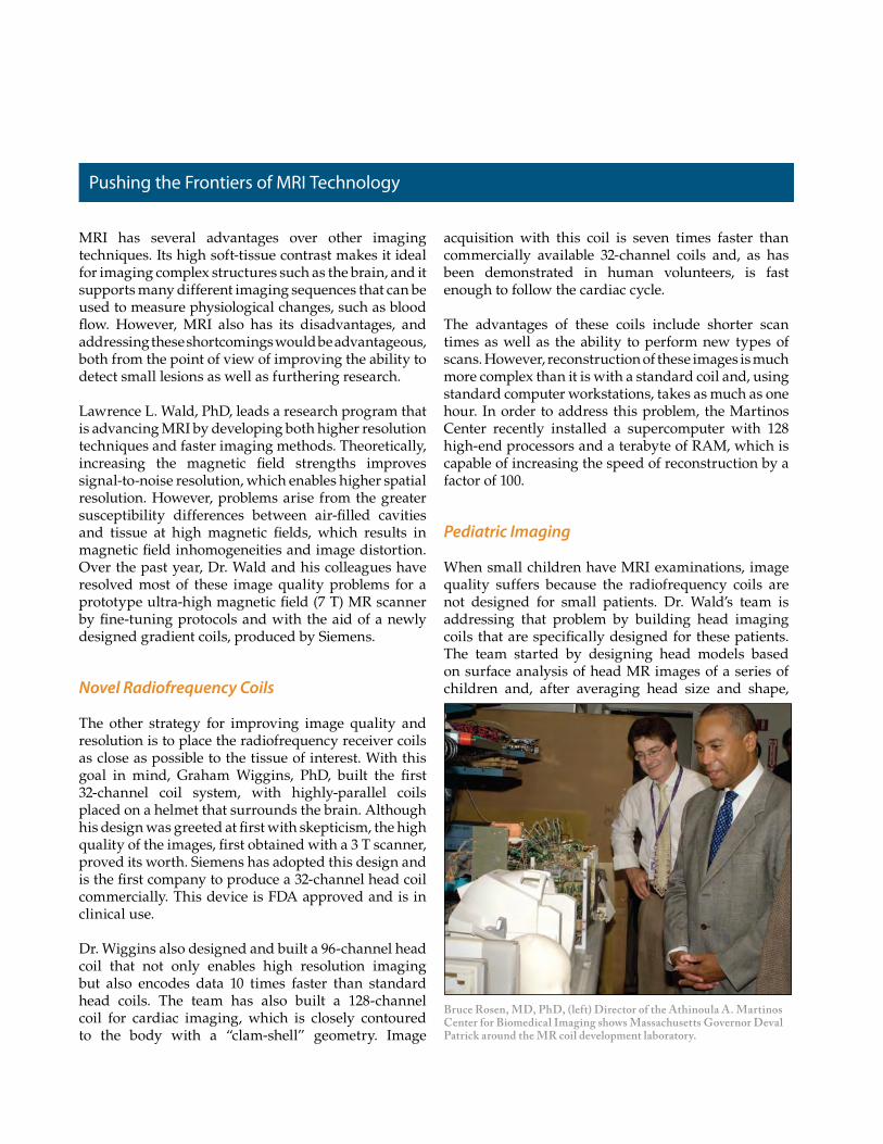
MRI has several advantages over other imaging techniques. Its high soft-tissue contrast makes it ideal for imaging complex structures such as the brain, and it supports many different imaging sequences that can be used to measure physiological changes, such as blood flow. However, MRI also has its disadvantages, and addressing these shortcomings would be advantageous, both from the point of view of improving the ability to detect small lesions as well as furthering research.
Lawrence L. Wald, PhD, leads a research program that is advancing MRI by developing both higher resolution techniques and faster imaging methods. Theoretically, increasing the magnetic field strengths improves signal-to-noise resolution, which enables higher spatial resolution. However, problems arise from the greater susceptibility differences between air-filled cavities and tissue at high magnetic fields, which results in magnetic field inhomogeneities and image distortion. Over the past year, Dr. Wald and his colleagues have resolved most of these image quality problems for a prototype ultra-high magnetic field (7 T) MR scanner by fine-tuning protocols and with the aid of a newly designed gradient coils, produced by Siemens.
Novel Radiofrequency Coils
The other strategy for improving image quality and resolution is to place the radiofrequency receiver coils as close as possible to the tissue of interest. With this goal in mind, Graham Wiggins, PhD, built the first 32-channel coil system, with highly-parallel coils placed on a helmet that surrounds the brain. Although his design was greeted at first with skepticism, the high quality of the images, first obtained with a 3 T scanner, proved its worth. Siemens has adopted this design and is the first company to produce a 32-channel head coil commercially. This device is FDA approved and is in clinical use.
Dr. Wiggins also designed and built a 96-channel head coil that not only enables high resolution imaging but also encodes data 10 times faster than standard head coils. The team has also built a 128-channel coil for cardiac imaging, which is closely contoured to the body with a “clam-shell” geometry. Image
acquisition with this coil is seven times faster than commercially available 32-channel coils and, as has been demonstrated in human volunteers, is fast enough to follow the cardiac cycle.
The advantages of these coils include shorter scan times as well as the ability to perform new types of scans. However, reconstruction of these images is much more complex than it is with a standard coil and, using standard computer workstations, takes as much as one hour. In order to address this problem, the Martinos Center recently installed a supercomputer with 128 high-end processors and a terabyte of RAM, which is capable of increasing the speed of reconstruction by a factor of 100.
Pediatric Imaging
When small children have MRI examinations, image quality suffers because the radiofrequency coils are not designed for small patients. Dr. Wald’s team is addressing that problem by building head imaging coils that are specifically designed for these patients. The team started by designing head models based on surface analysis of head MR images of a series of children and, after averaging head size and shape,
Pushing the Frontiers of MRI Technology
Bruce Rosen, MD, PhD, (left) Director of the Athinoula A. Martinos Center for Biomedical Imaging shows Massachusetts Governor Deval Patrick around the MR coil development laboratory.
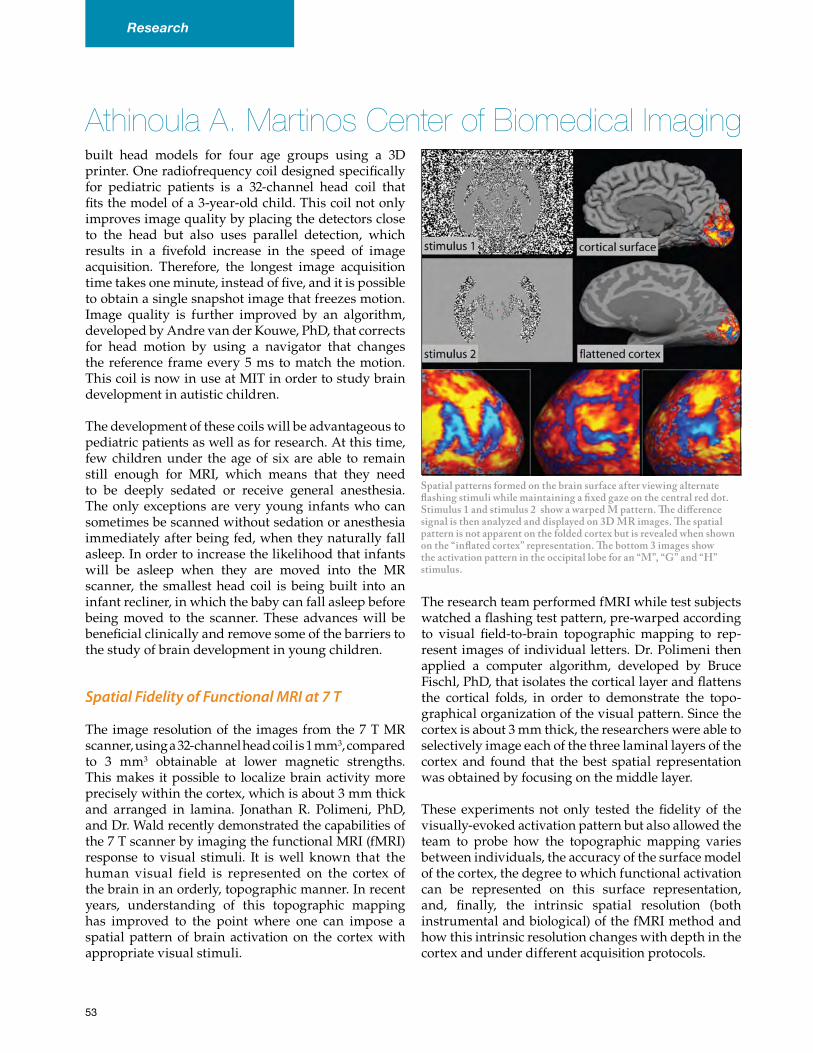
Athinoula A. Martinos Center of Biomedical Imagingbuilt head models for four age groups using a 3D printer. One radiofrequency coil designed specifically for pediatric patients is a 32-channel head coil that fits the model of a 3-year-old child. This coil not only improves image quality by placing the detectors close to the head but also uses parallel detection, which results in a fivefold increase in the speed of image acquisition. Therefore, the longest image acquisition time takes one minute, instead of five, and it is possible to obtain a single snapshot image that freezes motion. Image quality is further improved by an algorithm, developed by Andre van der Kouwe, PhD, that corrects for head motion by using a navigator that changes the reference frame every 5 ms to match the motion. This coil is now in use at MIT in order to study brain development in autistic children.
The development of these coils will be advantageous to pediatric patients as well as for research. At this time, few children under the age of six are able to remain still enough for MRI, which means that they need to be deeply sedated or receive general anesthesia. The only exceptions are very young infants who can sometimes be scanned without sedation or anesthesia immediately after being fed, when they naturally fall asleep. In order to increase the likelihood that infants will be asleep when they are moved into the MR scanner, the smallest head coil is being built into an infant recliner, in which the baby can fall asleep before being moved to the scanner. These advances will be beneficial clinically and remove some of the barriers to the study of brain development in young children.
Spatial Fidelity of Functional MRI at 7 T
The image resolution of the images from the 7 T MR scanner, using a 32-channel head coil is 1 mm3, compared to 3 mm3 obtainable at lower magnetic strengths. This makes it possible to localize brain activity more precisely within the cortex, which is about 3 mm thick and arranged in lamina. Jonathan R. Polimeni, PhD, and Dr. Wald recently demonstrated the capabilities of the 7 T scanner by imaging the functional MRI (fMRI) response to visual stimuli. It is well known that the human visual field is represented on the cortex of the brain in an orderly, topographic manner. In recent years, understanding of this topographic mapping has improved to the point where one can impose a spatial pattern of brain activation on the cortex with appropriate visual stimuli.
The research team performed fMRI while test subjects watched a flashing test pattern, pre-warped according to visual field-to-brain topographic mapping to rep-resent images of individual letters. Dr. Polimeni then applied a computer algorithm, developed by Bruce Fischl, PhD, that isolates the cortical layer and flattens the cortical folds, in order to demonstrate the topo-graphical organization of the visual pattern. Since the cortex is about 3 mm thick, the researchers were able to selectively image each of the three laminal layers of the cortex and found that the best spatial representation was obtained by focusing on the middle layer.
These experiments not only tested the fidelity of the visually-evoked activation pattern but also allowed the team to probe how the topographic mapping varies between individuals, the accuracy of the surface model of the cortex, the degree to which functional activation can be represented on this surface representation, and, finally, the intrinsic spatial resolution (both instrumental and biological) of the fMRI method and how this intrinsic resolution changes with depth in the cortex and under different acquisition protocols.
Spatial patterns formed on the brain surface after viewing alternate flashing stimuli while maintaining a fixed gaze on the central red dot. Stimulus 1 and stimulus 2 show a warped M pattern. The difference signal is then analyzed and displayed on 3D MR images. The spatial pattern is not apparent on the folded cortex but is revealed when shown on the “inflated cortex” representation. The bottom 3 images show the activation pattern in the occipital lobe for an “M”, “G” and “H” stimulus.
53
Research
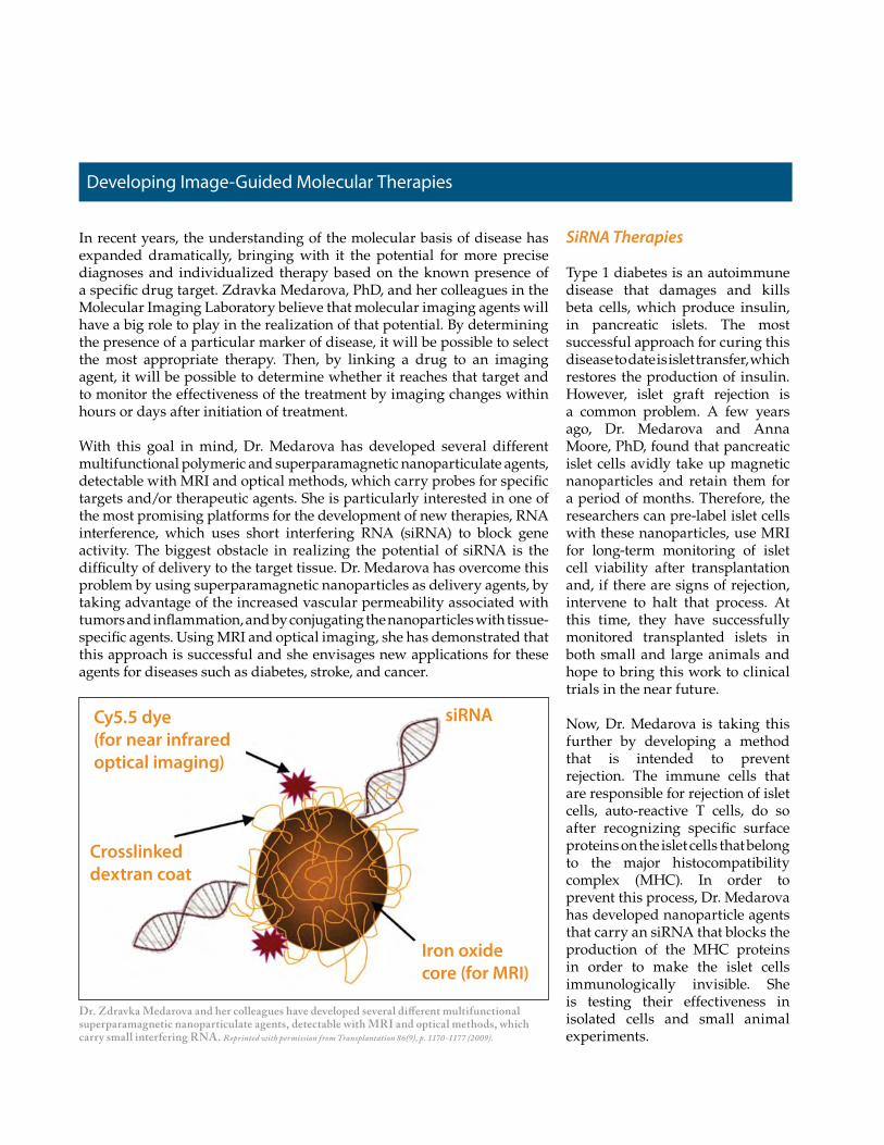
In recent years, the understanding of the molecular basis of disease has expanded dramatically, bringing with it the potential for more precise diagnoses and individualized therapy based on the known presence of a specific drug target. Zdravka Medarova, PhD, and her colleagues in the Molecular Imaging Laboratory believe that molecular imaging agents will have a big role to play in the realization of that potential. By determining the presence of a particular marker of disease, it will be possible to select the most appropriate therapy. Then, by linking a drug to an imaging agent, it will be possible to determine whether it reaches that target and to monitor the effectiveness of the treatment by imaging changes within hours or days after initiation of treatment.
With this goal in mind, Dr. Medarova has developed several different multifunctional polymeric and superparamagnetic nanoparticulate agents, detectable with MRI and optical methods, which carry probes for specific targets and/or therapeutic agents. She is particularly interested in one of the most promising platforms for the development of new therapies, RNA interference, which uses short interfering RNA (siRNA) to block gene activity. The biggest obstacle in realizing the potential of siRNA is the difficulty of delivery to the target tissue. Dr. Medarova has overcome this problem by using superparamagnetic nanoparticles as delivery agents, by taking advantage of the increased vascular permeability associated with tumors and inflammation, and by conjugating the nanoparticles with tissue-specific agents. Using MRI and optical imaging, she has demonstrated that this approach is successful and she envisages new applications for these agents for diseases such as diabetes, stroke, and cancer.
SiRNA Therapies
Type 1 diabetes is an autoimmune disease that damages and kills beta cells, which produce insulin, in pancreatic islets. The most successful approach for curing this disease to date is islet transfer, which restores the production of insulin. However, islet graft rejection is a common problem. A few years ago, Dr. Medarova and Anna Moore, PhD, found that pancreatic islet cells avidly take up magnetic nanoparticles and retain them for a period of months. Therefore, the researchers can pre-label islet cells with these nanoparticles, use MRI for long-term monitoring of islet cell viability after transplantation and, if there are signs of rejection, intervene to halt that process. At this time, they have successfully monitored transplanted islets in both small and large animals and hope to bring this work to clinical trials in the near future.
Now, Dr. Medarova is taking this further by developing a method that is intended to prevent rejection. The immune cells that are responsible for rejection of islet cells, auto-reactive T cells, do so after recognizing specific surface proteins on the islet cells that belong to the major histocompatibility complex (MHC). In order to prevent this process, Dr. Medarova has developed nanoparticle agents that carry an siRNA that blocks the production of the MHC proteins in order to make the islet cells immunologically invisible. She is testing their effectiveness in isolated cells and small animal experiments.
Developing Image-Guided Molecular Therapies
Dr. Zdravka Medarova and her colleagues have developed several different multifunctional superparamagnetic nanoparticulate agents, detectable with MRI and optical methods, which carry small interfering RNA. Reprinted with permission from Transplantation 86(9), p. 1170-1177 (2009).
Cy5.5 dye (for near infrared optical imaging)
Crosslinked dextran coat
siRNA
Iron oxide core (for MRI)
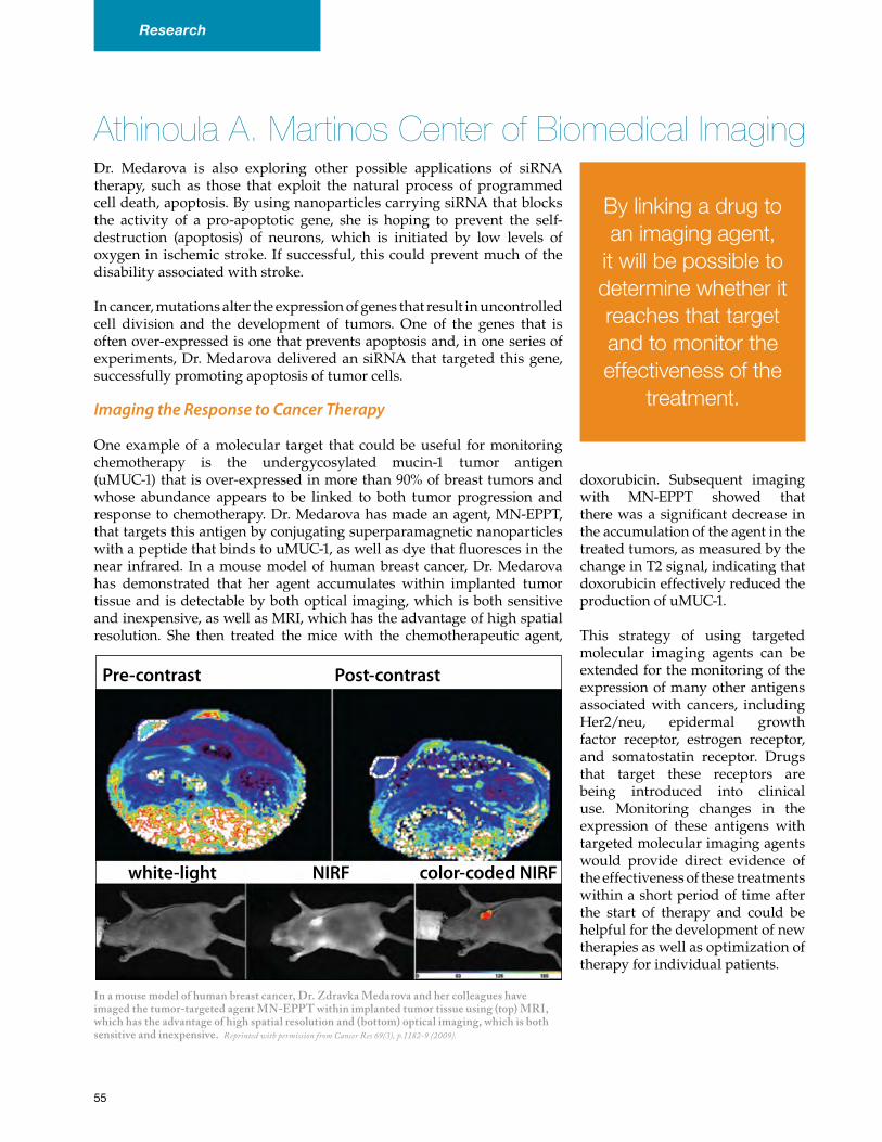
Athinoula A. Martinos Center of Biomedical ImagingDr. Medarova is also exploring other possible applications of siRNA therapy, such as those that exploit the natural process of programmed cell death, apoptosis. By using nanoparticles carrying siRNA that blocks the activity of a pro-apoptotic gene, she is hoping to prevent the self-destruction (apoptosis) of neurons, which is initiated by low levels of oxygen in ischemic stroke. If successful, this could prevent much of the disability associated with stroke.
In cancer, mutations alter the expression of genes that result in uncontrolled cell division and the development of tumors. One of the genes that is often over-expressed is one that prevents apoptosis and, in one series of experiments, Dr. Medarova delivered an siRNA that targeted this gene, successfully promoting apoptosis of tumor cells.
Imaging the Response to Cancer Therapy
One example of a molecular target that could be useful for monitoring chemotherapy is the undergycosylated mucin-1 tumor antigen (uMUC-1) that is over-expressed in more than 90% of breast tumors and whose abundance appears to be linked to both tumor progression and response to chemotherapy. Dr. Medarova has made an agent, MN-EPPT, that targets this antigen by conjugating superparamagnetic nanoparticles with a peptide that binds to uMUC-1, as well as dye that fluoresces in the near infrared. In a mouse model of human breast cancer, Dr. Medarova has demonstrated that her agent accumulates within implanted tumor tissue and is detectable by both optical imaging, which is both sensitive and inexpensive, as well as MRI, which has the advantage of high spatial resolution. She then treated the mice with the chemotherapeutic agent,
doxorubicin. Subsequent imaging with MN-EPPT showed that there was a significant decrease in the accumulation of the agent in the treated tumors, as measured by the change in T2 signal, indicating that doxorubicin effectively reduced the production of uMUC-1.
This strategy of using targeted molecular imaging agents can be extended for the monitoring of the expression of many other antigens associated with cancers, including Her2/neu, epidermal growth factor receptor, estrogen receptor, and somatostatin receptor. Drugs that target these receptors are being introduced into clinical use. Monitoring changes in the expression of these antigens with targeted molecular imaging agents would provide direct evidence of the effectiveness of these treatments within a short period of time after the start of therapy and could be helpful for the development of new therapies as well as optimization of therapy for individual patients.
By linking a drug to an imaging agent,
it will be possible to determine whether it reaches that target and to monitor the effectiveness of the
treatment.
In a mouse model of human breast cancer, Dr. Zdravka Medarova and her colleagues have imaged the tumor-targeted agent MN-EPPT within implanted tumor tissue using (top) MRI, which has the advantage of high spatial resolution and (bottom) optical imaging, which is both sensitive and inexpensive. Reprinted with permission from Cancer Res 69(3), p.1182-9 (2009).
Pre-contrast Post-contrast
white-light NIRF color-coded NIRF
55
Research
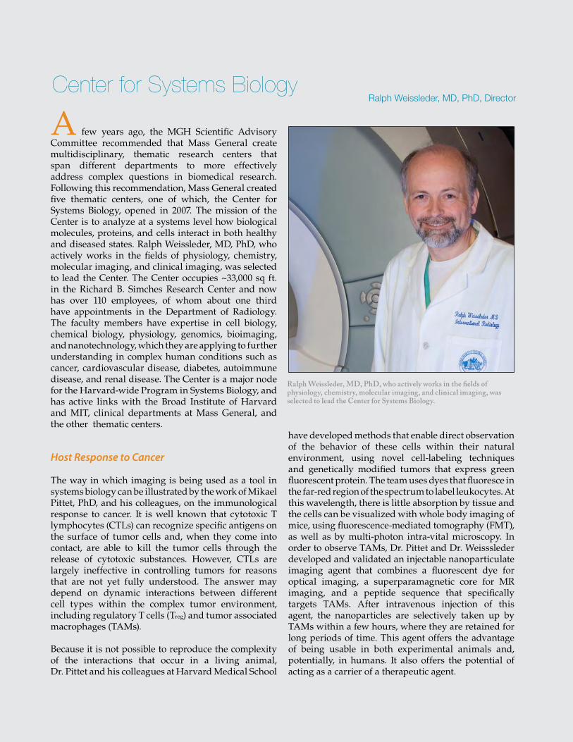
59
Center for Systems BiologyRalph Weissleder, MD, PhD, Director
A few years ago, the MGH Scientific Advisory Committee recommended that Mass General create multidisciplinary, thematic research centers that span different departments to more effectively address complex questions in biomedical research. Following this recommendation, Mass General created five thematic centers, one of which, the Center for Systems Biology, opened in 2007. The mission of the Center is to analyze at a systems level how biological molecules, proteins, and cells interact in both healthy and diseased states. Ralph Weissleder, MD, PhD, who actively works in the fields of physiology, chemistry, molecular imaging, and clinical imaging, was selected to lead the Center. The Center occupies ~33,000 sq ft. in the Richard B. Simches Research Center and now has over 110 employees, of whom about one third have appointments in the Department of Radiology. The faculty members have expertise in cell biology, chemical biology, physiology, genomics, bioimaging, and nanotechnology, which they are applying to further understanding in complex human conditions such as cancer, cardiovascular disease, diabetes, autoimmune disease, and renal disease. The Center is a major node for the Harvard-wide Program in Systems Biology, and has active links with the Broad Institute of Harvard and MIT, clinical departments at Mass General, and the other thematic centers.
Host Response to Cancer
The way in which imaging is being used as a tool in systems biology can be illustrated by the work of Mikael Pittet, PhD, and his colleagues, on the immunological response to cancer. It is well known that cytotoxic T lymphocytes (CTLs) can recognize specific antigens on the surface of tumor cells and, when they come into contact, are able to kill the tumor cells through the release of cytotoxic substances. However, CTLs are largely ineffective in controlling tumors for reasons that are not yet fully understood. The answer may depend on dynamic interactions between different cell types within the complex tumor environment, including regulatory T cells (Treg) and tumor associated macrophages (TAMs).
Because it is not possible to reproduce the complexity of the interactions that occur in a living animal, Dr. Pittet and his colleagues at Harvard Medical School
have developed methods that enable direct observation of the behavior of these cells within their natural environment, using novel cell-labeling techniques and genetically modified tumors that express green fluorescent protein. The team uses dyes that fluoresce in the far-red region of the spectrum to label leukocytes. At this wavelength, there is little absorption by tissue and the cells can be visualized with whole body imaging of mice, using fluorescence-mediated tomography (FMT), as well as by multi-photon intra-vital microscopy. In order to observe TAMs, Dr. Pittet and Dr. Weisssleder developed and validated an injectable nanoparticulate imaging agent that combines a fluorescent dye for optical imaging, a superparamagnetic core for MR imaging, and a peptide sequence that specifically targets TAMs. After intravenous injection of this agent, the nanoparticles are selectively taken up by TAMs within a few hours, where they are retained for long periods of time. This agent offers the advantage of being usable in both experimental animals and, potentially, in humans. It also offers the potential of acting as a carrier of a therapeutic agent.
Ralph Weissleder, MD, PhD, who actively works in the fields of physiology, chemistry, molecular imaging, and clinical imaging, was selected to lead the Center for Systems Biology.
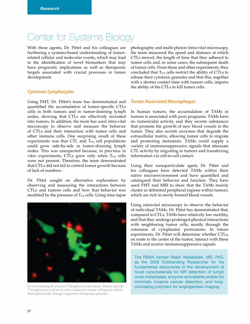
With these agents, Dr. Pittet and his colleagues are furthering a systems-based understanding of tumor-related cellular and molecular events, which may lead to the identification of novel biomarkers that may have prognostic implications as well as therapeutic targets associated with crucial processes in tumor development.
Cytotoxic Lymphocytes
Using FMT, Dr. Pittet’s team has demonstrated and quantified the accumulation of tumor-specific CTLs cells in both tumors and in tumor-draining lymph nodes, showing that CTLs are effectively recruited into tumors. In addition, the team has used intra-vital microscopy to observe and measure the behavior of CTLs and their interaction with tumor cells and other immune cells. One surprising result of these experiments was that CTL and Treg cell populations could grow side-by-side in tumor-draining lymph nodes. This was unexpected because, in previous in vitro experiments, CTLs grew only when Treg cells were not present. Therefore, the team demonstrated that CTLs did not fail to control tumor growth because of lack of numbers.
Dr. Pittet sought an alternative explanation by observing and measuring the interactions between CTLs and tumors cells and how that behavior was modified by the presence of Treg cells. Using time-lapse
photography and multi-photon intra-vital microscopy, the team measured the speed and distance at which CTLs moved, the length of time that they adhered to tumor cells and, in some cases, the subsequent death of tumor cells. From these and other experiments, they concluded that Treg cells restrict the ability of CTLs to release their cytotoxic granules and that this, together with a shorter contact time with tumors cells, impairs the ability of the CTLs to kill tumor cells.
Tumor Associated Macrophages
In human tumors, the accumulation of TAMs in tumors is associated with poor prognosis. TAMs have no tumoricidal activity and they secrete substances that promote the growth of new blood vessels in the tumor. They also secrete enzymes that degrade the extracellular matrix, allowing tumor cells to migrate and promoting metastasis. TAMs could supply a variety of immunosuppressive signals that attenuate CTL activity by migrating to tumors and transferring information via cell-to-cell contact.
Using their nanoparticulate agent, Dr. Pittet and his colleagues have detected TAMs within their native microenvironment and have quantified and catalogued their behavior and function. They have used FMT and MRI to show that the TAMs mainly cluster in delimited peripheral regions within tumors, which are rich in newly formed blood vessels.
Using intravital microscopy to observe the behavior of individual TAMs, Dr. Pittet has demonstrated that, compared to CTLs, TAMs have relatively low motility, and that they undergo prolonged physical interactions with neighboring tumor cells, mostly through the extension of cytoplasmic protrusions. In future experiments, Dr. Pittet will determine whether CTLs, en route to the center of the tumor, interact with these TAMs and receive immunosuppressive signals.
Center for Systems Biology
In vivo tracking of cytotoxic T lymphocytes in tumors. Tumor-specific T lymphocytes (red) are in close contact to a tumor cell (green), which shows phenotypic changes suggestive of ongoing apoptosis.
TheRSNA namedRalphWeissleder,MD, PhD,as the 2008 Outstanding Researcher for hisfundamental discoveries in the development ofnovel nanomaterials forMRdetection of lymphnodemetastases,enzyme-activatableprobesforminimally invasive cancer detection, and long-circulatingpolymersforangiogenesisimaging.
57
Research
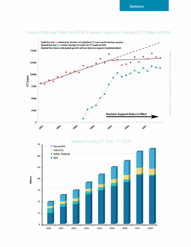
Statistics
Impact of Radiology Order Entry (ROE) & Decision Support on Outpatient CT Utilization at MGH
Research Funding FY 2000 - FY 2008
From
Sis
trom
, C. L
., et
al.
Rad
iolo
gy 2
009;
251:
147-
155,
rep
rodu
ced
wit
h pe
rmis
sion
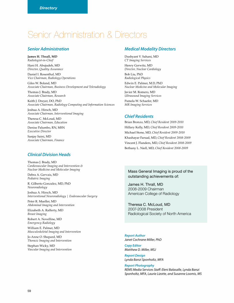
59
Senior Administration & Directors
Directory
Senior Administration
James H. Thrall, MD Radiologist-in-ChiefHani H. Abujudeh, MD Director, Quality Assurance
Daniel I. Rosenthal, MD Vice Chairman, Radiology Operations
Giles W. Boland, MD Associate Chairman, Business Development and Teleradiology
Thomas J. Brady, MD Associate Chairman, Research
Keith J. Dreyer, DO, PhD Associate Chairman, Radiology Computing and Information Sciences
Joshua A. Hirsch, MD Associate Chairman, Interventional Imaging
Theresa C. McLoud, MD Associate Chairman, Education
Denise Palumbo, RN, MSN Executive Director
Sanjay Saini, MD Associate Chairman, Finance
Clinical Division Heads
Thomas J. Brady, MD Cardiovascular Imaging and Intervention & Nuclear Medicine and Molecular Imaging
Debra A. Gervais, MD Pediatric Imaging
R. Gilberto Gonzalez, MD, PhD Neuroradiology
Joshua A. Hirsch, MD Interventional Neuroradiology | Endovascular Surgery
Peter R. Mueller, MD Abdominal Imaging and Intervention
Elizabeth A. Rafferty, MD Breast Imaging
Robert A. Novelline, MD Emergency Radiology
William E. Palmer, MD Musculoskeletal Imaging and Intervention
Jo-Anne O. Shepard, MD Thoracic Imaging and Intervention
Stephan Wicky, MD Vascular Imaging and Intervention
Medical Modality Directors
Dushyant V. Sahani, MD CT Imaging Services
Henry Gerwitz, MD Director, Nuclear Cardiology
Bob Liu, PhD Radiological Physics
Edwin E. Palmer, M.D, PhD Nuclear Medicine and Molecular Imaging
Javier M. Romero, MD Ultrasound Imaging Services
Pamela W. Schaefer, MD MR Imaging Services
Chief ResidentsBrian Bronzo, MD, Chief Resident 2009-2010
Hillary Kelly, MD, Chief Resident 2009-2010
Michael Stone, MD, Chief Resident 2009-2010
Khashayar Farsad, MD, Chief Resident 2008-2009
Vincent J. Flanders, MD, Chief Resident 2008-2009
Bethany L. Niell, MD, Chief Resident 2008-2009
MassGeneralImagingisproudofthe outstandingachievementsof:
JamesH.Thrall,MD 2008-2009 Chairman American College of Radiology
TheresaC.McLoud,MD 2007-2008 President Radiological Society of North America
Report Author Janet Cochrane Miller, PhD
Copy Editor Matthew D. Miller, MSJ
Report Design Lynda Banzi Sponholtz, MFA
Report Photography REMS Media Services Staff: Eleni Balasalle, Lynda Banzi Sponholtz, MFA, Laurie Lizotte, and Susanne Loomis, MS
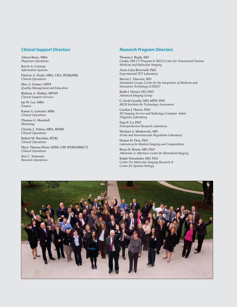
Research Program Directors
Thomas J. Brady, MD Cardiac MR CT Program & MGH Center for Transitional Nuclear Medicine and Molecular Imaging
Anna Liisa Brownell, PhD Experimental PET Laboratory
Steven L. Dawson, MD Simulation Group, Center for the Integration of Medicine and Innovative Technology (CIMIT)
Keith J. Dreyer, DO, PhD Advanced Imaging Group
G. Scott Gazelle, MD, MPH, PhD MGH Institute for Technology Assessment
Gordon J. Harris, PhD 3D Imaging Service and Radiology Computer Aided Diagnosis Laboratory
Eng H. Lo, PhD Neuroprotection Research Laboratory
Michael A. Moskowitz, MD Stroke and Neurovascular Regulation Laboratory
Homer H. Pien, PhD Laboratory for Medical Imaging and Computations
Bruce R. Rosen, MD, PhD Athinoula A. Martinos Center for Biomedical Imaging
Ralph Weissleder, MD, PhD Center For Molecular Imaging Research & Center for Systems Biology
Clinical Support Directors
Alison Berry, MBA Physician Operations
Kevin A. Conway Information Systems
Patricia A. Doyle, MBA, CRA, RT(R)(MR) Clinical Operations
Max A. Gomez, MPH Quality Management and Education
Barbara A. Hubley, MPAH Clinical Support Services
Jae W. Lee, MBA Finance
Karen A. Lemaire, MBA Clinical Operations
Thomas G. Marshall Marketing
Christy L. Palma, MBA, RDMS Clinical Operations
Robert M. Sheridan, RT(R) Clinical Operations
Mary Theresa Shore, MSM, CIIP, RT(R)(MR)(CT) Clinical Operations
Eric C. Tomasini Research Operations

64
www.massgeneralimaging.org