Embed Size (px)
Citation preview
Medical Art and Photographyin the History of Movement Disorders:
Part 13 of the MDS-Sponsored History of Movement DisordersExhibit, Barcelona, June 2000
Christopher G. Goetz,1* Teresa A. Chmura,1 and Douglas J. Lanska2
1Department of Neurological Sciences, Rush University/Rush-Presbyterian-St. Luke’s Medical Center, Chicago, Illinois, USA2Veterans Affairs Medical Center, Great Lakes VA Healthcare System, Tomah, Wisconsin, USA
Because movement disorders is a highly visual field inwhich diseases are recognized by distinctive static pos-tures and deformities as well as a gamut of involuntarymovements, medical communication has relied particu-larly on artistic and photographic documents. The earlydrawings and statues executed or commissioned byCharcot were important means of communication thatbridged language barriers on the phenomenology of Par-kinson’s disease. Time-lapse photography and othertechniques developed by Londe in Paris served to extendbeyond still photography and capture involuntary move-ments such as tics or chorea. Studying with Charcot,Marinescu returned to his native Romania and investedin the new technology of cinematography to capture agamut of gait disorders and parkinsonism. In the first halfof the twentieth century, individual investigators estab-lished large series of photographic collections of move-ment disorders, and Denny-Brown, Bucy, and researchcenters such as the Mayo Clinic regularly filmed pa-tients, and in some cases, experimental animals. Thesearchives serve as important documents on the naturalhistory of illnesses and on interventions, especially sur-gical. Herz used moving pictures and their frame-by-frame analysis to solidify a clinical description of dys-
tonia and to identify the patterns of twisting and gaitdysfunction typical of the disorder.
The first roentograms were developed in the nine-teenth century but were not specifically applied to move-ment disorders. Early pneumoencepholgrams aided cli-nicians in detecting lateral ventricular enlargement dueto presumed severe caudate atrophy in Huntington’s dis-ease. The advent of computed tompgraphy (CT) andmagnetic resonance imaging (MRI) have furthered diag-nostic precision in some entities, but functional imagingpromises more direct neuroimaging application to move-ment disorders.
The technology of videotapes was rapidly adopted bymovement disorder researchers and incorporated into thejournal format ofMovement Disordersfrom its incep-tion. These tapes communicate clinical findings that aresupplemented with written descriptions in the accompa-nying manuscripts similarly to theIcongraphie Pho-tographique de la Salpêtrièrethat was an early picture-based journal of still photographs with modest writtentext. In addition, videotapes have helped to establish in-ternational rating scales with standardized cases and ex-amples in order to facilitate multicenter research studiesand enhance inter-rater reliability. Film documents haveserved as important archives for disorders that are ex-tremely rare or no longer effectively exist, such asMPTP-induced parkinsonism, the movement disorders ofKuru, and postencephalitic parkinsonism.
SELECTED HISTORICAL REFERENCES
Barboi A, Goetz CG, Musetoiu R. 2000. Gheorghe Marinescu and theearly usage of cinematography in the analysis of neurological pa-tients. Neurology 54(Suppl. 3):A222.
Cantacuzene J. 1973. Soixante-quinze ans depuis la cre´ation du cine´made recherche me´dicale. Histoire des Sciences Me´dicales 7:291–302.
As part of the International Congress of the Movement DisorderSociety in Barcelona, June 2000, the society sponsored an exhibit de-voted to History of Movement Disorders. With the help of numerousmembers of the MDS and loans from libraries, private collections, andlaboratories, the authors developed a series of explanatory panels, ac-companied by photographs, diagrams and original artifacts that tracedthe early history of movement disorders from several perspectives.These materials have been adapted for publication inMovement Dis-ordersand are presented in an ongoing series.
*Correspondence to: C.G. Goetz, 1725 W. Harrison Street, Suite755, Chicago, IL 60612. E-mail: [email protected]
Received 25 August 2000; Accepted 25 August 2000Published online 14 September 2001; DOI 10.1002/mds.1190
Movement DisordersVol. 16, No. 5, 2001, pp. 947–953© 2001 Movement Disorder SocietyPublished by Wiley-Liss, Inc.
947
Charcot J-M. 1869. De la paralysie agitante (leçon 5) Oeuvres Com-plètes, volume 1, p 161–188. Paris: Bureaux du Progrès Me´dical;In English: On paralysis agitans (lecture 5) Lectures on the Dis-eases of the Nervous System (p 129–156). Trans. by G. Sigerson.London: New Sydenham’s Society; 1877.
Evidente VGH, Gwinn KA, Caviness JN, Muenter M, MulderDW.1998. Early cinematographic cases of postencephalitic parkin-sonism and other movement disorders. Mov Disord 13:167–169.
Goetz CG, Bonduelle M, Gelfand T. 1995. J-M Charcot: constructingneurology. New York: Oxford University Press.
Icongraphie Photographiqe de la Salpêtrière. Bourneville DM, RegnardP, editors. Bureaux du Progrès Me´dical; 1877–1879.
Kompoliti K, Goetz CG, Gajdusek DC, Cubo E. 1999. Movementdisorders in Kuru. Mov Disord 14:800–804.
Londe A. 1893. La photographie me´dicale. Paris: Gauthier-Villars.
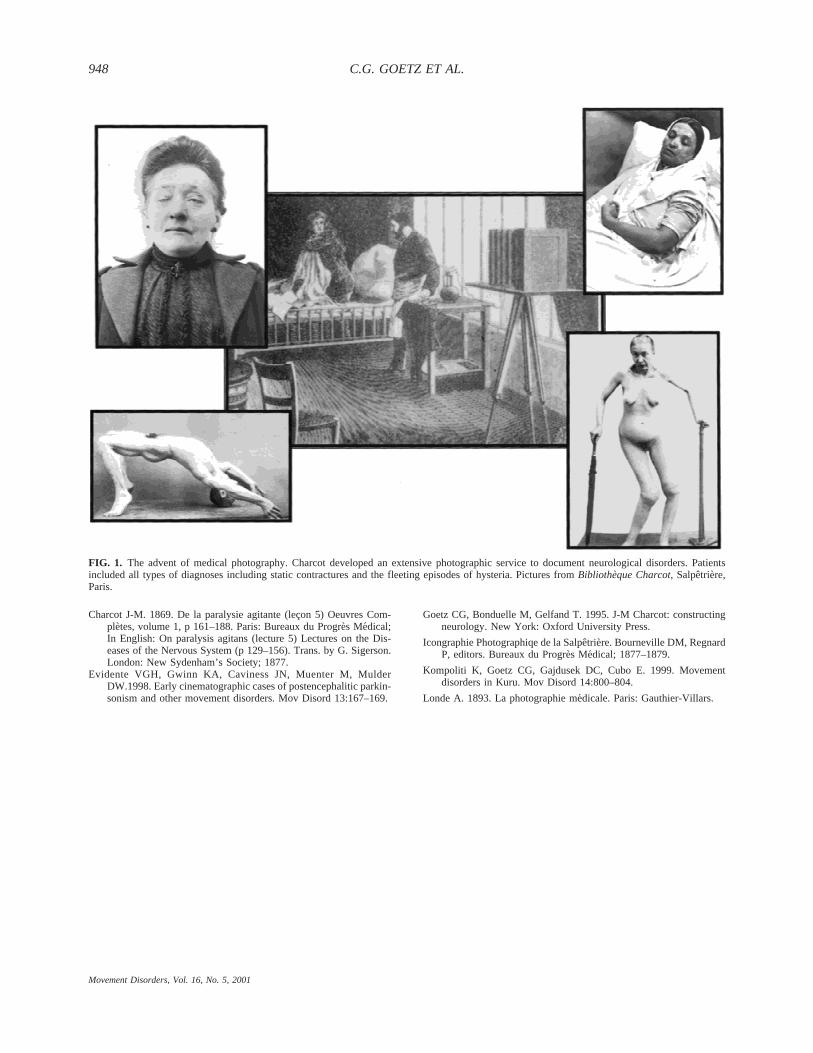
FIG. 1. The advent of medical photography. Charcot developed an extensive photographic service to document neurological disorders. Patientsincluded all types of diagnoses including static contractures and the fleeting episodes of hysteria. Pictures fromBibliothèque Charcot, Salpêtrière,Paris.
C.G. GOETZ ET AL.948
Movement Disorders, Vol. 16, No. 5, 2001
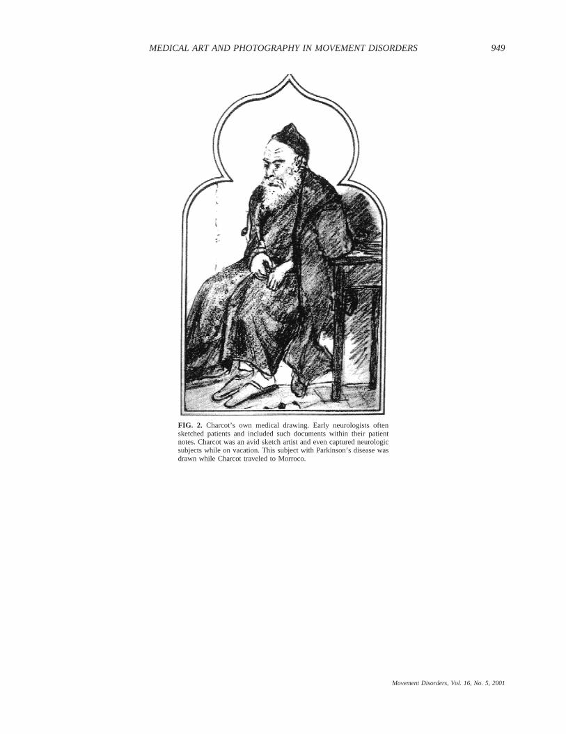
FIG. 2. Charcot’s own medical drawing. Early neurologists oftensketched patients and included such documents within their patientnotes. Charcot was an avid sketch artist and even captured neurologicsubjects while on vacation. This subject with Parkinson’s disease wasdrawn while Charcot traveled to Morroco.
MEDICAL ART AND PHOTOGRAPHY IN MOVEMENT DISORDERS 949
Movement Disorders, Vol. 16, No. 5, 2001
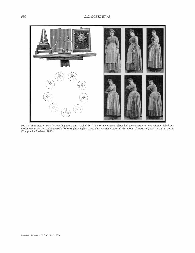
FIG. 3. Time lapse camera for recording movement. Applied by A. Londe, the camera utilized had several apertures electronically linked to ametronome to assure regular intervals between photographic shots. This technique preceded the advent of cinematography. From A. Londe,Photographie Me´dicale, 1893.
C.G. GOETZ ET AL.950
Movement Disorders, Vol. 16, No. 5, 2001
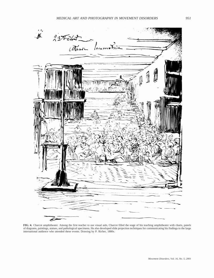
FIG. 4. Charcot amphitheater. Among the first teacher to use visual aids, Charcot filled the stage of his teaching amphitheater with charts, panelsof diagrams, paintings, statues, and pathological specimens. He also developed slide projection techniques for communicating his findings to the largeinternational audience who attended these events. Drawing by P. Richer, 1880s.
MEDICAL ART AND PHOTOGRAPHY IN MOVEMENT DISORDERS 951
Movement Disorders, Vol. 16, No. 5, 2001
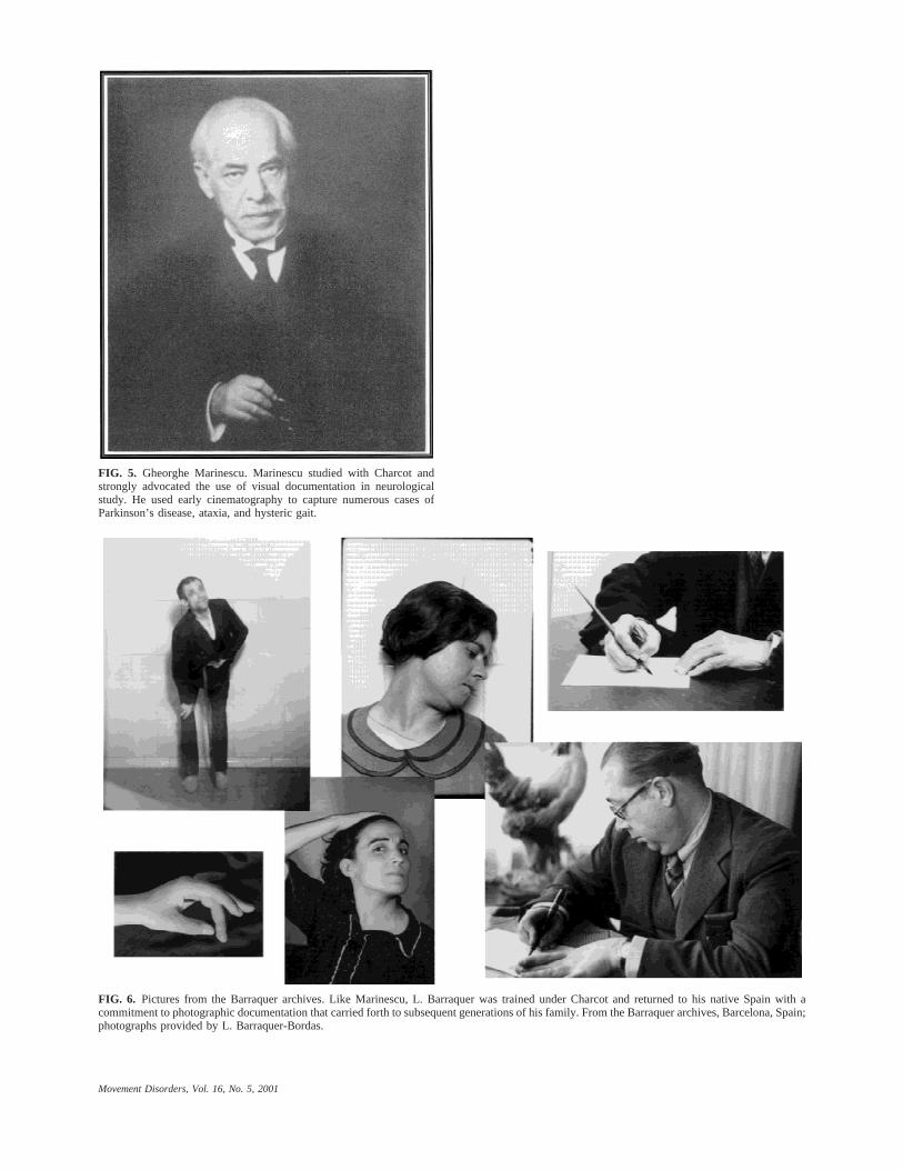
FIG. 5. Gheorghe Marinescu. Marinescu studied with Charcot andstrongly advocated the use of visual documentation in neurologicalstudy. He used early cinematography to capture numerous cases ofParkinson’s disease, ataxia, and hysteric gait.
FIG. 6. Pictures from the Barraquer archives. Like Marinescu, L. Barraquer was trained under Charcot and returned to his native Spain with acommitment to photographic documentation that carried forth to subsequent generations of his family. From the Barraquer archives, Barcelona, Spain;photographs provided by L. Barraquer-Bordas.
Movement Disorders, Vol. 16, No. 5, 2001
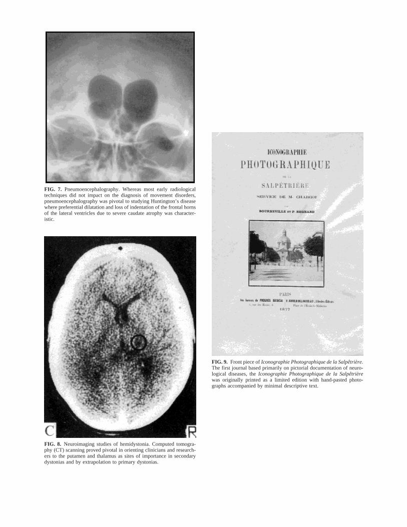
FIG. 7. Pneumoencephalography. Whereas most early radiologicaltechniques did not impact on the diagnosis of movement disorders,pneumoencephalography was pivotal to studying Huntington’s diseasewhere preferential dilatation and loss of indentation of the frontal hornsof the lateral ventricles due to severe caudate atrophy was character-istic.
FIG. 8. Neuroimaging studies of hemidystonia. Computed tomogra-phy (CT) scanning proved pivotal in orienting clinicians and research-ers to the putamen and thalamus as sites of importance in secondarydystonias and by extrapolation to primary dystonias.
FIG. 9. Front piece ofIconographie Photographique de la Salpêtrière.The first journal based primarily on pictorial documentation of neuro-logical diseases, theIconographie Photographique de la Salpêtrièrewas originally printed as a limited edition with hand-pasted photo-graphs accompanied by minimal descriptive text.