Embed Size (px)
Citation preview

METABOLISM OF CARBOHYDRATES
Biosynthesis of glycogen
In various life forms excess of glucose is converted to polymeric forms for storage and to disaccharides for transport. The main storage forms are glycogen in vertebrates, fungi & bacteria. Plants convert glucose to starch for storage. The transport form is sucrose in plants, glucose in animals & trehalose in insects.After a eal when more glucose is available, glycogen synthesis is acclerated. Under fasting conditions most of the glucose requirement it met by gluconcogenesis from non-carbohydrate precursors such as amino acids.Glycogen synthesis occurs virtually in all animal tissues but is specially prominent in liver & skeletal muscles. Liver in fact, is considered a reservoir of glucose in the form of glycogen. Liver’s capacity to store glycogen is only sufficient to supply the brain with glucose for half a day.Glycogen- is a polymer of ɑ (1-4) linked D-glucose with α (1-6) linked branches every 8-10 residues.
Glycogen occurs as intracellular granules of 100-400 A˚ diameter spheroidal molecules that each contains up to 120,000 glucose units. The granules are prominent in the cells that make the grestest use of glycogen; muscle-1-2% by wt. & liver up to 10% by wt. glycogen granules also contain. The enzymes that catalyze glycogen synthesis & degradation as well as many of the proteins that regulate these processes. It has only one reduing end & many non-reducing ends, one at the end of every branch. This permits rapid glucose mobilization thro’ simultaneous release of glucose units from thee end of every branch.
SYNTHESIS OF GLYCOGEN-The staring point of synthesis of glycogen is he conversion of glucose to glucose-6-phesphate.1). Phosphorylation of glucose to glucose-6- phosphate by the enzyme glucokinase or hexokinase. This conversion utilizes the energy in the form of ATP.
Glucose + ATP
Sometimes a (round about) indirect pathway is taken. Glucose is taken up by the erythrocytes & converted to lactate by the glycolytic sequence, which is then taken up by the liver & converted to G-6-p by gluconeogenesis.
Glucose → glycolysis lactateLactate → pyruvate → gluconeogenesis → g-s-p2).G-6-1 to G-I-P by the enzyme phosphoglucomutase.3) formation of UDP- glucose from G-I-P by the action of the enzyme UDP- glucose pyrophorylase. This is a key reaction in the synthesis of glycogen & the enzyme is named after the reserve reaction. In the cell, this reaction occurs in the direction of UDP- glucose formation because pyrophosphate formed
is rapidly hydrolyzed to inorganic PO4 by the enzyme inorganic pyrophosphates with a free energy of ∆ G’°= -25 KJ/ mol.Importance of sugar nucleotides-many reactions in which hexoses are polymerized invole sugar nucleotides. Here the anomertic carbon of a sugar is activated by attachment of a nucleotide thro’ a phosphodiester linkage, sugar nucleotides are substrates for polymerization of monosaccharides into disaccharides, glycogen, starch, cellulose & other complex polysaccharides, vitamin C etc. the role of sugar nucleotides was first discovered by Argentine biochemist luis leloir.
The formation of a sugar nucleotide is metabolically irreversible. The condensation of a nucleoside triphosphate with a hexose-1- phosphate to form a sugar nucleotide has a free energy change of near zero in the cell. The reaction releases pyrophosphate (PPi) which is hydrolyzed to inorganic phosphate. The large negative free energy change of this hydrolysis ∆ G’°= -25 KJ/mol, drives the synthesis reaction.Although the nucleotide itself does not take part in the chemical transformation, it has many groups that can undergo non-covalent interactions with the enzyme & thus contribute significantly to the catalytic activity. Also by ‘tagging’ some hexoses with nucleotidyl group, cells can set them aside for one purpose & separate them from a pool of other hexose phosphates meant for some other function.4). UDP- glucose is the immediate donor of glucose residues in the reaction catalyzed by the enzyme glycogen synthase. This enzyme brings about the transfer of a glucose residue UDP- glucose to a non- reducing end of a glycogen molecule, which acts as a primer. Thus glycogen synthase requires as a primer an α(1→4) polyglucose chain, which may be branched having at least 8 glucose residues.Glycogen synthase cannot make α(1→6) bonds found at the branch points of glycogen. It can only lengthen the branches by adding glucose units forming α(1→4) bonds. The overall equilibrium of the path from G-6-P to lengthened glycogen greatly favours the synthesis of glycogen.
5). The glycogen branching enzyme called amylo(1→4) to (1→6) transglycosylase or glycosy (4→6) transferase catalyzes the transfer of a terminal fragment of 6 or 7 glucose residues from the non-reducing end of a glycogen branch having at least 11 residues to the c-6 hydroxyl group of a glucose residue at a more interior position of the same glycogen chain or another, thus creating a new branch. The biological effect of branching is to make the glycogen molecule more soluble & to increase the number of non-reducing ends. This increases the number of sites for glycogen phosphorylase attack or glycogen synthase activity.
6). Glycogen synthase requires a primer to build up the glycogen molecule branching initiation of a brand new molecule, a protein called glycogenin in needed. This primer molecule acts as both a primer on which new chains are assembled & the enzyme the catalyzes their assembly.a. the first step in the synthesis of a new glycogen molecule is the covalent attachment ofa glucose residue to tyr194 of glycogenin. This attachment is catalyzed by the protien’s glucosyltransferase activity.
b. glycogenin then forms a tight complex with glycogen synthase.
c. the next few steps occurs within this complex & glucose residues are added to the nascent chain till up to 7 or more residues are linked. Each glucose residue is derived from UDP- glucose. The reactions are auto catalytic, mediated by the glucosyl transferase activity of glycogenin. At this point glycogen synthase takes over, extending the glycogen chain & ultimately dissociating from glycogenin.
d. the combined action of glycogen synthase & the branching enzyme builds up the glycogen molecule.
e. glycogenin remains buried within the particle, covalently attached to the single reducing end of the glycogen molecule.
Regulation – glycogen synthesis & glycogen phosphorylase are reciprocally regulated.
Glycogen synthase exists in phosphorylated & dephosphorylated forms. Its active form glycogen synthase –a is dephosphorylated. When it is phosphorylated at several ser-hydroxyl groups by specific protein kinases, glycogen synthase-a is converted to theless active form glycogen synthase-b. conversion of the ‘b’ form back to ‘a’ form is promoted by a phosphoprotein phosphatease, which removes the phosphate groups from the serine residues
On the other hand during breakdown of glycogen, the enzyme glycogen phosphorylase is regulated by both covalent & allosteric modulation.Phosphorylase –a the active form, which is phosphorylated at the ser14 on both of its subunits, is dephosphorylated by the enzyme phosphorylase-a phosohatase to yield phosphorylase-b, this ‘b’ form of the enzyme is less active. This conversion is stimulated by AMP, an allosteric modulator. Phosphorylase-b kinase can convert the inactive phosphorylase-b to phosphorylase-a by phosphorylating the ser14 residue with the help of ATP. Glycogen phosphorylase & glycogen synthase are therefore reciprocally regulated by the phosphorylation & dephosphorylation cycle. When one enzyme is stimulated the other is inhibited & it seems that, these two enzymes are never fully active simultancously.The balance between glycogen synthesis & breakdown in liver is controlled by the hormones glucagons & insulin. These hormones trigger a chain of events that lead to changes in phosphorylation of target proteins including glycogen synthase & glycogen phosphorylase, thus setting their levels pf activity. These hormones also regulate the concentration of F-2,6-BP, thereby balancing gluconeogenesis & glycolysis.
BIOSYNTHESIS OF STARCH-*. Starch is also a high mol. Wt. polymer of D-glucose. Starch is synthesized chloroplasts as one of the stable end products of photosynthesis. It is also synthesized other organelles & tissues like amyloplasts of the colourless parts of plants (seed, roots, tubers etc). The mechanism of hexose polymerization in the synthesis of starch is similar to that of glycogen. The sugar nucleotide in starch synthesis is ADP-glucose, which is formed by the condensation of G-I-P with ATP. Thus the first two reactions in starch synthesis are similar to that of glycogen synthesis viz.1). Glucose + ATP glucose-6- phosphate 2). Glucose-6-phosphate---------------------→ glucose-1-phosphate
3). Glucose-1- phosphate condenses with ATP forming ADP- glucose & inorganic pyrophosphate (PPi) which is then hydrolyzed by the enzyme inorganic pyrophosphatase to form 2 (Pi) with a large negative free energy change.
The enzyme that brings about the formation of ADP-glucose is ADP-glucose pyrophosphorylase. The enzyme is inhibited by Pi but actvated by PGA.4). Starch synthase- then transfers the glucose residues from ADP- glucose to the non-reducing end of →attacking C-4 hydroxyl of the primer, forming the characteristic α(1→4) linkage. The amylase of starch is unbranched, but amylopectin has numerous α(1→6) linkages though fewer than glycogen.
5) chloroplasts contain a branching enzyme similar to the glycogen branching enzyme, that introduces α(1→6) branches of amylopection.The overall reaction is- Starchn + glucose-1-phosphate + ATP →statch n+1 + ADP + 2Pi ∆G’°= -50KJ/ molStatch synthesis is regulated at the level of ADP- glucose formation.
Sucrose synthesis-The 3- carbon compounds generated by the photosynthetic CO2 fixation are converted to sucrose or starch. Sucrose, the disaccharide form is used for transport because of its unusual linkage between the C-1 of glucose & C-2 of fruvtose. This bond is not hydrolyzed by amylases, or other carbohydrate cleaving enzymes. Also as the anomeric carbons of both the monosaccharides are unavailable, this prevents sucrose from reacting non-enzymatically with amino acids & proteins.1). DHAP & GAP are transported from the chloroplast & condensed to form fructose -1, 6-bisphosphate . this reaction is catalyzed by the enzyme aldolase.
2). Hydrolysis of F-1, 6BP leads to F-6-P. this reaction is catalyzed by FB pase.3). Sucrose 6-phosphate synthase then catalyzes the reaction of F-6-P with UDP-glucose to form sucrose-6- phosphate.
4). Sucrose-6- phosphate phospahatase then removes the phosphate group thus making sugar available for transport. Sucrose synthesis is regulated & closely co-ordinated with starch synthesis.
Galactose is obtained from the hydrolysis of lactose. Galactose & glucose are epimerized that differ in their configuration at C-4. although hexokinase phosphorylates glucose fructose & mannose, it does not recognize galactose. Hence galaactose must be epimerized before it enters glycolysis. This conversion takes place with the help of its uridine diphosphate derivative.1). Galactose is first phosphorylated at C-1 by ATP in a reaction catalyzed by galactokinase.2). Galaactose-1- phosphate uridylyl transferase then transfers the uridylyi group of UDP-reversible cleavage of the phosphoryl bond.3). UDP-galactose-4-epimerase then converts UDP-GALACTOSE -4-epimerase then converts UDP-galactose to UDP-glucose. This enzyme has an associated NAD+.UDP-glucose enters the cycle again to converts yet another galactose-i-P.4). Glucose-1-P is converted to the glycolytic intermediate glucose-6-P by the catalytic action of phosphoglucomutase.
Galactosemia is a genetic disease characterized by the inability to convert galactose to glucose. Symptoms are failure to thrive, mental retardation & sometimes death due to liver damage.In the cases there is a deficiency of the enzyme galactose –1-P uridyly! Transferase. Thus formation of UDP-gluctose is prevented & galactose concentration in the blood builds up. This may sometimes lead to higher galactose concentration in the lens of the eye where the sugar is reduced to galactiol, which in turn leads to cataract formation.
BIOSYNTHESIS OF DEXTRANS-Dextrans are polysaccharides formed when various strains of leuconostoc mesenteroides & some other micro organisms are grown in sucrose solutions. The dextrans form ropy slimes & have various mol. Wts ., some as high as 4,000,000. they are all D-glucopyranose polymers but the molecular structure varies with the type & strain of organism forming them. They have branched structures involving α(1→6), α(1→4) & (1→3) linkages between glucose units though α(1→6) bonds generally dominate.Synthesis- dextran ssynthesis by micro organism consists of a cries of transglycosylations. Apparently the process starts something as follows, assuming α(1→6) linkage to be formed at first.
Glucose-1-fructose+ glucose-1-fructose--→glucose-,6-glucoside+2 fructose
Sucrose then continues to add to this new disaccharide with varying linkages to build up the execcdingly complex dextran molecule. The overall process may be represented as follows:-
N sucrose -----→ (C6H10O5)n +n fructoseDextran is of importance in mediagte because of its use as plasma substitute. They very great particle size of native dextran makes it unsuitable for this purpose & so preparations are obtained by hydrolyzing the native dextran with acid or enzymes to an average molecular wt. of 76000.(transglucosylase builds up dextrans; transfructosylase builds up levan or inulin).
BIOSYNTHESIS OF HEPARIN –Introduction- the extracellular spaces in the tissues of multicellular animals is fiiled with a gel like material, the extracellular matrix. This holds the cells together & provides a porous pathway for the diffusion of nutrients & oxygen to individual cells. Tthese tissues includes the connective tiissues such as cartilage, tendons, skin & blood vessles. This matrix is composed of interlocking meshwork of heterpoly – saccharides & fibrous proteins such as collagen, elastin, fibronectin &laminin. The heteropolysaccharides are largely glycosaminoglycans. These are a family of linear polymers composed of repeating disaccharide units. Once of the two monosaccharides is always either N-acetylglucosmine or N- acetylactosamine & the other monosaccharide is usually a uronic acid viz.-D-glucuronic acid or L- iduronic acid . in some glycosaminoglycans one or more of the hydroxyl groups of the amino sugar is esterified with sulphate. The combination of sulphate groups & the carboxylate groups of the uronic acid residues give the glycosaminoglycans a very high-ve charge. Some of the glycosaminoglycans are:-
1. hyaluronic acid→ D-glucuronate + N acetyl-D glucosamine.2. chondroitin-4- sulfate→ D-glucuronate + N- acetyl- D galaactosamine.3. dermatan sulphate → L- iduronate + N- acetyl-D- galactosamine4. keratin sulfaate → D- galactose + N- acetyl- D glucosamine -6- sulfate5. herarin→ D-iduronate – 2-sulfate + N-sulfo-D-glucosamine-6-sulfate.
A major function of the glycosaminoglycans is the formation of matrix to hold the protein components together to form proteoglycans. the proteglycans are widely distributed amongst tissues & are intimately associated with the extra cellular matrix. Heparin differs from the others by being present intra cellularly. It is present in the granules of mast cells, however its action accurs extra cellularly, after it is secreted from the cell.Herparin- is variably sulphated with an average of 2.5 sulfate residues per disaccharide unit, which makes it the most highly charged polymer in mammalian tissues. Heparin is not a constituent of the connective tissues but almost occufrs exclusively in the intra cellular granules of the mast cells that occur in the artcrital walls. Herparin was fecognized early because of its anticoagulant properties.Herparin is a polymer in which the repeating units consist of D-glucuronic acid usually with an O-SO4 group at C2 & D-glucosamine –N- SO4 with an additional O-SO4 at C6 both the linkages of the polymer are α(1→4).
enzymes. The synthesis of the two monosaccharides, N-acetylgluucosamine (GlcNAc) & glucuronic acid (GlcA) is a follows:-
1. synthesis of N- acetylglucosamine – (GlcNAc) glucose-6-P ↓
transaminate glutamine glutamate D- glucosdsamine-6-PGlucosamine -6-Pacetylase actyl CoA ↓ CoASH N-Acetyl-D-glucosamine-6-P ↓ Phosphosetyl glucosamine mutase N-Acetyl-D-glucosamine-1-PUDP-acetyl glucosamine UTP Pyrophosphorylase ↓ PPC UDP-N-Acetyl-D-glucosamine--------------→GalNAc
1. Fructose-6-P is isomerized & aminated to form D- glucosamine-6-P. the amino group donor being glutamine which is converted to glutamate.
2. D- glucosamine -6-P is then acetylated by acetyl CoA to form N-acetyl-D-glucosamine-6-P. CoA is reduced to CoASH.
3. N-acetyl-D-glucosamine-6-P is then converted to-1-P form by the enzyme mutase.4. this aminated, acetylated sugar is now converted to a sugar nucleotide by UTP. Pyrophosphate
is released & UDP-N-acetyl-D-gluosamine is formed.5. this can then be epimerized to GalNAc.
2. synthesis of Uronic acid- the specific oxidation of the primary alcohol group of aldoses yields uronic acid. Thus:-
3. sulfation-
POLYMERIZATION:-1. polysaccharide formation is initiated on a scrine residue of the core protein. Polypeptide of the
core protein, on which the polysaccharide chain is initiated consists of alternating serine & glycine residue. A xylose sugar is transferred from UDP-xylose to a serine residue by the enzyme xylose transferase.
2. the next step involves the transfer of two galactose units from the correcsponding UDP-nucleotides to the xylosylatedcore protein. Amino acid sequences flanking the serine residue to which xylose is attached, seem to act as singals for directing the assembling heparin. D-galactosyl transferase I & D-galactosyl transferase II bring about the linkage of the two galactose units.
3. the non-reducing end of the neutral trisaccharide xylosyl-galactosyl-galactose becomes the primer for the elongation of the polysaccharide. In case of heparin & heparin sulphate,
polymer formation occurs through an alternating transfer of glucuronic acid (GlcA) & N- acetylglucosamine (GlcNAc) units to the growing chain the mechanisms that control the length of the fully grown polysaccharides are not fully elucidated. In general, the length of the chain increases with the availability of the UDP-sugar precursor & decreases with the availability of the core protein. Glucouronin transferase I & GlcNAc transferase I bring about the respective linkages, further addition of GlcA & GlcNAc is brought about by a hetero oilgomeric dimmer complex EXTI & EXT2.
4. subsequent to the polymer formation, the repeating GlcA-GlcNAc disaccharide chain undergoes a number of enzymatic modifications. These occurs in the following orders:-
a. N-deacetylation of GlcNAc units give rise to the glucosamine ( GlcN) residues. The deacetylation is brought about by the enzyme deacetylase. This enzyme has a regulatory function.
b. N- sulfation of the newly formed GlcN residues by the enzyme N- sulfotransferase, which adds sulface groups to the free amino groups of glucosamine. PAPs is the sulphate donor.
c. C-5 epimerization of GlcA residues which leads to the formation of L-iduronic acid units (IdoA).
d. 2-0 sulfation of the newly formed IdoA by o-sulfotransferase.e. 6-0 sulfation of GlcN residues. Sulfation can also occur at C-3 of GlcN units & C-3 of GlcA
residues to a limited extent.
All these modifications are incomplete in vivo. In other words, not all the sugar residues that are potential substrates for the various enzymes are transformed into their relative products. Since 2-0 & 6-0 sulfations occur only after epimerization, the distribution of the sulphate groups is restricted to N- sulphated regions.It is proposed that sulfation proceeds at a much faster rate than chain clongation & when sulfation reaches the terminal Glc NAc it favours termination of the chain. It has been shown that GlcNAc sulphated at 4- position forms 87-95% of the terminal GlcNAc indicating that sulfation is important in the regulation of the chain length. Cleavage of herparin, packing & transport- The heparin polysaccharide formed by the activity of EXTI & EXTII is cleaved by an endo-β-D-glucuronidase to give fragments of relative mol. Wt. of 5000-30,000 D. these fragments are vesicularised in the golgi complex & transferred via blood to the mast cells for storage & secretion
SYNTHESIS OF THE PEPTIDO-GLYCAN FRAMEWORK OF BACTERIAL CELL WALL- Bacteria are surrounded by rigid cell walls that give them their characteristic shapes & permit them to live in hypotonic environments. Bacterial cell walls are of considerable significance because they are responsible for bacterial virulence. In fact, the symptoms of many bacterial disease can be elicited in animals merely by injection of bacterial cell walls. The characteristics antigens of bacteria are components of their cell walls & hence injecting bacterial cell wall preparations in animals often invokes immunity against bacteria. Bacteria are classified as gram +ve & gram –ve depending upon whether they take up gram stain. Gram +ve bacteria have thick cell wall whereas gram –ve bacteria have a thin cell wall. The cell walls of bacteria consist of covalently linked polysaccharide & stromiger is known as a peptidoglycan. The polysaccharide component consist of linear chains of alternating β(1→4) linked GlcNAc &N-acetylmuramic acid. They are further cross linked by short peptides attached to the Mur2Ac.Synthesis- during the assembly of the polysaccharide backbone of this complex macromolecule, both GlcAc & Mur2Ac are activated by attachment of uridine dinuccleotide to their anomeric carbons.
1. GlcNAc-1-P condenses with UTP to form UDP-GcNAc.2. this sugar nucleotide UDP-GlcNAc then reacts with phosphoenol pyruvate to form UDP-
Mur2Ac. This reaction needs the reducing power of NADPH, which is oxidized to NADP+.
3. five amino acids viz.- L-alanine, D-glutamate, L-lysine &2 molecules of D-alanine then form a pentapeptide with Mur2Ac.
4. the next reaction sees the transfer of Mur2Ac- pentapeptide from the uridine dinucleotide to a long chain isoprenoid alcohol- dolichol phosphate.
5. A GlcNAc unit is now donated by UDP-GlcNAc & a glycosidic linkage is formed between GlcNAc & Mur2Ac pentapeptide linked to dolichol.
6. in many bacteria 5 molecules of glycine linked by peptide linkages are linked to lysine residue of the pentapeptide.
7. finally this disaccharide decapeptide is added to the non-reducing end of an existing peptidoglycan molecule.
8. A transpeptidase cross links adjacent polysaccharide chains. Without the support of this peptidoglycan layers the bacterial cell is fragile & prone to osmotic lysis.
METABOLISM OF LIPIDSLipids play a variety of roles in the cells. They are-
1. the main form of stored energy.2. they are major constituents of cell membrances.3. specialized lipids serve as pigments (carotene, retinal), cofactors (vit K), detergents (bile
salts), transporters(doliclols), hormones (vit D derivatives & sex hormones), extra-& intra- cellular messengers (elcosanoids), etc.
thus synthesis of a variety of lipids is vital to all organisms.Like all other bioaynthetic pathways, lipids synthesis is also endergonic & reductive process. ATP is used asa source of metabolic energy & NADPH+H+ as a reducant.
SYNTHESIS OF PALMITIC ACID FROM Acetyl CoA-
David rittenberg & Conrad bloch in 1945, demonstrated that fatty acid biosynthesis occurs through 2C units condensation & that these 2C units are derived from acetic acid.When FA (fatty acid) oxidation was found to occur by the oidative removal of 2C units (acetyl CoA), it was thought that FA ynthesis might occur by simple reversal of the same steps. But that is not so!The breakdown ( oxidation) & synthesis pathways are catalyzed by different set of enzyme & take place in different parts of the cell. Moreover, a 3c intermediate (malonyl CoA) participates in the synthesis process but not in the breakdown.Fatty acids are synthesized by an extra-mitochondrial system which is responsible for the complete synthesis of palmitate from acetyl CoA in the cytosol.
STEP I- SYNTHESIS OF MALONYL CoA FROM ACETYL CoA-Acety’ CoA is the starting material for FA synthesis, it is generated in the mitochondria by the oxidative decarboxylation of pyruvate, the reaction being catalyzed by pyruvate the reaction being catalyzed by pyruvate, the reaction being catalyzed by pyruvate dehydrogenase. Acetyl CoA is also generaled from catabolism of amino acids, but acetyl CoA from FA oxidation is not a significant source for synthesis because oxidation & synthesis are reciprocally regulated.Acetyl CoA from the mitochondria cannot be transported out to the cytosol as the mitochondrial menbrance is impermeable to acetyl CoA.Hence, it first condenses with OAA to form citrate, which is then transport Via the citrate transporter to the cytosol.Acetyl CoA + OAA citrate syntiase citrate CoASH In the cytosol, citrate is cleaved by citrate lyase to regenerate acetyl CoA in an ATP dependent reaction.Citrate + CoA + ATP citratelyase Acetyl-CoA + ADP + PC + OAA
OAA MDH malateMalate ME pyruvate + Co2
NADP NADPH+H+
OAA cannot return to the mitohondria as it is due to the lack of a transporter, but is converted to malate by cytosolic malate dehydrogenase. Malate is then deerarboxylated to pyrovate by malic enzyme the NADPH+H+ produced is utilized in the FA synthesis reactions.The irreversible formation of malonyl CoA from Acetyl CoA & bicarbonate is catalyzed by acetvl CoA carboxylase.In bacteria this enzyme is made up of 3 different polypeptide subunits.In animals, all the three activities are a part of a single multifunctional polypeptide.In plant cells both forms of the enzymes are cited.In all cases, acetyl CoA carboxylase contains a biotin as prothetic group, which is covalently bound in an amide linkage to the ε amino group of a lys residue on one of the three domains of the enzyme molecule. The reaction is similar to other biotin dependent enzymes like pyruvate carboxylase & propionyl CoA carboxylase.

1. the carboxylase group derived from the HCO3 is first transferred to biotin in an ATP dependent reaction. Biotin carboxylase brings about this transfer.
2. the biotinyl group serves as a temporary carrier of CO2 transferring it to acetyl CoA by the transcarboxylase to yield malonyl CoA.
3. the enzyme acrtyl CoA carboxylase thus has biotin, biotin carrier protein, biotin carboxylase, transcarboxylase & an allosteric site, which has a regulatory function.
STEP II-The long chain FAS are assembled in a repeating 4-step sequence. The synthesis of FAs from acetyl CoA & malonyl CoA involves seven enzymatic reactions. These reactions were first studied in cell-free extracts of E.colli. in bacteria & plants the individual enzymes of the fatty acid synthase system are separate & the acyl radicals are found in combination with a protein called the acyl carrier protein-ACP.In yeast , mammals & birds, the synthase system is a multienzyme polypeptide complex that incorporates ACP, which takes over the role of CoA. This ACP contains the vitamins- pantotheenic acid in the form of 4’ phosphosantetheine, which forms a thioester with an acyl group. The phosphospantetheine is esterified with a ser-OH group of ACP. The use of one multinzyme polypeptide complex, that incorporates ACP, has the advantages of achieving the effect of compartmentalization of the process within the cell without the erection of permeability barriers. Moreover, synthesis of all the enzymes in the complex is coordinated since, it is a dimmer comprising of two identical monomers, each containg all seven, enzyme activities on one polypeptide chain arranged in a head to tail manner.
ACP-acyl carrier protein –caries acyl groups in thioester linkage.AT-actyl CoA-ACP transacetylase-transfer of acyl grp CoA—KSKS-β-ketoacyl-ACP synthase – condenses acyl & malonyl grps.MT- malonyl CoA-ACP transferase- transfers malonyl—ACPKR-β- ketoacyl-ACP reductase – reduces β-keto—β hydroxylHD- β – hydroxyacyl-ACP dehydrates- removes H2OER- enoyl-ACP reductase—reduces double bond forming sat. acyl ACP.

Fatty acid synthesis begis with the formation of malonyl CoA, but the growth of the FA chain is anchored to the acyl carrier protein (ACP).Hydrolysis of the thioester that links ACP to the fatty acly group is highly exergonic & the energy released when this bond is broken helps to makes thee first reaction thermodynamically favourable.The 4’phosphospantetheine of ACP is believed to serve as a flexible arm holding the growing fatty acyl chain to the surface of the FA synthase comlex & carrying the reaction intermediates from one enzyme active site to the next.
Priming reactions- before the 4 steps of the chain build up take place, two priming reactions or charging of the complex with the correct acyl groups is brought about.These are-
a. the acetyl group of acetyl CoA is transferred to the cys-SH group of the β- ketoacyl-ACP synthase (KS) by the enzyme acetyl CoA- ACP transacetylase (AT).
b. in the second reaction the malony group from malonyl CoA is transferred to the SH group of ACP. This is catalyzed by malonyl CoA-ACP transacetylase (MT).
With the attachment of the two acyl groups the enzyme is now “charged” or “loaded”.Reaction 1-condensation:-The first step in the formation of aFA chain is the condensation of the activated acetyl & malonyl groups to form the acetoacetyl-ACP, which is bound to the ACP through the pan-SH group. Simultaneously a molecule of CO2 is removed. The decarboxylation reaction drives the condensation reaction.

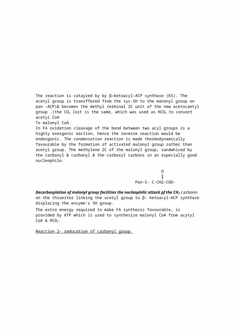
The reaction is catayzed by by β—ketoacyl-ACP synthase (KS). The acetyl group is transffered from the syc-SH to the manonyl group on pan –ACP)& becomes the methyl terminal 2C unit of the new acetocaetyl group .(the CO2 lost is the same, which was used as HCO3 to convert acetyi CoA To malonyl CoA.In FA oxidation cleavage of the bond between two acyl groups is a highly exergonic eaction, hence the reverse reaction would be endergonic. The condensation reaction is made thermodynamically favourable by the formation of activated malonyl group rather than acetyl group. The methylene 2C of the malonyl group, sandwhiced by the carbonyl & carbonyl & the carboxyl carbons in an especially good nucleophile. O ║ Pan-S- C-CH2-COO-
Decarboxylation of malonyl group facilities the nucleophilic attack pf the CH2 carbonn on the thioester linking the acetyl group to β- ketoacyl-ACP synthase displacing the enzyme’s SH group.
The extra energy required to make FA synthesis favourable, is provided by ATP which is used to synthesize malonyl CoA from acytyl CoA & HCO3.
Reaction 2- reducation of carbonyl group
The acetoacetyl- ACP formed after condensation undergoes reduction of the carbonl group at C-3 to from D-β hydroxybutyryl ACP. The reaction is cataylzed by β- ketoacyl reducatse (KR) & the e- donor is NADPH+H+. D-β- hydroxybutyryl group does not have the same stereoisomeric form as the L-β-htdroxybutyryl intermediate of FA oxidation.REACTION 3-DEHYDRATION-The elements of water are removed from C-2&C-3Oof D-β- hydroxybutyryl ACP to yield a double bond between C-2 & C-3 to form to form trans ∆2 – butenoyl ACP. The enzyme that catalyzes this this dehydration is β- hydroxyacyl-ACP dehydratase (HD).
REACTION-4-REDUCTION OF THE DOUBLE BOND-

Finally the double bond of ∆2 – butenoyl- ACP is reduced 9standard) to form butyryl- ACP. The reaction is catalyzed by enoyl-ACP reduatace (ER) & once again NADPH+H+ is the’s donor.The production of a 4C, saturated fattyacyl-ACP completes one pass through the fatty acid synthase complex.
STEP IIIThe butyryl group is now is ow transferred from the phosphopantetheine, SH group of ACP to the eyes-SH of β- ketoacyl synthase (KS) which initially bore the acetyl group.
Another malonyl group is now linked to the phosphosantetheine of the ACP.
Thus 7 cyvles later 16-C palmitoyl- ACP is formed which is bound to ACP.
STEP IV-
Free palmitate is released from ACP molecule by a hydrolytic activity in the synthase complex,
Small amonds of longer fatty acids such as stearate 18:0 are also formed. In certain plants like cocout & palm, chain termination occurs earlier & about 90% of the FAs in these oils are between 8-14 carbons long.THE OVERALL REACTION=
7 acetyl CoA + 7 CO2 + 7 ATP →7 malonyl CoA + 7ADP + 7Pi seven cycles later,Acetyl CoA + 7 molonyl CoA + 14 NADPH + 14 H+ → palmitate + 7 Co2 + 8 CoA + 14 NADP+ + 6 H2O
SOURCE OF NADPH+H+ -strong reducing environment for the reductive syntheis of fatty acids.Cytosolic NADH/NAD+ ratio is lower, so catabolism of glucose canocur in the same compartment at the same time as FA synthesis.In heptocytes & adipocytes, NADPH is generated by the pentose phosphate pathway & by NADP linked malic enzyme. Inside the mitochondria a high NADH/NAD+ ratio favours reduction of O2 via the respirtory chain.In plants fatty acid synthesis occurs in the chloroplast stroma & thus light reactions are the source of NADPH + H+
SOURCE OF ACETYL CoA-In non- photosynthetic eukaryotes, acetyl CoA is formed in the mitochondria from pyruvate oxidation & also from amino acid oxidation.Acetyl CoA from FA oxidation is not a significant source because oxidation & synthesis are regulated reciprocally.
REGULATION IN VERTEBRATES-i. the reaction catalyzed by by acetyl CoA carboxylase is the rate limiting step in
the biosynthesis of FA. Palmitoyl CoA, the principle end product of FA synthesis acts as a feed back inhibitor of the enzyme.
ii. Citrate is an allosteric activator & plays an omportant role in divering the cellular metabolism from consumption of metabolic fuel to its storage as FAs.
iii. Citrate is transported out of the mitochondria when there is an increase in the concentratiom of acetyl CoA & ATP. This citrate becomes the precursor of acetyl CoA in the cytosol; it alsoacts as an alosteric signal for the activation of acetyl CoA carboxylase & also inhibits the activity of PFK-I, thus reducing the rate of glycolysis.
iv. Acetyl CoA carboxylase is also regulated by covelent modification the homones glucangons & epinephrine trigger the ohosphorylation of acetyl CoA carboxylase, which is aan inactive form. This slows down the synthesis process. Phosphorylation also causes dissociation of the monomeric units & also loss of activity
In plants & bacteria regulation is not by citrate or phosphorylation. In plants activation is by increase in stromal pH & Mg2+. In bacteria the regulatory mechanism is much more complex. FA synthesis & β- oxidation do not occur simultaneouasly. Malonyl CoA inhibits β- oxidation by inhibiting the enzyme carnitine – acyltransferase I.
OVERVIEW-
BIOSYNTHEIS OF CHOLESTEROL FROM ACETYL CoA-Cholesterol is the most publicized lipid & is in focus because of the strong correlation between high cholesterol levels & cardiovascular disease. Cholesterol plays a crucial role in the structure of many membrances . it is also precursor of steroid hormones & bile acids. In mammalin diet it is not essential to includeit in the diet as mammals can synthesize it form simple precursors. Thus , in healthy organisms an intricate balance is maintanined between the biosynthesis, utilization & transport of cholesterol, keeping its harmful deposition to a minimum.
Structure- it is a 27 carbon compound & all its carbons are provided by acetate. Tis was revealed by the isotopic studies. The lengthly pathway of its synthesis was outlined by konrad bloch.
Study of the pathway of cholesterol synthesis has led to an understanding of: i. the transport of cholesterol & other lipids between organs .ii. the process by which cholesterol enters the cells.iii. the means by which intracellular cholesterol production is influenced by dietary cholesterol &iv. how failure to regulate cholesterol production affects health.
Synthesis-Acetate is converted to isoprene units, which are the key intermediates in the pathway to synthesis of cholesterol. Isoprene are also precursors to many other natural lipids.Cholesterol is synthesized from acetyl CoA in 4 stages. Like other long chain fatty acids, cholesterol is made from acetyl CoA but the basic assembly line is different. The 4 stages can be summarized as follows:
i. 3 acetate units condense to form 6C intermediate mevalonate.ii. Conversion of mevalonate to activated isoprene units.iii. Polymerization of six 5C isoprene units to form 30C linear structure of
squalence.iv. The cyclization of squalene forms the 4 rings of the steroid nucleus, which is
then follows by oxidation & removal or migration of methyl groups.
STEP I- SYNTHESIS OF MEVALONATE FROM ACETATE-2 molecules of acetyl CoA condense to form acetoacetyl CoA which then condenses with a third molecule acetyl CoA to yield a 6C compound known as β- hydroxyl-β- methyl glutaryl CoA (HMG CoA). These two teactions the catalyzed by thiolase & HMG CoA synthase. Both the reactions are reversible.In mitochondria, these two enzymes synthesize ketoric bodies, while the cytosolic isozymes synthesize HMG CoA that is used in cholesterol synthesis.The third reaction is irreversible in which HMG CoA is reduced to mevalonate. 2 molecules of NADPH+H+ donate 2e.g. the reaction is catalyzed by HMG CoA reductase, which is an integral
membrane protein of the smooth endoplamic reticulum. This enzyme is the major point of regulation in the synthesis of cholesterol.
STEPII- CONNVERTION OF MEVALONATE TO 2 ACTIVATED ISOPRENES-In the next stage of cholesterol synthesis, 3b phosphate groups are transffered from 3 ATP’s to mevalonate forming 3- phosphp-5-pyrophosphate mevalonate. Mevalonate 5- phosphor transferase brings aabout the first phosphorylation while phosphor-mevalonate kinase converts it pyrophosphate.In the next step 3 phospho-& the neighbouring carboxyl group, leave mevalonate producing a double bond In the 5C product- ∆3 – isopentenyl pyrophosphate. This reaction is catalyzed by pyrophospho mevalonate decarboxylase & leads to the formation of a molecule of CO2 & Pi.A reversible isomerization of the 5C product yields dimethyl allyl pyrophosphate. These two form the activated forms of the isoprene molecules.
STEPIII- CONDENSATION O0F SIX ACTIVATED ISOPRENE UNITS TO FORM SQUALEENE-Isopentenyl pyrophosphate & dimethyl allyl pyrophosphate undergo head to tail condensation. In this condensation one pyrophosphate group is displaces a 10c chain of geranyl pyrophosphate is formed. The enzyme that catalyzes this condensation is prenyl transferase.A seconds condensation of geranyl pyrophosphate with isopentenyl pyrophosphate yields a 15C intermediate known as franesyl pyrophosphate & a subsequent release of a pyrophosphate group. This condensation is also catalyzed by prenyl transferase. Finally two molecules of farnesyl pyrophosphate condense head to head with the elimination of both pyrophosphate groups forming squalene. The reaction is catalyzed by saqualene synthase.Farnesyl pyrophosphate is also the precursor of other isoprenoid compounds in mammals like ubiquinone.
STEPIV- CYCLIZATION OF SQUALEENE TO 4-RING STEROID NUCLEUS-All the sterols have 4 fused rings (steroid nucleus) & all are alcohols with a hydroxyl group at C-3, thus the name sterol.The cyclization of the linear hydrocarbon to form the tetracyclic steroid takes place in two steps.i. SQUALENE EPOXIDASE OR SQUALENE MONO- OXYGENASE- adds one oxygen
atom from O2 to the end of the aqualene chain forming an epoxide. NADPH+H+ reduces the remaining atom of O2 to H2O.
ii. the double bonds of the product, squalene 2,3-epoxide are positioned so that a remarkable concerted reaction can convert the linear squalene epoxide into a cyclic structure. In animals this cyclization results in lanosteril, which contains the characteristic 4 rings of the steroid nucleus. Lanosterol is converted, after a series of reactions (abouts 20) to cholesterol. The cyclization of squalene 2,3 epoxide is brought about by squalene oxidocyclase. The enzymes are embedded in the membrane of the endoplasmic reticulum.
Plants, fungi & protests make other related sterols instead of cholesterol using the same synthetic pathway as far as squalene 2,3 epoxide. The diversification in plants is to form other sterols like stigmasterol in some while ergosterols in fungi.
Cholesterol synthesized by the liver is either converted to bile acids or it is esterified by ACAT (acyl CoA cholesterol acyl transferase) to form cholesterol ester. These are highly hydrophobic & transported throughout the body as lipoprotein complexs. They are packaged into VLDL & enter the blood stream. As they circulate TAGs & apolipoprotiens are removed & VLDL is converted to IDL & LDL.