Embed Size (px)
Citation preview
The ANNALS of AFRICAN SURGERY. January 2016 Volume 13 Issue 132
CASE REPORT
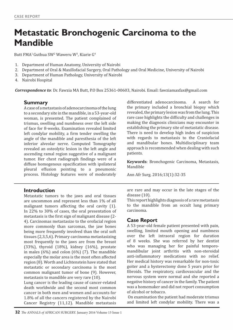
Metastatic Bronchogenic Carcinoma to the MandibleButt FMA1 Guthua SW2 Waweru W3, Kiarie G4
1. Department of Human Anatomy, University of Nairobi2. Department of Oral & Maxillofacial Surgery, Oral Pathology and Oral Medicine, University of Nairobi3. Department of Human Pathology, University of Nairobi4. Nairobi Hospital
Correspondence to: Dr. Fawzia MA Butt, P.O Box 25361-00603, Nairobi. Email: [email protected]
Summary A case of a metastasis of adenocarcinoma of the lung to a secondary site in the mandible, in a 53-year-old woman, is presented. The patient complained of trismus, swelling and numbness over the left side of face for 8-weeks. Examination revealed limited left condylar mobility, a firm tender swelling the angle of the mandible and paresthesia of the left inferior alveolar nerve. Computed Tomography revealed an osteolytic lesion in the left angle and ascending ramal region suggestive of a malignant tumor. Her chest radiograph findings were of a diffuse homogenous opacification with ipsilateral pleural effusion pointing to a pneumonic process. Histology features were of moderately
differentiated adenocarcinoma. A search for the primary included a bronchial biopsy which revealed, the primary lesion was from the lung. This rare case highlights the difficulty and challenges in making the diagnosis clinicians may encounter in establishing the primary site of metastatic disease. There is need to develop high index of suspicion with regards to metastasis to the Craniofacial and mandibular bones. Multidisciplinary team approach is recommended when dealing with such patients.
Keywords: Bronchogenic Carcinoma, Metastasis, Mandible
Ann Afr Surg. 2016;13(1):32-35
Introduction Metastatic tumors to the jaws and oral tissues are uncommon and represent less than 1% of all malignant tumors affecting the oral cavity (1). In 22% to 30% of cases, the oral presentation of metastasis is the first sign of malignant disease (2-4). Carcinomas metastasize to the orofacial region more commonly than sarcomas, the jaw bones being more frequently involved than the oral soft tissues (2,3,5,6). Primary carcinoma metastasizing most frequently to the jaws are from the breast (33%), thyroid (18%), kidney (16%), prostate in males (6%) and colon (6%) (7). The mandible especially the molar area is the most often affected region (8). Worth and Lichtenstein have stated that metastatic or secondary carcinoma is the most common malignant tumor of bone (9). However, metastasis to mandible are very rare (10).Lung cancer is the leading cause of cancer-related death worldwide and the second most common cancer in both men and women and accounts for 1.8% of all the cancers registered by the Nairobi Cancer Registry (11,12). Mandible metastasis
are rare and may occur in the late stages of the disease (10). This report highlights diagnosis of a rare metastasis to the mandible from an occult lung primary carcinoma.
Case ReportA 53-year-old female patient presented with pain, swelling, limited mouth opening and numbness over the left intraoral region for duration of 8 weeks. She was referred by her dentist who was managing her for painful temporo-mandibular joint arthritis with non-steroidal anti-inflammatory medications with no relief. Her medical history was remarkable for non-toxic goiter and a hysterectomy done 5 years prior for fibroids. The respiratory, cardiovascular and the nervous system were normal and she reported a negative history of cancer in the family. The patient was a homemaker and did not report consumption of alcohol or tobacco. On examination the patient had moderate trismus and limited left condylar mobility. There was a
The ANNALS of AFRICAN SURGERY | www.annalsofafricansurgery.com
The ANNALS of AFRICAN SURGERY. January 2016 Volume 13 Issue 1 33
ORIGINAL PAPER
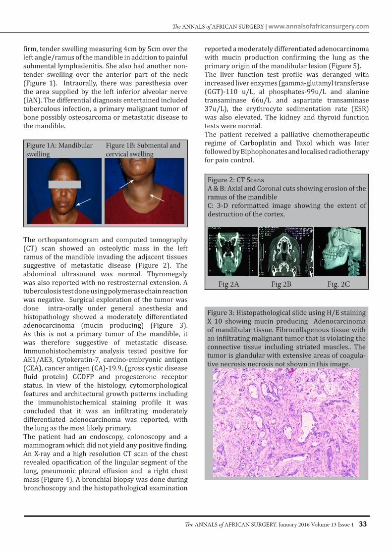
firm, tender swelling measuring 4cm by 5cm over the left angle/ramus of the mandible in addition to painful submental lymphadenitis. She also had another non-tender swelling over the anterior part of the neck (Figure 1). Intraorally, there was paresthesia over the area supplied by the left inferior alveolar nerve (IAN). The differential diagnosis entertained included tuberculous infection, a primary malignant tumor of bone possibly osteosarcoma or metastatic disease to the mandible.
Figure 1A: Mandibular Figure 1B: Submental and swelling cervical swelling
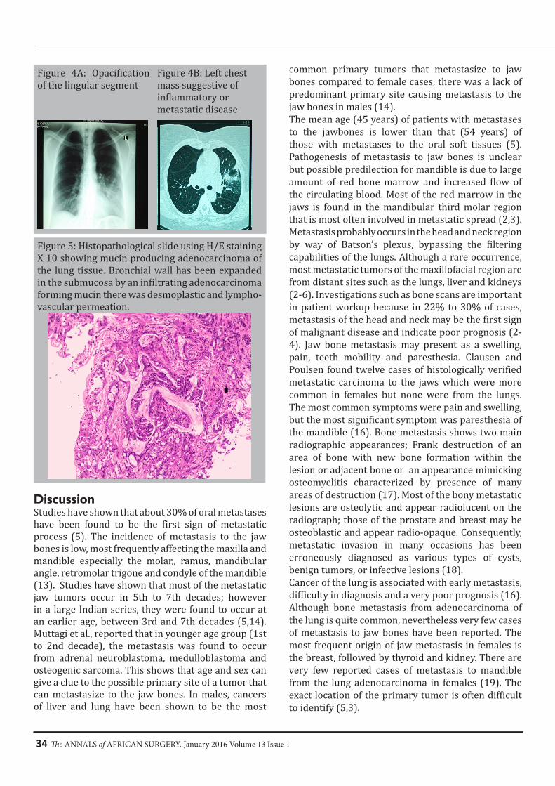
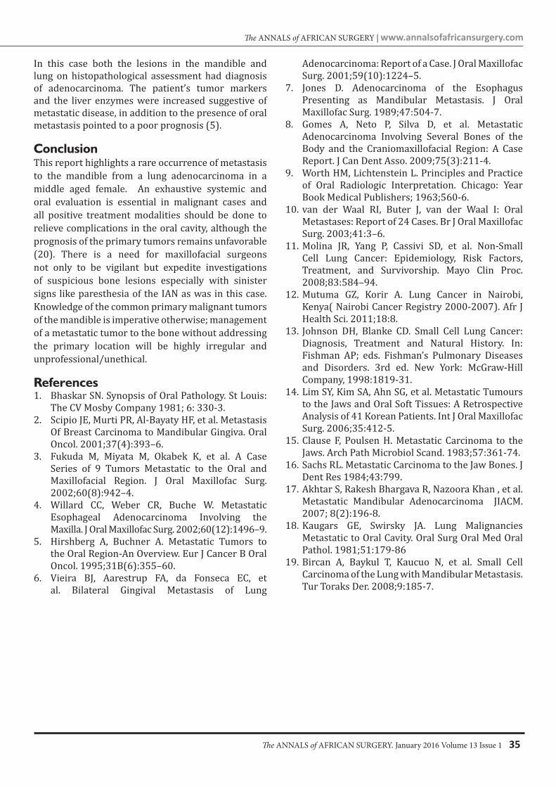
The orthopantomogram and computed tomography (CT) scan showed an osteolytic mass in the left ramus of the mandible invading the adjacent tissues suggestive of metastatic disease (Figure 2). The abdominal ultrasound was normal. Thyromegaly was also reported with no restrosternal extension. A tuberculosis test done using polymerase chain reaction was negative. Surgical exploration of the tumor was done intra-orally under general anesthesia and histopathology showed a moderately differentiated adenocarcinoma (mucin producing) (Figure 3). As this is not a primary tumor of the mandible, it was therefore suggestive of metastatic disease. Immunohistochemistry analysis tested positive for AE1/AE3, Cytokeratin-7, carcino-embryonic antigen (CEA), cancer antigen (CA)-19.9, (gross cystic disease fluid protein) GCDFP and progesterone receptor status. In view of the histology, cytomorphological features and architectural growth patterns including the immunohistochemical staining profile it was concluded that it was an infiltrating moderately differentiated adenocarcinoma was reported, with the lung as the most likely primary. The patient had an endoscopy, colonoscopy and a mammogram which did not yield any positive finding. An X-ray and a high resolution CT scan of the chest revealed opacification of the lingular segment of the lung, pneumonic pleural effusion and a right chest mass (Figure 4). A bronchial biopsy was done during bronchoscopy and the histopathological examination
reported a moderately differentiated adenocarcinoma with mucin production confirming the lung as the primary origin of the mandibular lesion (Figure 5).The liver function test profile was deranged with increased liver enzymes (gamma-glutamyl transferase (GGT)-110 u/L, al phosphates-99u/L and alanine transaminase 66u/L and aspartate transaminase 37u/L), the erythrocyte sedimentation rate (ESR) was also elevated. The kidney and thyroid function tests were normal.The patient received a palliative chemotherapeutic regime of Carboplatin and Taxol which was later followed by Biphophonates and localised radiotherapy for pain control.
Figure 2: CT ScansA & B: Axial and Coronal cuts showing erosion of the ramus of the mandibleC: 3-D reformatted image showing the extent of destruction of the cortex.
Fig 2A Fig 2B Fig. 2C
Figure 3: Histopathological slide using H/E staining X 10 showing mucin producing Adenocarcinoma of mandibular tissue. Fibrocollagenous tissue with an infiltrating malignant tumor that is violating the connective tissue including striated muscles.. The tumor is glandular with extensive areas of coagula-tive necrosis necrosis not shown in this image.
The ANNALS of AFRICAN SURGERY. January 2016 Volume 13 Issue 134
Figure 4A: Opacification of the lingular segment
Figure 4B: Left chest mass suggestive of inflammatory or metastatic disease
Figure 5: Histopathological slide using H/E staining X 10 showing mucin producing adenocarcinoma of the lung tissue. Bronchial wall has been expanded in the submucosa by an infiltrating adenocarcinoma forming mucin there was desmoplastic and lympho-vascular permeation.
DiscussionStudies have shown that about 30% of oral metastases have been found to be the first sign of metastatic process (5). The incidence of metastasis to the jaw bones is low, most frequently affecting the maxilla and mandible especially the molar,, ramus, mandibular angle, retromolar trigone and condyle of the mandible (13). Studies have shown that most of the metastatic jaw tumors occur in 5th to 7th decades; however in a large Indian series, they were found to occur at an earlier age, between 3rd and 7th decades (5,14). Muttagi et al., reported that in younger age group (1st to 2nd decade), the metastasis was found to occur from adrenal neuroblastoma, medulloblastoma and osteogenic sarcoma. This shows that age and sex can give a clue to the possible primary site of a tumor that can metastasize to the jaw bones. In males, cancers of liver and lung have been shown to be the most
common primary tumors that metastasize to jaw bones compared to female cases, there was a lack of predominant primary site causing metastasis to the jaw bones in males (14).The mean age (45 years) of patients with metastases to the jawbones is lower than that (54 years) of those with metastases to the oral soft tissues (5). Pathogenesis of metastasis to jaw bones is unclear but possible predilection for mandible is due to large amount of red bone marrow and increased flow of the circulating blood. Most of the red marrow in the jaws is found in the mandibular third molar region that is most often involved in metastatic spread (2,3). Metastasis probably occurs in the head and neck region by way of Batson’s plexus, bypassing the filtering capabilities of the lungs. Although a rare occurrence, most metastatic tumors of the maxillofacial region are from distant sites such as the lungs, liver and kidneys (2-6). Investigations such as bone scans are important in patient workup because in 22% to 30% of cases, metastasis of the head and neck may be the first sign of malignant disease and indicate poor prognosis (2-4). Jaw bone metastasis may present as a swelling, pain, teeth mobility and paresthesia. Clausen and Poulsen found twelve cases of histologically verified metastatic carcinoma to the jaws which were more common in females but none were from the lungs. The most common symptoms were pain and swelling, but the most significant symptom was paresthesia of the mandible (16). Bone metastasis shows two main radiographic appearances; Frank destruction of an area of bone with new bone formation within the lesion or adjacent bone or an appearance mimicking osteomyelitis characterized by presence of many areas of destruction (17). Most of the bony metastatic lesions are osteolytic and appear radiolucent on the radiograph; those of the prostate and breast may be osteoblastic and appear radio-opaque. Consequently, metastatic invasion in many occasions has been erroneously diagnosed as various types of cysts, benign tumors, or infective lesions (18). Cancer of the lung is associated with early metastasis, difficulty in diagnosis and a very poor prognosis (16). Although bone metastasis from adenocarcinoma of the lung is quite common, nevertheless very few cases of metastasis to jaw bones have been reported. The most frequent origin of jaw metastasis in females is the breast, followed by thyroid and kidney. There are very few reported cases of metastasis to mandible from the lung adenocarcinoma in females (19). The exact location of the primary tumor is often difficult to identify (5,3).
The ANNALS of AFRICAN SURGERY | www.annalsofafricansurgery.com
The ANNALS of AFRICAN SURGERY. January 2016 Volume 13 Issue 1 35
ORIGINAL PAPER
In this case both the lesions in the mandible and lung on histopathological assessment had diagnosis of adenocarcinoma. The patient’s tumor markers and the liver enzymes were increased suggestive of metastatic disease, in addition to the presence of oral metastasis pointed to a poor prognosis (5).
ConclusionThis report highlights a rare occurrence of metastasis to the mandible from a lung adenocarcinoma in a middle aged female. An exhaustive systemic and oral evaluation is essential in malignant cases and all positive treatment modalities should be done to relieve complications in the oral cavity, although the prognosis of the primary tumors remains unfavorable (20). There is a need for maxillofacial surgeons not only to be vigilant but expedite investigations of suspicious bone lesions especially with sinister signs like paresthesia of the IAN as was in this case. Knowledge of the common primary malignant tumors of the mandible is imperative otherwise; management of a metastatic tumor to the bone without addressing the primary location will be highly irregular and unprofessional/unethical.
References1. Bhaskar SN. Synopsis of Oral Pathology. St Louis:
The CV Mosby Company 1981; 6: 330-3.2. Scipio JE, Murti PR, Al-Bayaty HF, et al. Metastasis
Of Breast Carcinoma to Mandibular Gingiva. Oral Oncol. 2001;37(4):393–6.
3. Fukuda M, Miyata M, Okabek K, et al. A Case Series of 9 Tumors Metastatic to the Oral and Maxillofacial Region. J Oral Maxillofac Surg. 2002;60(8):942–4.
4. Willard CC, Weber CR, Buche W. Metastatic Esophageal Adenocarcinoma Involving the Maxilla. J Oral Maxillofac Surg. 2002;60(12):1496–9.
5. Hirshberg A, Buchner A. Metastatic Tumors to the Oral Region-An Overview. Eur J Cancer B Oral Oncol. 1995;31B(6):355–60.
6. Vieira BJ, Aarestrup FA, da Fonseca EC, et al. Bilateral Gingival Metastasis of Lung
Adenocarcinoma: Report of a Case. J Oral Maxillofac Surg. 2001;59(10):1224–5.
7. Jones D. Adenocarcinoma of the Esophagus Presenting as Mandibular Metastasis. J Oral Maxillofac Surg. 1989;47:504-7.
8. Gomes A, Neto P, Silva D, et al. Metastatic Adenocarcinoma Involving Several Bones of the Body and the Craniomaxillofacial Region: A Case Report. J Can Dent Asso. 2009;75(3):211-4.
9. Worth HM, Lichtenstein L. Principles and Practice of Oral Radiologic Interpretation. Chicago: Year Book Medical Publishers; 1963;560-6.
10. van der Waal RI, Buter J, van der Waal I: Oral Metastases: Report of 24 Cases. Br J Oral Maxillofac Surg. 2003;41:3–6.
11. Molina JR, Yang P, Cassivi SD, et al. Non-Small Cell Lung Cancer: Epidemiology, Risk Factors, Treatment, and Survivorship. Mayo Clin Proc. 2008;83:584–94.
12. Mutuma GZ, Korir A. Lung Cancer in Nairobi, Kenya( Nairobi Cancer Registry 2000-2007). Afr J Health Sci. 2011;18:8.
13. Johnson DH, Blanke CD. Small Cell Lung Cancer: Diagnosis, Treatment and Natural History. In: Fishman AP; eds. Fishman’s Pulmonary Diseases and Disorders. 3rd ed. New York: McGraw-Hill Company, 1998:1819-31.
14. Lim SY, Kim SA, Ahn SG, et al. Metastatic Tumours to the Jaws and Oral Soft Tissues: A Retrospective Analysis of 41 Korean Patients. Int J Oral Maxillofac Surg. 2006;35:412-5.
15. Clause F, Poulsen H. Metastatic Carcinoma to the Jaws. Arch Path Microbiol Scand. 1983;57:361-74.
16. Sachs RL. Metastatic Carcinoma to the Jaw Bones. J Dent Res 1984;43:799.
17. Akhtar S, Rakesh Bhargava R, Nazoora Khan , et al. Metastatic Mandibular Adenocarcinoma JIACM. 2007; 8(2):196-8.
18. Kaugars GE, Swirsky JA. Lung Malignancies Metastatic to Oral Cavity. Oral Surg Oral Med Oral Pathol. 1981;51:179-86
19. Bircan A, Baykul T, Kaucuo N, et al. Small Cell Carcinoma of the Lung with Mandibular Metastasis. Tur Toraks Der. 2008;9:185-7.