Embed Size (px)
Citation preview
Microbial Diseases of the Respiratory System
Chapter 24
Microbial Respiratory Infections
• INTRODUCTION
– Infections of the upper respiratory system are the most common type of human infection.
– Pathogens that enter the respiratory system may infect other parts of the body by hematogenousspread (Ex: septicemia, meningitis, distant focal infection).
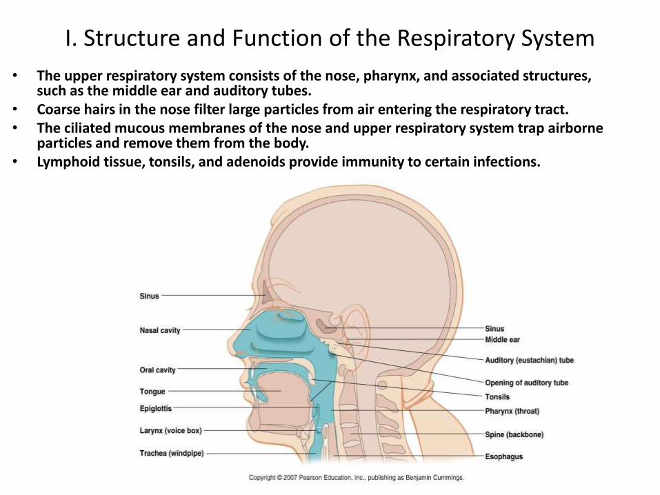
I. Structure and Function of the Respiratory System
• The upper respiratory system consists of the nose, pharynx, and associated structures, such as the middle ear and auditory tubes.
• Coarse hairs in the nose filter large particles from air entering the respiratory tract. • The ciliated mucous membranes of the nose and upper respiratory system trap airborne
particles and remove them from the body. • Lymphoid tissue, tonsils, and adenoids provide immunity to certain infections.
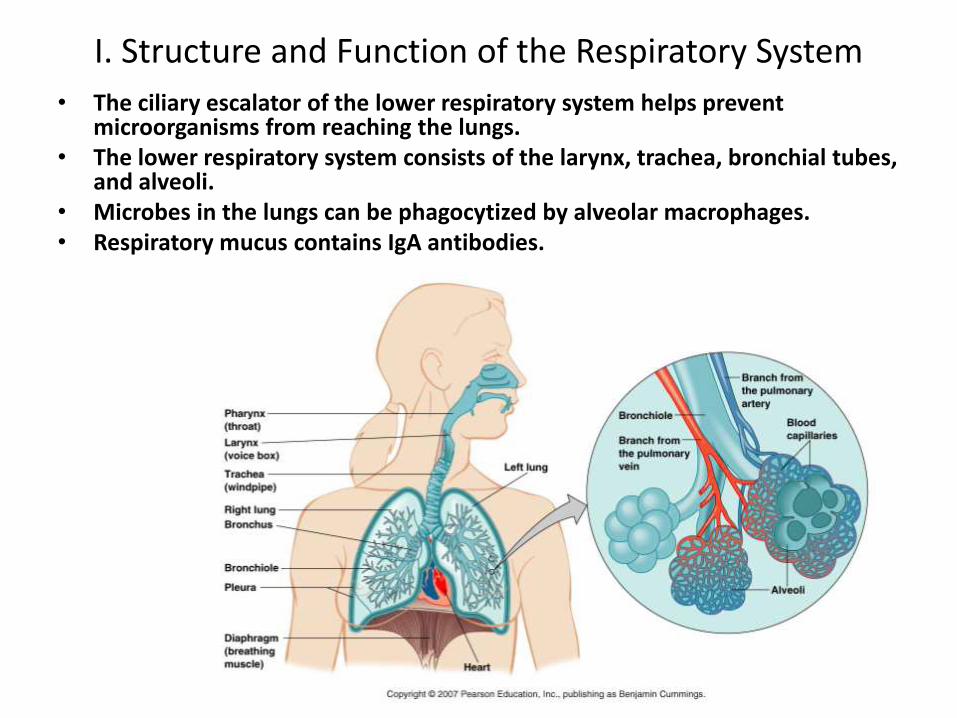
I. Structure and Function of the Respiratory System
• The ciliary escalator of the lower respiratory system helps prevent microorganisms from reaching the lungs.
• The lower respiratory system consists of the larynx, trachea, bronchial tubes, and alveoli.
• Microbes in the lungs can be phagocytized by alveolar macrophages. • Respiratory mucus contains IgA antibodies.
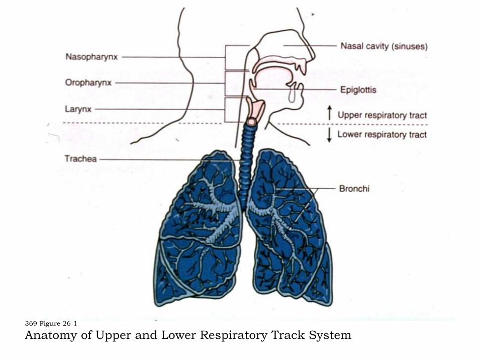
369 Figure 26-1
Anatomy of Upper and Lower Respiratory Track System

371 Plate I. A
Expectorated sputum, smear, Gram stain, light microscopy, low power
view (LPV). Purulence none. Contaminating bacteria and epithelial cells
heavy. No pathogens seen. Please submit carefully collected sample of
lower respiratory tree material. The sample is saliva, not sputum.
There could be several reasons for submission of this sample to the
laboratory. The patient could have been poorly directed and simply
"spit" into the collection container, or the patient's cough may not be
productive of sputum.
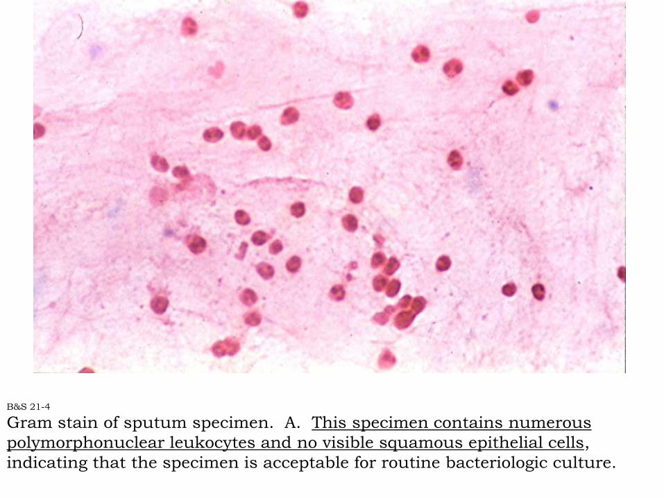
B&S 21-4
Gram stain of sputum specimen. A. This specimen contains numerous
polymorphonuclear leukocytes and no visible squamous epithelial cells,
indicating that the specimen is acceptable for routine bacteriologic culture.
II. Normal Microbiota of the Respiratory System
• The normal microbiota of the nasal cavity and throat can include pathogenic microorganisms in a carrier status.
• Don’t cause disease because of competition with predominant microorganisms.
• The lower respiratory system is usually sterile because of the action of the ciliary escalator.
III. Microbial Diseases of the Upper Respiratory System
• Specific areas of the upper respiratory system can become infected to produce pharyngitis, laryngitis, tonsillitis, sinusitis, and epiglottitis. – Pharyngitis – sore throat– Laryngitis – infected larynx– Tonsillitis – inflamed tonsils– Sinusitis – infected sinus– Epiglotittis – inflammation of the flap like structures of
cartilage that prevents swallowed material from entering the larynx – possible life-threatening when inflamed and occludes airway. H. influenzae type b can cause epiglottitis.
• These infections may be caused by several bacteria and viruses, often in combination.
• Most respiratory tract infections are self-limiting.
III. Microbial Diseases of the Upper Respiratory System
• A. Streptococcal Pharyngitis (Strep Throat)– This infection is caused by group A -hemolytic
streptococci, the group that consists of the species Streptococcus pyogenes.
– Symptoms of this infection are inflammation of the mucous membrane and fever, tonsillitis, and otitis media may also occur. At least half of pharyngitis cases are caused by viruses.
– Preliminary rapid clinic diagnosis is made by indirect agglutination tests or next day culture in the micro lab. Definitive diagnosis is based on a rise in IgM antibodies.
– Penicillin is used to treat streptococcal pharyngitis.
– Immunity to streptococcal infections is type-specific.
– Strep throat is usually transmitted by droplets but at one time was commonly associated with unpasteurized milk.
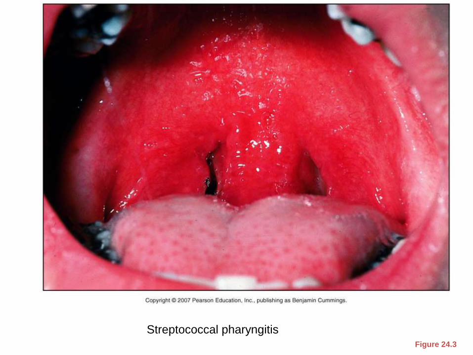
Figure 24.3
Streptococcal pharyngitis
III. Microbial Diseases of the Upper Respiratory System
• B. Scarlet Fever
– Strep throat, caused by an erythrogenic toxin-producing S. pyogenes, results in scarlet fever.
– S. pyogenes produces erythrogenic toxin when lysogenized by a phage.
• Means Strep A has to have a bacterial phage carrying the toxin gene.
– Symptoms include a red rash, high fever, and a red, enlarged tongue, peeled skin. Death is a possible outcome.
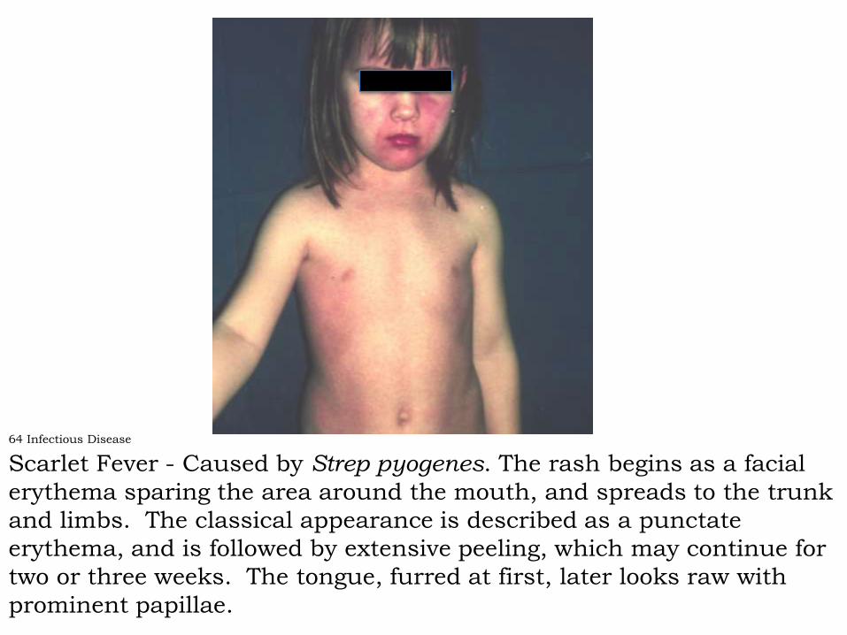
64 Infectious Disease
Scarlet Fever - Caused by Strep pyogenes. The rash begins as a facial
erythema sparing the area around the mouth, and spreads to the trunk
and limbs. The classical appearance is described as a punctate
erythema, and is followed by extensive peeling, which may continue for
two or three weeks. The tongue, furred at first, later looks raw with
prominent papillae.
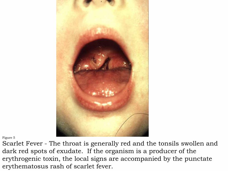
Figure 5
Scarlet Fever - The throat is generally red and the tonsils swollen and
dark red spots of exudate. If the organism is a producer of the
erythrogenic toxin, the local signs are accompanied by the punctate
erythematosus rash of scarlet fever.
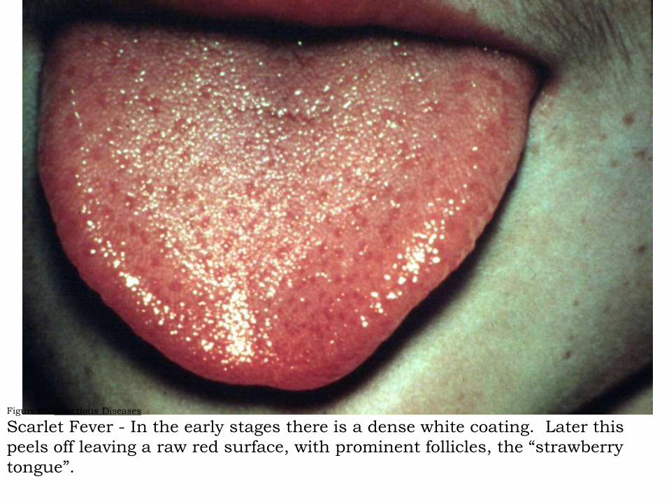
Figure 6 - Infectious Diseases
Scarlet Fever - In the early stages there is a dense white coating. Later this
peels off leaving a raw red surface, with prominent follicles, the “strawberry
tongue”.
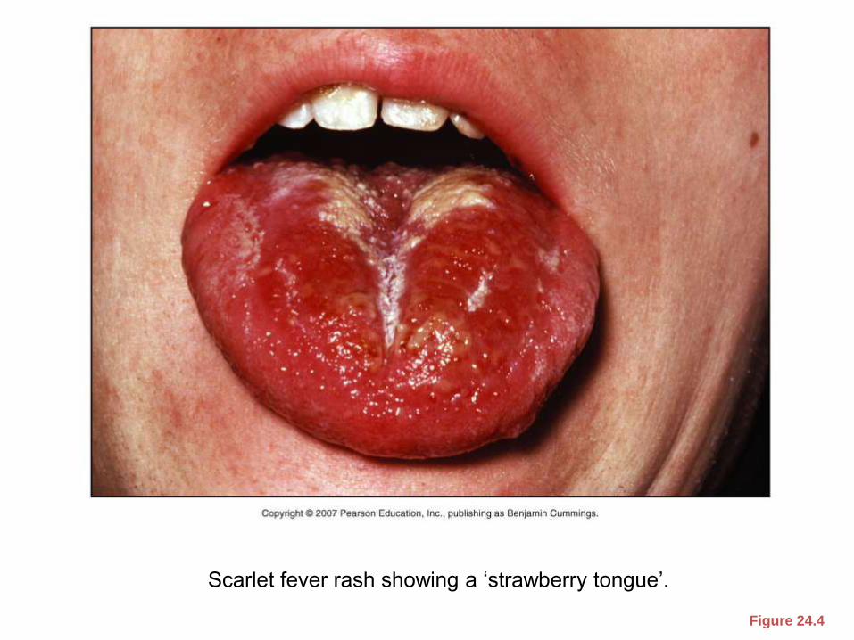
Figure 24.4
Scarlet fever rash showing a ‘strawberry tongue’.
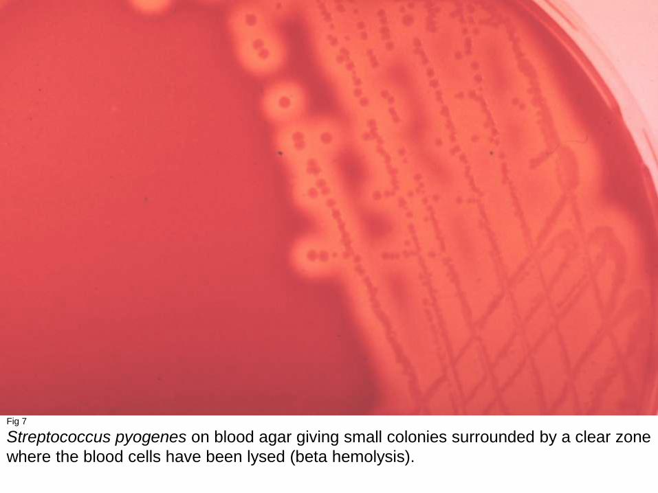
Fig 7
Streptococcus pyogenes on blood agar giving small colonies surrounded by a clear zone
where the blood cells have been lysed (beta hemolysis).
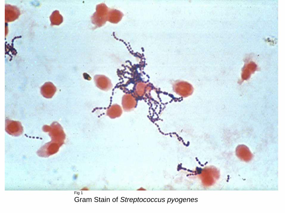
Fig 1
Gram Stain of Streptococcus pyogenes
III. Microbial Diseases of the Upper Respiratory System
• C. Diphtheria - Corynebacterium diphtheriae
– Diphtheria is caused by exotoxin-producing Corynebacterium diphtheriae.
– Gram positive non-spore forming pleomorphic rod. Dividing cells often fold into V and Y shapes.
– Exotoxin is produced when the bacteria are lysogenized by a phage.
– Many well people are symptomless carriers.
– A membrane, containing fibrin and dead human and bacterial cells, forms in the throat and can block the passage of air. “diphtheria” means leather
– The exotoxin inhibits protein synthesis, and heart, kidney, or nerve damage may result.
III. Microbial Diseases of the Upper Respiratory System
• C. Diphtheria - Corynebacterium diphtheriae (cont.)
– Laboratory diagnosis is based on isolation of the bacteria and the appearance of growth on differential media.
– Antitoxin must be administered to neutralize the toxin, and antibiotics can stop growth of the bacteria.
– Routine immunization in the U.S. includes diphtheria toxoid in the DTaP vaccine. Prior to this diphtheria was the leading killer of children.
– Slow-healing skin ulcerations are characteristic of cutaneous diphtheria.
• Cutaneous diphtheria characterized by skin lesions is fairly common in tropical countries. In US affects mainly lower socio- economic groups.
• There is minimal dissemination of the exotoxin in the bloodstream.
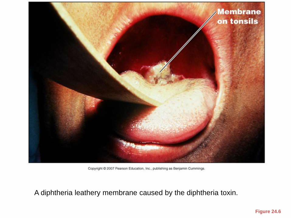
Figure 24.6
A diphtheria leathery membrane caused by the diphtheria toxin.
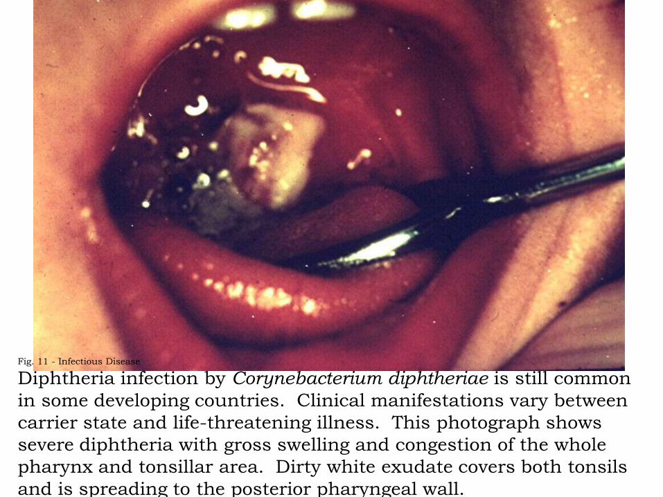
Fig. 11 - Infectious Disease
Diphtheria infection by Corynebacterium diphtheriae is still common
in some developing countries. Clinical manifestations vary between
carrier state and life-threatening illness. This photograph shows
severe diphtheria with gross swelling and congestion of the whole
pharynx and tonsillar area. Dirty white exudate covers both tonsils
and is spreading to the posterior pharyngeal wall.
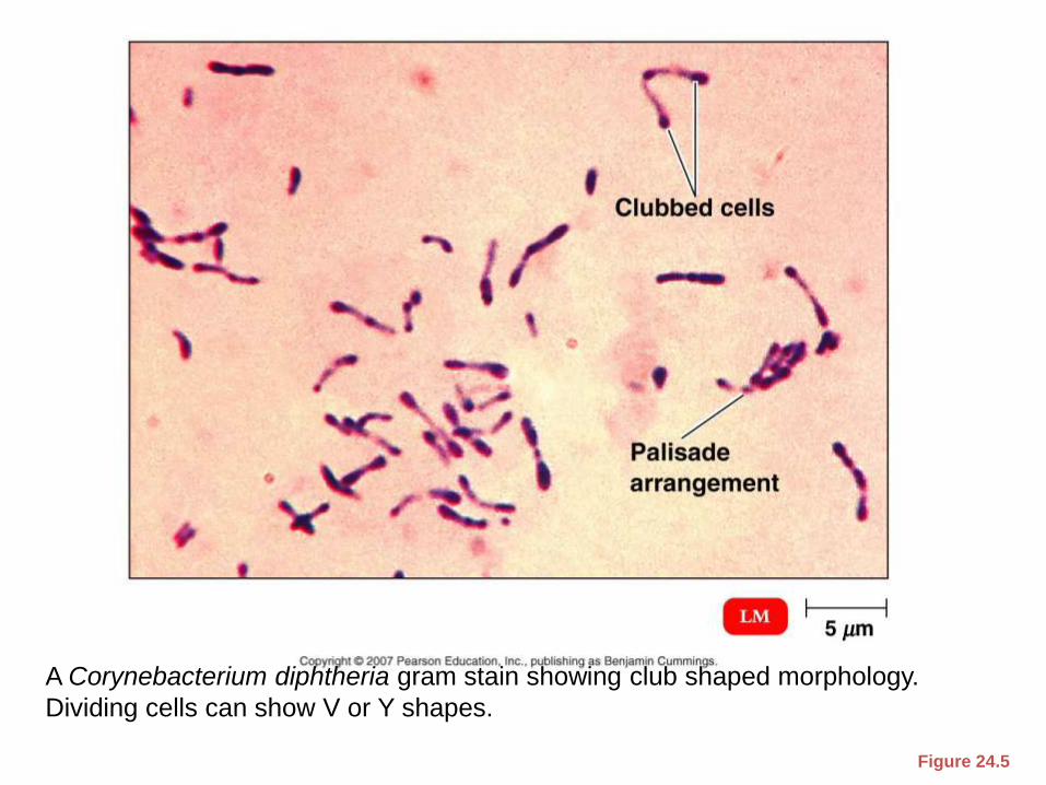
Figure 24.5
A Corynebacterium diphtheria gram stain showing club shaped morphology.
Dividing cells can show V or Y shapes.
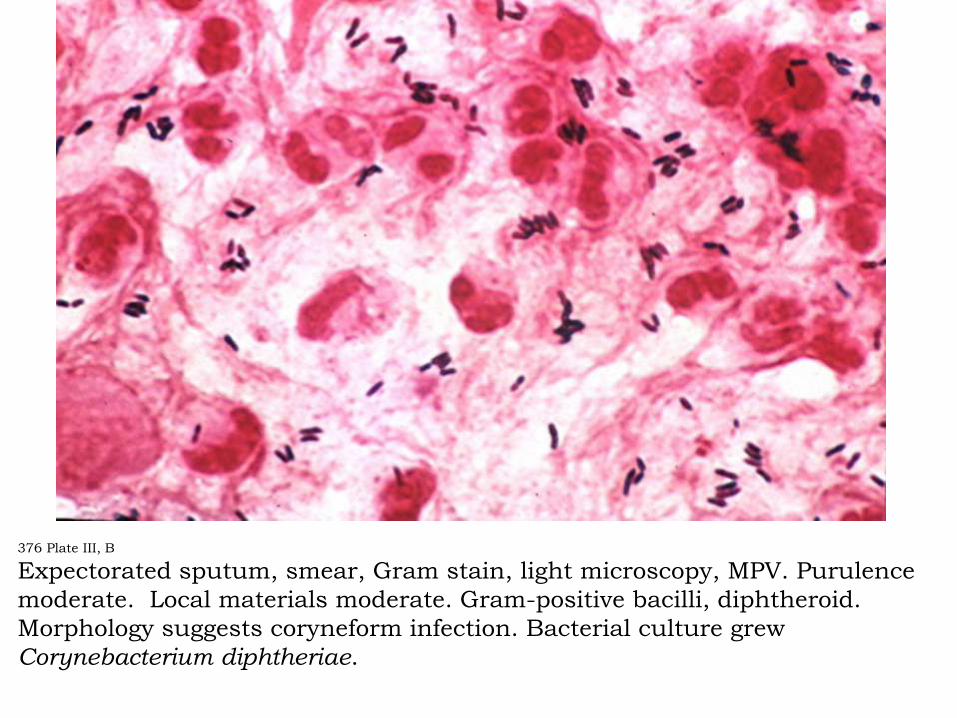
376 Plate III, B
Expectorated sputum, smear, Gram stain, light microscopy, MPV. Purulence
moderate. Local materials moderate. Gram-positive bacilli, diphtheroid.
Morphology suggests coryneform infection. Bacterial culture grew
Corynebacterium diphtheriae.

III. Microbial Diseases of the Upper Respiratory System
• D. Otitis Media: an uncomfortable infections of the middle ear.
– Earache, or otitis media, can occur as a complication of nose and throat infections.
– Pus accumulation causes pressure on the eardrum. 8 million cases/yr.
– Bacterial causes include Streptococcus pneumoniae, Hemophilus influenzae, Moraxella (Branhamella) catarrhalis, Streptococcus pyogenes,and Staphylococcus aureus.

B&S 24-2
The ear anatomy
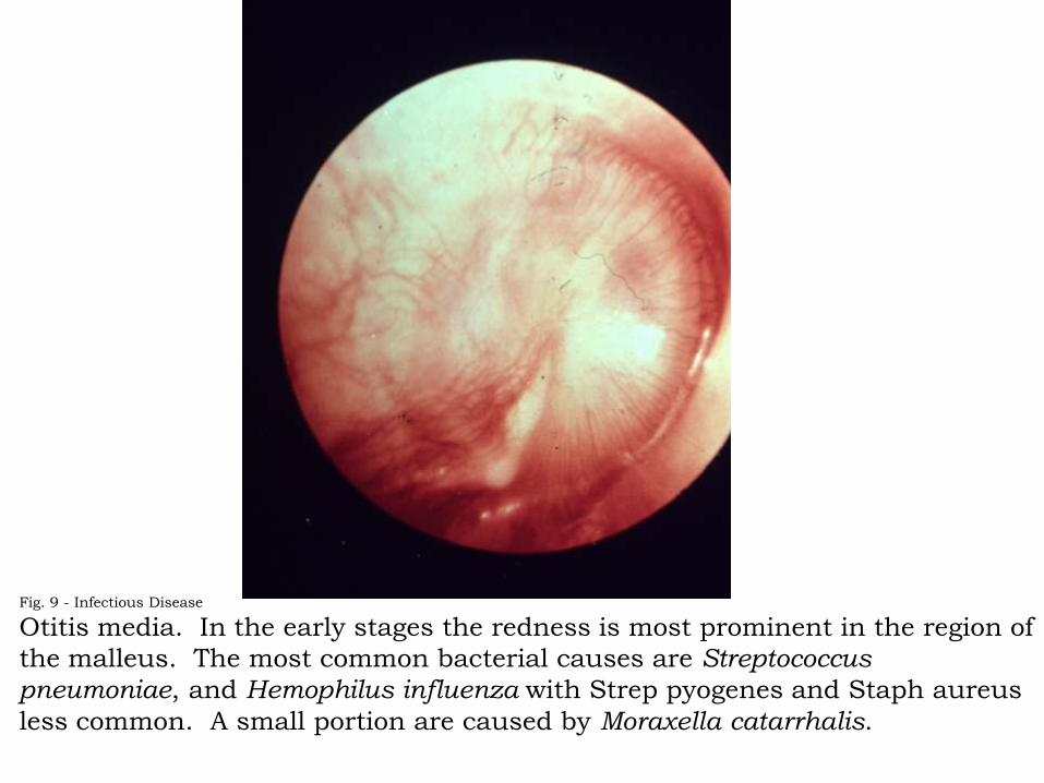
Fig. 9 - Infectious Disease
Otitis media. In the early stages the redness is most prominent in the region of
the malleus. The most common bacterial causes are Streptococcus
pneumoniae, and Hemophilus influenza with Strep pyogenes and Staph aureus
less common. A small portion are caused by Moraxella catarrhalis.
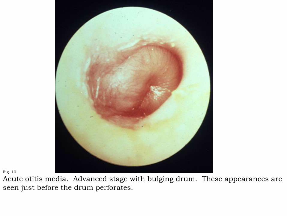
Fig. 10
Acute otitis media. Advanced stage with bulging drum. These appearances are
seen just before the drum perforates.
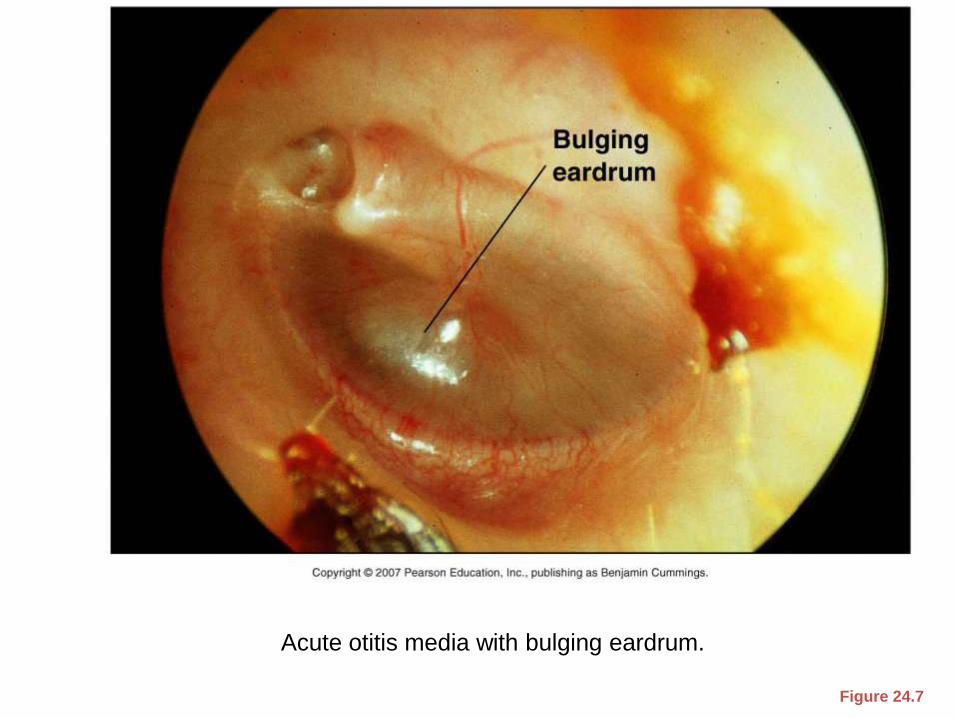
Figure 24.7
Acute otitis media with bulging eardrum.
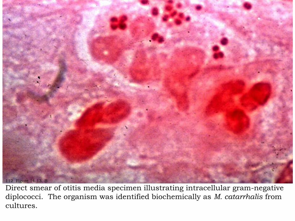
112 Figure 14-13. B
Direct smear of otitis media specimen illustrating intracellular gram-negative
diplococci. The organism was identified biochemically as M. catarrhalis from
cultures.
IV. Viral Disease of the Upper Respiratory System
• A. The Common Cold - rhinoviruses (50%), corona viruses (15-20%)– Any one of approximately 200 different viruses can cause
the common cold; rhinoviruses cause about 50% of all colds.
• Symptoms include sneezing, nasal secretions, and congestion.
• Sinus infections, lower respiratory tract infections, laryngitis, and otitis media can occur as complications of a cold. Usually no fever in uncomplicated cases.
• Colds are most often transmitted by indirect contact.
• Rhinoviruses prefer temperatures slightly lower than body temperature.
• The incidence of colds increases during cold weather, possibly because of increased interpersonal indoor contact or physiological changes.
• Antibodies are produced against the specific viruses.
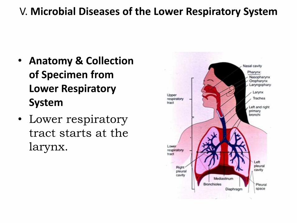
V. Microbial Diseases of the Lower Respiratory System
• Anatomy & Collection of Specimen from Lower Respiratory System
• Lower respiratory
tract starts at the
larynx.
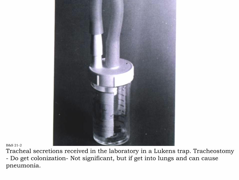
B&S 21-2
Tracheal secretions received in the laboratory in a Lukens trap. Tracheostomy
- Do get colonization- Not significant, but if get into lungs and can cause
pneumonia.
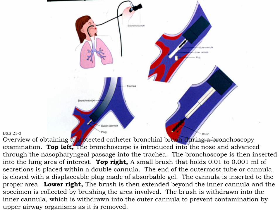
B&S 21-3
Overview of obtaining a protected catheter bronchial brush during a bronchoscopy
examination. Top left, The bronchoscope is introduced into the nose and advanced
through the nasopharyngeal passage into the trachea. The bronchoscope is then inserted
into the lung area of interest. Top right, A small brush that holds 0.01 to 0.001 ml of
secretions is placed within a double cannula. The end of the outermost tube or cannula
is closed with a displaceable plug made of absorbable gel. The cannula is inserted to the
proper area. Lower right, The brush is then extended beyond the inner cannula and the
specimen is collected by brushing the area involved. The brush is withdrawn into the
inner cannula, which is withdrawn into the outer cannula to prevent contamination by
upper airway organisms as it is removed.
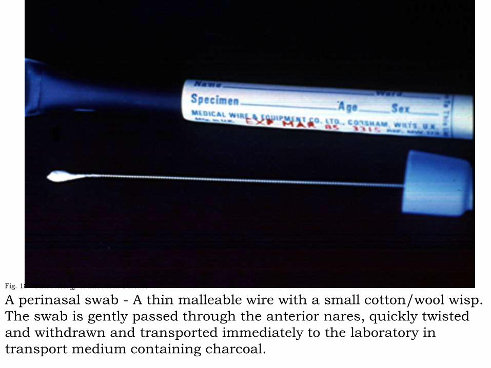
Fig. 18 - Microbiology of Infectious Disease
A perinasal swab - A thin malleable wire with a small cotton/wool wisp.
The swab is gently passed through the anterior nares, quickly twisted
and withdrawn and transported immediately to the laboratory in
transport medium containing charcoal.
VI. Bacterial Diseases of the Lower Respiratory System
• Many of the same microorganisms that infect the upper respiratory system also infect the lower respiratory system.
• Diseases of the lower respiratory system include bronchitis and pneumonia.
• Pneumonia is a term applied to severe complications of bronchitis with the alveoli involved.
VI. Bacterial Diseases of the Lower Respiratory System
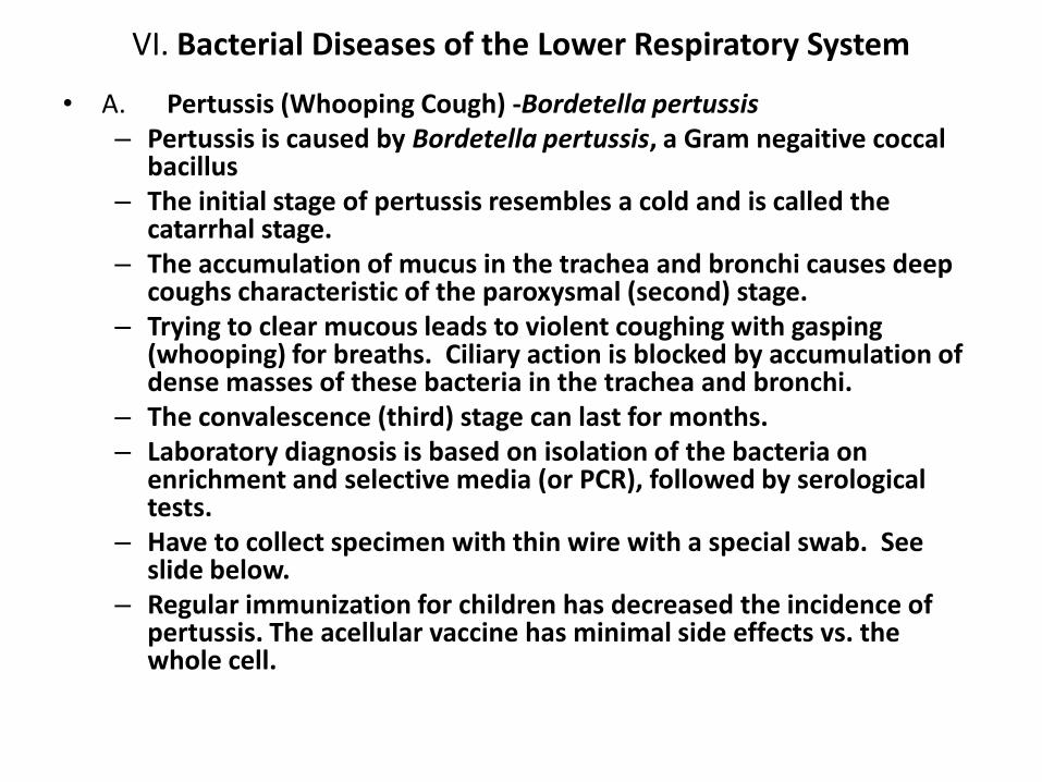
• A. Pertussis (Whooping Cough) -Bordetella pertussis– Pertussis is caused by Bordetella pertussis, a Gram negaitive coccal
bacillus– The initial stage of pertussis resembles a cold and is called the
catarrhal stage. – The accumulation of mucus in the trachea and bronchi causes deep
coughs characteristic of the paroxysmal (second) stage. – Trying to clear mucous leads to violent coughing with gasping
(whooping) for breaths. Ciliary action is blocked by accumulation of dense masses of these bacteria in the trachea and bronchi.
– The convalescence (third) stage can last for months. – Laboratory diagnosis is based on isolation of the bacteria on
enrichment and selective media (or PCR), followed by serological tests.
– Have to collect specimen with thin wire with a special swab. See slide below.
– Regular immunization for children has decreased the incidence of pertussis. The acellular vaccine has minimal side effects vs. the whole cell.
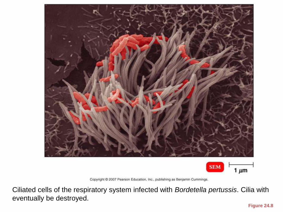
Figure 24.8
Ciliated cells of the respiratory system infected with Bordetella pertussis. Cilia with
eventually be destroyed.
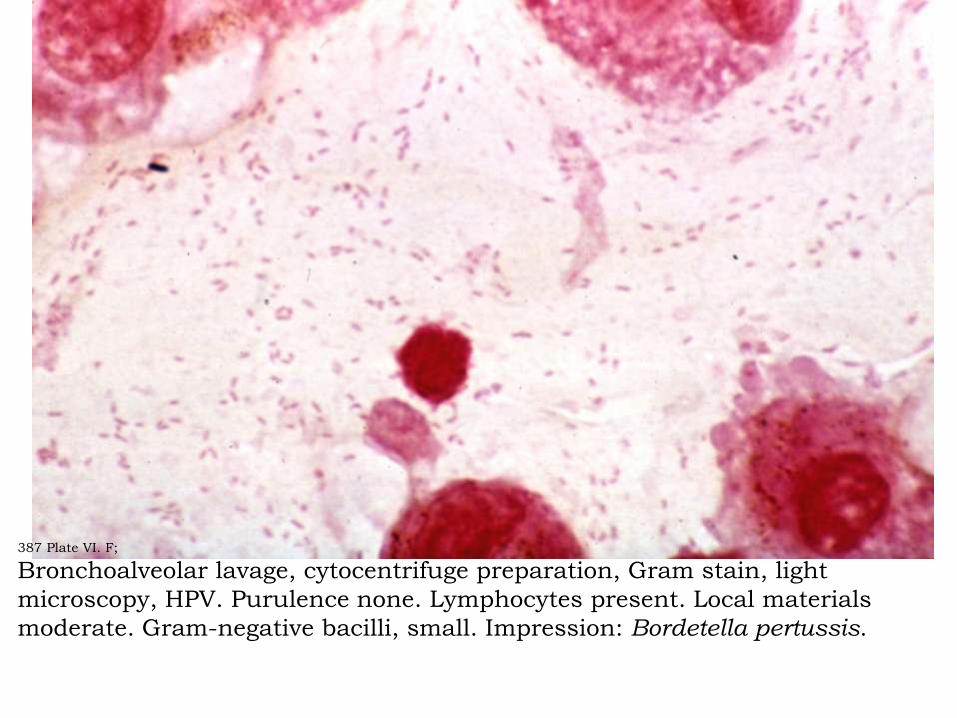
387 Plate VI. F;
Bronchoalveolar lavage, cytocentrifuge preparation, Gram stain, light
microscopy, HPV. Purulence none. Lymphocytes present. Local materials
moderate. Gram-negative bacilli, small. Impression: Bordetella pertussis.
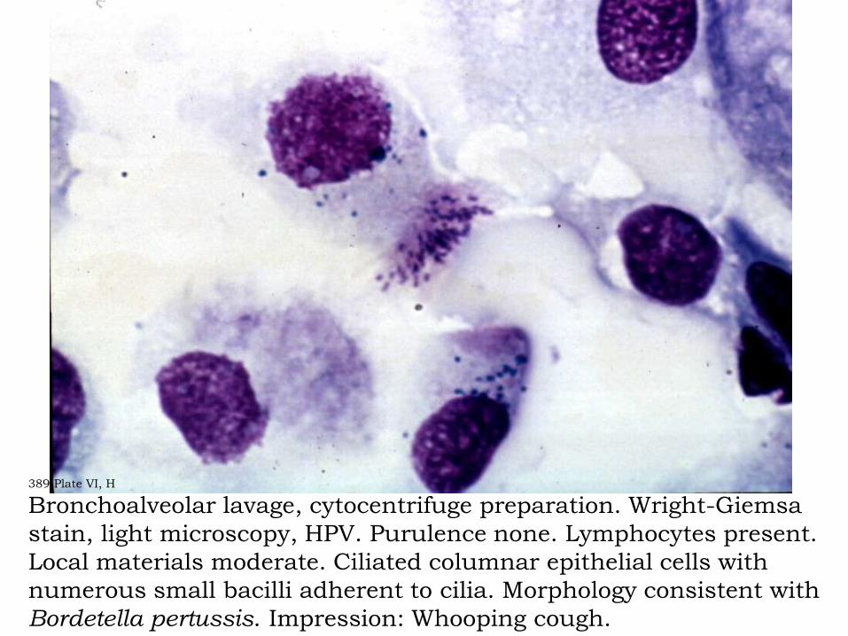
389 Plate VI, H
Bronchoalveolar lavage, cytocentrifuge preparation. Wright-Giemsa
stain, light microscopy, HPV. Purulence none. Lymphocytes present.
Local materials moderate. Ciliated columnar epithelial cells with
numerous small bacilli adherent to cilia. Morphology consistent with
Bordetella pertussis. Impression: Whooping cough.
VI. Bacterial Diseases of the Lower Respiratory System
• B. Tuberculosis -Mycobacterium tuberculosis.– Tuberculosis is caused by Mycobacterium tuberculosis, a slow growing
acid-fast bacillus
– Large amounts of lipids in the cell wall account for the bacterium' s acid-fast characteristic as well as its resistance to drying and disinfectants.
– M. tuberculosis may be ingested by alveolar macrophages. If not killed, the bacteria reproduce in the macrophages. Resistant to phagocytosis.
– Lesions formed by M. tuberculosis are called tubercles; dead macrophages and bacteria form the caseous lesion that might calcify and appear in an X ray as a Ghon complex. Ghon complex means calcified caseous lesions. Caused by hypersensitivity reaction.
– Liquefaction of the caseous lesion results in a tuberculous cavity in which M. tuberculosis can grow.
– New foci of infection can develop when a caseous lesion ruptures and releases bacteria into blood or lymph vessels; this is called miliary tuberculosis.
– Miliary (characterized by lesions resembling millet seeds) tuberculosis is characterized by weight loss, coughing, and loss of vigor.
VI. Bacterial Diseases of the Lower Respiratory System
• B. Tuberculosis -Mycobacterium tuberculosis (cont.).– Chemotherapy usually involves two drugs taken for 1- 2 years;
multidrug-resistant M. tuberculosis is becoming prevalent. DOT therapy.
– Drugs are Streptomycin, rifampin, isoniazid (INH) and ethanbutol.– Intracellular growth shields TB from antibiotics.– Stress and genetic differences contribute to susceptibility.– A positive tuberculin skin test can indicate either an active case of TB,
or prior infection, or vaccination and immunity to the disease. – Sensitized T-cells are present at site of infection or skin test.– Laboratory diagnosis is based on the presence of acid-fast bacilli and
isolation of the bacteria, which requires incubation of up to 8 weeks.– Mycobacterium bovis causes bovine tuberculosis and can be
transmitted to humans by unpasteurized milk. – M. bovis infections usually affect the bones or lymphatic system. – BCG vaccine for tuberculosis consists of a live, avirulent culture of M.
bovis. Useful if given early in childhood. Mainly outside of US>– M. avium-intracellulare complex infects patients in the late stages of
HIV infection.

Figure 24.9
Mycobacterium tuberculosis acid fast stain, showing filamentous red-stained
funguslike growth. Smear from lung tissue.
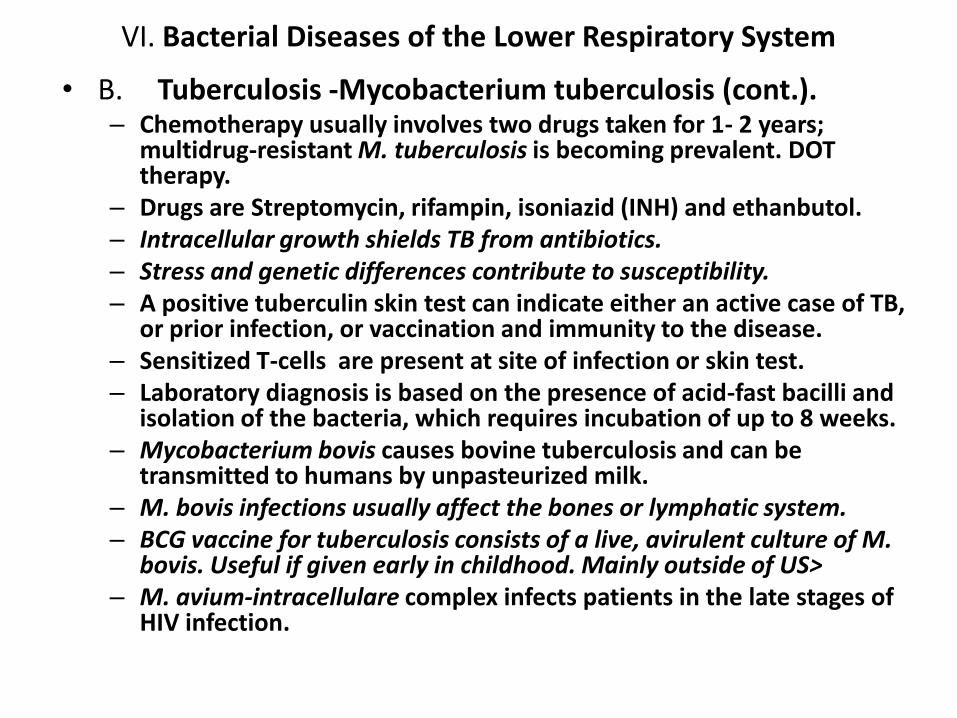
376 Plate IV. D
Expectorated sputum, concentrated smear, fluorochrome acid-fast
stain, fluorescent microscopy, MPV. Typical acid-fast bacteria,
numerous. Impression: Tuberculosis. The patient had been placed in
respiratory isolation following physical examination and history and
was immediately begun on antituberculosis therapy following receipt of
the direct examination report. Mycobacterium tuberculosis was
identified from culture.
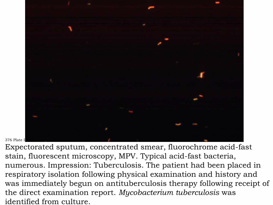
225 - Fig. 22-13
Mycobacterium tuberculosis growing on Lowenstein-Jensen (LJ) medium. The
medium contains egg, mineral salts and malachite (green). Growth appears
after about 4 weeks incubation as granular buff-colored colonies.

Figure 24.10 - Overview
Progression of a
Mycobacteria tuberculosis
infection of the lung.
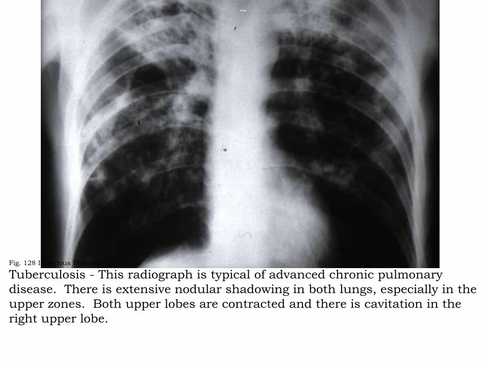
Fig. 128 Infectious Diseases
Tuberculosis - This radiograph is typical of advanced chronic pulmonary
disease. There is extensive nodular shadowing in both lungs, especially in the
upper zones. Both upper lobes are contracted and there is cavitation in the
right upper lobe.
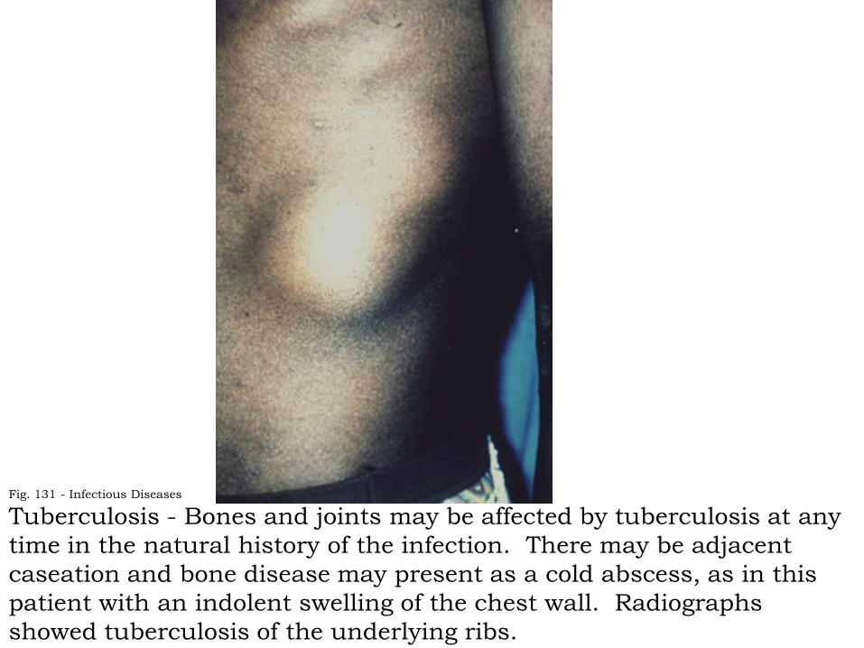
Fig. 131 - Infectious Diseases
Tuberculosis - Bones and joints may be affected by tuberculosis at any
time in the natural history of the infection. There may be adjacent
caseation and bone disease may present as a cold abscess, as in this
patient with an indolent swelling of the chest wall. Radiographs
showed tuberculosis of the underlying ribs.
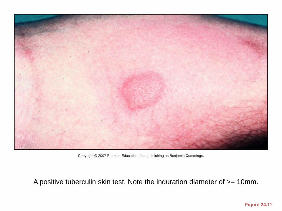
Figure 24.11
A positive tuberculin skin test. Note the induration diameter of >= 10mm.

Figure 24.12 - Overview
US distribution of tuberculosis, 2003.
VI. Bacterial Diseases of the Lower Respiratory System
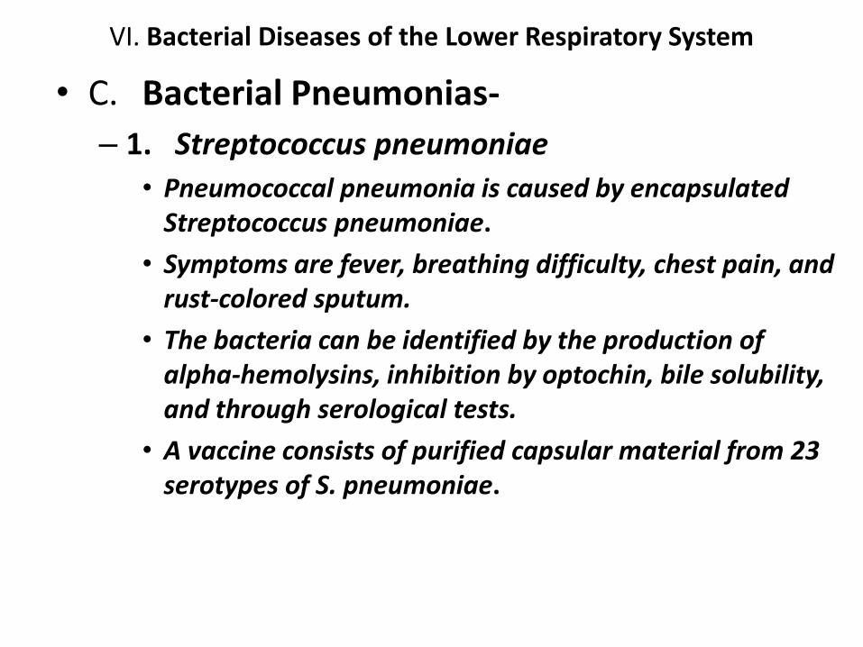
• C. Bacterial Pneumonias-
– 1. Streptococcus pneumoniae
• Pneumococcal pneumonia is caused by encapsulated Streptococcus pneumoniae.
• Symptoms are fever, breathing difficulty, chest pain, and rust-colored sputum.
• The bacteria can be identified by the production of alpha-hemolysins, inhibition by optochin, bile solubility, and through serological tests.
• A vaccine consists of purified capsular material from 23 serotypes of S. pneumoniae.
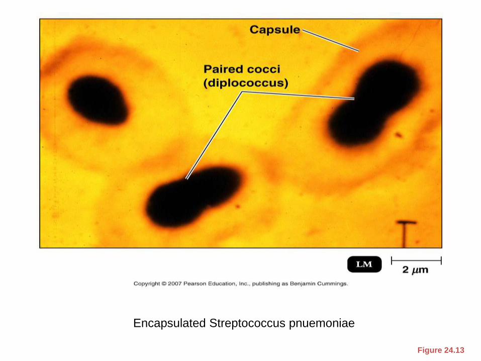
Figure 24.13
Encapsulated Streptococcus pnuemoniae
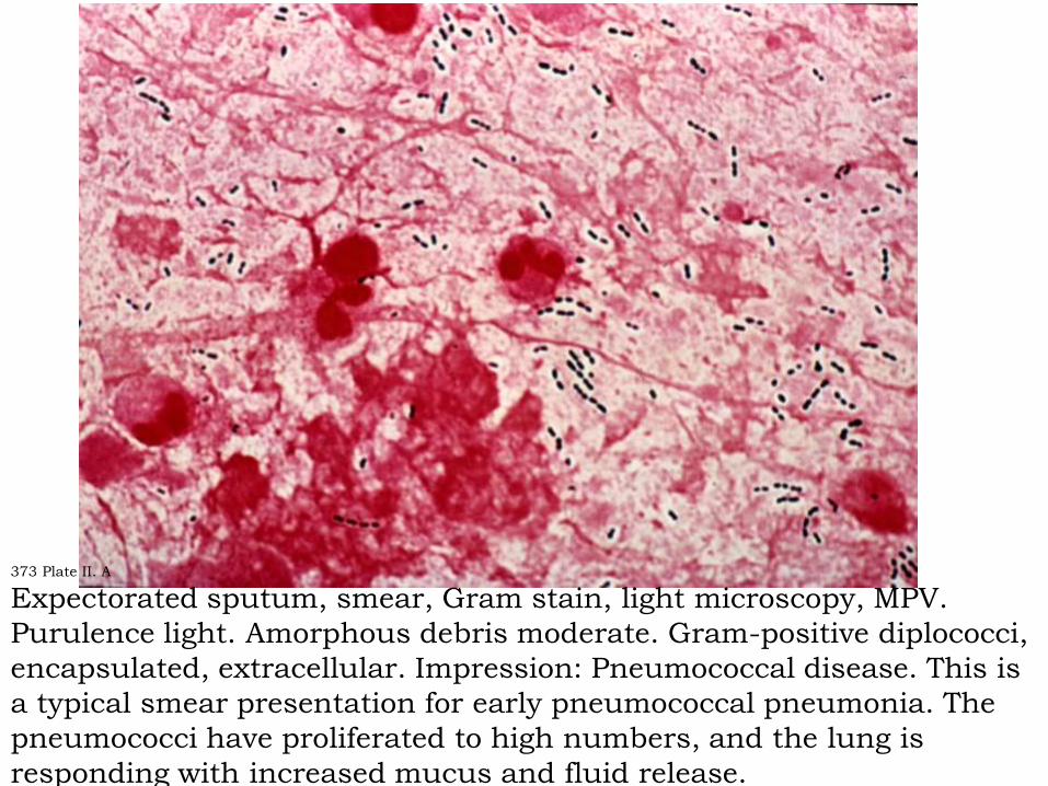
373 Plate II. A
Expectorated sputum, smear, Gram stain, light microscopy, MPV.
Purulence light. Amorphous debris moderate. Gram-positive diplococci,
encapsulated, extracellular. Impression: Pneumococcal disease. This is
a typical smear presentation for early pneumococcal pneumonia. The
pneumococci have proliferated to high numbers, and the lung is
responding with increased mucus and fluid release.
VI. Bacterial Diseases of the Lower Respiratory System
• C. Bacterial Pneumonias-
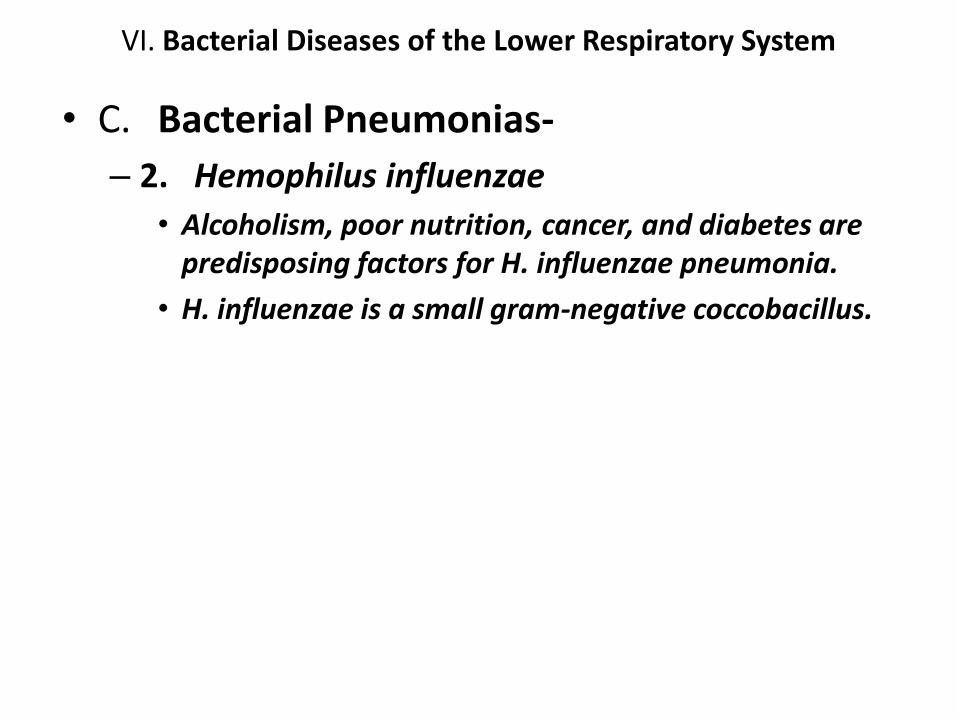
– 2. Hemophilus influenzae
• Alcoholism, poor nutrition, cancer, and diabetes are predisposing factors for H. influenzae pneumonia.
• H. influenzae is a small gram-negative coccobacillus.
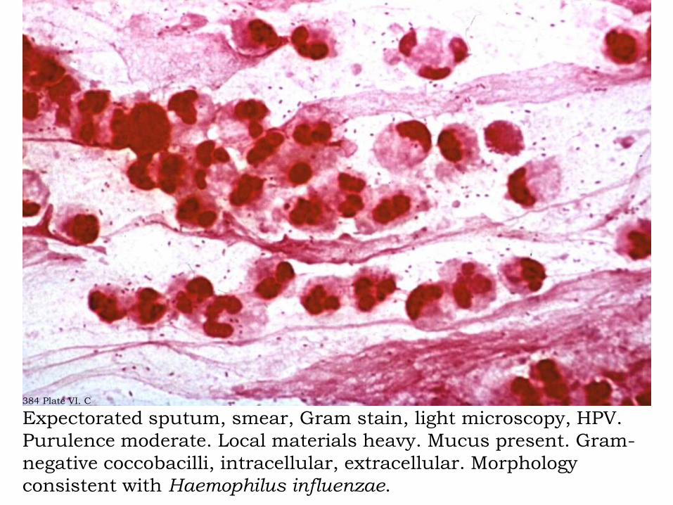
384 Plate VI. C
Expectorated sputum, smear, Gram stain, light microscopy, HPV.
Purulence moderate. Local materials heavy. Mucus present. Gram-
negative coccobacilli, intracellular, extracellular. Morphology
consistent with Haemophilus influenzae.
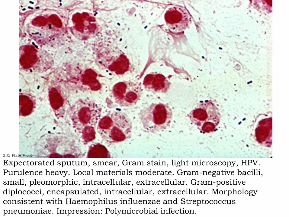
385 Plate VI. D
Expectorated sputum, smear, Gram stain, light microscopy, HPV.
Purulence heavy. Local materials moderate. Gram-negative bacilli,
small, pleomorphic, intracellular, extracellular. Gram-positive
diplococci, encapsulated, intracellular, extracellular. Morphology
consistent with Haemophilus influenzae and Streptococcus
pneumoniae. Impression: Polymicrobial infection.
VI. Bacterial Diseases of the Lower Respiratory System
• C. Bacterial Pneumonias-
– 3. Mycoplasmal Pneumonia -Mycoplasma pneumoniae
• Mycoplasmal pneumonia is common in children and young adults; as many as 20% of cases. Lowgrade fever, cough of long 2-3 weeks duration.
• Organism has no cell wall so is difficult to grow so presents like a viral infection.
• Described as “atypical “ or “walking pneumonia”.
• M. pneumoniae produces small fried-egg colonies after 2 weeks' incubation on enriched media containing horse serum and yeast extract.
• A complement-fixation test, used to diagnose the disease, is based on the rising of antibody titer.
• Treated with tetracyclines.
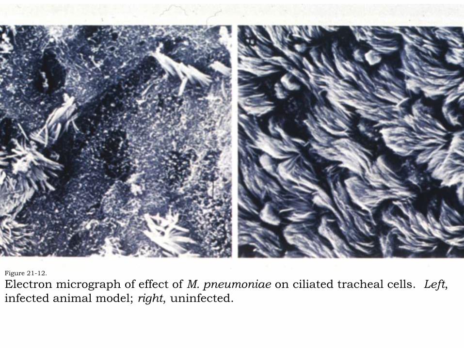
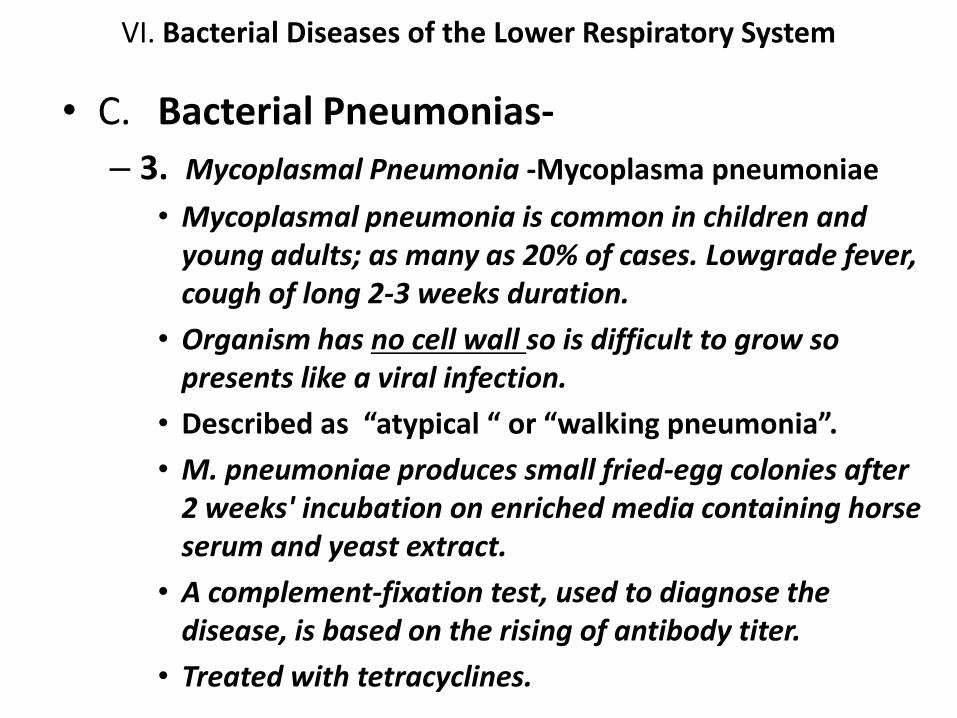
Figure 21-12.
Electron micrograph of effect of M. pneumoniae on ciliated tracheal cells. Left,
infected animal model; right, uninfected.
211 Figure 21-14.
Typical chest radiograph of a patient with a 3-week course of “atypical”
pneumonia. Note nonspecific interstitial pneumonia, patch infiltrate delineated
by “feathery outline”.
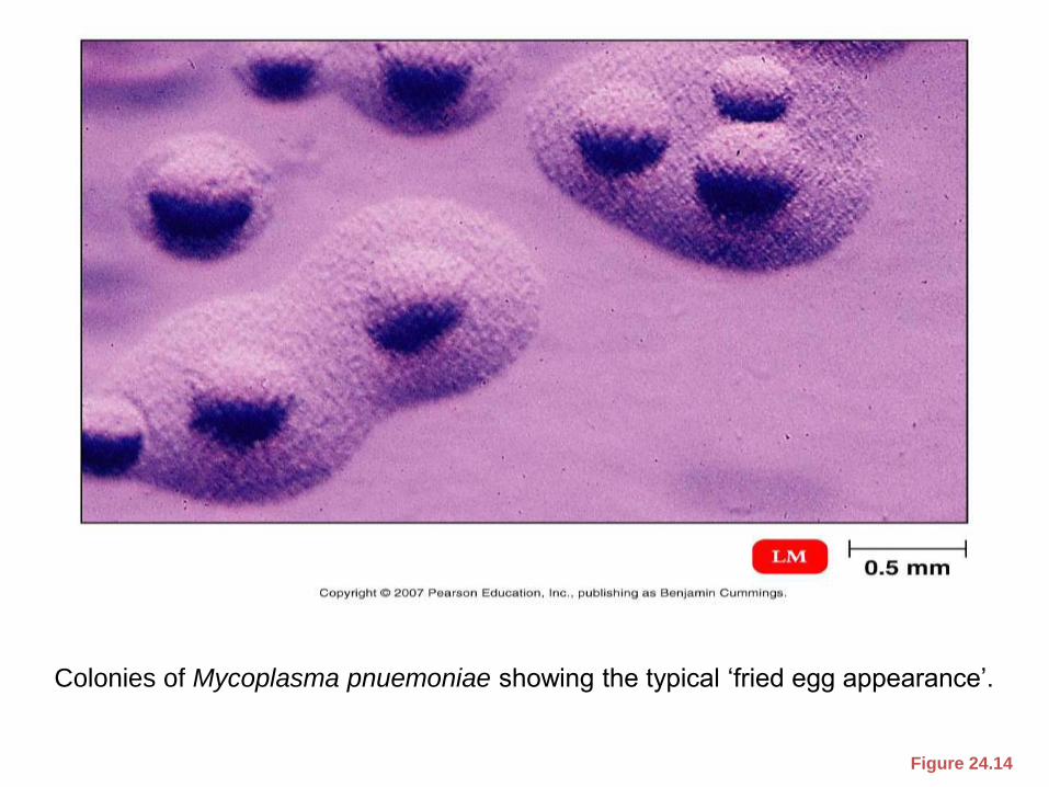
Figure 24.14
Colonies of Mycoplasma pnuemoniae showing the typical ‘fried egg appearance’.
VI. Bacterial Diseases of the Lower Respiratory System
• D. Legionellosis -Legionella pneumophila– The disease is caused by the aerobic gram-negative rod
Legionella pneumophila.
– Legionnaires Disease due to 1976 outbreak of the American Legion meeting.
– The bacterium can grow in water, such as air-conditioning cooling towers, slow plumbing, and then be disseminated in the air.
– This pneumonia does not appear to be transmitted from person to person. High fever, cough, general symptoms of pneumonia.
– Bacterial culture, FA tests, and DNA probes are used for laboratory diagnosis.
– Treated with erythromycin and rifampin
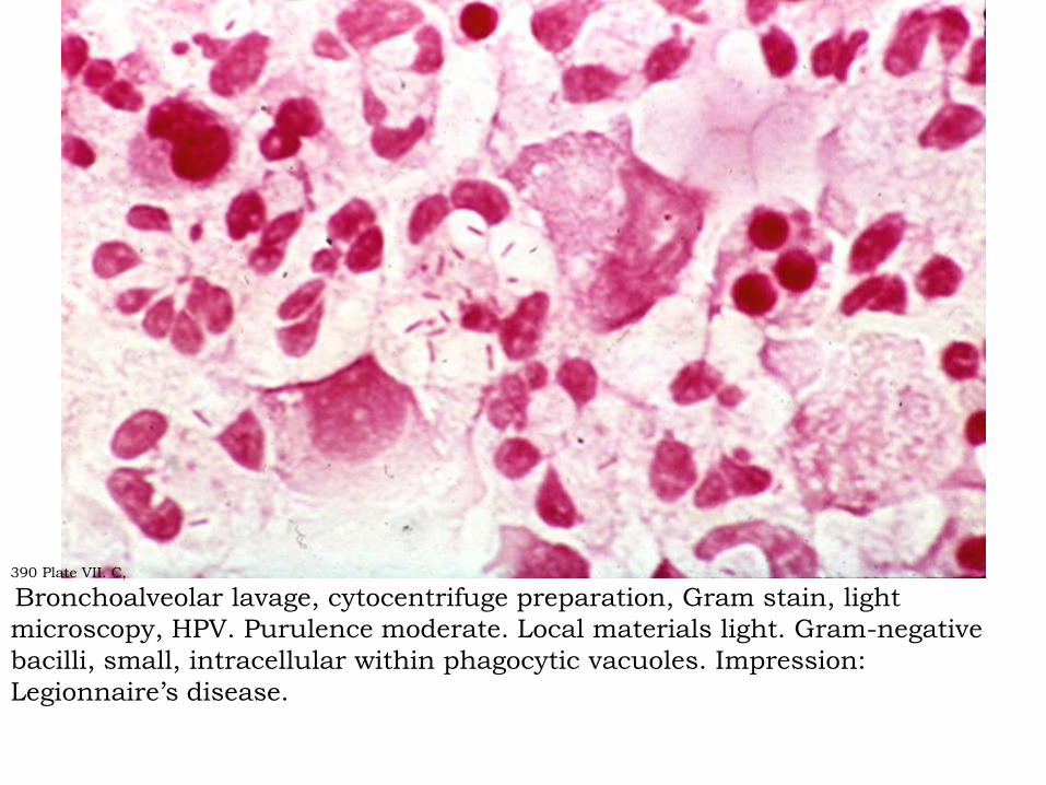
390 Plate VII. C,
Bronchoalveolar lavage, cytocentrifuge preparation, Gram stain, light
microscopy, HPV. Purulence moderate. Local materials light. Gram-negative
bacilli, small, intracellular within phagocytic vacuoles. Impression:
Legionnaire’s disease.
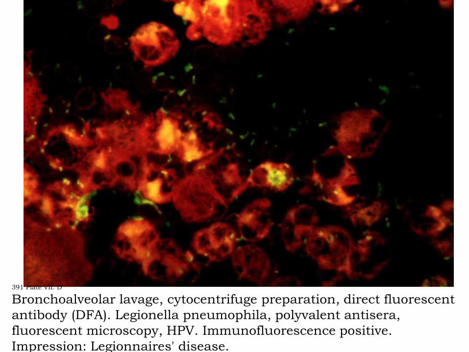
391 Plate VII. D
Bronchoalveolar lavage, cytocentrifuge preparation, direct fluorescent
antibody (DFA). Legionella pneumophila, polyvalent antisera,
fluorescent microscopy, HPV. Immunofluorescence positive.
Impression: Legionnaires' disease.
VI. Bacterial Diseases of the Lower Respiratory System
• E. Psittacosis (Ornithosis) Chlamydia psittaci– Chlamydia psittaci is transmitted by contact with
contaminated droppings and exudates of fowl.
– Psittacine birds such as parakeets and parrots but other birds are infected too.
– Are obligate intracellular parasites but have an elementary body stage that allows the bacteria to survive outside a host.
– Commercial bird handlers are most susceptible to this disease. Example: chicken and turkey farms
– The bacteria are isolated in embryonated eggs, mice, or cell culture; identification is based on FA staining.
– Treated with tetracycline.
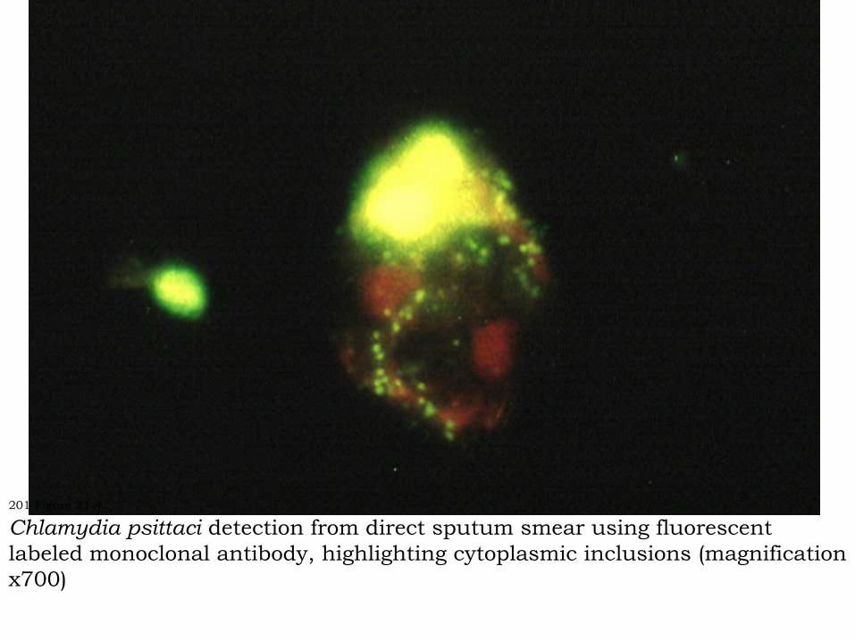
201 Figure 21-4.
Chlamydia psittaci detection from direct sputum smear using fluorescent
labeled monoclonal antibody, highlighting cytoplasmic inclusions (magnification
x700)
VI. Bacterial Diseases of the Lower Respiratory System
• F. Other Bacterial Pneumonias– Gram-positive bacteria that cause pneumonia include Staph
aureus and Strep pyogenes.
– Gram-negative bacteria that cause pneumonia include Moraxella .(B.) catarrhalis, Klebsiella pneumoniae, and Pseudomonas species.
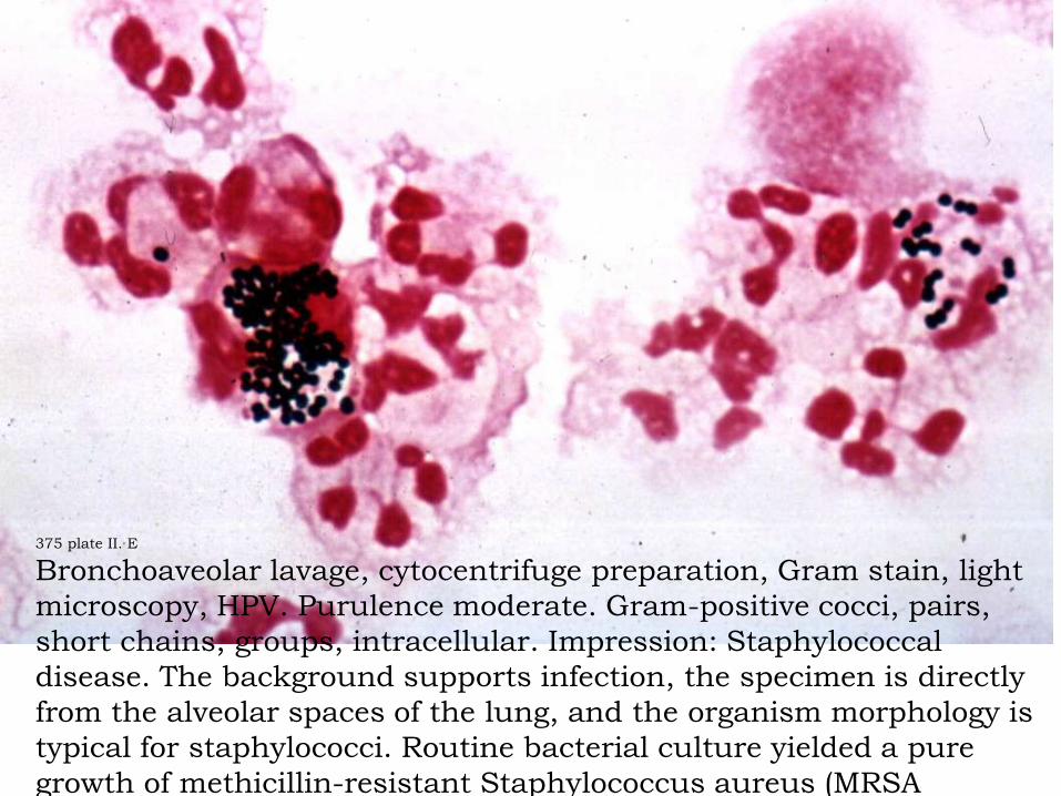
375 plate II. E
Bronchoaveolar lavage, cytocentrifuge preparation, Gram stain, light
microscopy, HPV. Purulence moderate. Gram-positive cocci, pairs,
short chains, groups, intracellular. Impression: Staphylococcal
disease. The background supports infection, the specimen is directly
from the alveolar spaces of the lung, and the organism morphology is
typical for staphylococci. Routine bacterial culture yielded a pure
growth of methicillin-resistant Staphylococcus aureus (MRSA
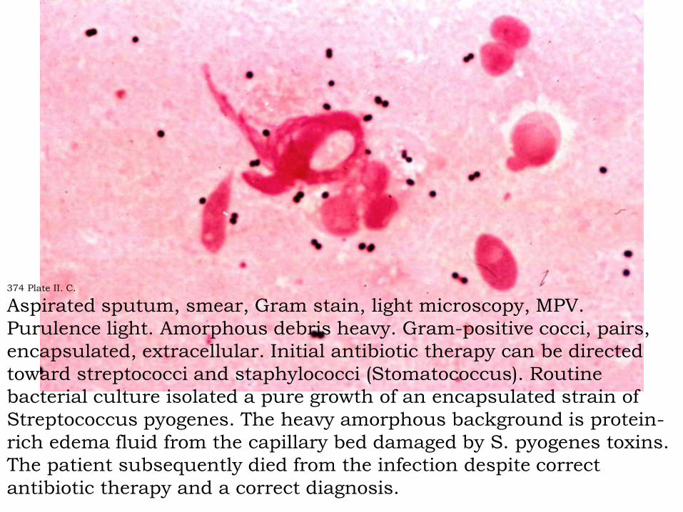
374 Plate II. C.
Aspirated sputum, smear, Gram stain, light microscopy, MPV.
Purulence light. Amorphous debris heavy. Gram-positive cocci, pairs,
encapsulated, extracellular. Initial antibiotic therapy can be directed
toward streptococci and staphylococci (Stomatococcus). Routine
bacterial culture isolated a pure growth of an encapsulated strain of
Streptococcus pyogenes. The heavy amorphous background is protein-
rich edema fluid from the capillary bed damaged by S. pyogenes toxins.
The patient subsequently died from the infection despite correct
antibiotic therapy and a correct diagnosis.
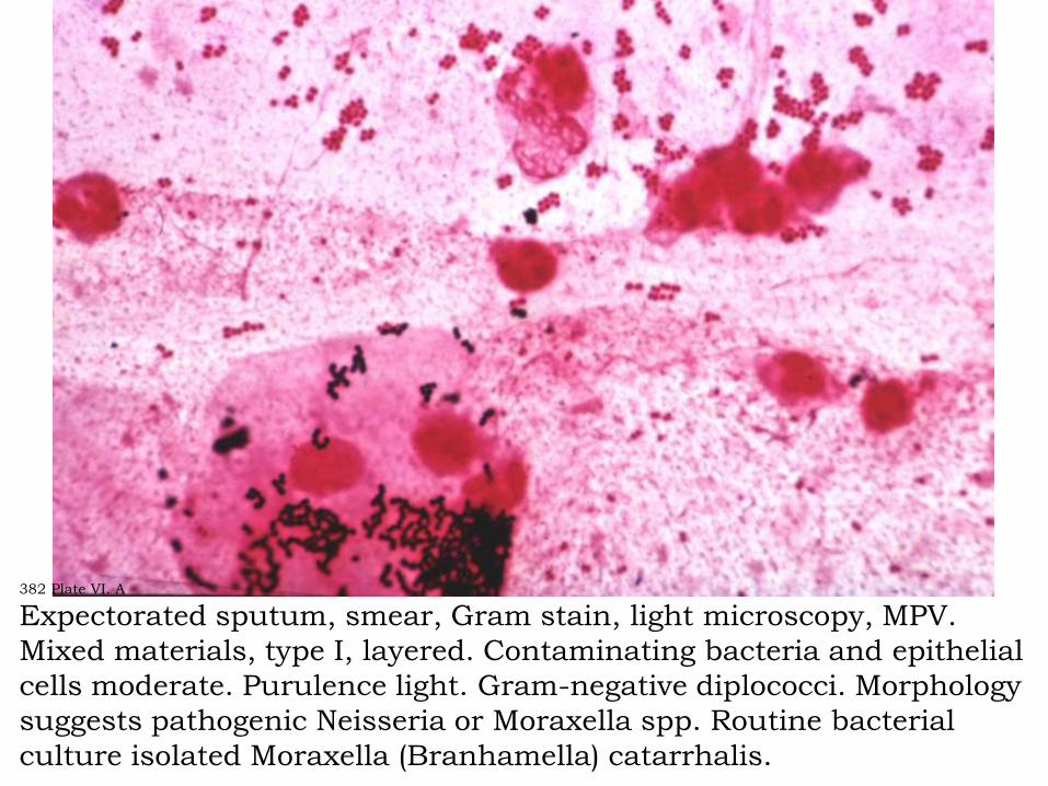
382 Plate VI. A
Expectorated sputum, smear, Gram stain, light microscopy, MPV.
Mixed materials, type I, layered. Contaminating bacteria and epithelial
cells moderate. Purulence light. Gram-negative diplococci. Morphology
suggests pathogenic Neisseria or Moraxella spp. Routine bacterial
culture isolated Moraxella (Branhamella) catarrhalis.

392 Plate VII. E
Expectorated sputum, smear, Gram stain, light microscopy, MPV. Purulence
light. Local materials light. Mucus moderate. Gram-negative bacilli, medium.
Impression: Enteric bacillary infection.
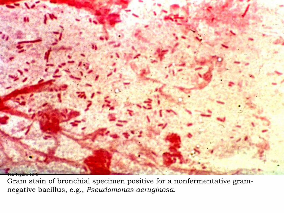
162 Figure 18-6
Gram stain of bronchial specimen positive for a nonfermentative gram-
negative bacillus, e.g., Pseudomonas aeruginosa.

VII. Viral Diseases of the Lower Respiratory System
• A. Viral Pneumonia
– A number of viruses can cause pneumonia as a complication of infections such as with viral influenza.
– The etiologies viral pneumonia are not usually identified in a clinical laboratory because of the difficulty in isolating and identifying viruses.
• If no other cause is found, for example negative bacteria and fungus cultures and mycoplasmalpneumonia has been ruled out, a viral etiology may be assumed.
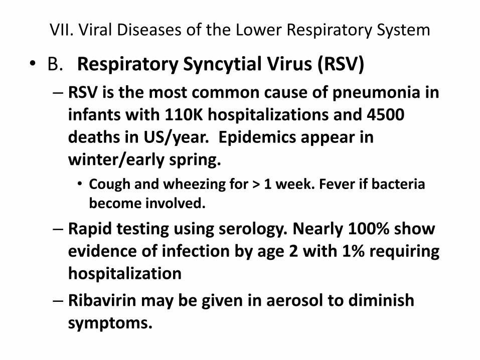
VII. Viral Diseases of the Lower Respiratory System
• B. Respiratory Syncytial Virus (RSV)
– RSV is the most common cause of pneumonia in infants with 110K hospitalizations and 4500 deaths in US/year. Epidemics appear in winter/early spring.
• Cough and wheezing for > 1 week. Fever if bacteria become involved.
– Rapid testing using serology. Nearly 100% show evidence of infection by age 2 with 1% requiring hospitalization
– Ribavirin may be given in aerosol to diminish symptoms.
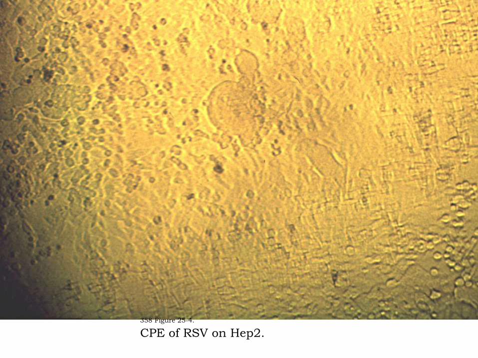
358 Figure 25-4.
CPE of RSV on Hep2.
VII. Viral Diseases of the Lower Respiratory System
• C. Influenza (Flu) Influenza virus– Influenza is caused by influenza virus and is characterized by chills, fever,
headache, and general muscular aches.
– Hemagglutinin (H) and neuraminidase (N) spikes project from the outer lipid bilayer of the virus.
– Viral strains are identified by antigenic differences in the H and N spikes; they are also divided by antigenic differences in their protein coats (A, B, and C).
– Viral isolates are identified by hemagglutination-inhibition tests and immunofluorescence testing with monoclonal antibodies.
– Antigenic shifts that alter the antigenic nature of the H and N spikes make natural immunity and vaccination of questionable value and may lead to pandemics. Minor antigenic changes are caused by antigenic drift and are usually covered by yearly vaccines.
– Deaths during an influenza epidemic are usually from secondary bacterial infections.
– Multivalent vaccines are available for the elderly and other high-risk groups that are 70-90% effective for no more than 3 years for that strain.
– Amantadine and rimantadine given early, reduce symptoms against A-type Influenzavirus. Also zanamivir Relenza) and oseltamivir (Tamiflu).
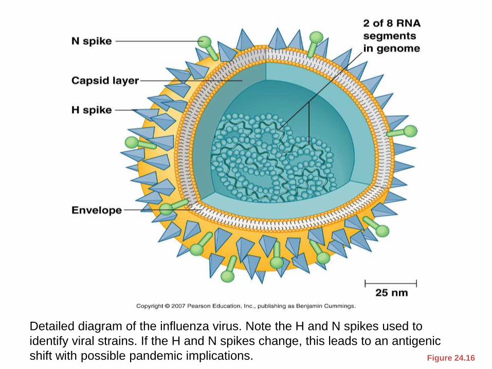
Figure 24.16
Detailed diagram of the influenza virus. Note the H and N spikes used to
identify viral strains. If the H and N spikes change, this leads to an antigenic
shift with possible pandemic implications.
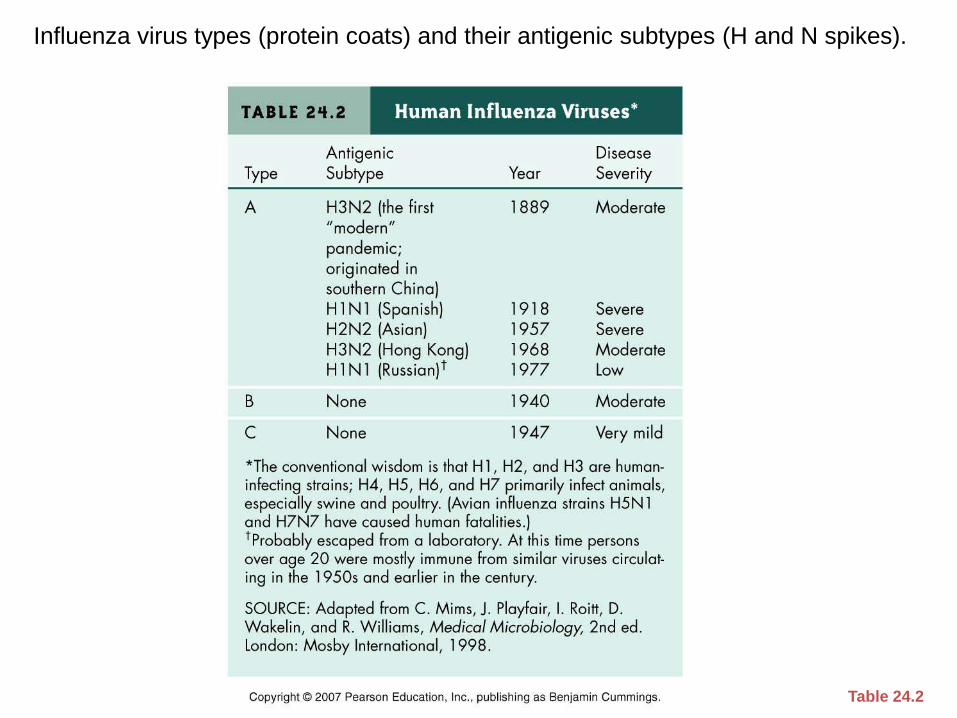
Table 24.2
Influenza virus types (protein coats) and their antigenic subtypes (H and N spikes).
VIII. Fungal Diseases of the Lower Respiratory System
• Fungal spores are easily inhaled; they may germinate in the lower respiratory tract.
• The incidence of fungal diseases has been increasing in recent years.
• The mycoses below can be treated with imidazole drugs (itraconazole) and amphotericin B (serious life threatening cases as this drug is very toxic).
VIII. Fungal Diseases of the Lower Respiratory System
• A. Coccidioidomycosis: San Joaquin Valley Fever
– Inhalation of the airborne arthrospores of Coccidioidesimmitis can result in coccidioidomycosis, typically a limited minor lung infection. Found in dry soils of SW US.
– Most cases are subclinical with about 1% of cases progressing to a TB like course of illness. Predisposing factors such as fatigue and poor nutrition, older age, HIV may lead to disseminated disease.
– See dimorphic forms in tissue: spherules with yeastlikeendospores.
– Same treatment as Histo using fluconazole (diflucan), ketoconazole, and itraconazole. Sometimes use Amphotericin B to treat serious cases but it is very toxic.
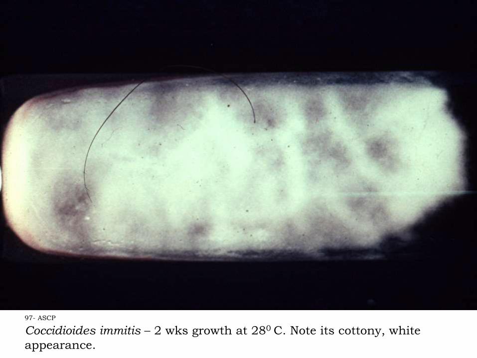
97- ASCP
Coccidioides immitis – 2 wks growth at 280 C. Note its cottony, white
appearance.
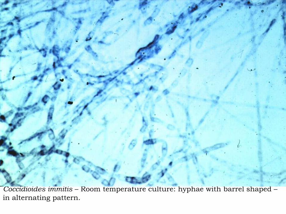
98- ASCP
Coccidioides immitis – Room temperature culture: hyphae with barrel shaped –
in alternating pattern.
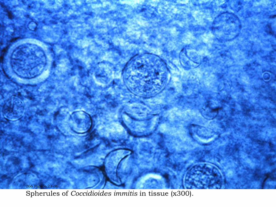
266 Figure 23-36.
Spherules of Coccidioides immitis in tissue (x300).
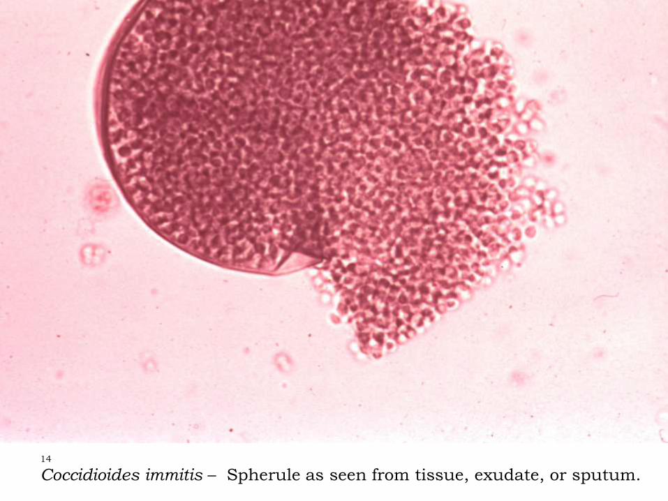
14
Coccidioides immitis – Spherule as seen from tissue, exudate, or sputum.

Figure 24.20
US endemic area for coccidiodomycosis. Note the high incidence that occurs in
the San Joaquin Valley.
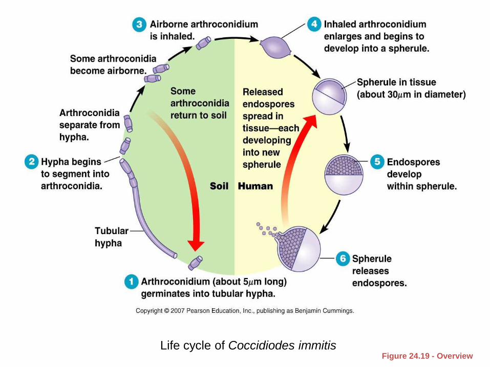
Figure 24.19 - OverviewLife cycle of Coccidiodes immitis
VIII. Fungal Diseases of the Lower Respiratory System
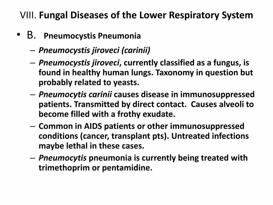
• B. Pneumocystis Pneumonia
– Pneumocystis jiroveci (carinii)
– Pneumocystis jiroveci, currently classified as a fungus, is found in healthy human lungs. Taxonomy in question but probably related to yeasts.
– Pneumocytis carinii causes disease in immunosuppressedpatients. Transmitted by direct contact. Causes alveoli to become filled with a frothy exudate.
– Common in AIDS patients or other immunosuppressedconditions (cancer, transplant pts). Untreated infections maybe lethal in these cases.
– Pneumocytis pneumonia is currently being treated with trimethoprim or pentamidine.
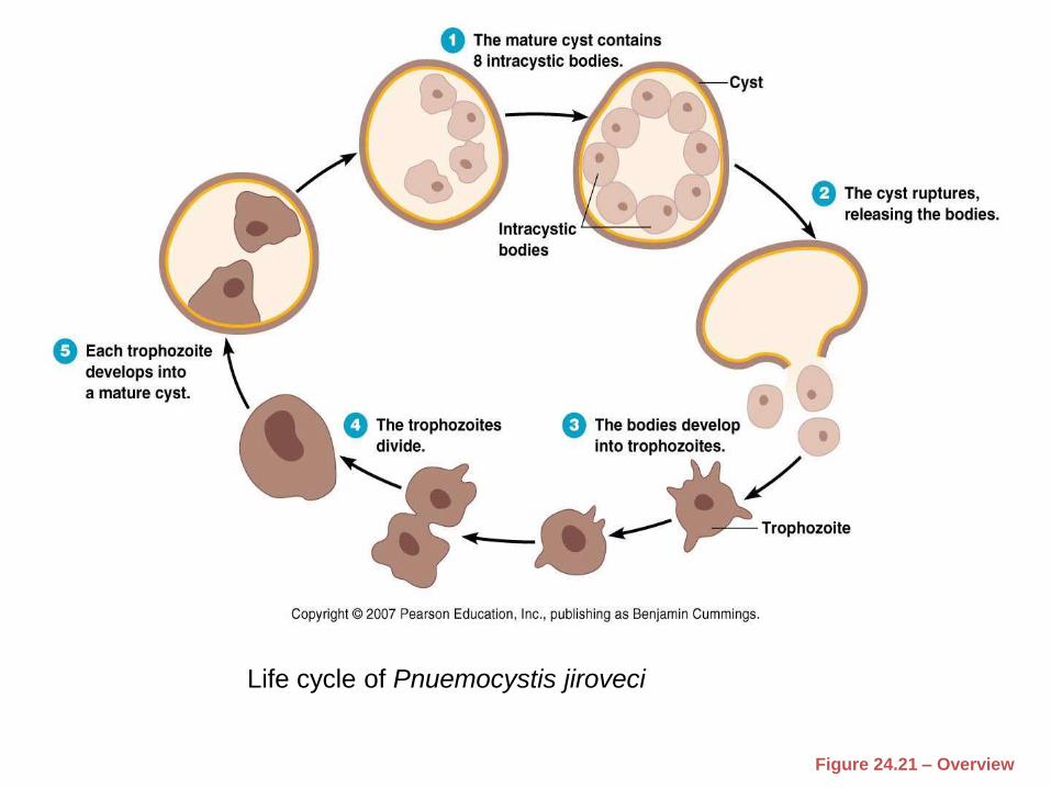
Figure 24.21 – Overview
Life cycle of Pnuemocystis jiroveci
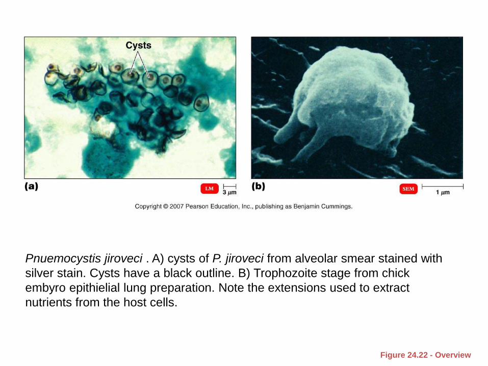
Figure 24.22 - Overview
Pnuemocystis jiroveci . A) cysts of P. jiroveci from alveolar smear stained with
silver stain. Cysts have a black outline. B) Trophozoite stage from chick
embyro epithielial lung preparation. Note the extensions used to extract
nutrients from the host cells.
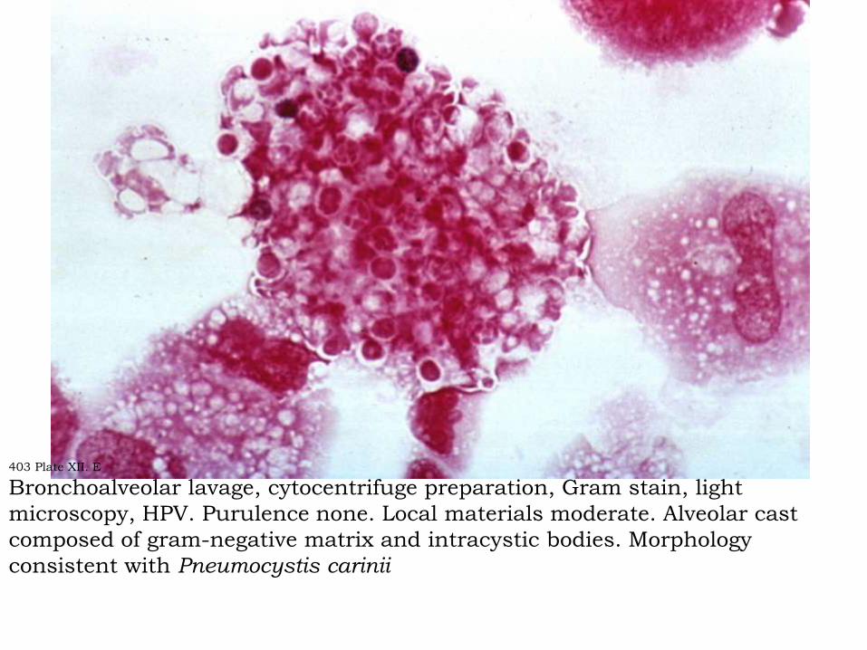
403 Plate XII. E
Bronchoalveolar lavage, cytocentrifuge preparation, Gram stain, light
microscopy, HPV. Purulence none. Local materials moderate. Alveolar cast
composed of gram-negative matrix and intracystic bodies. Morphology
consistent with Pneumocystis carinii