Embed Size (px)
Citation preview
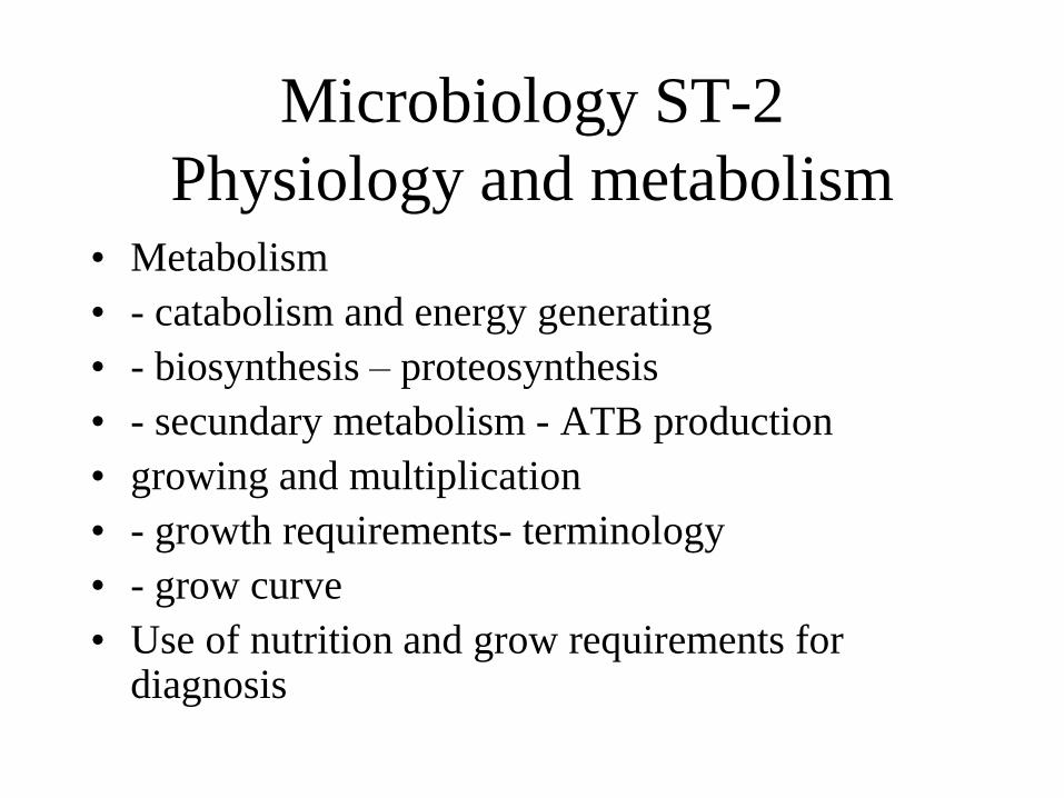
Microbiology ST-2
Physiology and metabolism • Metabolism
• - catabolism and energy generating
• - biosynthesis – proteosynthesis
• - secundary metabolism - ATB production
• growing and multiplication
• - growth requirements- terminology
• - grow curve
• Use of nutrition and grow requirements for diagnosis
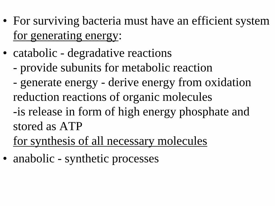
• For surviving bacteria must have an efficient system
for generating energy:
• catabolic - degradative reactions
- provide subunits for metabolic reaction
- generate energy - derive energy from oxidation
reduction reactions of organic molecules
-is release in form of high energy phosphate and
stored as ATP
for synthesis of all necessary molecules
• anabolic - synthetic processes
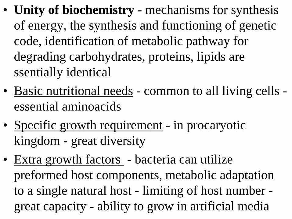
• Unity of biochemistry - mechanisms for synthesis
of energy, the synthesis and functioning of genetic
code, identification of metabolic pathway for
degrading carbohydrates, proteins, lipids are
ssentially identical
• Basic nutritional needs - common to all living cells -
essential aminoacids
• Specific growth requirement - in procaryotic
kingdom - great diversity
• Extra growth factors - bacteria can utilize
preformed host components, metabolic adaptation
to a single natural host - limiting of host number -
great capacity - ability to grow in artificial media
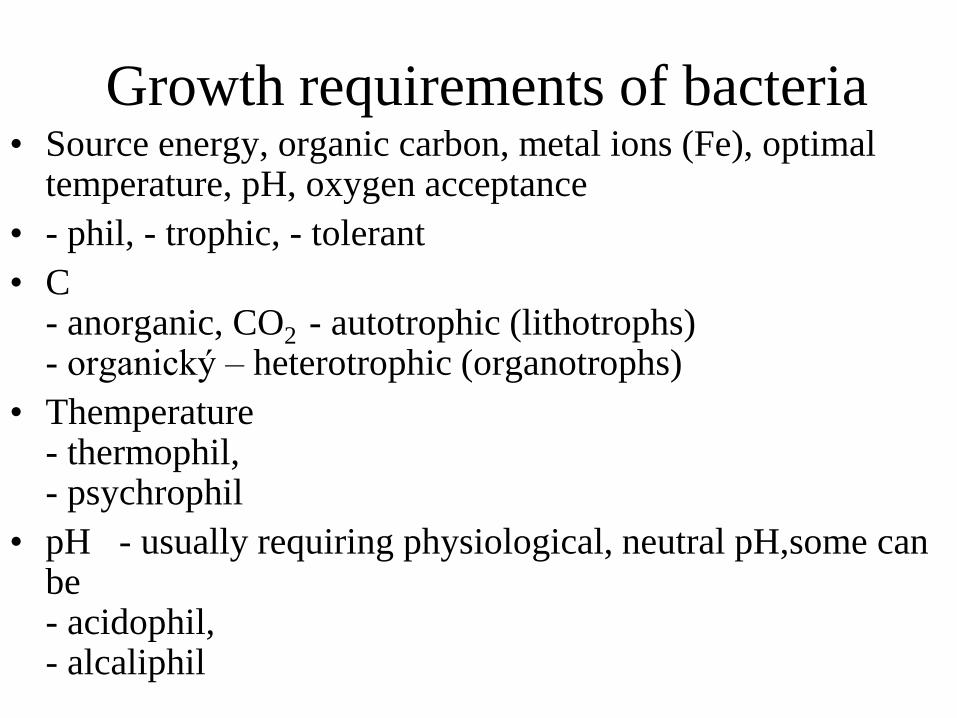
Growth requirements of bacteria • Source energy, organic carbon, metal ions (Fe), optimal
temperature, pH, oxygen acceptance
• - phil, - trophic, - tolerant
• C - anorganic, CO2 - autotrophic (lithotrophs) - organický – heterotrophic (organotrophs)
• Themperature - thermophil, - psychrophil
• pH - usually requiring physiological, neutral pH,some can be - acidophil, - alcaliphil
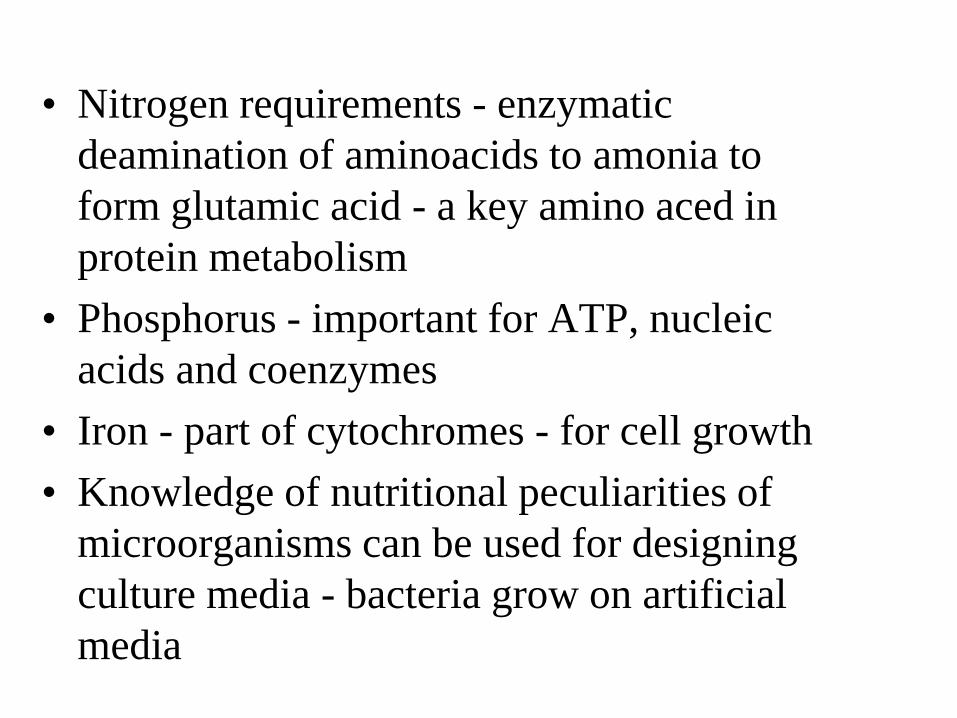
• Nitrogen requirements - enzymatic
deamination of aminoacids to amonia to
form glutamic acid - a key amino aced in
protein metabolism
• Phosphorus - important for ATP, nucleic
acids and coenzymes
• Iron - part of cytochromes - for cell growth
• Knowledge of nutritional peculiarities of
microorganisms can be used for designing
culture media - bacteria grow on artificial
media
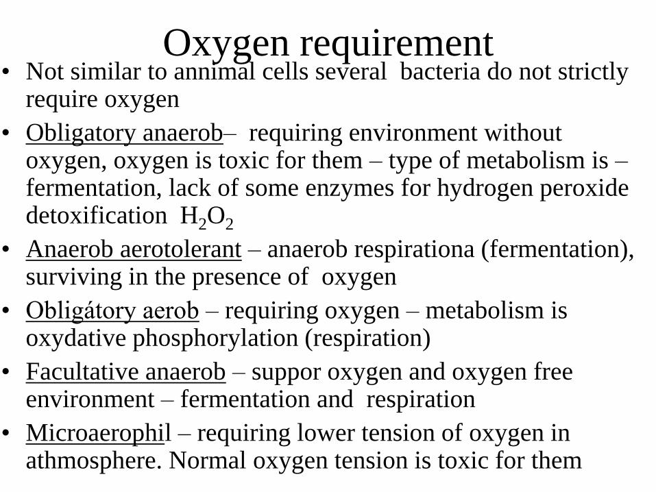
Oxygen requirement • Not similar to annimal cells several bacteria do not strictly
require oxygen
• Obligatory anaerob– requiring environment without oxygen, oxygen is toxic for them – type of metabolism is – fermentation, lack of some enzymes for hydrogen peroxide detoxification H2O2
• Anaerob aerotolerant – anaerob respirationa (fermentation), surviving in the presence of oxygen
• Obligátory aerob – requiring oxygen – metabolism is oxydative phosphorylation (respiration)
• Facultative anaerob – suppor oxygen and oxygen free environment – fermentation and respiration
• Microaerophil – requiring lower tension of oxygen in athmosphere. Normal oxygen tension is toxic for them
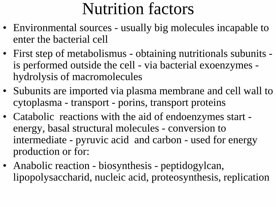
Nutrition factors • Environmental sources - usually big molecules incapable to
enter the bacterial cell
• First step of metabolismus - obtaining nutritionals subunits - is performed outside the cell - via bacterial exoenzymes - hydrolysis of macromolecules
• Subunits are imported via plasma membrane and cell wall to cytoplasma - transport - porins, transport proteins
• Catabolic reactions with the aid of endoenzymes start - energy, basal structural molecules - conversion to intermediate - pyruvic acid and carbon - used for energy production or for:
• Anabolic reaction - biosynthesis - peptidogylcan, lipopolysaccharid, nucleic acid, proteosynthesis, replication
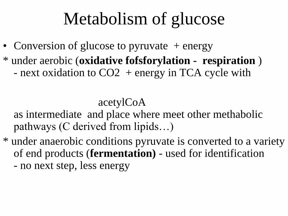
Metabolism of glucose
• Conversion of glucose to pyruvate + energy
* under aerobic (oxidative fofsforylation - respiration ) - next oxidation to CO2 + energy in TCA cycle with
acetylCoA as intermediate and place where meet other methabolic pathways (C derived from lipids…)
* under anaerobic conditions pyruvate is converted to a variety of end products (fermentation) - used for identification - no next step, less energy
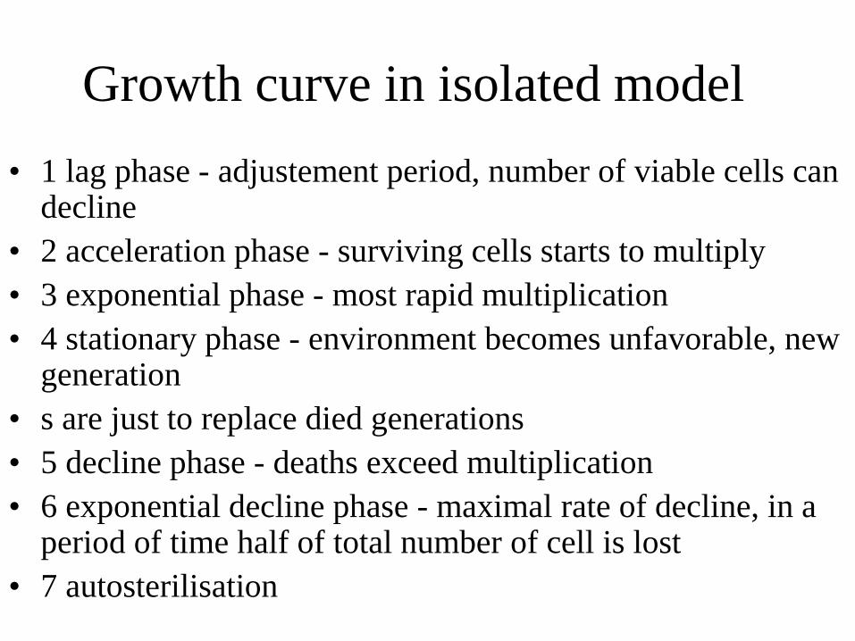
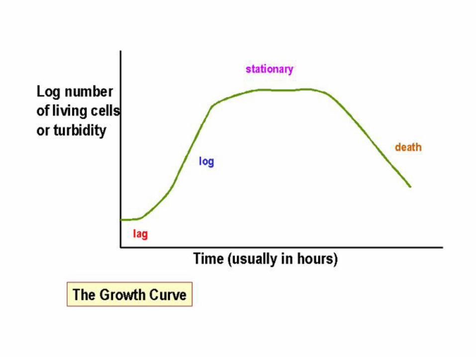
Growth curve in isolated model
• 1 lag phase - adjustement period, number of viable cells can decline
• 2 acceleration phase - surviving cells starts to multiply
• 3 exponential phase - most rapid multiplication
• 4 stationary phase - environment becomes unfavorable, new generation
• s are just to replace died generations
• 5 decline phase - deaths exceed multiplication
• 6 exponential decline phase - maximal rate of decline, in a period of time half of total number of cell is lost
• 7 autosterilisation
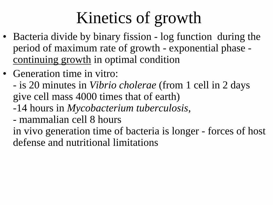
Kinetics of growth • Bacteria divide by binary fission - log function during the
period of maximum rate of growth - exponential phase - continuing growth in optimal condition
• Generation time in vitro: - is 20 minutes in Vibrio cholerae (from 1 cell in 2 days give cell mass 4000 times that of earth) -14 hours in Mycobacterium tuberculosis, - mammalian cell 8 hours in vivo generation time of bacteria is longer - forces of host defense and nutritional limitations
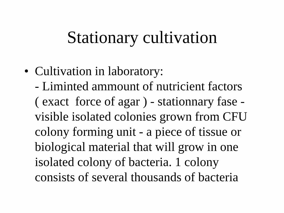
Stationary cultivation
• Cultivation in laboratory:
- Liminted ammount of nutricient factors
( exact force of agar ) - stationnary fase -
visible isolated colonies grown from CFU
colony forming unit - a piece of tissue or
biological material that will grow in one
isolated colony of bacteria. 1 colony
consists of several thousands of bacteria
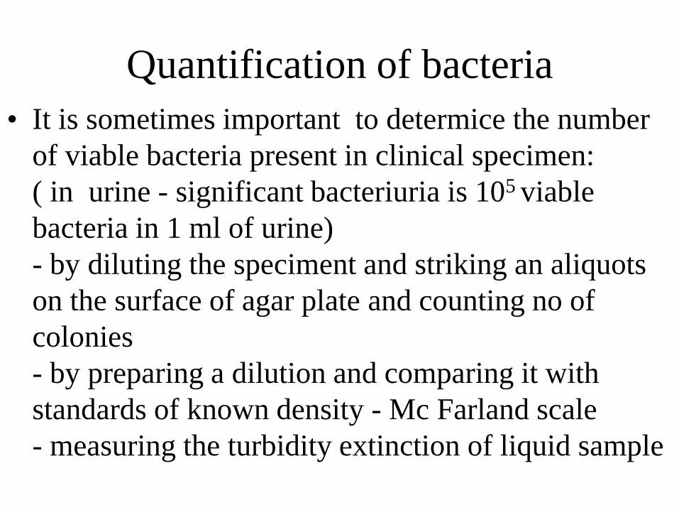
Quantification of bacteria
• It is sometimes important to determice the number
of viable bacteria present in clinical specimen:
( in urine - significant bacteriuria is 105 viable
bacteria in 1 ml of urine)
- by diluting the speciment and striking an aliquots
on the surface of agar plate and counting no of
colonies
- by preparing a dilution and comparing it with
standards of known density - Mc Farland scale
- measuring the turbidity extinction of liquid sample
Cultivation
• To identify a bacterial pathogen it is necessary to transfere
it as a biological sample from site of infection on artificial medium simulating its requirement for growth and isolate grown bacteria in pure culture
• A panel of tests are applied to identify the unknown colony
• This is possible in great majority of bacteria and some yeast - growing on artificial media and being biochemically active - direct detection of pathogen - visualisation
• Not available for viruses - need vital medium for replication (continuous cell lines, annimal model). Indirect detection is more frquently used - via Ab detection.
Steps in identification of
unknown colony
• requirement of oxygen
• Macroscopy of colonies
• Microscopy native (movement) or Gram stain (morphology, cell wall structure) G+,G-,rod, coccus, spiral
- Cell arrangement diplococcus, regular alignement,
- Detection of capsule (agglutination, Burri)
• Ability to ferment certain substrate - sugar, aminoacids - (biochemical properties)
• identification of enzymes - (physiology)
-susceptibility to ATB, and lysis by bacteriophage
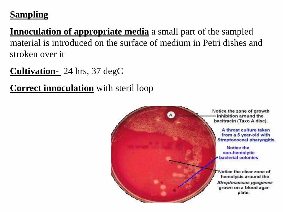
Sampling
Innoculation of appropriate media a small part of the sampled
material is introduced on the surface of medium in Petri dishes and
stroken over it
Cultivation- 24 hrs, 37 degC
Correct innoculation with steril loop
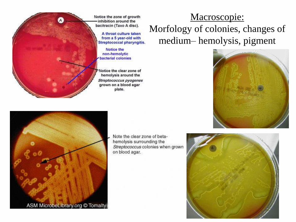
Macroscopie:
Morfology of colonies, changes of
medium– hemolysis, pigment
Microscopy
• - magnification,
• - distinquishing
types – according to the method used for
visualisation:
- light microscopy,
- fluorescein mikroscopy,
- elektron microscopy
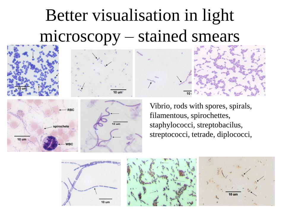
Better visualisation in light
microscopy – stained smears
Vibrio, rods with spores, spirals,
filamentous, spirochettes,
staphylococci, streptobacilus,
streptococci, tetrade, diplococci,
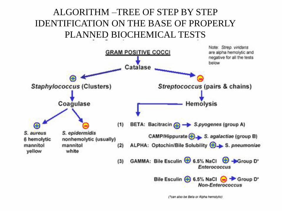
ALGORITHM –TREE OF STEP BY STEP
IDENTIFICATION ON THE BASE OF PROPERLY
PLANNED BIOCHEMICAL TESTS
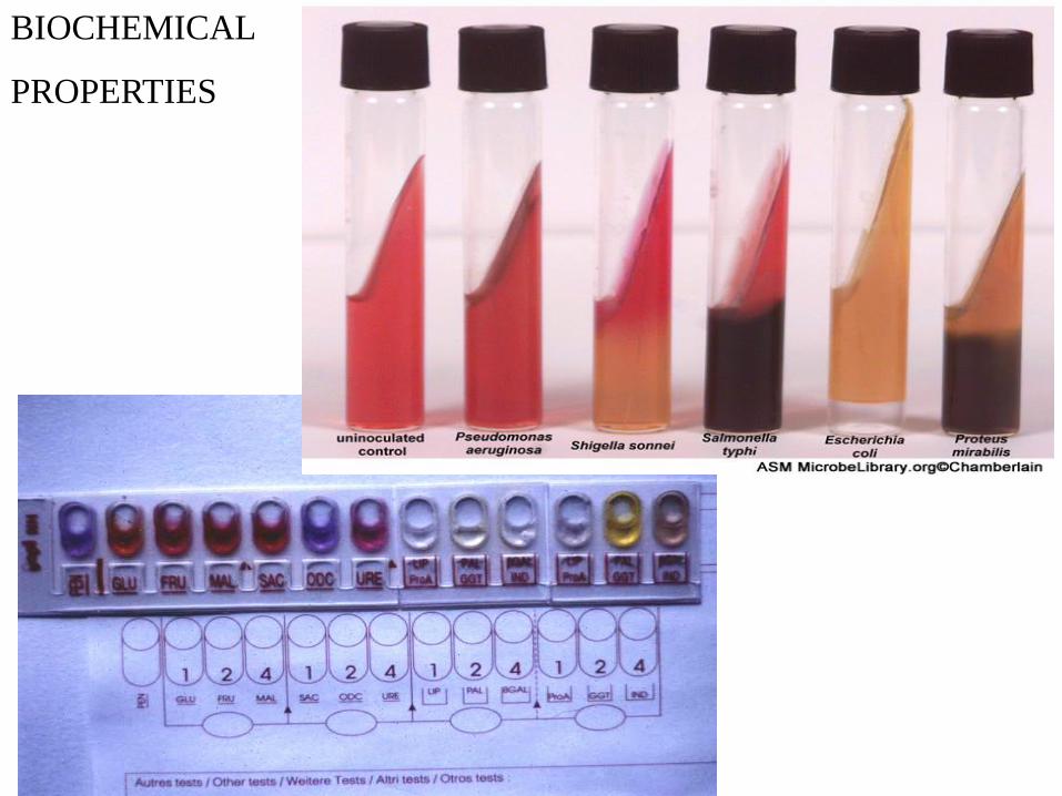
BIOCHEMICAL
PROPERTIES
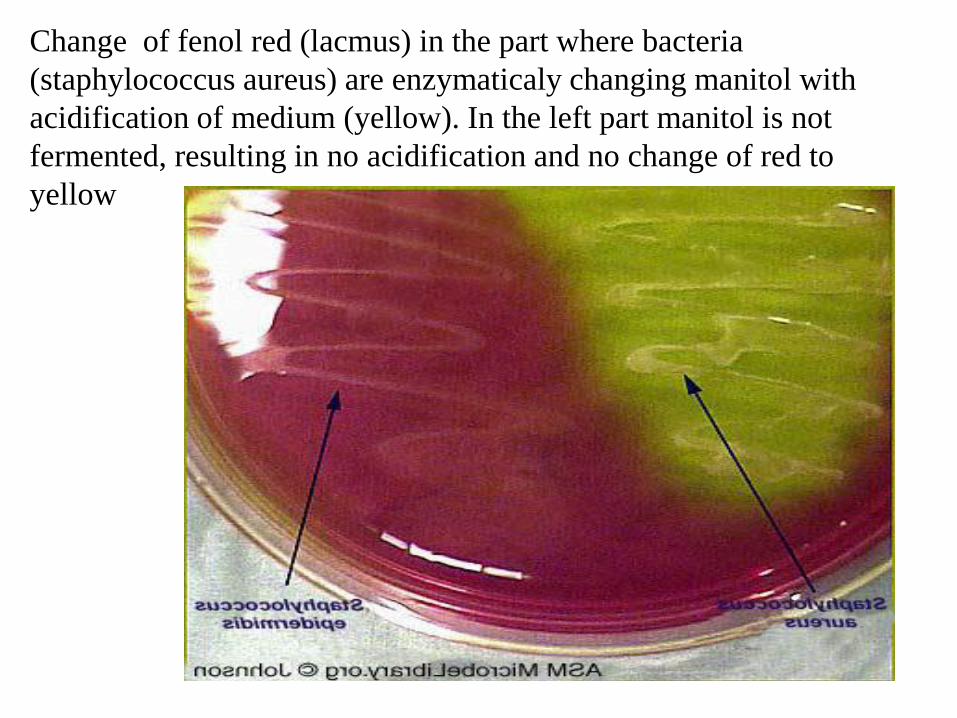
Change of fenol red (lacmus) in the part where bacteria
(staphylococcus aureus) are enzymaticaly changing manitol with
acidification of medium (yellow). In the left part manitol is not
fermented, resulting in no acidification and no change of red to
yellow
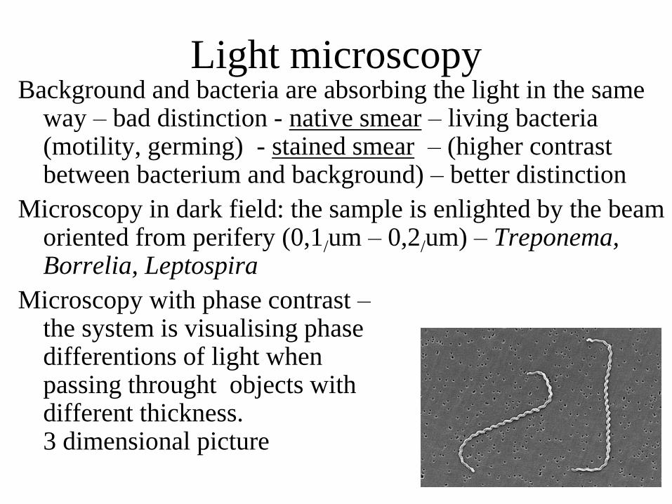
Light microscopy Background and bacteria are absorbing the light in the same
way – bad distinction - native smear – living bacteria (motility, germing) - stained smear – (higher contrast between bacterium and background) – better distinction
Microscopy in dark field: the sample is enlighted by the beam oriented from perifery (0,1/um – 0,2/um) – Treponema, Borrelia, Leptospira
Microscopy with phase contrast – the system is visualising phase differentions of light when passing throught objects with different thickness. 3 dimensional picture
Light microscop Magnification: 2 systems of lenses
– senses of objective
– 10 x general overview,
– 40 x parasits, cysts, molds
–100 x with imersion oil bacteria – lenses of ocular 10x
Overall magnification is ocular multiplied by objective magnification.
Distinction capacity: wave lenght of the light beam and the angle in which the light beam enter the lense of objective – numeric aperture
Light microscope: 1 - 2 /um – the smallest distinguishable
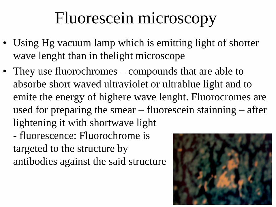
Fluorescein microscopy
• Using Hg vacuum lamp which is emitting light of shorter
wave lenght than in thelight microscope
• They use fluorochromes – compounds that are able to
absorbe short waved ultraviolet or ultrablue light and to
emite the energy of highere wave lenght. Fluorocromes are
used for preparing the smear – fluorescein stainning – after
lightening it with shortwave light
- fluorescence: Fluorochrome is
targeted to the structure by
antibodies against the said structure
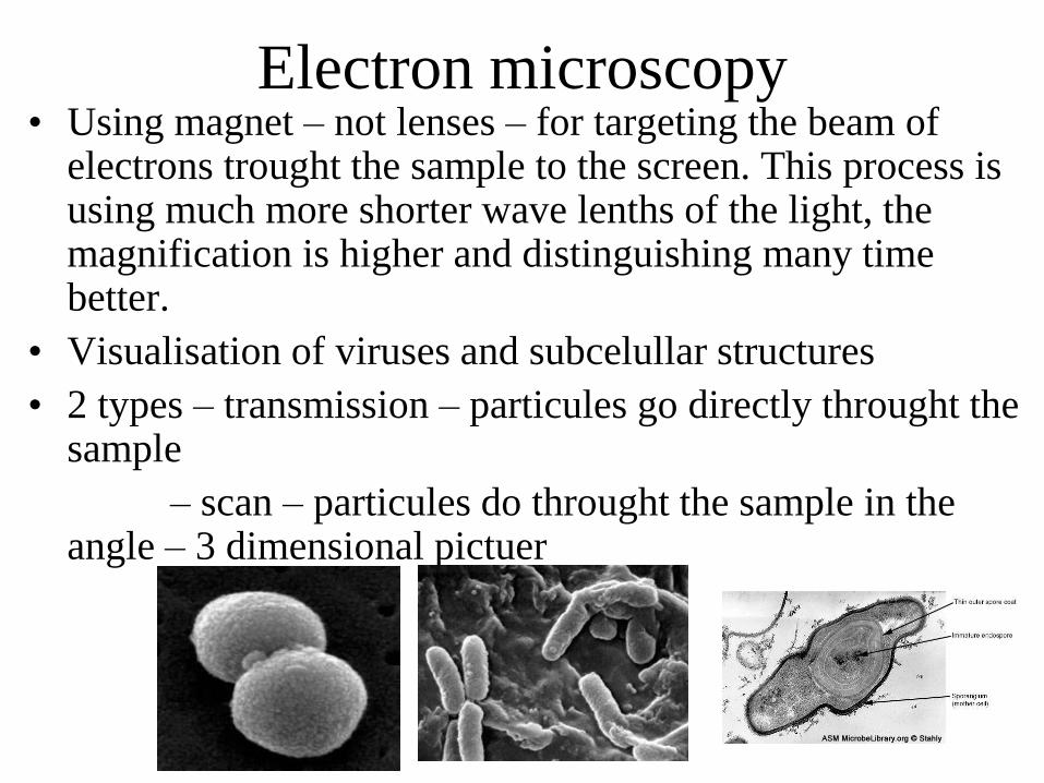
Electron microscopy • Using magnet – not lenses – for targeting the beam of
electrons trought the sample to the screen. This process is using much more shorter wave lenths of the light, the magnification is higher and distinguishing many time better.
• Visualisation of viruses and subcelullar structures
• 2 types – transmission – particules go directly throught the sample
– scan – particules do throught the sample in the angle – 3 dimensional pictuer
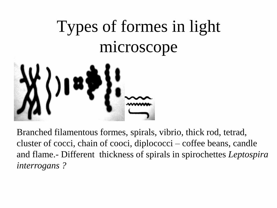
Types of formes in light
microscope
Branched filamentous formes, spirals, vibrio, thick rod, tetrad,
cluster of cocci, chain of cooci, diplococci – coffee beans, candle
and flame.- Different thickness of spirals in spirochettes Leptospira
interrogans ?
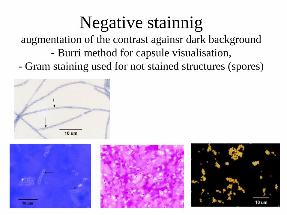
Negative stainnig augmentation of the contrast againsr dark background
- Burri method for capsule visualisation,
- Gram staining used for not stained structures (spores)
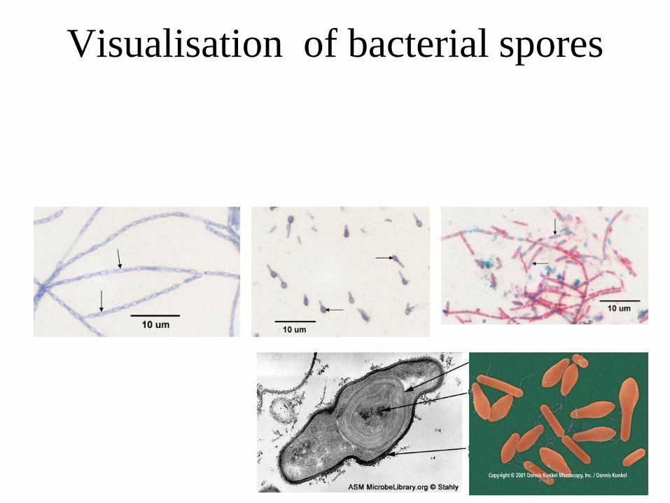
Visualisation of bacterial spores
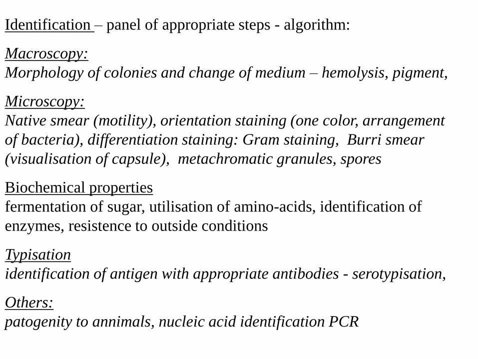
Identification – panel of appropriate steps - algorithm:
Macroscopy:
Morphology of colonies and change of medium – hemolysis, pigment,
Microscopy:
Native smear (motility), orientation staining (one color, arrangement
of bacteria), differentiation staining: Gram staining, Burri smear
(visualisation of capsule), metachromatic granules, spores
Biochemical properties
fermentation of sugar, utilisation of amino-acids, identification of
enzymes, resistence to outside conditions
Typisation
identification of antigen with appropriate antibodies - serotypisation,
Others:
patogenity to annimals, nucleic acid identification PCR