Embed Size (px)
Citation preview
Microphthalmia-associated Transcription Factor (MITF)Promotes Differentiation of Human Retinal PigmentEpithelium (RPE) by Regulating microRNAs-204/211Expression*□S
Received for publication, February 20, 2012, and in revised form, April 18, 2012 Published, JBC Papers in Press, April 20, 2012, DOI 10.1074/jbc.M112.354761
Jeffrey Adijanto‡, John J. Castorino‡, Zi-Xuan Wang‡, Arvydas Maminishkis§, Gerald B. Grunwald‡,and Nancy J. Philp‡1
From the ‡Department of Pathology, Anatomy, and Cell Biology, Thomas Jefferson University, Philadelphia, Pennsylvania 19107and the §NEI, National Institutes of Health, Bethesda, Maryland 20892
Background:microRNAs 204/211 regulate retinal pigment epithelial cell phenotype.Results: In RPE,MITF regulatesmiR-204/211 expression and down-regulation ofMITF results in loss of RPE phenotype, whichcan be prevented by overexpressing miR-204/211.Conclusion:MITF-mediated expression of miR-204/211 directs RPE differentiation.Significance:miR-204/211-based therapeutics may be effective treatments for diseases that involve loss of RPE phenotype.
The retinal pigment epithelium (RPE) plays a fundamentalrole in maintaining visual function and dedifferentiation ofRPE contributes to the pathophysiology of several ocular dis-eases. To identifymicroRNAs (miRNAs) thatmay be involved inRPE differentiation, we compared the miRNA expression pro-files of differentiated primary human fetal RPE (hfRPE) cells todedifferentiated hfRPE cells. We found that miR-204/211, thetwo most highly expressed miRNAs in the RPE, were signifi-cantly down-regulated in dedifferentiated hfRPE cells. Impor-tantly, transfection of pre-miR-204/211 into hfRPE cells pro-moted differentiation whereas adding miR-204/211 inhibitorsled to their dedifferentiation. Microphthalmia-associated tran-scription factor (MITF) is a key regulator of RPE differentiationthat was also down-regulated in dedifferentiated hfRPE cells.MITF knockdown decreased miR-204/211 expression andcaused hfRPE dedifferentiation. Significantly, co-transfectionof MITF siRNAwith pre-miR-204/211 rescued RPE phenotype.Collectively, our data show that miR-204/211 promote RPE dif-ferentiation, suggesting that miR-204/211-based therapeuticsmay be effective treatments for diseases that involve RPE dedif-ferentiation such as proliferative vitreoretinopathy.
The retinal pigment epithelium (RPE)2 is amonolayer of cellsthat forms the outer blood retinal barrier and performs a num-
ber of specialized functions that are critical for photoreceptorhealth and excitability (1). The RPE possesses long apicalmicrovilli that ensheath the photoreceptor outer segments.These two structures are separated by a small volume of space(subretinal space� 10�l) that serves as a conduit for the trans-fer of nutrients and metabolic wastes between photoreceptorsand the choroidal blood vessels (2). The RPE hosts a unique setof enzymes such as lecithin retinol acyltransferase (LRAT) andRPE65 that participates in the visual cycle by catalyzing theconversion of all-trans-retinol to 11-cis-retinal, the latter ofwhich is critical for photoreceptor excitability (3). In addition,RPE cells phagocytose shed photoreceptor outer segments (4)and secrete growth factors to nourish the retina (5) and thechoroidal blood vessels (6). Thus, loss of RPE functions, asoccurs in age-related and proliferative diseases, invariably leadsto photoreceptor degeneration and visual impairment (7).Damage to the RPE or retinal detachment caused by trauma
or intraocular diseases can trigger a repair process, in whichRPE cells lose cell-cell contact and epithelial phenotype tobecome proliferative and motile fibroblast-like cells. In prolif-erative vitreoretinopathy (PVR), for example, unchecked pro-liferation of RPE cells and theirmigration into retinal layers andthe vitreous result in formation of epiretinal membranes thatcan contract and cause retinal detachment and visual impair-ment (8). This switch from an epithelial to mesenchymal-likephenotype involves complex cellular reprogramming with sig-nificant alterations in core cellular functions (e.g. metabolism,cell-cell junctions, cell-cycle progression, cytoskeletal rear-rangement) as well as gene and protein expression (9). In thesearch for potential regulators of this process, microRNAs(miRNAs) appeared to be excellent candidates because eachmiRNApotentially regulates expression of a large array of genes(�300) that may be involved in a variety of cellular functionssuch as proliferation and metabolism (10). Recent studies inother biological systems have also established a role of miRNAsin cellular differentiation as positive and negative regulators ofepithelial-to-mesenchymal transition (EMT) (11, 12).
* This work was supported, in whole or in part, by National Institutes of HealthGrant EY-012042 (to N. J. P.).
□S This article contains supplemental Figs. 1–3 and supplemental Tables 1–3.1 To whom correspondence should be addressed: Dept. of Anatomy, Pathol-
ogy, and Cell Biology, Thomas Jefferson University, 1020 Locust St., Rm.534, Philadelphia, PA 19107. Tel.: 215-503-7854; E-mail: [email protected].
2 The abbreviations used are: RPE, retinal pigment epithelium; AAV, adeno-associated virus; EMT, epithelial-to-mesenchymal transition; EVOM, epi-thelial volt-ohm meter; hfRPE, human fetal RPE; KD, knockdown; miRNA,microRNA; MITF, microphthalmia-associated transcription factor; PVR, pro-liferative vitreoretinopathy; qPCR, quantitative PCR; TER, transepithelialresistance; TRPM, melastatin, transient receptor potential cation channelsubfamily M member; P0, passage 0.
THE JOURNAL OF BIOLOGICAL CHEMISTRY VOL. 287, NO. 24, pp. 20491–20503, June 8, 2012Published in the U.S.A.
JUNE 8, 2012 • VOLUME 287 • NUMBER 24 JOURNAL OF BIOLOGICAL CHEMISTRY 20491
by guest on April 26, 2019
http://ww
w.jbc.org/
Dow
nloaded from
MiRNAs are small (�23 nucleotides) regulatory RNAs thatsuppress gene expression by binding to specific sequences inthe 3�-untranslated region of their target mRNA. Studies invarious organ systems revealed that certain miRNAs are highlyenriched in a tissue-specific pattern (13–16). Furthermore,transfection of such miRNAs into stem cells (17, 18) or evenfibroblasts (19) can induce differentiation into the cell type thatnormally expresses the miRNA at high levels. These findingssupport the notion that specific miRNAs may direct cell speci-fication and differentiation of cells that normally express themat high levels (reviewed in Ref. 20). Because down-regulation oftissue-specific miRNAs is commonly associated with disease,their restoration may slow or inhibit disease progression. Forexample, miR-145 directs smooth muscle differentiation, andits expression was down-regulated in vascular walls withneointimal lesions induced by arterial injury (15). Formation ofthese lesions was inhibited when injured arteries were trans-fected with miR-145.In the RPE, miR-204 and 211 are the two most highly
enrichedmiRNAs, and their expression is critical for maintain-ing barrier properties and function (16). miR-211 resides in thesixth intron ofTRPM1 (melastatin, transient receptor potentialcation channel subfamily M member 1), the transcription ofwhich is regulated by microphthalmia-associated transcriptionfactor (MITF) (21), a master regulator of melanocyte and RPEdifferentiation (22, 23). Mice with homozygous null mutationin the MITF gene have white coats and microphthalmia (24).Furthermore, histological analysis of microphthalmia mouseeyes demonstrated that the absence of MITF prevented RPEdifferentiation (25). Because MITF and miR-204/211 areimportant regulators of RPE development and function, it wasof interest to determine whether MITF regulates miR-204/211expression in the RPE and whether expressing high levels ofmiR-204/211 alone is sufficient to direct RPE differentiation.In this study, we used primary cultures of human fetal RPE
cells (hfRPE) developed byMaminishkis et al. as amodel system(26, 27). These cells exhibit properties (morphology, physiol-ogy, protein andmRNAprofiles) characteristic of native fetal oradult human RPE. In our experiments, we mimicked RPEdetachment and dedifferentiation (as occurs in PVR) by subcul-turing cells at low cell density and found that this processresulted in significant down-regulation ofMITF and miR-204/211. Using this in vitro model of RPE dedifferentiation, wefound that introduction of pre-miR-204/211 promoted RPEdifferentiation and protected them from dedifferentiation. Ourfindings may help facilitate development of miR-204/211-based therapies for human ocular diseases that involve RPEdedifferentiation such as age-relatedmacular degeneration andPVR.
EXPERIMENTAL PROCEDURES
hfRPE Culture Model—hfRPE monolayers were cultured onT25 flasks (P0 hfRPE) as described previously (26). Briefly,hfRPE cells were trypsinized from a T25 flask and seeded onto12-well Transwells at �1.25 � 105 cells/well. P1 hfRPE cellswere cultured for 3–4 weeks to reach maturity (transepithelialresistance (TER) �500 ohms�cm2) prior to experimentation.TER was measured with an epithelial volt-ohmmeter (EVOM)
(WPI, Sarasota, FL) at room temperature.Media and Transwellresistances were taken into account by subtracting 122ohms�cm2 from the EVOMreadout. To test for choroidal fibro-blast contamination, hfRPE cells were stained with collagentype I/procollagen antibody (Cell Sciences; Canton, MA).Human fetal choroidal fibroblast cells were used as positivecontrols and were cultured in the same medium as hfRPE cells.Total mRNA Extraction—Total mRNA of samples was
extracted using mirVana miRNA extraction kit (Ambion, Aus-tin, TX) according to the manufacturer’s protocol. RNA boundin the column matrix was treated with RQ1 DNase (5 units/sample; Promega) at 37 °C for 30min followed bymultiplewashsteps according to the manufacturer’s protocol. RNA waseluted with diethylpyrocarbonate-treated water preheated to85 °C. Total RNA concentration was measuring using Qubit�fluorometer (Invitrogen).miRNA Microarray and Data Analysis—Total mRNA of
differentiated and dedifferentiated hfRPE samples were pre-pared using TRIzol (Invitrogen) as described previously (28),and 100 ng of total mRNA from each sample was labeled andhybridized to a human miRNA microarray (V2) from AgilentTechnologies (Santa Clara, CA) according to the manufactur-er’s protocol. The microarray was scanned with an AgilentMicroarray Scanner, and the data were processed using FeatureExtraction software v10.7.3.1 (Agilent). The microarray wasnormalized to miR-24 and miR-130a, whose expression levelswere the least different between the two RPE cell phenotypes.The normalized array was analyzed using Significance Analysisof Microarrays (SAM 4.0 with R2.14.1) (29) for two-classunpaired statistical analysis with � � 5.0 and -fold change �2.miRNAs with fluorescence�50 in both RPE sample types wereeliminated. LOG2 fluorescence intensities of miRNAs wererepresented with a heatmap generated in MultiExperimentViewer (MeV v4.8). The normalized version of the microarraydata can be downloaded from NCBI GEO database (accessionnumber GSE36137).Reverse Transcription and Real-time Quantitative PCR
(qPCR)—RNA (1 �g/sample) was reverse transcribed using oli-go(dT)20 primers and SuperScript III (Invitrogen). qPCRs forgene expression studies were performed using ITaq SYBRGreen Supermix with ROX (Bio-Rad) in 20-�l reactions (10 ngof cDNA/RxN). qPCRwas performed using EppendorfMaster-cycler� ep realplex2. Primerswere designed according to guide-lines set by Dieffenbach et al. (30). Custom oligonucleotideswere purchased from EurofinsMGWOperon (Huntsville, AL).Sequences for all primers used in this study are listed in supple-mental Table 1.qRT-PCR Using TaqMan miRNA Assays—miR-204, miR-
211, miR-125b, let-7g, miR-21, and miR-31 TaqMan primersand probes were purchased from Applied Biosystems. 10 ng oftotal RNAwas used in reverse transcription, and the PCRswereperformed according to the manufacturer’s protocol. qPCRdata were analyzed using the comparative 2-��Ct method (31).qPCR Data Analysis—For SYBRGreen qRT-PCR, ribosomal
protein S18 (RPS18) gene was used as reference gene becausethe 2-Ct values of RPS18 from differentiated versus dedifferen-tiated hfRPE samples were statistically insignificant. For Taq-Man assays, U18 snoRNA was used as reference gene because
MITF Promotes RPE Differentiation by Regulating miR-204/211
20492 JOURNAL OF BIOLOGICAL CHEMISTRY VOLUME 287 • NUMBER 24 • JUNE 8, 2012
by guest on April 26, 2019
http://ww
w.jbc.org/
Dow
nloaded from
U18 lies within the intron of RPL4 and themeanCt values (2-Ct)of RPL4 of dedifferentiated versus differentiated hfRPE sampleswere statistically insignificant. 2-�Ct of treated versus controlsamples was analyzed for statistical significance using Student’st test (two-tailed; unpaired samples, unequal variances). p val-ues of � 0.05 were considered statistically significant.siRNA, Pre-miRNA, and Anti-miRNA Transfection—Am-
bion pre-miR miRNA precursors and anti-miRNA were pur-chased from Applied Biosystems. All siRNAs were purchasedfrom Santa Cruz Biotechnology (Santa Cruz, CA). In all exper-iments, pre-miRNA were transfected upon seeding and on the3rd day after seeding. Dharmafect 4 was used as transfectionreagent (0.2%) in antibiotic-free complete MEM containing 5%serum.WesternBlotting—hfRPE cells onTranswell filterswere lysed
and homogenized as described previously (28). 15 �g of totalprotein lysates was loaded onto a NuPAGE� 4–12% Tris-ace-tate gel (Invitrogen) for electrophoresis. Proteins were subse-quently transferred onto PVDF membranes using XCell IITM
Blot Module (Invitrogen). Nonspecific binding sites wereblocked with TBS (0.1% Tween 20) containing 5% w/v pow-dered milk. Antibodies used in this study are listed in supple-mental Table 2.Immunofluorescence and Imaging—hfRPE cells on Tran-
swell filters were fixed with 4% formaldehyde in 1� PBS for 5min at room temperature followed by 20 min at 4 °C. Sampleswere permeabilized for 5 min with 0.3% Triton X-100 andblockedwith PBS 0.1%Tween 20 (PBST) containing BSA (5%w/v). Samples were incubated overnight with antibodiesagainst MCT3 and ZO-1 (clone ZO1-1A12; Invitrogen). Sam-ples were washedwith PBST and incubated for 1 h in secondaryantibodies (Invitrogen). After washing with PBST, sampleswere stainedwith phalloidin (1 h at 1:100; Invitrogen) andDAPI(5 min at 1:1000) prior to mounting with gelvatol onto micro-scope slides. Confocal images on Figs. 3G and 5G were takenwith Zeiss LSM 510 confocal microscope at �40 (Plan-Neo-fluar �40/1.3 oil differential interference contrast) with �2scanner zoom (�80 final) and 0.5-�m Z-stack intervals. Allother images were taken usingNikonA1R confocalmicroscopeat�60 (Plan ApoVC�60WI differential interference contrastN2) with �1.33 scanner zoom (�80 final) and 0.5-�m Z-stackintervals. Images were extracted from NIS-Elements and ana-lyzed using Adobe Photoshop 7.0.miRNA Target Prediction Analysis—Putative miR-204/211
targets were obtained from TargetScan, miRanda, PicTar, andmiRDB. Because predictionsmade by TargetScan (and PicTar),among other algorithms, have been shown to be among themost accurate (as analyzed by proteomics) (32), miRNA targetgenes ranked in the “top 100” list of TargetScan were scoredhigher. Each gene was annotated with their respective ontol-ogy profiles and pathway profile (GenMAPP and KEGG databases) that were extracted from the annotation files ofAffymetrix human microarray chipset (HG-U133 Plus 2).This list of miR-204/211 targets is available in supplementalTable 3, and a selection of these targets was categorized andis listed in Fig. 6.
RESULTS
RPEDedifferentiation Involves Loss of RPE-specific Genes andmiR-204/211—The RPE is normally quiescent and nonmigra-tory, but in disease conditions such as PVR, it can undergodedifferentiation into fibroblast-like cells that are proliferativeand motile. This phenomenon was observed in vitro as cells atthe free edge of differentiated hfRPEmonolayer dedifferentiate,migrate, and establish a new population of sparsely pigmentedfibroblast-like cells (28). To study the role of miRNAs in RPEdifferentiation, we compared the miRNA expression profile ofdedifferentiated versus differentiated hfRPE cells using miRNAmicroarray analysis (Fig. 1A). In this experiment, we alsoincluded a sample of partially differentiated hfRPE cells (pig-mented but lost epithelial morphology) to represent RPE cellsin an earlier stage of dedifferentiation. From this array, wefound that the three most highly expressed miRNAs (miR-204,miR-211, and miR-125b) in the RPE (16) were significantlydown-regulated in dedifferentiated hfRPE cells. miR-200a andmiR-200b, which suppress EMT by targeting Zeb1 and Zeb2transcription factors (33), were also down-regulated in dedif-ferentiated RPE cells. In addition, expression of miRNAs thatare commonly down-regulated (let-7 family) or up-regulated(miR-21 and miR-31) in cancer was also altered. This model ofRPE dedifferentiation, however, was difficult to manipulatebecause dedifferentiation and migration of cells from the edgeof the RPE monolayer occur sporadically and therefore cannotbe experimentally induced and controlled. Thus, we developedan alternative in vitromodel of RPE dedifferentiation in whichwe passaged P1 hfRPE cells at low density (P2 at 1%, P3 at 30%)twice to produce a homogeneous population of dedifferenti-ated hfRPE cells (Fig. 1B). However, such seeding conditionsalso promote growth of choroidal fibroblast contaminants thatmay be present in the hfRPE culture. To address this concern,we examined the purity of our hfRPE cultures and showed thatour dedifferentiated hfRPE cells did not express collagen type I(gene and protein), which was highly expressed in choroidalfibroblast cells (supplemental Fig. 2). Using this model of RPEdedifferentiation, we validated our microarray data with Taq-Man qRT-PCR miRNA assay (Fig. 1C). In agreement with themicroarray data, we found that miR-204/211 were among themost significantly down-regulated miRNAs in dedifferentiatedRPE cells.To further understand the biological role of miR-204/211 in
the RPE, putative miR-204/211 target genes were identifiedusing in silicomiRNA target prediction tools (see Fig. 6). Next,we compared the expression of these genes in differentiatedversus dedifferentiated RPE cells by qRT-PCR analysis. Wefound that several of the predicted miR-204/211 target genes(CDH2, CREB5, TCF3 (TCF7L1), TGFBR1, RAB22A, ELOVL6,TCF12, TCF4 (TCF7L2), SMAD4, and SIRT1) were up-regu-lated in dedifferentiated hfRPE cells, consistent with the loss ofmiR-204/211 (Fig. 1C). In addition, dedifferentiated hfRPE cellsexpress low levels of genes that are known to be important forRPE function: blood-retinal barrier (CDH1,CLDN10,CLDN19,OCLN), ion and nutrient transport (BEST1, SLC16A1 (MCT1),SLC16A8 (MCT3), SLC2A1 (GLUT1)), and retinal cycle(RPE65, CRALBP) (Fig. 1E). Accompanying these changes was
MITF Promotes RPE Differentiation by Regulating miR-204/211
JUNE 8, 2012 • VOLUME 287 • NUMBER 24 JOURNAL OF BIOLOGICAL CHEMISTRY 20493
by guest on April 26, 2019
http://ww
w.jbc.org/
Dow
nloaded from
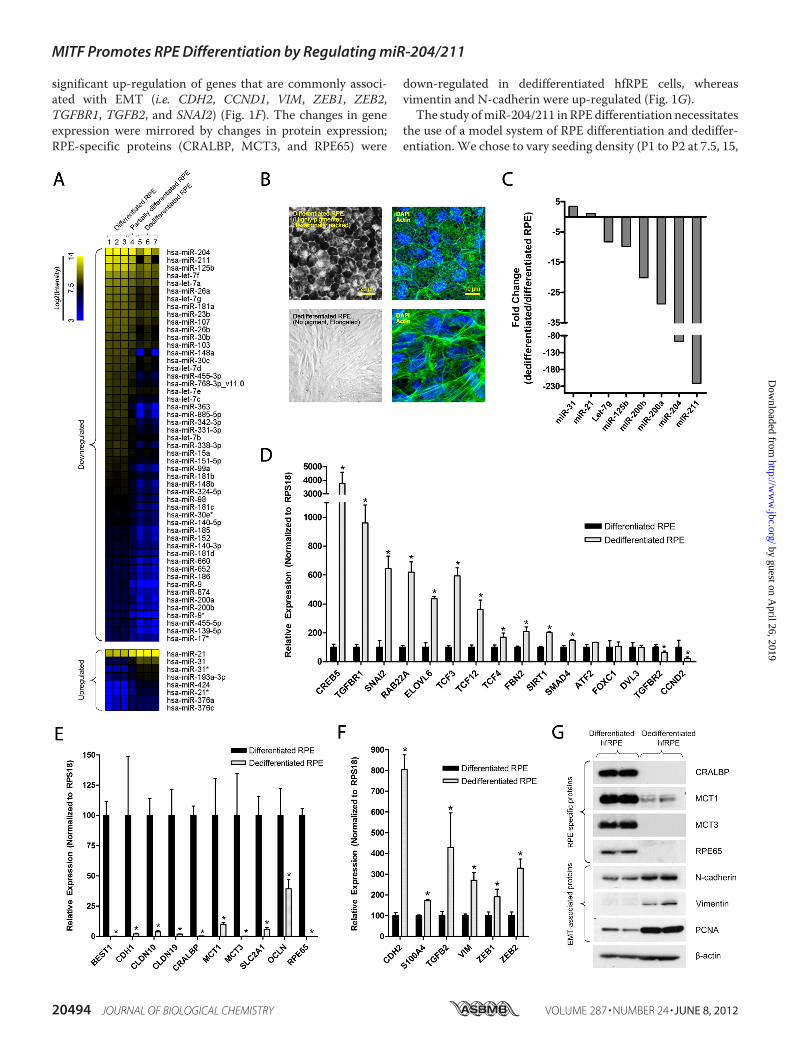
significant up-regulation of genes that are commonly associ-ated with EMT (i.e. CDH2, CCND1, VIM, ZEB1, ZEB2,TGFBR1, TGFB2, and SNAI2) (Fig. 1F). The changes in geneexpression were mirrored by changes in protein expression;RPE-specific proteins (CRALBP, MCT3, and RPE65) were
down-regulated in dedifferentiated hfRPE cells, whereasvimentin and N-cadherin were up-regulated (Fig. 1G).The study ofmiR-204/211 inRPEdifferentiation necessitates
the use of a model system of RPE differentiation and dediffer-entiation.We chose to vary seeding density (P1 to P2 at 7.5, 15,
MITF Promotes RPE Differentiation by Regulating miR-204/211
20494 JOURNAL OF BIOLOGICAL CHEMISTRY VOLUME 287 • NUMBER 24 • JUNE 8, 2012
by guest on April 26, 2019
http://ww
w.jbc.org/
Dow
nloaded from
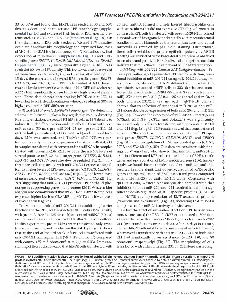
30, or 60%) and found that hfRPE cells seeded at 30 and 60%densities developed characteristic RPE morphology (supple-mental Fig. 1A) and expressed high levels of RPE-specific pro-teins such as MCT3 and CRALBP (supplemental Fig. 1B). Onthe other hand, hfRPE cells seeded at 7.5 and 15% densitiesexhibited fibroblast-like morphology and expressed low levelsofMCT3 andCRALBP. In addition, qRT-PCR results show thatexpression of miR-204/211 (supplemental Fig. 1D) and RPE-specific genes (BEST1, CLDN19, CRALBP,MCT3, and RPE65)(supplemental Fig. 1E) were generally higher in RPE cellsseeded at 60 versus 15% density. This trendwas also observed atall three time points tested (3, 7, and 15 days after seeding). By15 days, the expression of several RPE-specific genes (BEST1,CLDN19, and MCT3) in hfRPE cells seeded at 60% densityreached levels comparable with that of P1 hfRPE cells, whereasRPE65 took significantly longer to achieve high levels of expres-sion. These data showed that seeding hfRPE cells at 15% orlower led to RPE dedifferentiation whereas seeding at 30% orhigher resulted in RPE differentiation.miR-204/211 Promote Epithelial Phenotype—To determine
whether miR-204/211 play a key regulatory role in directingRPE differentiation, we seeded P2 hfRPE cells at 15% density toinduce RPE dedifferentiation and transfected them with pre-miR-control (50 nM), pre-miR-204 (25 nM), pre-miR-211 (25nM), or both pre-miR-204/211 (25 nM each) and cultured for 7days. RNA was extracted, and TaqMan qRT-PCR was per-formed to verify increased expression of mature miR-204/211in samples transfectedwith correspondingmiRNAs. In samplestreated with pre-miR-204, -211, or both, the mRNA levels ofseveral putative miR-204/211 target genes (CREB5, RAB22A,ELOVL6, and TCF12) were also down-regulated (Fig. 2B). Fur-thermore, cells transfected with miR-204/211 expressed signif-icantly higher levels of RPE-specific genes (CLDN10, CLDN19,BEST1,MCT3,RPE65, andCRALBP) (Fig. 2C), and lower levelsof genes associated with EMT (CDH2, VIM, and SNAI2) (Fig.2D), suggesting that miR-204/211 promote RPE epithelial phe-notype by suppressing genes that promote EMT. Western blotanalysis also demonstrated that miR-204/211-transfected cellsexpressed higher levels of CRALBP andMCT3 and lower levelsof N-cadherin (Fig. 2E).To evaluate the role of miR-204/211 in establishing barrier
functions of the RPE, we transfected hfRPE cells (15% density)with pre-miR-204/211 (25 nM each) or control miRNA (50 nM)on Transwell filters andmeasured TER after 21 days in culture.In this experiment, pre-miRNAs were transfected only twice(once upon seeding and another on the 3rd day). Fig. 2F showsthat at the end of the 3rd week, hfRPE cells transfected withmiR-204/211 had higher TER (79 23 ohms�cm2) comparedwith control (35 8 ohms�cm2; n � 4; p � 0.03). Immuno-staining of these cells revealed that hfRPE cells transfected with
control miRNA formed multiple layered fibroblast-like cellswith stress fibers that did not expressMCT3 (Fig. 2G, upper). Incontrast, hfRPE cells transfectedwith pre-miR-204/211 formeda monolayer of hexagonally packed cells with circumferentialbundles of actin filaments at the lateral junctions and apicalmicrovilli as revealed by phalloidin staining. Furthermore,these cells reestablished proper epithelial polarity as MCT3labelingwas restricted to the basolateralmembrane as observedin a mature and polarized RPE in situ. Taken together, our dataindicate that miR-204/211 can prevent RPE dedifferentiation.Inhibiting miR-204/211 Caused RPE Dedifferentiation—Be-
cause pre-miR-204/211 prevented RPE dedifferentiation, func-tional inhibition of miR-204/211 using miR-204/211 antagom-ers (anti-miRs) should block RPE differentiation. To test thishypothesis, we seeded hfRPE cells at 30% density and trans-fected them with anti-miR-204 (25 nM 25 nM control anti-miR), 25 nM anti-miR-211 (25 nM 25 nM control anti-miR), orboth anti-miR-204/211 (25 nM each). qRT-PCR analysisshowed that transfection of either anti-miR-204 or anti-miR-211 alone decreased expression of both miR-204 and miR-211(Fig. 3A). However, the expression ofmiR-204/211 target genes(CREB5, ELOVL6, TCF12, and RAB22A) was significantlyincreased only in cells co-transfected with both anti-miR-204and -211 (Fig. 3B). qRT-PCR results showed that transfection ofanti-miR-204 or -211 resulted in down-regulation of RPE-spe-cific genes (BEST1, CLDN10, CLDN19, MCT3, and RPE65)(Fig. 3C) and up-regulation of EMT-associated genes (CDH2,VIM, and SNAI2) (Fig. 3D). Our data are consistent with find-ings by Wang et al., who showed that inhibiting miR-204 or-211 in differentiated RPE cells resulted in loss of RPE-specificgenes and up-regulation of EMT-associated genes (16). Impor-tantly, we found that co-transfection of anti-miR-204 and -211resulted in more significant down-regulation of RPE-specificgenes and up-regulation of EMT-associated genes comparedwith anti-miR-204 or anti-miR-211 alone. Consistent withqRT-PCR data, Western blot analysis also demonstrated thatinhibition of both miR-204 and -211 resulted in the most sig-nificant down-regulation of RPE-specific proteins (CRALBPand MCT3) and up-regulation of EMT-associated proteins(vimentin and N-cadherin) (Fig. 3E), indicating that miR-204compensated for miR-211 activity and vice versa.To test the effect of anti-miR-204/211 on RPE barrier func-
tion, we measured the TER of hfRPE cells cultured at 30% den-sity transfectedwith anti-miR-204, -211, or both anti-miR-204/211 (two transfections over 14 days). After 14 days in culture,control hfRPE cells established a resistance of �250 ohms�cm2,whereas cells transfected with anti-miR-204, -211, or both 204/211 had significantly lower resistances (�120, 180, and 80ohms�cm2, respectively) (Fig. 3F). The morphology of cellstransfected with either anti-miR-204 or -211 alone was not sig-
FIGURE 1. RPE dedifferentiation is characterized by loss of epithelial phenotype, changes in miRNA profile, and significant alterations in mRNA andprotein expression. Differentiated hfRPE cells (passage 1 (P1)) were grown on Transwell filters over 4 weeks to obtain a differentiated RPE monolayer. A,dedifferentiated RPE cells that migrated from the free edge of confluent hfRPE monolayer were isolated, and microRNA microarray was performed to comparetheir miRNA expression levels with that of differentiated RPE cells. B, in a different model, dedifferentiated RPE cells were obtained by passaging P1 hfRPE cellsat low cell density twice (P1 to P2 at 1%, P2 to P3 at 30%) on 100-mm culture dishes. C, the expression of several miRNAs that were significantly altered in themicroarray analysis was verified using TaqMan microRNA assay. D–F, to compare mRNA expression of differentiated versus dedifferentiated RPE cells, qRT-PCRwas performed to evaluate the expression of miR-204/211 targets (D), genes involved in barrier, nutrient/ion transport, and RPE-specific functions (E), andgenes that are commonly up-regulated in EMT (F). G, Western blot shows that RPE dedifferentiation involves a loss of RPE-specific proteins and an increase inEMT-associated proteins. Statistically significant changes (p � 0.05) are marked with asterisks. Error bars, S.D.
MITF Promotes RPE Differentiation by Regulating miR-204/211
JUNE 8, 2012 • VOLUME 287 • NUMBER 24 JOURNAL OF BIOLOGICAL CHEMISTRY 20495
by guest on April 26, 2019
http://ww
w.jbc.org/
Dow
nloaded from
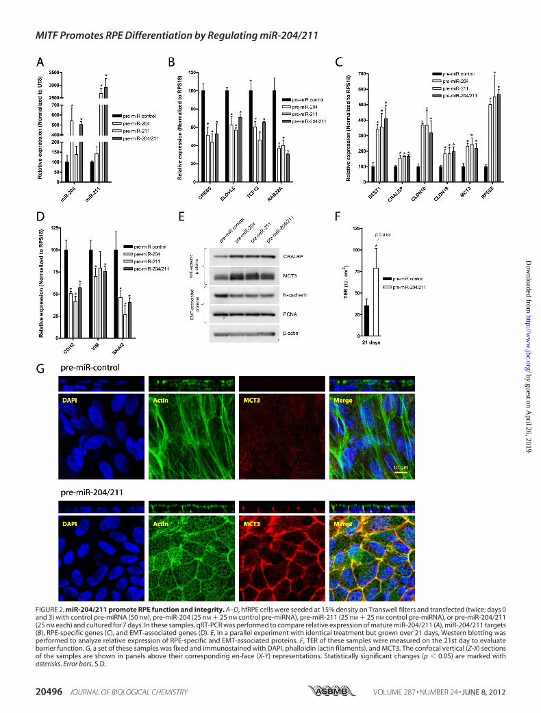
FIGURE 2. miR-204/211 promote RPE function and integrity. A–D, hfRPE cells were seeded at 15% density on Transwell filters and transfected (twice; days 0and 3) with control pre-miRNA (50 nM), pre-miR-204 (25 nM 25 nM control pre-miRNA), pre-miR-211 (25 nM 25 nM control pre-miRNA), or pre-miR-204/211(25 nM each) and cultured for 7 days. In these samples, qRT-PCR was performed to compare relative expression of mature miR-204/211 (A), miR-204/211 targets(B), RPE-specific genes (C), and EMT-associated genes (D). E, in a parallel experiment with identical treatment but grown over 21 days, Western blotting wasperformed to analyze relative expression of RPE-specific and EMT-associated proteins. F, TER of these samples were measured on the 21st day to evaluatebarrier function. G, a set of these samples was fixed and immunostained with DAPI, phalloidin (actin filaments), and MCT3. The confocal vertical (Z-X) sectionsof the samples are shown in panels above their corresponding en-face (X-Y) representations. Statistically significant changes (p � 0.05) are marked withasterisks. Error bars, S.D.
MITF Promotes RPE Differentiation by Regulating miR-204/211
20496 JOURNAL OF BIOLOGICAL CHEMISTRY VOLUME 287 • NUMBER 24 • JUNE 8, 2012
by guest on April 26, 2019
http://ww
w.jbc.org/
Dow
nloaded from
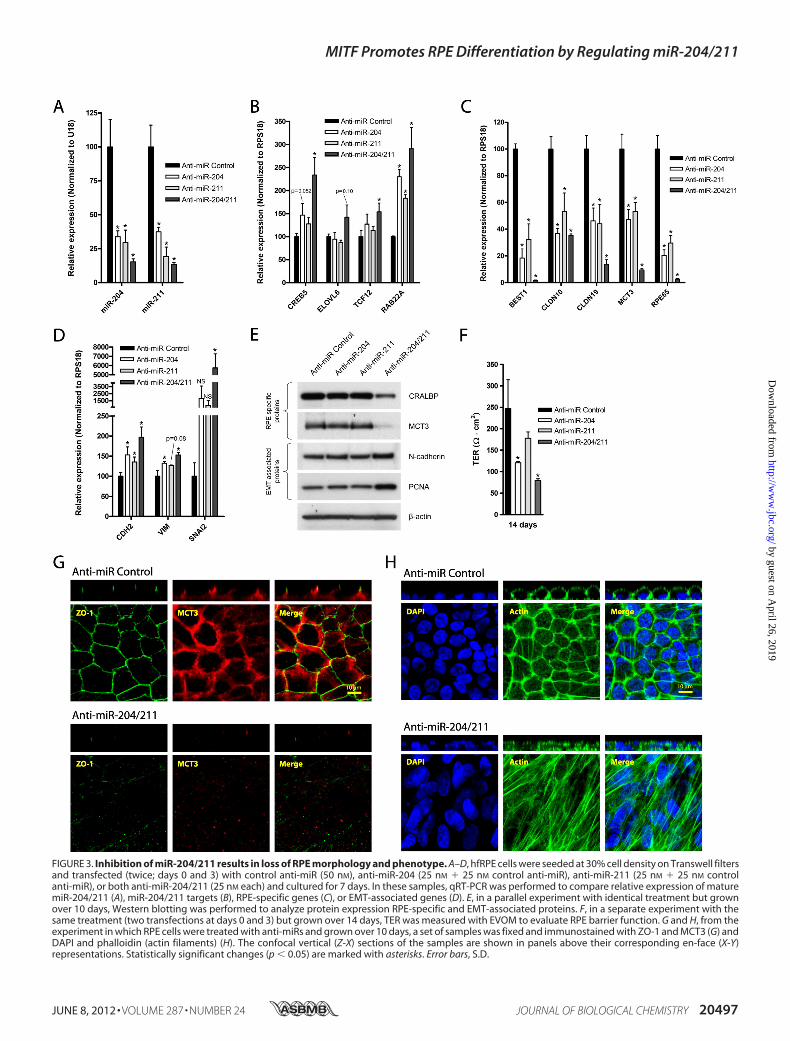
FIGURE 3. Inhibition of miR-204/211 results in loss of RPE morphology and phenotype. A–D, hfRPE cells were seeded at 30% cell density on Transwell filtersand transfected (twice; days 0 and 3) with control anti-miR (50 nM), anti-miR-204 (25 nM 25 nM control anti-miR), anti-miR-211 (25 nM 25 nM controlanti-miR), or both anti-miR-204/211 (25 nM each) and cultured for 7 days. In these samples, qRT-PCR was performed to compare relative expression of maturemiR-204/211 (A), miR-204/211 targets (B), RPE-specific genes (C), or EMT-associated genes (D). E, in a parallel experiment with identical treatment but grownover 10 days, Western blotting was performed to analyze protein expression RPE-specific and EMT-associated proteins. F, in a separate experiment with thesame treatment (two transfections at days 0 and 3) but grown over 14 days, TER was measured with EVOM to evaluate RPE barrier function. G and H, from theexperiment in which RPE cells were treated with anti-miRs and grown over 10 days, a set of samples was fixed and immunostained with ZO-1 and MCT3 (G) andDAPI and phalloidin (actin filaments) (H). The confocal vertical (Z-X) sections of the samples are shown in panels above their corresponding en-face (X-Y)representations. Statistically significant changes (p � 0.05) are marked with asterisks. Error bars, S.D.
MITF Promotes RPE Differentiation by Regulating miR-204/211
JUNE 8, 2012 • VOLUME 287 • NUMBER 24 JOURNAL OF BIOLOGICAL CHEMISTRY 20497
by guest on April 26, 2019
http://ww
w.jbc.org/
Dow
nloaded from
nificantly different from control anti-miRNA-transfected cells(data not shown), but cells transfected with both anti-miR-204and -211 exhibited dramatic loss of RPE phenotype as charac-terized by the complete loss ofMCT3 and ZO-1 and the forma-tion of multilayered cells with stress fibers (Fig. 3, G and H).Collectively, our data indicate that inhibition of both miR-204and -211 is required to induce RPE dedifferentiation.MITF Knockdown Decreased Expression of miR-204/211 and
Their Host Genes, TRPM1 and TRPM3—Because down-regu-lation of miR-204/211 caused RPE dedifferentiation, we exam-ined upstreammechanisms that regulate miR-204/211 expres-sion. miR-204 and miR-211 lie within the introns of TRPM3and TRPM1, respectively, and early studies in melanocytesshowed that transcription of miR-211 and its host gene,TRPM1, are coordinately regulated by MITF. Thus, we com-paredMITF gene expression in differentiated versus dedifferenti-ated RPE cells and found thatMITF and its target genes (TRPM1,TRPM3, TYR, and TYRP1) were significantly down-regulated indedifferentiatedRPE cells (Fig. 4A). To further examine the role ofMITF inmiR-204/211expression,we transfectedhfRPEcells (30%density) with MITF siRNA (30 nM) versus control siRNA (30 nM)and found that MITF knockdown caused significant down-regu-lation of its target genes (TRPM1, TRPM3, TYR, and TYRP1) andmiR-204/211 (Fig. 4,B andC). TheMITFKD-induced decrease inmiR-204/211 expression was accompanied by a concomitant up-regulation of miR-204/211 target genes (CREB5, RAB22A,ELOVL6, SNAI2, and TCF12) (Fig. 4D).
To determine whether miR-204/211 down-regulation wasthe primary cause for the loss of RPE phenotype in MITF KDcells, we examined whether addition of pre-miR-204/211 couldprevent RPE dedifferentiation caused byMITF KD. hfRPE cells(30% density) were transfected with MITF siRNA (30 nM) control miRNA (30 nM), MITF siRNA (30 nM) pre-miR-204/211 (15 nM each), or control miRNA and siRNA (30 nM each)(once at time of seeding and again 3 days later) and cultured for7 days. Consistent with miR-204/211 levels (Fig. 5A), expres-sion of miR-204/211 targets (CREB5, ELOVL6, TCF12, andRAB22A) was up-regulated in MITF KD cells, and these geneswere suppressed in hfRPE cells transfected with both MITFsiRNA and pre-miR-204/211 (Fig. 5B). MITF siRNA alsodecreased expression of RPE-specific genes (BEST1, CRALBP,CLDN19,MCT3, and RPE65), and this effect was prevented byco-transfection with pre-miR-204/211 (Fig. 5C). Expression ofEMT-associated genes that were up-regulated in MITF KDcells was also suppressed by pre-miR-204/211 (Fig. 5D). Theseeffects were confirmed at the protein level by Western blotanalysis (Fig. 5E; hfRPE cultured for 21 days).Next, we examined whether hfRPE cells transfected with
both MITF siRNA and miR-204/211 could reestablish barrierfunctions. TER was measured on the 14th and 21st day, and weobserved that hfRPE cells with MITF KD had no detectableresistance at either time point (Fig. 5F). However, hfRPE cellstransfected with both MITF siRNA and pre-miR-204/211 hadresistances of �240 ohms�cm2 on the 21st day, demonstrating
FIGURE 4. MITF regulates miR-204/211 expression in RPE. A, qRT-PCR of was performed to compare expression of MITF and its target genes (TRPM1, TRPM3,TYR, TYRP1) in differentiated versus dedifferentiated RPE cells. B–D, hfRPE cells were seeded at 30% density on Transwell filters and transfected twice (days 0 and3) with control versus MITF siRNA (30 nM each), and qRT-PCR was performed to determine relative expression of MITF and its target genes (B), maturemiR-204/211 (C), and miR-204/211 target genes (D). Statistically significant changes (p � 0.05) are marked with asterisks. Error bars, S.D.
MITF Promotes RPE Differentiation by Regulating miR-204/211
20498 JOURNAL OF BIOLOGICAL CHEMISTRY VOLUME 287 • NUMBER 24 • JUNE 8, 2012
by guest on April 26, 2019
http://ww
w.jbc.org/
Dow
nloaded from
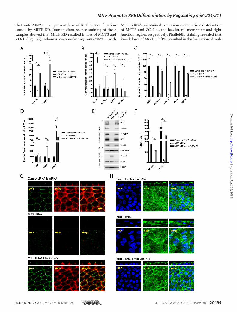
that miR-204/211 can prevent loss of RPE barrier functioncaused by MITF KD. Immunofluorescence staining of thesesamples showed that MITF KD resulted in loss of MCT3 andZO-1 (Fig. 5G), whereas co-transfecting miR-204/211 with
MITF siRNAmaintained expression and polarized distributionof MCT3 and ZO-1 to the basolateral membrane and tightjunction region, respectively. Phalloidin staining revealed thatknockdown ofMITF in hfRPE resulted in the formation ofmul-
MITF Promotes RPE Differentiation by Regulating miR-204/211
JUNE 8, 2012 • VOLUME 287 • NUMBER 24 JOURNAL OF BIOLOGICAL CHEMISTRY 20499
by guest on April 26, 2019
http://ww
w.jbc.org/
Dow
nloaded from
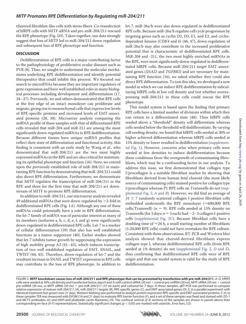
tilayered fibroblast-like cells with stress fibers. Co-transfectionof hfRPE cells withMITF siRNA and pre-miR-204/211 rescuedthe RPE phenotype (Fig. 5H). Taken together, our data stronglysuggest that loss of MITF led to miR-204/211 down-regulationand subsequent loss of RPE phenotype and function.
DISCUSSION
Dedifferentiation of RPE cells is a major contributing factorto the pathophysiology of proliferative ocular diseases such asPVR (8). Thus, we sought to understand the molecular mecha-nisms underlying RPE dedifferentiation and identify potentialtherapeutics that could inhibit this process. We focused oursearch to microRNAs because they are important regulators ofgene expression and havewell established roles inmany biolog-ical processes including development and differentiation (17,34–37). Previously, we and others demonstrated that RPE cellsat the free edge of an intact monolayer can proliferate andmigrate, giving rise tomesenchymal cells that express low levelsof RPE-specific proteins and increased levels of EMT-associ-ated proteins (28, 38). Microarray analysis comparing themiRNA profile of these samples with that of differentiated RPEcells revealed that miR-204 and miR-211 are among the mostsignificantly down-regulatedmiRNAs in RPE dedifferentiation.Because different tissues have unique miRNA profiles thatreflect their state of differentiation and functional activity, thisfinding is consistent with an early study by Wang et al., whodemonstrated that miR-204/211 are the two most highlyexpressedmiRNAs in theRPE and are also critical formaintain-ing its epithelial phenotype and function (16). Here, we extendupon the previously established role of miR-204/211 in main-tainingRPE function by demonstrating thatmiR-204/211 couldalso direct RPE differentiation. Furthermore, we demonstratethat MITF regulates the transcription of miR-204/211 in theRPE and show for the first time that miR-204/211 act down-stream of MITF to promote RPE differentiation.In addition tomiR-204/211, ourmicroarray analysis revealed
49 additional miRNAs that were down-regulated by �2-fold indedifferentiated RPE cells (Fig. 1A). Although any one of thesemiRNAs could potentially have a role in RPE differentiation,the let-7 family of miRNA was of particular interest as many ofits members (isoforms a, b, c, d, e, f, and g) were significantlydown-regulated in dedifferentiated RPE cells. Let-7 is a markerof cellular differentiation (39) that also has well establishedfunctions as a tumor suppressor (40). Earlier studies showedthat let-7 inhibits tumor growth by suppressing the expressionof high mobility group A2 (41–43), which induces transcrip-tion of two well established regulators of EMT, SNAIL, andTWIST (44, 45). Therefore, down-regulation of let-7 and theresultant increase in SNAIL andTWISTexpression inRPE cellsmay contribute to the loss of RPE phenotype. In addition to
let-7, miR-26a/b were also down-regulated in dedifferentiatedRPE cells. BecausemiR-26a/b regulate cell cycle progression bytargeting genes such as cyclin D2, D3, E1, and E2, and cyclin-dependent kinases (CDK4 and 6) (46, 47), down-regulation ofmiR-26a/b may also contribute to the increased proliferativepotential that is characteristic of dedifferentiated RPE cells.MiR-204 and -211, the two most highly enriched miRNAs inthe RPE, were most significantly down-regulated in dedifferen-tiated hfRPE cells. Because miR-204/211 target EMT-associ-ated genes (SNAI2 and TGFBR2) and are necessary for main-taining RPE function (16), we asked whether they could alsodirect RPE differentiation. To test this idea, we developed a newmodel in which we can induce RPE dedifferentiation by subcul-turing hfRPE cells at low cell density and test whether overex-pressing miR-204/211 in these cells could rescue the RPEphenotype.This model system is based upon the finding that primary
RPE cells have a limited number of divisions within which theycan return to a differentiated state (48). Thus hfRPE cellsseeded above a “threshold” density will differentiate whereascells seeded below the threshold will dedifferentiate. By varyingcell seeding density, we found that hfRPE cells seeded at 30% orhigher achieved differentiation whereas hfRPE cells seeded at15% density or lower resulted in dedifferentiation (supplemen-tal Fig. 1). However, concerns arise when primary cells wereseeded at low densities to induce dedifferentiation becausethese conditions favor the overgrowth of contaminating fibro-blasts, which may be a confounding factor in our analysis. Toaddress this issue, we first demonstrated that collagen typeI/procollagen is a suitable fibroblast marker by showing thatfibroblasts derived from human fetal choroid (the most likelysource of contaminating cells) stained positive for collagen typeI/procollagen whereas P1 RPE cells on Transwells do not (sup-plemental Fig. 2, A and B). However, we did find an average of31 7 randomly scattered collagen I-positive fibroblast cellsembedded underneath the RPE monolayer (�600,000 RPEcells/Transwell) (n � 9). RPE cells seeded at 15% density onTranswells (for 3 days;n� 3 each) had�2–3 collagen I-positivecells (supplemental Fig. 2C). Because fibroblast cells have adoubling time of �24 h, a small starting number of fibroblasts(1:20,000 RPE cells) could not have overtaken the RPE culture.Consistent with these observations, RT-PCR andWestern blotanalysis showed that choroid-derived fibroblasts expresscollagen type I, whereas dedifferentiated RPE cells (from RPEseeded at 1% density) do not (supplemental Fig. 2, D and E),thus confirming that dedifferentiated RPE cells were of RPEorigin and that our model system is valid for the study of RPEdedifferentiation.
FIGURE 5. MITF knockdown causes loss of miR-204/211 and RPE phenotype that can be prevented by transfection with pre-miR-204/211. A–D, hfRPEcells were seeded at 30% cell density and transfected (twice; days 0 and 3) with control siRNA (30 nM) control pre-miRNA (30 nM), MITF siRNA (30 nM) controlpre-miRNA (30 nM), or MITF siRNA (30 nM) pre-miR-204/211 (15 nM each) and cultured for 7 days. In these samples, qRT-PCR was performed to comparerelative expression of mature miR-204/211 (A), miR-204/211 targets (B), RPE-specific genes (C), and EMT-associated genes (D). E, in a parallel experiment withidentical treatment but grown over 21 days, Western blotting was performed to analyze protein expression RPE-specific and EMT-associated proteins. F–H, inthese samples TER was measured with EVOM (14 and 21 days) to evaluate RPE barrier function (F), and a set of these samples was fixed and stained with ZO-1and MCT3 antibodies (G) and DAPI and phalloidin (actin filaments) (H). The confocal vertical (Z-X) sections of the samples are shown in panels above theircorresponding en-face (X-Y) representations. Statistically significant changes (p � 0.05) are marked with asterisks. Error bars, S.D.
MITF Promotes RPE Differentiation by Regulating miR-204/211
20500 JOURNAL OF BIOLOGICAL CHEMISTRY VOLUME 287 • NUMBER 24 • JUNE 8, 2012
by guest on April 26, 2019
http://ww
w.jbc.org/
Dow
nloaded from
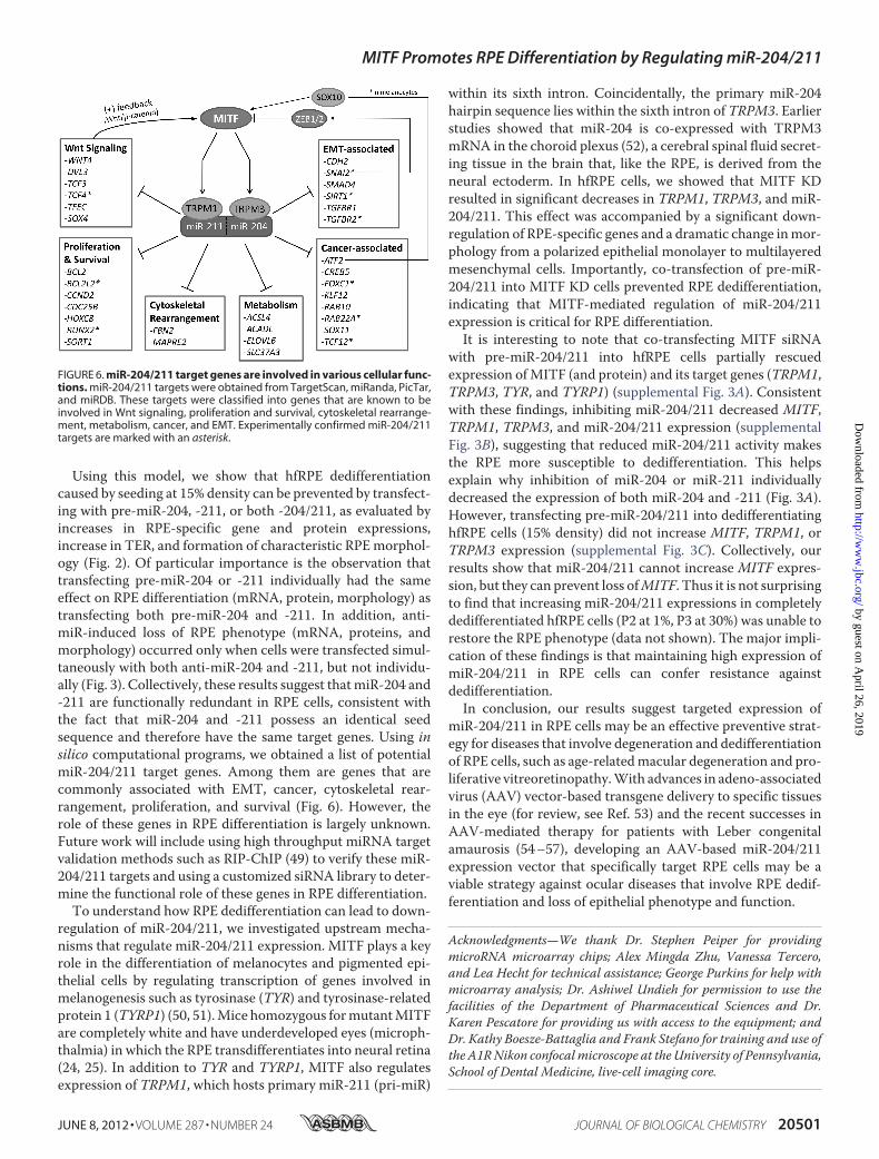
Using this model, we show that hfRPE dedifferentiationcaused by seeding at 15% density can be prevented by transfect-ing with pre-miR-204, -211, or both -204/211, as evaluated byincreases in RPE-specific gene and protein expressions,increase in TER, and formation of characteristic RPEmorphol-ogy (Fig. 2). Of particular importance is the observation thattransfecting pre-miR-204 or -211 individually had the sameeffect on RPE differentiation (mRNA, protein, morphology) astransfecting both pre-miR-204 and -211. In addition, anti-miR-induced loss of RPE phenotype (mRNA, proteins, andmorphology) occurred only when cells were transfected simul-taneously with both anti-miR-204 and -211, but not individu-ally (Fig. 3). Collectively, these results suggest thatmiR-204 and-211 are functionally redundant in RPE cells, consistent withthe fact that miR-204 and -211 possess an identical seedsequence and therefore have the same target genes. Using insilico computational programs, we obtained a list of potentialmiR-204/211 target genes. Among them are genes that arecommonly associated with EMT, cancer, cytoskeletal rear-rangement, proliferation, and survival (Fig. 6). However, therole of these genes in RPE differentiation is largely unknown.Future work will include using high throughput miRNA targetvalidation methods such as RIP-ChIP (49) to verify these miR-204/211 targets and using a customized siRNA library to deter-mine the functional role of these genes in RPE differentiation.To understand how RPE dedifferentiation can lead to down-
regulation of miR-204/211, we investigated upstream mecha-nisms that regulate miR-204/211 expression. MITF plays a keyrole in the differentiation of melanocytes and pigmented epi-thelial cells by regulating transcription of genes involved inmelanogenesis such as tyrosinase (TYR) and tyrosinase-relatedprotein 1 (TYRP1) (50, 51).Mice homozygous formutantMITFare completely white and have underdeveloped eyes (microph-thalmia) in which the RPE transdifferentiates into neural retina(24, 25). In addition to TYR and TYRP1, MITF also regulatesexpression of TRPM1, which hosts primary miR-211 (pri-miR)
within its sixth intron. Coincidentally, the primary miR-204hairpin sequence lies within the sixth intron of TRPM3. Earlierstudies showed that miR-204 is co-expressed with TRPM3mRNA in the choroid plexus (52), a cerebral spinal fluid secret-ing tissue in the brain that, like the RPE, is derived from theneural ectoderm. In hfRPE cells, we showed that MITF KDresulted in significant decreases in TRPM1, TRPM3, and miR-204/211. This effect was accompanied by a significant down-regulation of RPE-specific genes and a dramatic change inmor-phology from a polarized epithelial monolayer to multilayeredmesenchymal cells. Importantly, co-transfection of pre-miR-204/211 into MITF KD cells prevented RPE dedifferentiation,indicating that MITF-mediated regulation of miR-204/211expression is critical for RPE differentiation.It is interesting to note that co-transfecting MITF siRNA
with pre-miR-204/211 into hfRPE cells partially rescuedexpression ofMITF (and protein) and its target genes (TRPM1,TRPM3, TYR, and TYRP1) (supplemental Fig. 3A). Consistentwith these findings, inhibiting miR-204/211 decreased MITF,TRPM1, TRPM3, and miR-204/211 expression (supplementalFig. 3B), suggesting that reduced miR-204/211 activity makesthe RPE more susceptible to dedifferentiation. This helpsexplain why inhibition of miR-204 or miR-211 individuallydecreased the expression of both miR-204 and -211 (Fig. 3A).However, transfecting pre-miR-204/211 into dedifferentiatinghfRPE cells (15% density) did not increase MITF, TRPM1, orTRPM3 expression (supplemental Fig. 3C). Collectively, ourresults show that miR-204/211 cannot increase MITF expres-sion, but they can prevent loss ofMITF. Thus it is not surprisingto find that increasing miR-204/211 expressions in completelydedifferentiated hfRPE cells (P2 at 1%, P3 at 30%) was unable torestore the RPE phenotype (data not shown). The major impli-cation of these findings is that maintaining high expression ofmiR-204/211 in RPE cells can confer resistance againstdedifferentiation.In conclusion, our results suggest targeted expression of
miR-204/211 in RPE cells may be an effective preventive strat-egy for diseases that involve degeneration and dedifferentiationof RPE cells, such as age-relatedmacular degeneration and pro-liferative vitreoretinopathy.With advances in adeno-associatedvirus (AAV) vector-based transgene delivery to specific tissuesin the eye (for review, see Ref. 53) and the recent successes inAAV-mediated therapy for patients with Leber congenitalamaurosis (54–57), developing an AAV-based miR-204/211expression vector that specifically target RPE cells may be aviable strategy against ocular diseases that involve RPE dedif-ferentiation and loss of epithelial phenotype and function.
Acknowledgments—We thank Dr. Stephen Peiper for providingmicroRNA microarray chips; Alex Mingda Zhu, Vanessa Tercero,and Lea Hecht for technical assistance; George Purkins for help withmicroarray analysis; Dr. Ashiwel Undieh for permission to use thefacilities of the Department of Pharmaceutical Sciences and Dr.Karen Pescatore for providing us with access to the equipment; andDr. Kathy Boesze-Battaglia and Frank Stefano for training and use ofthe A1RNikon confocalmicroscope at the University of Pennsylvania,School of Dental Medicine, live-cell imaging core.
FIGURE 6. miR-204/211 target genes are involved in various cellular func-tions. miR-204/211 targets were obtained from TargetScan, miRanda, PicTar,and miRDB. These targets were classified into genes that are known to beinvolved in Wnt signaling, proliferation and survival, cytoskeletal rearrange-ment, metabolism, cancer, and EMT. Experimentally confirmed miR-204/211targets are marked with an asterisk.
MITF Promotes RPE Differentiation by Regulating miR-204/211
JUNE 8, 2012 • VOLUME 287 • NUMBER 24 JOURNAL OF BIOLOGICAL CHEMISTRY 20501
by guest on April 26, 2019
http://ww
w.jbc.org/
Dow
nloaded from
REFERENCES1. Strauss, O. (2005) The retinal pigment epithelium in visual function.
Physiol. Rev. 85, 845–8812. Adijanto, J., Banzon, T., Jalickee, S., Wang, N. S., and Miller, S. S. (2009)
CO2-induced ion and fluid transport in human retinal pigment epithe-lium. J. Gen. Physiol. 133, 603–622
3. Lamb, T. D., and Pugh, E. N., Jr. (2004) Dark adaptation and the retinoidcycle of vision. Prog. Retin. Eye Res. 23, 307–380
4. Kevany, B. M., and Palczewski, K. (2010) Phagocytosis of retinal rod andcone photoreceptors. Physiology 25, 8–15
5. Steele, F. R., Chader, G. J., Johnson, L. V., and Tombran-Tink, J. (1993)Pigment epithelium-derived factor: neurotrophic activity and identifica-tion as a member of the serine protease inhibitor gene family. Proc. Natl.Acad. Sci. U.S.A. 90, 1526–1530
6. Witmer, A. N., Vrensen, G. F., Van Noorden, C. J., and Schlingemann,R. O. (2003) Vascular endothelial growth factors and angiogenesis in eyedisease. Prog. Retin. Eye Res. 22, 1–29
7. Sparrow, J. R., Hicks, D., and Hamel, C. P. (2010) The retinal pigmentepithelium in health and disease. Curr. Mol. Med. 10, 802–823
8. Hiscott, P., Sheridan, C., Magee, R. M., and Grierson, I. (1999) Matrix andthe retinal pigment epithelium in proliferative retinal disease. Prog. Retin.Eye Res. 18, 167–190
9. Kalluri, R., andWeinberg, R. A. (2009) The basics of epithelial-mesenchy-mal transition. J. Clin. Invest. 119, 1420–1428
10. Bartel, D. P. (2009) MicroRNAs: target recognition and regulatory func-tions. Cell 136, 215–233
11. Zavadil, J., Narasimhan, M., Blumenberg, M., and Schneider, R. J. (2007)Transforming growth factor-� and microRNA: mRNA regulatory net-works in epithelial plasticity. Cells Tissues Organs 185, 157–161
12. Gregory, P. A., Bert, A. G., Paterson, E. L., Barry, S. C., Tsykin, A., Farshid,G., Vadas,M.A., Khew-Goodall, Y., andGoodall, G. J. (2008) ThemiR-200family and miR-205 regulate epithelial to mesenchymal transition by tar-geting ZEB1 and SIP1. Nat. Cell Biol. 10, 593–601
13. Liang, Y., Ridzon, D., Wong, L., and Chen, C. (2007) Characterization ofmicroRNA expression profiles in normal human tissues. BMC Genomics8, 166
14. Fineberg, S. K., Kosik, K. S., and Davidson, B. L. (2009) MicroRNAs po-tentiate neural development. Neuron 64, 303–309
15. Cheng, Y., Liu, X., Yang, J., Lin, Y., Xu, D. Z., Lu, Q., Deitch, E. A., Huo, Y.,Delphin, E. S., and Zhang, C. (2009)MicroRNA-145, a novel smoothmus-cle cell phenotypic marker and modulator, controls vascular neointimallesion formation. Circ. Res. 105, 158–166
16. Wang, F. E., Zhang, C., Maminishkis, A., Dong, L., Zhi, C., Li, R., Zhao, J.,Majerciak, V., Gaur, A. B., Chen, S., and Miller, S. S. (2010) MicroRNA-204/211 alters epithelial physiology. FASEB J. 24, 1552–1571
17. Cordes, K. R., Sheehy, N. T., White, M. P., Berry, E. C., Morton, S. U.,Muth, A. N., Lee, T. H., Miano, J. M., Ivey, K. N., and Srivastava, D. (2009)miR-145 and miR-143 regulate smooth muscle cell fate and plasticity.Nature 460, 705–710
18. Cheng, L. C., Pastrana, E., Tavazoie, M., and Doetsch, F. (2009) miR-124regulates adult neurogenesis in the subventricular zone stem cell niche.Nat. Neurosci. 12, 399–408
19. Yoo, A. S., Sun, A. X., Li, L., Shcheglovitov, A., Portmann, T., Li, Y., Lee-Messer, C., Dolmetsch, R. E., Tsien, R. W., and Crabtree, G. R. (2011)MicroRNA-mediated conversion of human fibroblasts to neurons.Nature476, 228–231
20. Kosik, K. S. (2010) MicroRNAs and cellular phenotypy. Cell 143, 21–2621. Mazar, J., DeYoung, K., Khaitan, D.,Meister, E., Almodovar, A., Goydos, J.,
Ray, A., and Perera, R. J. (2010) The regulation of miRNA-211 expressionand its role in melanoma cell invasiveness. PLoS One 5, e13779
22. Steingrímsson, E., Copeland, N. G., and Jenkins, N. A. (2004)Melanocytesand the microphthalmia transcription factor network. Annu. Rev. Genet.38, 365–411
23. Tsukiji, N., Nishihara, D., Yajima, I., Takeda, K., Shibahara, S., andYamamoto, H. (2009) Mitf functions as an in ovo regulator for cell differ-entiation and proliferation during development of the chick RPE. Dev.Biol. 326, 335–346
24. Hodgkinson, C. A., Moore, K. J., Nakayama, A., Steingrímsson, E., Cope-land, N. G., Jenkins, N. A., and Arnheiter, H. (1993) Mutations at themouse microphthalmia locus are associated with defects in a gene encod-ing a novel basic-helix-loop-helix-zipper protein. Cell 74, 395–404
25. Bumsted, K. M., and Barnstable, C. J. (2000) Dorsal retinal pigment epi-thelium differentiates as neural retina in the microphthalmia (mi/mi)mouse. Invest. Ophthalmol. Vis. Sci. 41, 903–908
26. Maminishkis, A., Chen, S., Jalickee, S., Banzon, T., Shi, G., Wang, F. E.,Ehalt, T., Hammer, J. A., and Miller, S. S. (2006) Confluent monolayers ofcultured human retinal pigment epithelium exhibit morphology andphysiology of native tissue. Invest. Ophthalmol. Vis. Sci. 47, 3612–3624
27. Strunnikova, N. V., Maminishkis, A., Barb, J. J., Wang, F., Zhi, C., Sergeev,Y., Chen, W., Edwards, A. O., Stambolian, D., Abecasis, G., Swaroop, A.,Munson, P. J., and Miller, S. S. (2010) Transcriptome analysis and molec-ular signature of human retinal pigment epithelium.Hum.Mol. Genet. 19,2468–2486
28. Gallagher-Colombo, S., Maminishkis, A., Tate, S., Grunwald, G. B., andPhilp, N. J. (2010)Modulation ofMCT3 expression during wound healingof the retinal pigment epithelium. Invest. Ophthalmol. Vis. Sci. 51,5343–5350
29. Tusher, V. G., Tibshirani, R., and Chu, G. (2001) Significance analysis ofmicroarrays applied to the ionizing radiation response. Proc. Natl. Acad.Sci. U.S.A. 98, 5116–5121
30. Dieffenbach, C. W., Lowe, T. M., and Dveksler, G. S. (1993) General con-cepts for PCR primer design. PCR Methods Appl. 3, S30–37
31. Schmittgen, T. D., and Livak, K. J. (2008) Analyzing real-time PCR data bythe comparative C(T) method. Nat. Protoc. 3, 1101–1108
32. Baek, D., Villén, J., Shin, C., Camargo, F. D., Gygi, S. P., and Bartel, D. P.(2008) The impact of microRNAs on protein output. Nature 455, 64–71
33. Korpal, M., Lee, E. S., Hu, G., and Kang, Y. (2008) The miR-200 familyinhibits epithelial-mesenchymal transition and cancer cell migration bydirect targeting of E-cadherin transcriptional repressors ZEB1 and ZEB2.J. Biol. Chem. 283, 14910–14914
34. Chen, C. Z., Li, L., Lodish, H. F., and Bartel, D. P. (2004) MicroRNAsmodulate hematopoietic lineage differentiation. Science 303, 83–86
35. Makeyev, E. V., Zhang, J., Carrasco, M. A., and Maniatis, T. (2007) ThemicroRNA miR-124 promotes neuronal differentiation by triggeringbrain-specific alternative pre-mRNA splicing.Mol. Cell 27, 435–448
36. Lu, J., Guo, S., Ebert, B. L., Zhang, H., Peng, X., Bosco, J., Pretz, J.,Schlanger, R., Wang, J. Y., Mak, R. H., Dombkowski, D. M., Preffer, F. I.,Scadden, D. T., and Golub, T. R. (2008) MicroRNA-mediated control ofcell fate inmegakaryocyte-erythrocyte progenitors.Dev. Cell 14, 843–853
37. King, I. N., Qian, L., Liang, J., Huang, Y., Shieh, J. T., Kwon, C., and Sriv-astava, D. (2011) A genome-wide screen reveals a role for microRNA-1 inmodulating cardiac cell polarity. Dev. Cell 20, 497–510
38. Tamiya, S., Liu, L., and Kaplan, H. J. (2010) Epithelial-mesenchymal tran-sition and proliferation of retinal pigment epithelial cells initiated uponloss of cell-cell contact. Invest. Ophthalmol. Vis. Sci. 51, 2755–2763
39. Thomson, J. M., Parker, J., Perou, C. M., and Hammond, S. M. (2004) Acustom microarray platform for analysis of microRNA gene expression.Nat. Methods 1, 47–53
40. Park, S. M., Shell, S., Radjabi, A. R., Schickel, R., Feig, C., Boyerinas, B.,Dinulescu, D. M., Lengyel, E., and Peter, M. E. (2007) Let-7 prevents earlycancer progression by suppressing expression of the embryonic geneHMGA2. Cell Cycle 6, 2585–2590
41. Lee, Y. S., and Dutta, A. (2007) The tumor suppressor microRNA let-7represses the HMGA2 oncogene. Genes Dev. 21, 1025–1030
42. Mayr, C., Hemann, M. T., and Bartel, D. P. (2007) Disrupting the pairingbetween let-7 and Hmga2 enhances oncogenic transformation. Science315, 1576–1579
43. Shell, S., Park, S.M., Radjabi, A. R., Schickel, R., Kistner, E. O., Jewell, D. A.,Feig, C., Lengyel, E., and Peter, M. E. (2007) Let-7 expression defines twodifferentiation stages of cancer. Proc. Natl. Acad. Sci. U.S.A. 104,11400–11405
44. Tan, E. J., Thuault, S., Caja, L., Carletti, T., Heldin, C. H., and Moustakas,A. (2012) Regulation of transcription factor Twist expression by the DNAarchitectural protein high mobility group A2 during epithelial-to-mesen-chymal transition. J. Biol. Chem. 287, 7134–7145
MITF Promotes RPE Differentiation by Regulating miR-204/211
20502 JOURNAL OF BIOLOGICAL CHEMISTRY VOLUME 287 • NUMBER 24 • JUNE 8, 2012
by guest on April 26, 2019
http://ww
w.jbc.org/
Dow
nloaded from

45. Thuault, S., Tan, E. J., Peinado, H., Cano, A., Heldin, C. H., andMoustakas,A. (2008) HMGA2 and Smads co-regulate SNAIL1 expression during in-duction of epithelial-to-mesenchymal transition. J. Biol. Chem. 283,33437–33446
46. Lu, J., He,M. L.,Wang, L., Chen, Y., Liu, X., Dong,Q., Chen, Y. C., Peng, Y.,Yao, K. T., Kung, H. F., and Li, X. P. (2011) MiR-26a inhibits cell growthand tumorigenesis of nasopharyngeal carcinoma through repression ofEZH2. Cancer Res. 71, 225–233
47. Zhu, Y., Lu, Y., Zhang, Q., Liu, J. J., Li, T. J., Yang, J. R., Zeng, C., andZhuang, S. M. (2011) MicroRNA-26a/b and their host genes cooperate toinhibit the G1/S transition by activating the pRb protein. Nucleic AcidsRes., in press
48. Grisanti, S., and Guidry, C. (1995) Transdifferentiation of retinal pigmentepithelial cells from epithelial to mesenchymal phenotype. Invest. Oph-thalmol. Vis. Sci. 36, 391–405
49. Keene, J. D., Komisarow, J. M., and Friedersdorf, M. B. (2006) RIP-Chip:the isolation and identification of mRNAs, microRNAs and protein com-ponents of ribonucleoprotein complexes from cell extracts.Nat. Protoc. 1,302–307
50. Yasumoto, K., Yokoyama, K., Takahashi, K., Tomita, Y., and Shibahara, S.(1997) Functional analysis of microphthalmia-associated transcriptionfactor in pigment cell-specific transcription of the human tyrosinase fam-ily genes. J. Biol. Chem. 272, 503–509
51. Cheli, Y., Ohanna, M., Ballotti, R., and Bertolotto, C. (2010) Fifteen-yearquest for microphthalmia-associated transcription factor target genes.Pigment Cell Melanoma Res. 23, 27–40
52. Deo, M., Yu, J. Y., Chung, K. H., Tippens, M., and Turner, D. L. (2006)Detection of mammalian microRNA expression by in situ hybridization
with RNA oligonucleotides. Dev. Dyn. 235, 2538–254853. Liu, M. M., Tuo, J., and Chan, C. C. (2011) Gene therapy for ocular dis-
eases. Br. J. Ophthalmol. 95, 604–61254. Bainbridge, J. W., Smith, A. J., Barker, S. S., Robbie, S., Henderson, R.,
Balaggan, K., Viswanathan, A., Holder, G. E., Stockman, A., Tyler, N.,Petersen-Jones, S., Bhattacharya, S. S., Thrasher, A. J., Fitzke, F.W., Carter,B. J., Rubin, G. S., Moore, A. T., and Ali, R. R. (2008) Effect of gene therapyon visual function in Leber’s congenital amaurosis. N. Engl. J. Med. 358,2231–2239
55. Cideciyan, A. V., Hauswirth, W.W., Aleman, T. S., Kaushal, S., Schwartz,S. B., Boye, S. L., Windsor, E. A., Conlon, T. J., Sumaroka, A., Pang, J. J.,Roman, A. J., Byrne, B. J., and Jacobson, S. G. (2009) Human RPE65 genetherapy for Leber congenital amaurosis: persistence of early visual im-provements and safety at 1 year. Hum. Gene Ther. 20, 999–1004
56. Maguire, A. M., Simonelli, F., Pierce, E. A., Pugh, E. N., Jr., Mingozzi, F.,Bennicelli, J., Banfi, S., Marshall, K. A., Testa, F., Surace, E. M., Rossi, S.,Lyubarsky, A., Arruda, V. R., Konkle, B., Stone, E., Sun, J., Jacobs, J.,Dell’Osso, L., Hertle, R., Ma, J. X., Redmond, T. M., Zhu, X., Hauck, B.,Zelenaia, O., Shindler, K. S., Maguire, M. G., Wright, J. F., Volpe, N. J.,McDonnell, J. W., Auricchio, A., High, K. A., and Bennett, J. (2008) Safetyand efficacy of gene transfer for Leber’s congenital amaurosis. N. Engl.J. Med. 358, 2240–2248
57. Simonelli, F., Maguire, A. M., Testa, F., Pierce, E. A., Mingozzi, F., Benni-celli, J. L., Rossi, S., Marshall, K., Banfi, S., Surace, E. M., Sun, J., Redmond,T. M., Zhu, X., Shindler, K. S., Ying, G. S., Ziviello, C., Acerra, C., Wright,J. F., McDonnell, J. W., High, K. A., Bennett, J., and Auricchio, A. (2010)Gene therapy for Leber’s congenital amaurosis is safe and effectivethrough 1.5 years after vector administration.Mol. Ther. 18, 643–650
MITF Promotes RPE Differentiation by Regulating miR-204/211
JUNE 8, 2012 • VOLUME 287 • NUMBER 24 JOURNAL OF BIOLOGICAL CHEMISTRY 20503
by guest on April 26, 2019
http://ww
w.jbc.org/
Dow
nloaded from

Grunwald and Nancy J. PhilpJeffrey Adijanto, John J. Castorino, Zi-Xuan Wang, Arvydas Maminishkis, Gerald B.
Expressionof Human Retinal Pigment Epithelium (RPE) by Regulating microRNAs-204/211
Microphthalmia-associated Transcription Factor (MITF) Promotes Differentiation
doi: 10.1074/jbc.M112.354761 originally published online April 20, 20122012, 287:20491-20503.J. Biol. Chem.
10.1074/jbc.M112.354761Access the most updated version of this article at doi:
Alerts:
When a correction for this article is posted•
When this article is cited•
to choose from all of JBC's e-mail alertsClick here
Supplemental material:
http://www.jbc.org/content/suppl/2012/04/20/M112.354761.DC1
http://www.jbc.org/content/287/24/20491.full.html#ref-list-1
This article cites 56 references, 19 of which can be accessed free at
by guest on April 26, 2019
http://ww
w.jbc.org/
Dow
nloaded from