Embed Size (px)
Citation preview
Molekulare Diagnostik
1Institute for Genomics and Bioinformatics, TU Graz / Austria Dr. Andreas Prokesch
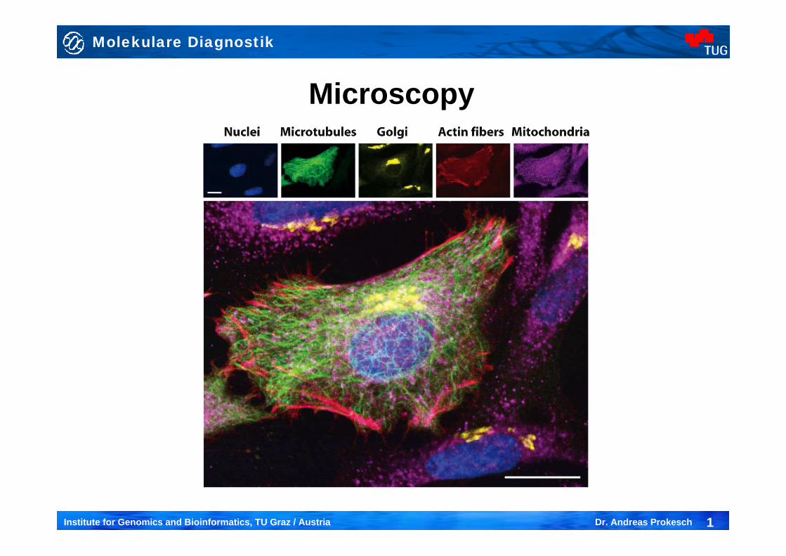
Microscopy
Molekulare Diagnostik
2Institute for Genomics and Bioinformatics, TU Graz / Austria Dr. Andreas Prokesch
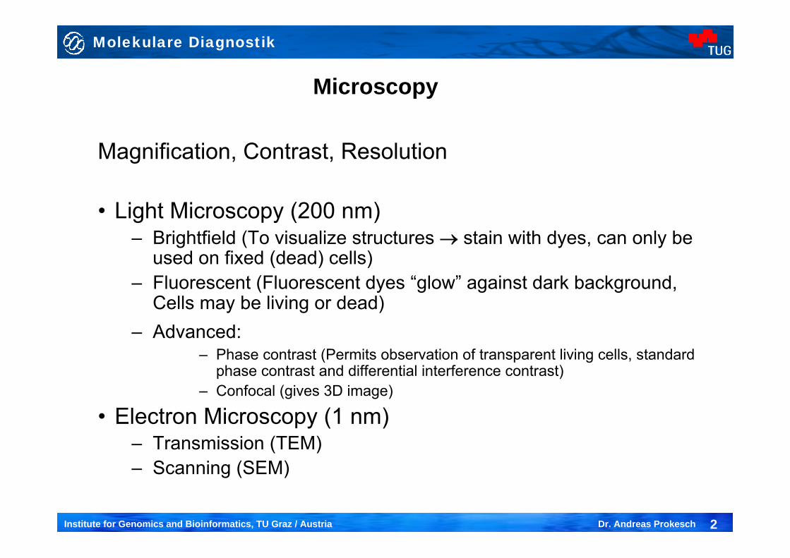
Microscopy
Magnification, Contrast, Resolution
• Light Microscopy (200 nm)– Brightfield (To visualize structures → stain with dyes, can only be
used on fixed (dead) cells)– Fluorescent (Fluorescent dyes “glow” against dark background,
Cells may be living or dead)– Advanced:
– Phase contrast (Permits observation of transparent living cells, standard phase contrast and differential interference contrast)
– Confocal (gives 3D image)
• Electron Microscopy (1 nm)– Transmission (TEM)– Scanning (SEM)
Molekulare Diagnostik
3Institute for Genomics and Bioinformatics, TU Graz / Austria Dr. Andreas Prokesch
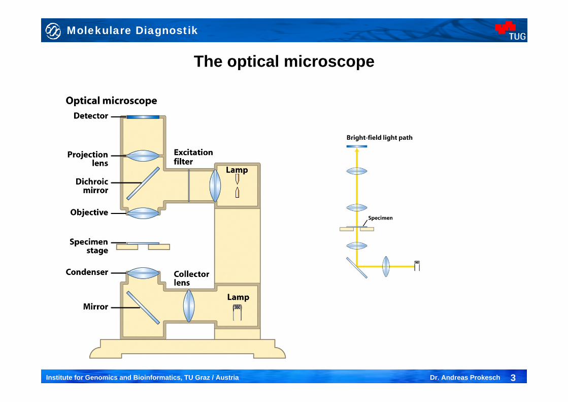
The optical microscope
Molekulare Diagnostik
4Institute for Genomics and Bioinformatics, TU Graz / Austria Dr. Andreas Prokesch
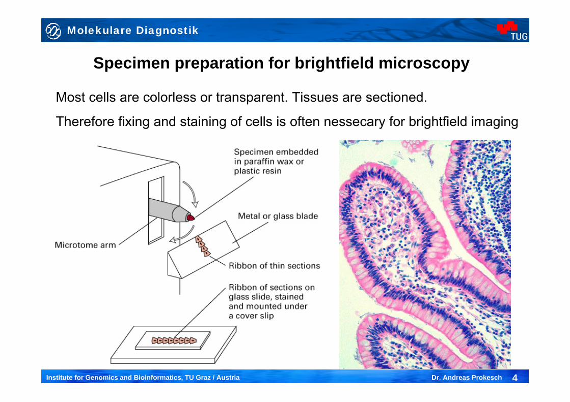
Specimen preparation for brightfield microscopy
Most cells are colorless or transparent. Tissues are sectioned.
Therefore fixing and staining of cells is often nessecary for brightfield imaging
Molekulare Diagnostik
5Institute for Genomics and Bioinformatics, TU Graz / Austria Dr. Andreas Prokesch
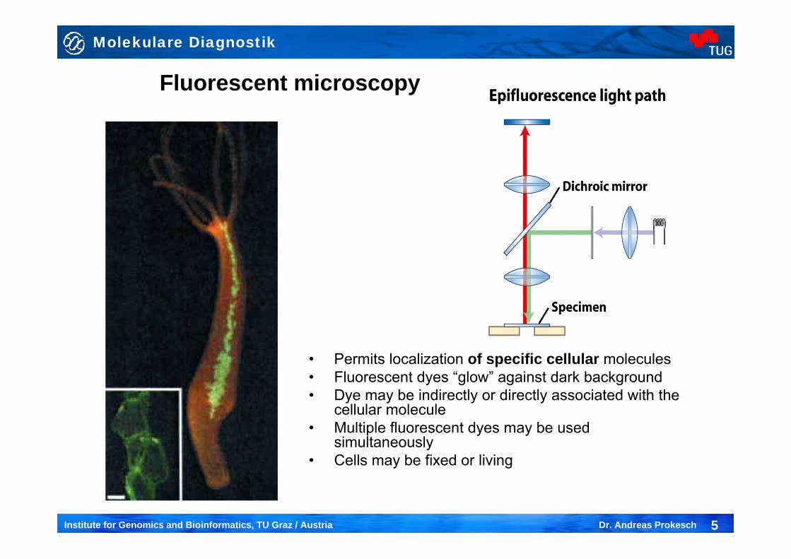
Fluorescent microscopy
• Permits localization of specific cellular molecules• Fluorescent dyes “glow” against dark background• Dye may be indirectly or directly associated with the
cellular molecule• Multiple fluorescent dyes may be used
simultaneously• Cells may be fixed or living

Molekulare Diagnostik
6Institute for Genomics and Bioinformatics, TU Graz / Austria Dr. Andreas Prokesch
Immunofluorescense microscopy3T
3-L1
cel
ls d
8
DAPI Anti-ArxesAnti-Calnexin(ER) Merge
DAPI Anti-ArxesAnti-Pparγ Merge
bar: 10µm
Molekulare Diagnostik
7Institute for Genomics and Bioinformatics, TU Graz / Austria Dr. Andreas Prokesch
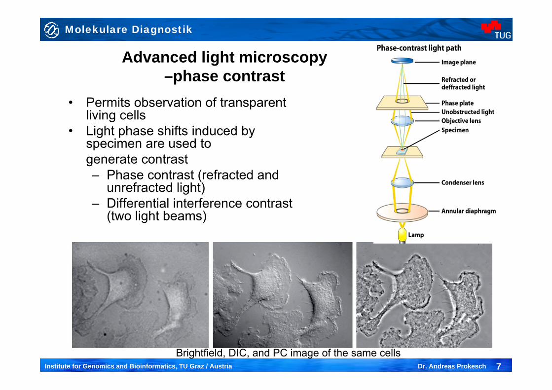
Advanced light microscopy –phase contrast
• Permits observation of transparent living cells
• Light phase shifts induced by specimen are used to generate contrast– Phase contrast (refracted and
unrefracted light)– Differential interference contrast
(two light beams)
Brightfield, DIC, and PC image of the same cells
Molekulare Diagnostik
8Institute for Genomics and Bioinformatics, TU Graz / Austria Dr. Andreas Prokesch
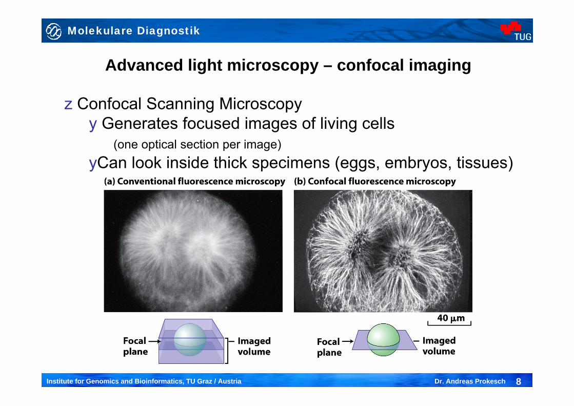
Advanced light microscopy – confocal imaging
z Confocal Scanning Microscopyy Generates focused images of living cells
(one optical section per image)yCan look inside thick specimens (eggs, embryos, tissues)
Molekulare Diagnostik
9Institute for Genomics and Bioinformatics, TU Graz / Austria Dr. Andreas Prokesch
Advanced light microscopy – confocal imaging
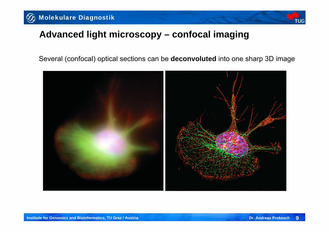
Several (confocal) optical sections can be deconvoluted into one sharp 3D image
Molekulare Diagnostik
10Institute for Genomics and Bioinformatics, TU Graz / Austria Dr. Andreas Prokesch
Molekulare Diagnostik
11Institute for Genomics and Bioinformatics, TU Graz / Austria Dr. Andreas Prokesch
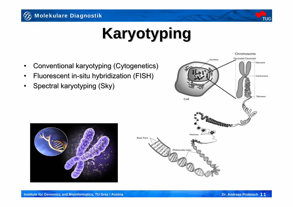
KaryotypingKaryotyping
•• Conventional karyotyping (Cytogenetics)Conventional karyotyping (Cytogenetics)•• Fluorescent inFluorescent in--situ hybridization (FISH)situ hybridization (FISH)•• Spectral karyotyping (Sky)Spectral karyotyping (Sky)
Molekulare Diagnostik
12Institute for Genomics and Bioinformatics, TU Graz / Austria Dr. Andreas Prokesch
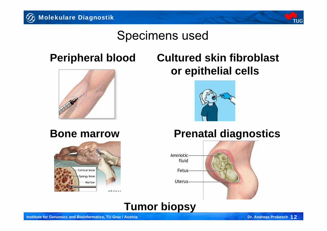
Specimens used
Peripheral blood Cultured skin fibroblast or epithelial cells
Bone marrow Prenatal diagnostics
Tumor biopsy
Molekulare Diagnostik
13Institute for Genomics and Bioinformatics, TU Graz / Austria Dr. Andreas Prokesch
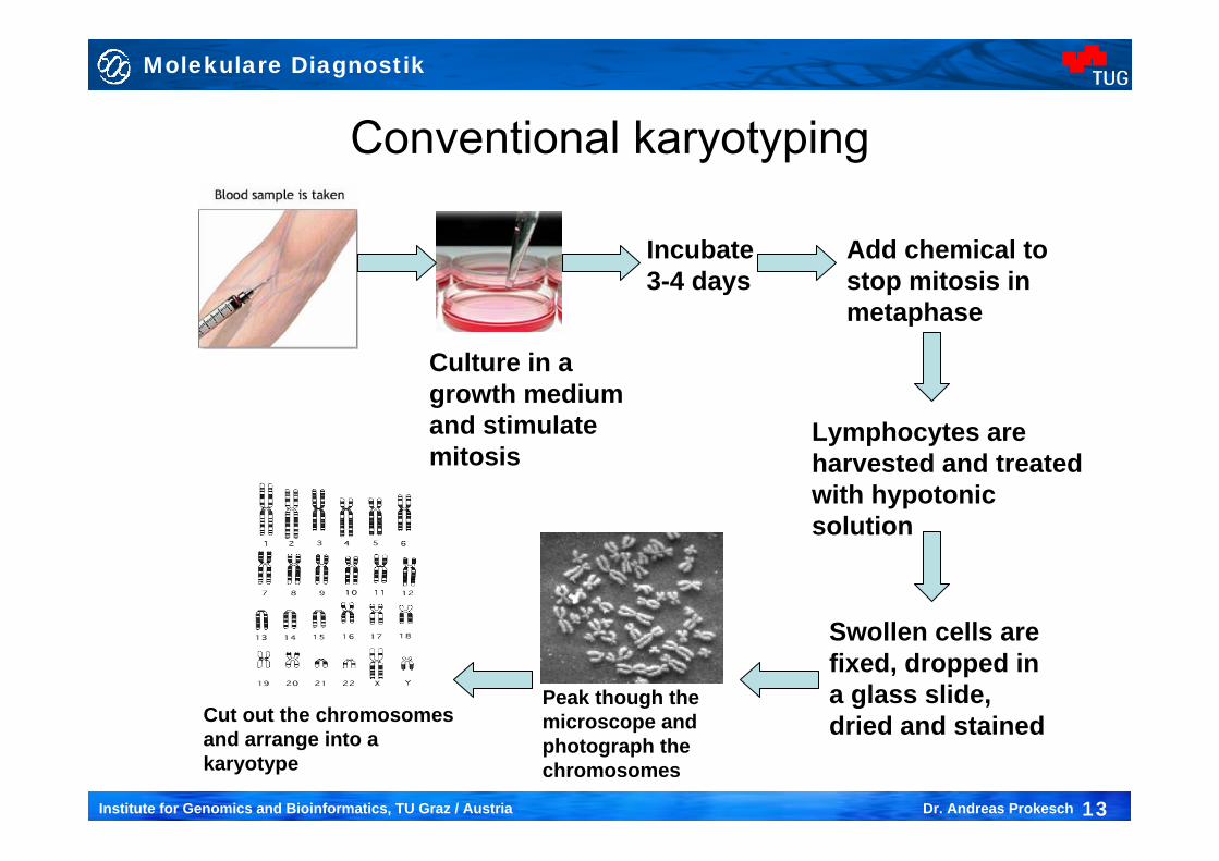
Conventional karyotyping
Incubate 3-4 days
Culture in a growth medium and stimulate mitosis
Add chemical to stop mitosis in metaphase
Lymphocytes are harvested and treated with hypotonic solution
Swollen cells are fixed, dropped in a glass slide, dried and stained
Peak though the microscope and photograph the chromosomes
Cut out the chromosomes and arrange into a karyotype
Molekulare Diagnostik
14Institute for Genomics and Bioinformatics, TU Graz / Austria Dr. Andreas Prokesch
Conventional karyotyping
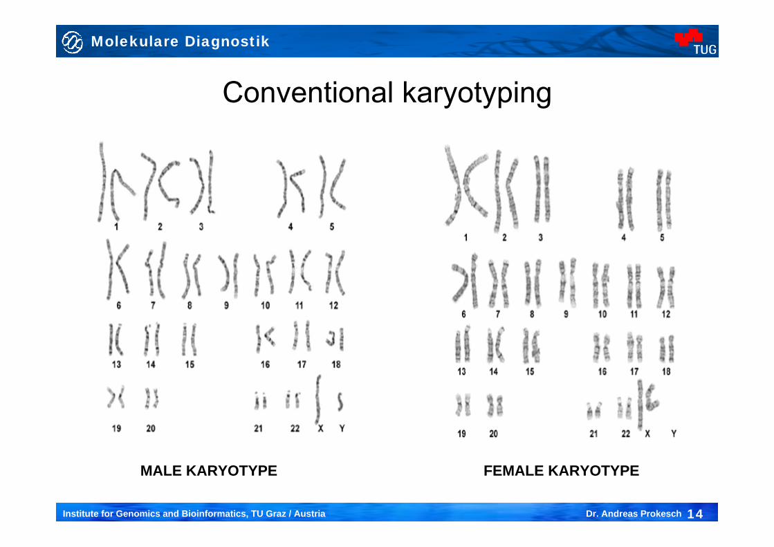
MALE KARYOTYPE FEMALE KARYOTYPE
Molekulare Diagnostik
15Institute for Genomics and Bioinformatics, TU Graz / Austria Dr. Andreas Prokesch
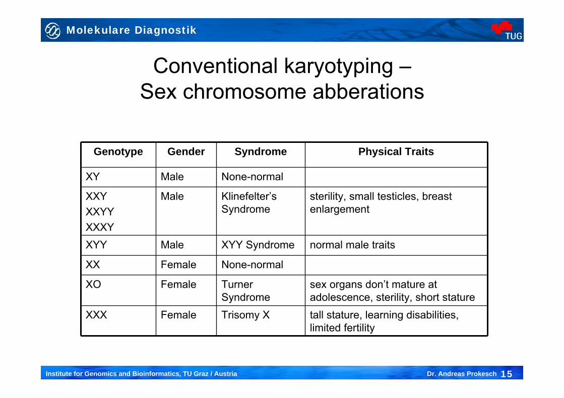
Genotype Gender Syndrome Physical Traits
XY Male None-normal
XXYXXYYXXXY
Male Klinefelter’s Syndrome
sterility, small testicles, breast enlargement
XYY Male XYY Syndrome normal male traits
XX Female None-normal
XO Female Turner Syndrome
sex organs don’t mature at adolescence, sterility, short stature
XXX Female Trisomy X tall stature, learning disabilities, limited fertility
Conventional karyotyping –Sex chromosome abberations
Molekulare Diagnostik
16Institute for Genomics and Bioinformatics, TU Graz / Austria Dr. Andreas Prokesch
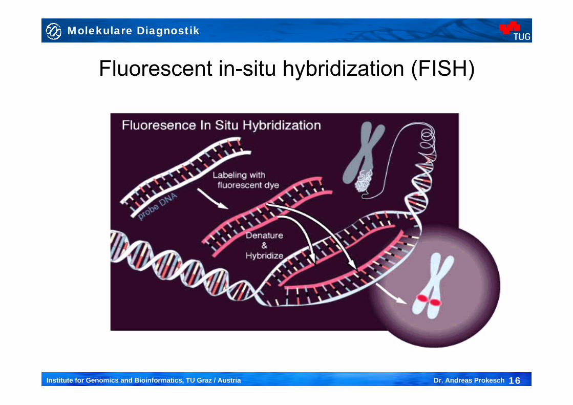
Fluorescent in-situ hybridization (FISH)
Molekulare Diagnostik
17Institute for Genomics and Bioinformatics, TU Graz / Austria Dr. Andreas Prokesch
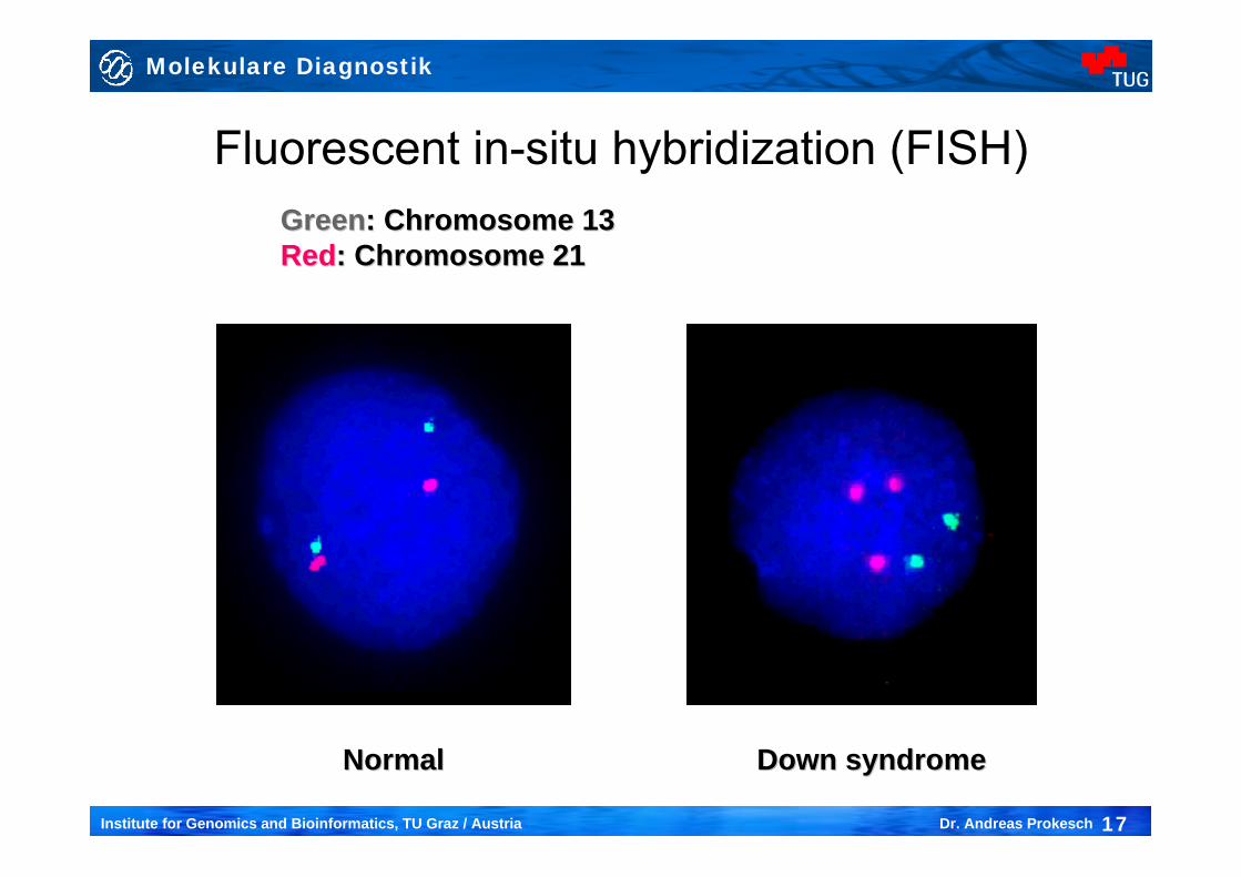
Fluorescent in-situ hybridization (FISH)
NormalNormal Down syndromeDown syndrome
GreenGreen: Chromosome 13: Chromosome 13RedRed: Chromosome 21: Chromosome 21
Molekulare Diagnostik
18Institute for Genomics and Bioinformatics, TU Graz / Austria Dr. Andreas Prokesch
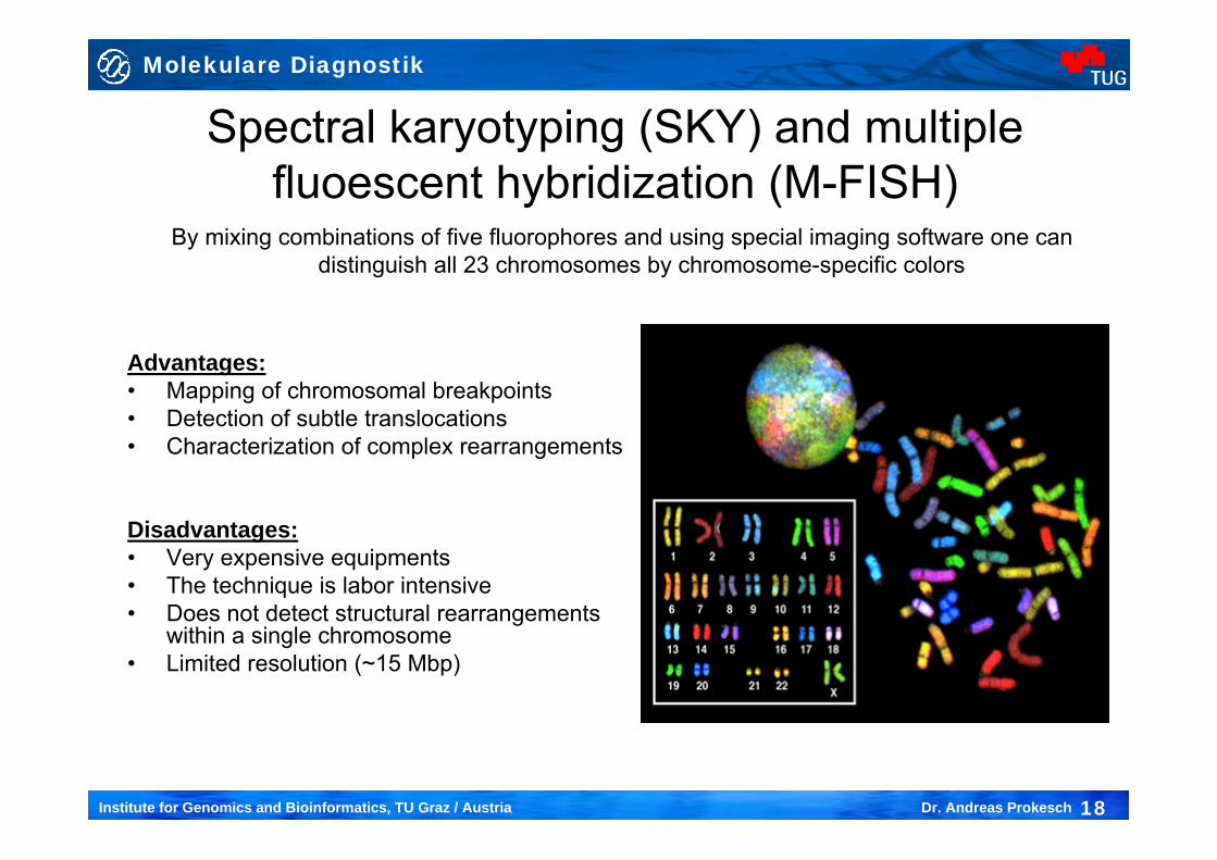
Spectral karyotyping (SKY) and multiple fluoescent hybridization (M-FISH)
By mixing combinations of five fluorophores and using special imaging software one can distinguish all 23 chromosomes by chromosome-specific colors
Advantages:• Mapping of chromosomal breakpoints• Detection of subtle translocations• Characterization of complex rearrangements
Disadvantages:• Very expensive equipments• The technique is labor intensive• Does not detect structural rearrangements
within a single chromosome • Limited resolution (~15 Mbp)
Molekulare Diagnostik
19Institute for Genomics and Bioinformatics, TU Graz / Austria Dr. Andreas Prokesch
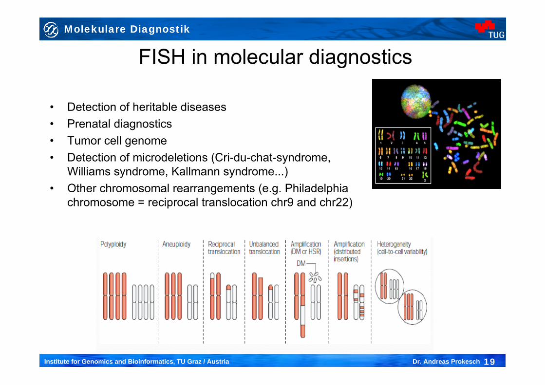
FISH in molecular diagnostics
• Detection of heritable diseases• Prenatal diagnostics• Tumor cell genome • Detection of microdeletions (Cri-du-chat-syndrome,
Williams syndrome, Kallmann syndrome...)• Other chromosomal rearrangements (e.g. Philadelphia
chromosome = reciprocal translocation chr9 and chr22)