Embed Size (px)
Citation preview
ARCHIVES OF BIOCHEMISTRY AND BIOPHYSICS
Vol. 282, No. 2, November 1, pp. 270-274,199O
Microsomal Iron-Dependent NADPH Oxidation: Evidence for the Involvement of Membrane-Bound Nonheme Iron in NADPH Oxidation by Rat Heart Microsomes
Giorgio Minotti**’ and Marco Di Gennaro? Institutes of *General Pathology and tceriatrics, Catholic University School of Medicine, Largo F. Vito I,00168 Rome, Italy
Received March 29,1990, and in revised form June 1,199O
Rat heart microsomes were found to contain nonheme iron and two lines of evidence suggested that this iron was involved in NADPH oxidation. As first evidence, pretreatment of rats with iron gluconate increased mi- crosomal iron content and NADPH oxidation. As sec- ond evidence, treatment of microsomes with nonionic detergent Triton N- 10 1 decreased membrane iron con- tent and NADPH oxidation. Triton N-lOl-solubilized nonheme iron was nondialyzable and ammonium sul- fate-precipitable, indicative of association with pro- tein(s). This protein-bound iron per se did not oxidize NADPH but its addition to detergent-treated micro- somes restored very high rates of NADPH oxidation, that were abolished by inhibiting NADPH-cytochrome P450 reductase with p-hydroxymercuribenzoate. Since heart microsomes djd not contain cytochrome P450, these results suggested that stimulation of NADPH oxidation was mediated by direct electron transfer from reductase to iron. Purified rat heart fer- ritin and hemosiderin did not stimulate NADPH oxida- tion and the stimulation observed with detergent-solu- bilized microsomal iron was much higher than that observed with EDTA-Fe3+, a very effective electron acceptor for the reductase. This suggested that (i) microsomal iron was different from other intracellular iron-storage proteins, and (ii) microsomal iron was un- usually permissive to one-electron transfer from reductase. 0 1990 Academic Press, Inc.
Microsomes contain a flavoenzyme that utilizes NADPH as electron donor and juxtaposed cytochrome (cyt)’ P450 or exogenously added cyt c as electron accep-
i To whom all correspondence should be addressed at: Department of Physiology and Biophysics, Case Western Reserve University School of Medicine, 2119 Abington Road, Cleveland, OH 44106.
*Abbreviations used: cyt, cytochrome; HMW, high molecular weight; p-HMB, p-hydroxymercuribenzoate; 0; superoxide.
270
tors. This enzyme can therefore be referred to as NADPH-cyt P450 or -cyt c reductase (1). Morehouse et al. (2) and Winston et al. (3) have shown that ferric nonheme iron can substitute for cyt P450 or cyt c as elec- tron acceptor for the reductase. Addition of iron to puri- fied reductase will therefore stimulate NADPH oxida- tion (2,3). Chelators must be used to maintain ferric iron in solution (2) but chelators can also influence the redox potential of iron and hence its efficiency as electrophilic substrate for the reductase (2, 3). For example, EDTA facilitates electron transfer from reductase to iron whereas ADP does not (2, 3). NADPH oxidation will therefore be enhanced by EDTA-Fe3+ but not by ADP- Fe3+ (2,3).
Previous studies by Montgomery et al. (4), Thomas and Aust (5), and Minotti (6,7) have shown that micro- somes contain substantial amounts of ferric nonheme iron, which cannot be ascribed to contamination by fer- ritin, hemosiderin, or adventitious iron present in labo- ratory buffers and solutions. In light of previous consid- erations, one possible function of this microsome-bound iron might be to serve as electron acceptor for the reduc- tase, as does EDTA-Fe3+ or any other chelate with an appropriate redox potential. To validate this specula- tion, we have studied NADPH oxidation by rat heart mi- crosomes in which the content of nonheme iron was in- creased or decreased by means of in viva or in vitro treatments. We have chosen to work with heart micro- somes because they are known not to contain cyt P450 (8) which might compete as electron acceptor for the re- ductase and thus interfere with quantitation of reduc- tase-nonheme iron interaction(s) (3).
MATERIALS AND METHODS
Chemicals. Sephadex G-25 and Sephadex G-200 were from Phar- macia (Uppsala, Sweden), and iron gluconate (Ferlixit) and desferri- oxamine B methanesulfonate (Desferal) were from Rhone Poulenc Pharma Italia (Milano, Italy) and Ciba-Geigy (Basel, Switzerland), respectively. EDTA and nicotinamide were purchased from Merck
0003.9861/90 $3.00 Copyright 0 1990 by Academic Press, Inc.
All rights of reproduction in any form reserved.
RAT HEART MICROSOMES, IRON, AND NADPH OXIDATION 271
(Darmstadt, West Germany) and ultrapure ammonium sulfate (Fe, ~0.5 ppm) was purchased from Schwartz-Mann Biotech. (Cleveland, OH). All other chemicals were purchased from Sigma Chemical Co. (St. Louis, MO) with exception of Chelex 100 ion-exchange resin, which was from Bio-Rad (Richmond, CA) and was used to remove contaminating metals from solutions and reagents. The experiments were performed in 50 mM NaCl, carefully adjusted to pH 7.0 just prior to use. This was done to avoid artifactual interactions between iron and most common laboratory buffers (9). Although unbuffered, the reaction mixtures remained at pH 7.0 throughout the experiment time.
Preparation of microsomes. Microsomes were isolated from heart ventricles of six-week-old male Wistar rats (140-g average weight). The isolation procedure was that described by Pederson and Aust (lo), with exception that a 25,000g centrifugation was included prior to ul- tracentrifugation (5). Microsomes were washed twice in 0.15 M KCI- 0.2% nicotinamide and then chromatographed on Sepharose CL-XB as described in details elsewhere (5). Chromatographed microsomes were washed again in the KCl-nicotinamide medium, suspended in 50 mM NaCl-20% glycerol, pH 7.0, and assayed for proteins according to Lowry (11). In selected experiments, microsomes were isolated from rats pretreated with iron gluconate (50 mg of elemental iron im./kg body wt, on alternate days for 1 week) and sacrificed 4 days after the last injection. Microsomes from untreated or iron-treated rats were referred to as “control” or “iron-loaded” microsomes, respectively. Neither control nor iron-loaded microsomes were found to contain cyt P450, measured as described by Omura and Sato (12). Where indi- cated, microsomes were incubated with chelators (EDTA, desferriox- amine, citrate) or detergents (sodium deoxycolate, Trit,on N-101) in 50 mM NaCl, pH 7.0,4”C. After 30 min, the incubation mixtures were centrifuged at 105,OOOg and supernatants were analyzed for reductase and nonheme iron content. In experiments with Triton N-101, sedi- mented microsomes were washed twice in the KCl-nicotinamide me- dium, suspended in the NaCl-glycerol medium, and used for later ex- periments.
Preparation of HMW microsomal iron and iron-free HMW micro- somal extract. Aliquots of the 105,OOOg supernatants of incubations containing iron-loaded microsomes and 0.1% Triton N-101 were as- sayed for nonheme iron, placed in dialysis bags with a 10.kDa exclu- sion limit, and dialyzed overnight against 500 ml of 50 mM NaCl, pH 7.0. The dialyzed sample, which retained all of the iron present in 105,OOOg supernatants, was incubated on ice with increasing amounts of ammonium sulfate and then centrifuged at 10,OOOg. As the satura- tion in ammonium sulfate approached 40’%, all of the iron was found to disappear from 10,OOOg supernatants and precipitate in the form of a brownish pellet. This was dissolved in 50 mM NaC1, dialyzed over- night as described above, and concentrated under nitrogen in Amicon ultrafiltration cells (Danvers, MA) equipped with lo-kDa exclusion limit filters. Following treatment with Chelex 100 to remove any loosely bound iron, these final preparations were adjusted to pH 7.0, assayed for nonheme iron and proteins, and referred to as “HMW mi- crosomal iron.” In other experiments, 105,OOOg supernatants were in- cubated with dithionite (5 pi/ml of a saturat,ed anaerobic solution) and bathophenanthroline (0.25 mM). Following this treatment, which was intended to achieve reduction of Fe”+ and chelation of Fe’+ in a low molecular weight form, samples were processed as described for prepa- ration of HMW microsomal iron. These final preparations contained proteins but not nonheme iron, and were therefore referred to as “iron- free HMW microsomal extract.”
Preparation of iron chelates, ferritin, and hemosiderin. Iron che- lates were prepared as described in Ref. (13). Chelator:iron ratios were 1O:l for ADP-Fe3’ and 1.1:1 for EDTA-Fe3+. Ferritin and hemosid- erin were isolated and purified from the heart of iron-treated rats ac- cording to Kontoghiorghes et al. (14).
Assays. The reductase was measured as cyt c reduction according to Dignam and Strobe1 (l), 1 unit (U) of activity being defined as 1 Fmol cyt c reduced/ml/min. NADPH oxidation was measured as the decrease in absorbance at 340 nm and rates were determined within
C’ 1
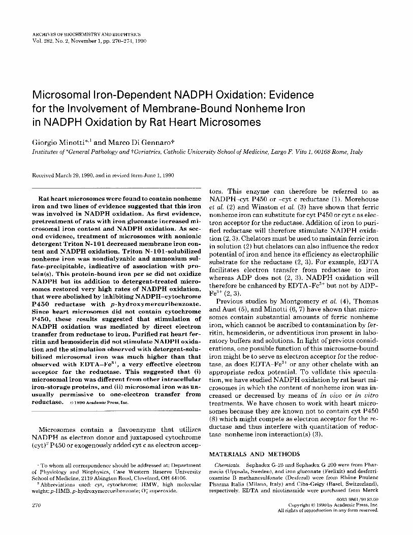
FIG. 1. Nonheme iron, reductase, and NADPH oxidation in control or iron-loaded heart microsomes. (A) Nonheme iron and reductase of control or iron-loaded heart microsomes were measured as described under Materials and Methods. (B) NADPH oxidation was measured in incubations (l-ml final volume) containing microsomes (0.05-0.15 mg protein/ml) in 50 mM NaCl, pH 7.0, 37°C. Reactions were started by addition of NADPH (0.25 mM) and the decrease in absorbance at 340 nm was monitored continuously.
the linear phase of reaction (usually 8-10 min). Values were given as nmoles/milliter/minute to permit direct comparison between different microsome preparations and different experimental conditions. Mi- crosome-bound nonheme iron, HMW microsomal iron, and ferritin- or hemosiderin-bound iron were measured as described by Brumby and Massey (15).
Other experimental conditions are specified in the legends of the figures and the tables. Unless otherwise indicated, values are given as means (-+ standard errors, SE) of three separate experiments that were performed in triplicate with microsomes from 5 to 15 hearts.
RESULTS
As shown in Fig. lA, rat heart microsomes were found to contain nonheme iron, which increased upon pre- treatment of rats with iron gluconate. This treatment did not modify the reductase content of heart micro- somes yet NADPH oxidation was higher with iron- loaded microsomes than with control microsomes, irre- spective of the microsome concentration being used (see Figs. 1A and 1B).
Chelators like EDTA, desferrioxamine, and citrate did not remove iron from iron-loaded microsomes (Table I). Iron was quite effectively removed by detergents, but the use of these substances was made difficult by simulta- neous solubilization of the reductase, which was recov- ered with iron in the 105,OOOg supernatant of incuba- tions. For example, 0.1% (w/v) deoxycolate, an ionic detergent, removed 25% of microsome-bound nonheme iron but also removed 18% of the reductase (Table I). With 0.1% (v/v) Triton N-101, a nonionic detergent, ap- prox 50% of iron was removed from iron-loaded micro- somes without concomitant solubilization of the reduc- tase. Concentrations of Triton N-101 above 0.1% removed higher amounts of nonheme iron but also caused extensive solubilization of the reductase (see also Table I). Keeping these results in mind, we combined in vivo treatment with iron gluconate and in vitro treat-
272 MINOTTI AND DI GENNARO
TABLE I TABLE III
Chelator- or Detergent-Dependent Removal of Iron and Reductase from Iron-Loaded Heart Microsomes
HMW Microsomal Iron-Dependent Oxidation of NADPH by Triton/Control Heart Microsomes
Addition Nonheme iron
(nmol/ml) Reductase (mU/mU
None Chelators (EDTA,
desferrioxamine, citrate) Detergents
Deoxycolate (0.1% w/v) Triton N-101
0.1% v/v 0.5% v/v 1.0% v/v
0.0 0.0
0.0 0.0
5.1 (25) 8.4 (18)
10.6 (52) 0.0 12.9 (63) 11.5 (24) 14.1 (69) 37.5 (81)
Note. Reaction mixtures (3.ml final volume) contained iron-loaded microsomes (1.5 mg protein/ml, corresponding to 20.3 nmol of non- heme iron and 46.5 mU reductase/ml of incubation) in 50 mM NaCl, pH 7.0, 4°C. Where indicated, chelators (1 mM) or detergents were included. After 30 min, reaction mixtures were centrifuged at 105,OOOg and supernatants were assayed for nonheme iron and reductase. Num- bers in parenthesca indicate percentage of iron or reductase removal from microsomes. Values are means of two separate determinations, which agreed by 80-93%.
NADPH oxidation System (nmol/ml/min)
Complete 0.12 * 0.01 +HMW microsomal iron 2.7 20.3 +HMW microsomal iron, -microsomes 0.0 +HMW microsomal iron, +p-HMB 0.0 +Iron-free HMW microsomal extract 0.12 + 0.01
Note. Incubations (l-ml final volume) contained Triton-control mi- crosomes (0.075 mg protein/ml, corresponding to 3.1 mU reductase/ ml) in 50 mM NaCl, pH 7.0, 37°C. Reactions were started by addition of NADPH (0.25 mM). Where indicated HMW microsomal iron (10 nmol Fe, 0.36 mg protein), iron-free HMW microsomal extract (0.36 mg protein) andp-HMB (0.05 mM) were included in the reaction mix- ture.
ment with 0.1% Triton N-101 to obtain microsomes that retained their reductase activity but varied with respect to iron content and hence iron:reductase ratio. As shown in Table II, iron loaded microsomes were those with the highest iron:reductase ratio whereas Tritonliron-loaded microsomes were those in which the iron:reductase ratio was lowered to that of the control microsomes. Finally,
Triton/control microsomes were those with the lowest iron:reductase ratio. Table II shows that rates of NADPH oxidation by these four types of microsomes were directly proportional to iron:reductase ratios. Thus, NADPH oxidation was maximal with iron-loaded microsomes and was minimal with Triton/control mi- crosomes. Intermediate rates of NADPH oxidation were observed with either Triton/iron-loaded or control mi- crosomes, i.e., microsomes which also exhibited interme- diate iron:reductase ratios.
TABLE II
NADPH Oxidation by Heart Microsomes with Different Iron: Reductase Ratios
Microsomes 1ron:reductase
(nmol/mU) NADPH oxidation
(nmol/ml/min)
Iron-loaded Triton/iron-loaded Control Triton/control
0.44 t 0.05 1.4 t 0.13 0.21 +- 0.02 0.71 + 0.09 0.20 -t 0.01 0.65 -+ 0.07 0.04 -t 0.01 0.12 -t 0.01
Note. Treatment of microsomes with 0.1% Triton N-101 decreased total protein recovery by 25-35%. Since the reductase was not solubi- lized under these conditions, detergent-treated microsomes were char- acterized by apparent increase of enzyme activity. For this same rea- son, values of nonheme iron content per milligram of protein of Tri- ton-microsomes were higher than expected on the basis of iron solubilized by the detergent (cf. Table I). To permit direct comparison between untreated and detergent-treated microsomes, values of iron content are therefore given as iron:reductase ratios. NADPH oxida- tion was measured in incubations (l-ml final volume) containing a fixed amount of reductase (3.1 mu) which corresponded to 0.1 or 0.075-0.065 mg protein of untreated or detergent-treated microsomes, respectively. Other conditions for NADPH oxidation assay were as described in the legend to Fig. 1B.
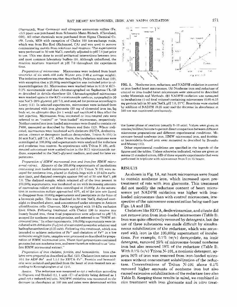
As shown in Table III, NADPH oxidation by Triton/ control microsomes was stimulated by addition of HMW microsomal iron but not by addition of an iron-free HMW microsomal extract. High molecular weight mi- crosomal iron did not promote NADPH oxidation in in- cubations lacking microsomes or in incubations contain- ing microsomes and p-HMB, an inhibitor of the reductase (16) (see also Table III). NADPH oxidation was enhanced by HMW microsomal iron in a concen- tration-dependent manner (Fig. 2). This was also ob- served with EDTA-Fe3+ but the extent of stimulation was minimal as compared to that elicited by equimolar HMW microsomal iron (see also Fig. 2). Neither ferritin, hemosiderin or ADP-Fe”+ were found to stimulate NADPH oxidation within the range of iron concentra- tions examined in this study (see also Fig. 2).
DISCUSSION
Previous studies by Vile and Winterbourn (17) have shown that microsomes can bind exogenously added nonheme iron by virtue of the polar head groups of phos- pholipids. These authors have also observed that chela- tors like EDTA, desferrioxamine, or citrate compete with phospholipids for iron and can therefore be used to obtain iron-free microsomes (17). In our study rat heart microsomes were isolated and washed in iron-free solu-
RAT HEART MICROSOMES, IRON. AND NADPH OXIDATION 273
HMW mlcrosomal Iron
E 5
T 4
0 5 10 15 20
Nonheme Iron (PM)
FIG. 2. The effect of HMW microsomal iron vs other forms of non- heme iron on the oxidation of NADPH by Triton/control heart micro- somes. Incubations (l-ml final volume) were prepared as described in Table III and NADPH oxidation was measured in the presence of in- creasing concentrations of the indicated forms of nonheme iron. For values without bars, the SE was less than 4%.
tions yet they were found to contain nonheme iron which could not be removed by any of the aforementioned che- lators (cf. Fig. IA and Table I). Iron was removed by ionic or nonionic detergents such as deoxycolate or Tri- ton N-101, respectively (cf. Table I). With 0.1% Triton N-101 we have been able to remove substantial amounts of iron without concomitant solubilization of the reduc- tase. This has helped us to manipulate iron to reductase availability and thus establish a possible role for this iron in reductase-catalyzed NADPH oxidation. Two lines of evidence indicate that microsome-bound non- heme iron is involved in NADPH oxidation. As first evi- dence, we have observed that pretreating rats with iron gluconate caused an approximate twofold increase in both iron content and NADPH oxidation, as measured with three different microsome concentrations (cf. Figs. 1A and 1B). As second evidence, we have observed that removing iron by means of 0.1% Triton N-101 decreased NADPH oxidation by either control or iron-loaded mi- crosomes (cf. Table II). By combining in uiuo treatment with iron gluconate and in vitro treatment with 0.1% Triton N-101 we have therefore obtained four different types of microsomes, in which NADPH oxidation was found to increase or decrease wit,h increasing or decreas- ing the availability of iron to reductase (cf. Table II).
Our finding that Triton N-101 decreases the micro- somal oxidation of NADPH should not be considered as unprecedented. For example, Bachur et al. (18) have de- scribed model systems in which Triton N-lOl-treated heart microsomes did not oxidize NADPH unless artifi- cial electron acceptors for the reductase were included in the reaction mixture. This suggested that Triton N- 101 had removed an electron-transport component other than the reductase. Despite some differences in ex- perimental conditions, we propose that our study con- firms and extends those earlier observations, because it indicates that appropriate concentrations of Triton N- 101 will selectively remove nonheme iron involved in
NADPH oxidation (cf. the effects of 0.1% Triton N-101 in Tables I and II).
The nonheme iron solubilized by 0.1% Triton N-101 was nondialyzable and ammonium sulfate-precipitable, indicative of association with protein(s) (cf. Materials and Met,hods). This iron was referred to as HMW micro- somal iron and was examined for its ability to stimulate NADPH oxidation by Triton/control microsomes, i.e., microsomes which contained the reductase but had been cleaned of almost all of their bound iron and thus exhib- ited the lowest rates of NADPH oxidation. The results obtained with this reconstituted system showed that HMW microsomal iron was capable of stimulating NADPH oxidation but failed to promote NADPH oxida- tion in incubations lacking microsomes or in incubations containing microsomes plus p-HMB, a powerful inhibi- tor of the reductase (cf. Table III). These observations provided direct evidence that membrane iron-dependent NADPH oxidation was mediated by the reductase. On the other hand, control experiments showed that NADPH oxidation was not stimulated by an iron-free HMW microsomal extract, which consisted of deter- gent-solubilized protein(s) that had been depleted of their bound iron by combined treatment with a ferric reductant (dithionite) and a ferrous chelator (batho- phenanthroline) (cf. Materials and Methods and Table III). This latter finding confirmed that protein-bound iron was required for stimulation of NADPH oxidation and showed that iron-binding protein(s) per se had no effect on reductase activity.
In this study we have not pursued any further charac- terization of microsomal iron nor have we attempted to establish whether this iron may at least in part be ac- counted for by putative nonheme iron-sulfur protein(s) of the microsomal milieu (4,19,20). Our data, however, strongly support an earlier contention that microsomal iron is different from hemosiderin and ferritin, which are found predominantly in lysosomes or cytosol, respec- tively, but also contaminate microsomes isolated by usual methods of ultracentrifugation (4-7). First, cosed- imentation of lysosomes with microsomes was prevented by including a 25,OOOg centrifugation prior to ultracen- trifugation (cf. Materials and Methods and Ref. (5)). Second, all microsome preparations were chromato- graphed on Sepharose CL-2B, which very effectively re- moves ferritin and several other loosely bound proteins (5-7). Finally, and perhaps more importantly, neither ferritin nor hemosiderin were found to substitute for HMW microsomal iron in stimulation of NADPH oxi- dation (see Fig. 2).
Comparative experiments have shown that micro- somal iron must be highly permissive to one-electron transfer from the reductase. As a matter of fact, stimula- tion of NADPH oxidation by increasing amounts of HMW microsomal iron was much higher than that achieved by equimolar EDTA-Fe”‘, which is a very effective electron acceptor for the reductase (cf. Fig. 2
274 MINOTTI AND DI GENNARO
and Refs. (2, 3)). ADP-Fe3+ did not stimulate NADPH oxidation, in agreement with a previous finding that it may not serve as a substrate for the reductase (cf. Fig. 2 and Refs. (2,3)).
Possible consequences of very facile electron transfer from reductase to juxtaposed nonheme iron remain to be established. One possibility might be that Fe3+ reduction is followed by Fe’+ autoxidation, which thus regenerates membrane-bound Fe3+ and concomitantly reduces mo- lecular oxygen to 0,. In this respect, previous studies with purified microsomal enzymes have shown that the reductase is a rather poor electron donor for molecular oxygen and that formation of 0; is strictly contingent on the intermediacy of cyt P450, which first incorporates one electron from the reductase and then autoxidizes at expense of oxygen (2, 3, 21). However, studies with in- tact microsomes have shown that cytochrome P450 de- ficiency may not preclude 0, formation (22, 23). This has led to speculation that the microsomal milieu con- tains some type of redox-active component which can substitute for cyt P450 as electron carrier from the re- ductase to oxygen (23). Redox cycling of membrane- bound nonheme iron might conceivably account for these cyt P450-independent mechanism(s) of 0, forma- tion. Interestingly, previous (6, 7) and current (24) work in this laboratory would suggest that the microsomal content of nonheme iron is inversely related to that of cyt P450. For example, the nonheme iron content of cyt P450-free heart microsomes is 50% higher than that of liver microsomes, which contain substantial amounts of the hemeprotein (23). Although the precise mecha- nism(s) for this inverse relationship have not conclu- sively been established, these observations raise the pos- sibility that nonheme iron can substitute for cyt P450 from both a mechanistic and a quantitative viewpoint.
One other possible consequence of reductase to iron electron transfer might be that Fe3’ reduction is fol- lowed by release of some Fe2+. This Fe’+ would in turn promote lipid peroxidation either via decomposition of preformed lipid peroxides to lipid alkoxyl radicals or via autoxidation at expense of oxygen and formation of lipid oxidants such as hydroxyl radical or equally reactive iron-oxygen complexes (25). This latter possibility might account for observations that NADPH-depen- dent microsomal lipid peroxidation is mediated by re- duction of exogenously added iron (2) but may also occur upon reductive release of what has been referred to as “endogenous iron” (5,26,27). Irrespective of separate or simultaneous occurrence of these and other possibilities, it is noteworthy that microsomal iron increases substan- tially upon pretreatment of rats with iron gluconate (cf. Fig. 1A). Increased availability of microsomal iron for reaction with oxygen, lipids, or other biomolecules might have importance in toxicities associated with iron over- load.
ACKNOWLEDGMENTS
We thank Dr. Antonio Sgadari and Mr. Giovanni Vagni for assis- tance during some phases of this work. G.M. thanks Farmitalia- Carlo Erba (Gruppo Erbamont, Milano, Italy) for financial support.
REFERENCES
1.
2.
3.
4.
5.
6.
7.
8.
9.
10.
11.
12.
13.
14.
15.
16.
17.
18.
19.
20.
21.
22.
23.
24.
25.
26.
27.
Strobel, H. W., and Dignam, J. D. (1978) in Methods in Enzymol- ogy (Fleischer, S., and Packer, L., Eds.), Vol. 52,89996, Academic Press, New York.
Morehouse, L. A., Thomas, C. E., and Aust, S. D. (1984) Arch. Biochem. Biophys. 232,366-377.
Winston, G., Feierman, D., and Cederbaum, A. I. (1984) Arch. Bio- them. Biophys. 232,378-390.
Montgomery, M. R., Clark, C., and Holtzman, J. L. (1974) Arch. Biochem. Biophys. 160,113-118.
Thomas, C. E., and Aust, S. D. (1985) J. Free Radicals Biol. Med. 1,293-300.
Minotti, G. (1989) Arch. Biochem Biophys. 268,398-403.
Minotti, G. (1989) Arch. Biochem. BCOghys. 273,337-343.
Hrycay, E. G., and O’Brien, P. J. (1974) Arch. Biochem. Biophys. 160,230-245.
Lambeth, D., Ericson, G. R., Yorek, M. A., and Ray, P. D. (1982) Biochim. Biophys. Acta 719,501~508.
Pederson, T. C., and Aust, S. D. (1969) B&hem. Pharmacol. 19, 2221-2230.
Lowry, 0. H., Rosebrough, N. J., Farr, A. L., and Randall, R. J. (1951) J. Biol. Chem. 193,265-275.
Omura, T., and Sato, R. (1964) J. Biol. Chem. 239,2370-2378.
Minotti, G., and Aust, S. D. (1987) Arch. Biochem. Biophys. 253, 257-266.
Kontoghiorghes, G. J., Chambers, S., and Hoffbrand, A. V. (1987) Biochem J. 24 1,87-92.
Brumby, D. E., and Massey, V. (1967) in Methods in Enzymology (Estabrook, R. W., and Pullman, M. E., Eds.), Vol. 10, pp. 463- 474, Academic Press, New York.
Moreno, S. N. d., Schreiber, J., and Mason, R. (1988) J. Biol. Chem. 261,7811b7815.
Vile, G. F., and Winterbourn, C. (1987) FEBSLett. 215,151-154.
Bachur, N. R., Gordon, S. L., and Gee, M. V. (1978) Cancer Res. 38,1745-1750.
Holtzman, J. L., Gram, T. E., Gigon, P. L., and Gillette, J. R. (1968) Biochem. J. 110,4077412.
Mason, H. S., North, J. C., and Vanneste, M. (1965) Fed. Proc. 24,1172~1180.
Parkinson, A., Thomas, P. E., Ryan, D. E., Gorsky, L. P., Shively, J. E., Sayer, J. M., Jerina, D. M., and Levin, W. (1986) J. Biol. Chem. 261,11,487-11,495.
Galeotti, T., Bartoli, G. M., Bartoli, S., and Bertoli, E. (1980) in Biological and Clinical Aspects of Superoxide and Superoxide Dis- mutase (Bannister, W. H., and Bannister, J. V., Eds.), pp. 106- 117, Elsevier/North Holland, Amsterdam/New York.
Turrens, J. F., Freeman, B. A., and Crapo, J. D. (1982) Arch. Bio- them. Biophys. 217,411-421.
Minotti, G., and Di Gennaro, M. (1990), unpublished observa- tions.
Minotti, G., and Aust, S. D. (1987) J. Biol. Chem. f&62,1098-1104.
Krikun, J., and Cederbaum, A. I. (1986) FEBS Lett. 208, 292- 296.
Puntarulo, S., Turrens, J. F., and Cederbaum, A. I. (1989) Free Radical Biol. Med. 7,269-273.