Embed Size (px)
Citation preview
MIF allele-dependent regulation of the MIF coreceptorCD44 and role in rheumatoid arthritisSeung-Ah Yooa,1, Lin Lengb,1, Bum-Joon Kimb,2, Xin Dub, Pathricia V. Tilstamb, Kyung Hee Kimb, Jin-Sun Konga,Hyung-Ju Yoona, Aihua Liub, Tian Wangb, Yan Songb, Maor Saulerb, Jurgen Bernhagenc, Christopher T. Ritchlind,Patty Leeb, Chul-Soo Choa, Wan-Uk Kima,3, and Richard Bucalab,3
aCollege of Medicine, The Catholic University of Korea, Seoul St. Mary’s Hospital, Seoul 06591, Korea; bDepartment of Medicine, Yale University School ofMedicine, New Haven, CT 06510; cVascular Biology, Institute for Stroke and Dementia Research, Klinikum der Universität München, Ludwig-Maximilians-Universität München, 80539 Munich, Germany; and dDepartment of Medicine, University of Rochester Medical Center, Rochester, NY 14642
Edited by Dennis A. Carson, University of California, San Diego, La Jolla, CA, and approved October 26, 2016 (received for review August 1, 2016)
Fibroblast-like synoviocytes mediate joint destruction in rheumatoidarthritis and exhibit sustained proinflammatory and invasive prop-erties. CD44 is a polymorphic transmembrane protein with definedroles in matrix interaction and tumor invasion that is also a signalingcoreceptor for macrophage migration inhibitory factor (MIF), whichengages cell surface CD74. High-expression MIF alleles (rs5844572)are associated with rheumatoid joint erosion, but whether MIF sig-naling through the CD74/CD44 receptor complex promotes up-stream autoimmune responses or contributes directly to synovialjoint destruction is unknown. We report here the functional regu-lation of CD44 by an autocrine pathway in synovial fibroblasts thatis driven by high-expressionMIF alleles to up-regulate an inflamma-tory and invasive phenotype. MIF increases CD44 expression, pro-motes its recruitment into a functional signal transduction complex,and stimulates alternative exon splicing, leading to expression ofthe CD44v3–v6 isoforms associated with oncogenic invasion. CD44recruitment into the MIF receptor complex, downstream MAPK andRhoA signaling, and invasive phenotype require MIF and CD74 andare reduced by MIF pathway antagonists. These data support afunctional role for high-MIF expression alleles and the two-compo-nent CD74/CD44 MIF receptor in rheumatoid arthritis and suggestthat pharmacologic inhibition of this pathway may offer a specificmeans to interfere with progressive joint destruction.
immunogenetics | autoimmunity | MIF | CD44 | CD74
The pathologic hallmark of rheumatoid arthritis (RA) is a sy-novial pannus that comprises proliferating and invasive fibro-
blast-like synoviocytes (FLSs), infiltrating leukocytes, and anassociated neoangiogenic response (1, 2). Rheumatoid FLSsdestroy cartilage and underlying bone by producing matrixmetalloproteinases (MMPs), inflammatory and growth-promotingcytokines, and prostaglandins (2). Rheumatoid synoviocytes alsoresist apoptosis and show increased adhesive and invasive prop-erties; for instance, when implanted into immunodeficient mice,they readily migrate to distant tissue sites (3). The tumor-like featuresof these stromal lineage cells persist during long-term culture andmayresult from epigenetic and genetic alterations, including mutations inthe tumor suppressor p53 that augment prosurvival pathways (2, 4, 5).Macrophage migration inhibitory factor (MIF) is an immu-
noregulatory cytokine that is expressed by different cell types andinhibits activation-induced apoptosis to sustain the survival andinflammatory activation of monocytes/macrophages (6, 7). Humangenetic studies have established the presence of functional poly-morphisms in the MIF promoter that occur commonly in thepopulation, with high-expression alleles linked to the severity ofrheumatoid joint erosions (8, 9). Immunoneutralization or geneticdeletion of MIF also inhibits arthritis development and joint de-struction in different experimental models of disease (10–14). MIFis expressed in elevated levels in the plasma and synovium of RApatients (8, 15), where it induces sustained MAPK activation (16,17), suppresses the proapoptotic action of p53 (11, 18), and in-creases the production of arachidonic acid (19), which enhances
the translational stability of proinflammatory cytokine mRNAsand contributes to high levels of prostaglandin release (17). MIFalso is produced by rheumatoid T lymphocytes, where it has beenshown to stimulate MMP expression in synovial fibroblasts (20)and up-regulate RANKL to promote osteoclastogenesis (21).MIF initiates signal transduction by binding to cell surface CD74
(22), leading to the intracytoplasmic phosphorylation of its coreceptorCD44 and activation of Src-family kinases (23). CD44 is a poly-morphic glycoprotein that mediates cell–cell adhesion and cell–matrix interactions, and it has been implicated in cellular homing,tumor invasiveness and metastasis, and angiogenesis (24–26).The CD44 gene comprises 19 exons, of which 10 participate inalternative splicing to produce variants with an extended ec-todomain structure (e.g., CD44v1–10) (27). Oncogenic activationinitiates alternative splicing, and the CD44v3–v6 isoforms, inparticular, have been implicated in enhancing cellular migration,adhesion, and invasion by mechanisms that involve increasedmatrix interaction and the creation of neodomains for growthfactors and MMPs (27). Whether CD44 expression, signaltransduction, or alternative splicing is functionally regulated byhigh-genotypic MIF expression and has a direct pathogenic rolein rheumatoid pannus formation is unknown. The precise mecha-nisms underlying MIF’s role in autoimmune tissue damage are ofinterest and are being focused by the entry into clinical testing
Significance
High-expression alleles of the cytokine macrophage migration in-hibitory factor (MIF) are associated with severe joint destruction inautoimmune arthritis, but themechanism for this effect is unknown.High-genotypic MIF-expressing joint fibroblasts produce high levelsof MIF under inflammatory stimulation to up-regulate the surfaceexpression of the MIF signaling coreceptor CD44 and promote itsalternative splicing into invasive, tumor-associated isoforms, whichcontribute to the invasive and tissue-destructive character of therheumatoid joint synovium. These findings support a precisionmedicine approach to the treatment of rheumatoid arthritis bypharmacologically targeting the MIF pathway in high-genotypicMIF-expressing patients.
Author contributions: L.L., W.-U.K., and R.B. designed research; S.-A.Y., L.L., B.-J.K., X.D.,P.V.T., K.H.K., J.-S.K., H.-J.Y., A.L., T.W., Y.S., M.S., and C.-S.C. performed research; J.B. andC.T.R. contributed new reagents/analytic tools; L.L., B.-J.K., K.H.K., M.S., J.B., P.L., W.-U.K.,and R.B. analyzed data; and W.-U.K. and R.B. wrote the paper.
Conflict of interest statement: R.B. and J.B. are listed as co-inventors on patents describingMIF inhibitors.
This article is a PNAS Direct Submission.1S.-A.Y. and L.L. contributed equally to this work.2Present address: Department of Microbiology and Immunology, College of Medicine,Seoul National University, Seoul 03080, Korea.
3To whom correspondence may be addressed. Email: [email protected] or [email protected].
This article contains supporting information online at www.pnas.org/lookup/suppl/doi:10.1073/pnas.1612717113/-/DCSupplemental.
www.pnas.org/cgi/doi/10.1073/pnas.1612717113 PNAS | Published online November 21, 2016 | E7917–E7926
MED
ICALSC
IENCE
SPN
ASPL
US
Dow
nloa
ded
by g
uest
on
Feb
ruar
y 18
, 202
0
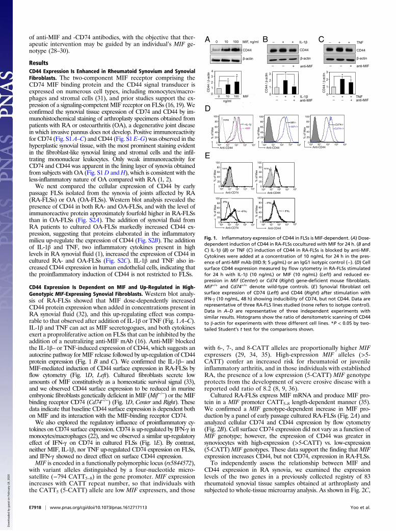
of anti-MIF and -CD74 antibodies, with the objective that ther-apeutic intervention may be guided by an individual’s MIF ge-notype (28–30).
ResultsCD44 Expression Is Enhanced in Rheumatoid Synovium and SynovialFibroblasts. The two-component MIF receptor comprising theCD74 MIF binding protein and the CD44 signal transducer isexpressed on numerous cell types, including monocytes/macro-phages and stromal cells (31), and prior studies support the ex-pression of a signaling-competent MIF receptor on FLSs (16, 19). Weconfirmed the synovial tissue expression of CD74 and CD44 by im-munohistochemical staining of arthroplasty specimens obtained frompatients with RA or osteoarthritis (OA), a degenerative joint diseasein which invasive pannus does not develop. Positive immunoreactivityfor CD74 (Fig. S1 A–C) and CD44 (Fig. S1 E–G) was observed in thehyperplastic synovial tissue, with the most prominent staining evidentin the fibroblast-like synovial lining and stromal cells and the infil-trating mononuclear leukocytes. Only weak immunoreactivity forCD74 and CD44 was apparent in the lining layer of synovia obtainedfrom subjects with OA (Fig. S1D andH), which is consistent with theless-inflammatory nature of OA compared with RA (1, 2).We next compared the cellular expression of CD44 by early
passage FLSs isolated from the synovia of joints affected by RA(RA-FLSs) or OA (OA-FLSs). Western blot analysis revealed thepresence of CD44 in both RA- and OA-FLSs, and with the level ofimmunoreactive protein approximately fourfold higher in RA-FLSsthan in OA-FLSs (Fig. S2A). The addition of synovial fluid fromRA patients to cultured OA-FLSs markedly increased CD44 ex-pression, suggesting that proteins elaborated in the inflammatorymilieu up-regulate the expression of CD44 (Fig. S2B). The additionof IL-1β and TNF, two inflammatory cytokines present in highlevels in RA synovial fluid (1), increased the expression of CD44 incultured RA- and OA-FLSs (Fig. S2C). IL-1β and TNF also in-creased CD44 expression in human endothelial cells, indicating thatthe proinflammatory induction of CD44 is not restricted to FLSs.
CD44 Expression Is Dependent on MIF and Up-Regulated in High-Genotypic MIF-Expressing Synovial Fibroblasts. Western blot analy-sis of RA-FLSs showed that MIF dose-dependently increasedCD44 protein expression when added in concentrations present inRA synovial fluid (32), and this up-regulating effect was compa-rable to that observed after addition of IL-1β or TNF (Fig. 1 A–C).IL-1β and TNF can act as MIF secretogogues, and both cytokinesexert a proproliferative action on FLSs that can be inhibited by theaddition of a neutralizing anti-MIF mAb (16). Anti-MIF blockedthe IL-1β– or TNF-induced expression of CD44, which suggests anautocrine pathway for MIF release followed by up-regulation of CD44protein expression (Fig. 1 B and C). We confirmed the IL-1β– andMIF-mediated induction of CD44 surface expression in RA-FLSs byflow cytometry (Fig. 1D, Left). Cultured fibroblasts secrete lowamounts of MIF constitutively as a homeostatic survival signal (33),and we observed CD44 surface expression to be reduced in murineembryonic fibroblasts genetically deficient in MIF (Mif−/−) or the MIFbinding receptor CD74 (Cd74−/−) (Fig. 1D, Center and Right). Thesedata indicate that baseline CD44 surface expression is dependent bothon MIF and its interaction with the MIF-binding receptor CD74.We also explored the regulatory influence of proinflammatory cy-
tokines on CD74 surface expression. CD74 is up-regulated by IFN-γ inmonocytes/macrophages (22), and we observed a similar up-regulatoryeffect of IFN-γ on CD74 in cultured FLSs (Fig. 1E). By contrast,neither MIF, IL-1β, nor TNF up-regulated CD74 expression on FLSs,and IFN-γ showed no direct effect on surface CD44 expression.MIF is encoded in a functionally polymorphic locus (rs5844572),
with variant alleles distinguished by a four-nucleotide micro-satellite (−794 CATT5–8) in the gene promoter. MIF expressionincreases with CATT repeat number, so that individuals withthe CATT5 (5-CATT) allele are low MIF expressers, and those
with 6-, 7-, and 8-CATT alleles are proportionally higher MIFexpressers (29, 34, 35). High-expression MIF alleles (>5-CATT) confer an increased risk for rheumatoid or juvenileinflammatory arthritis, and in those individuals with establishedRA, the presence of a low expression (5-CATT) MIF genotypeprotects from the development of severe erosive disease with areported odd ratio of 8.2 (8, 9, 36).Cultured RA-FLSs express MIF mRNA and produce MIF pro-
tein in a MIF promoter CATT5–8 length-dependent manner (35).We confirmed a MIF genotype-dependent increase in MIF pro-duction by a panel of early passage cultured RA-FLSs (Fig. 2A) andanalyzed cellular CD74 and CD44 expression by flow cytometry(Fig. 2B). Cell surface CD74 expression did not vary as a function ofMIF genotype; however, the expression of CD44 was greater insynoviocytes with high-expression (>5-CATT) vs. low-expression(5-CATT)MIF genotypes. These data support the finding thatMIFexpression increases CD44, but not CD74, expression in RA-FLSs.To independently assess the relationship between MIF and
CD44 expression in RA synovia, we examined the expressionlevels of the two genes in a previously collected registry of 83rheumatoid synovial tissue samples obtained at arthroplasty andsubjected to whole-tissue microarray analysis. As shown in Fig. 2C,
A B CD44
-actin
MIF, ng/ml 0 10 100
CD44
-actin
+ + IL-1
+ anti-MIF
CD44
-actin
_ _
_ _
+ + TNF
+ anti-MIF _ _
C
D
100
20
40
60
80
100
0
% o
f Max
104 101 102 103
+IL-1+MIF
none
Anti-CD44
20
40
60
80
100
0
% o
f Max
Anti-CD44 100
Cd74-/-Cd74+/+
104 101 102 103
20
40
60
80
100
0
% o
f Max
Anti-CD44 100
Mif-/-Mif+/+
104 101 102 103
Anti-CD74
20
40
60
80
100
0
% o
f Max
104 100 101 102 103
Anti-CD44
20
40
60
80
100
0
% o
f Max
104 100 101 102 103
Anti-CD74 104 100 101 102 103
20
40
60
80
100
0
% o
f Max
+IFN
20
40
60
80
100
0
% o
f Max
104
Anti-CD44 100 101 102 103
+ IFN
E
- IFN - IFN
none none
1
0
2
3
0 10 100 MIF _ + anti-MIF _
ng/ml
1
2
_ + anti-MIF_
1
0
2
3 3 * * * 4
*
CD
44 /
-act
in
CD
44 /
-act
in
CD
44 /
-act
in
0 _ + + TNF _ + + IL-1
Fig. 1. Inflammatory expression of CD44 in FLSs is MIF-dependent. (A) Dose-dependent induction of CD44 in RA-FLSs cocultured with MIF for 24 h. (B andC) IL-1β (B) or TNF (C) induction of CD44 in RA-FLSs is blocked by anti-MIF.Cytokines were added at a concentration of 10 ng/mL for 24 h in the pres-ence of anti-MIF mAb (IIID.9; 5 μg/mL) or an IgG1 isotypic control (−). (D) Cellsurface CD44 expression measured by flow cytometry in RA-FLSs stimulatedfor 24 h with IL-1β (10 ng/mL) or MIF (10 ng/mL) (Left) and reduced ex-pression in Mif (Center) or Cd74 (Right) gene-deficient mouse fibroblasts.Mif+/+ and Cd74+/+ denote wild-type controls. (E) Synovial fibroblast cellsurface expression of CD74 (Left) and CD44 (Right) after stimulation withIFN-γ (10 ng/mL, 48 h) showing inducibility of CD74, but not CD44. Data arerepresentative of three RA-FLS lines studied (none refers to isotype control).Data in A–D are representative of three independent experiments withsimilar results. Histograms show the ratio of densitometric scanning of CD44to β-actin for experiments with three different cell lines. *P < 0.05 by two-tailed Student’s t test for the comparisons shown.
E7918 | www.pnas.org/cgi/doi/10.1073/pnas.1612717113 Yoo et al.
Dow
nloa
ded
by g
uest
on
Feb
ruar
y 18
, 202
0
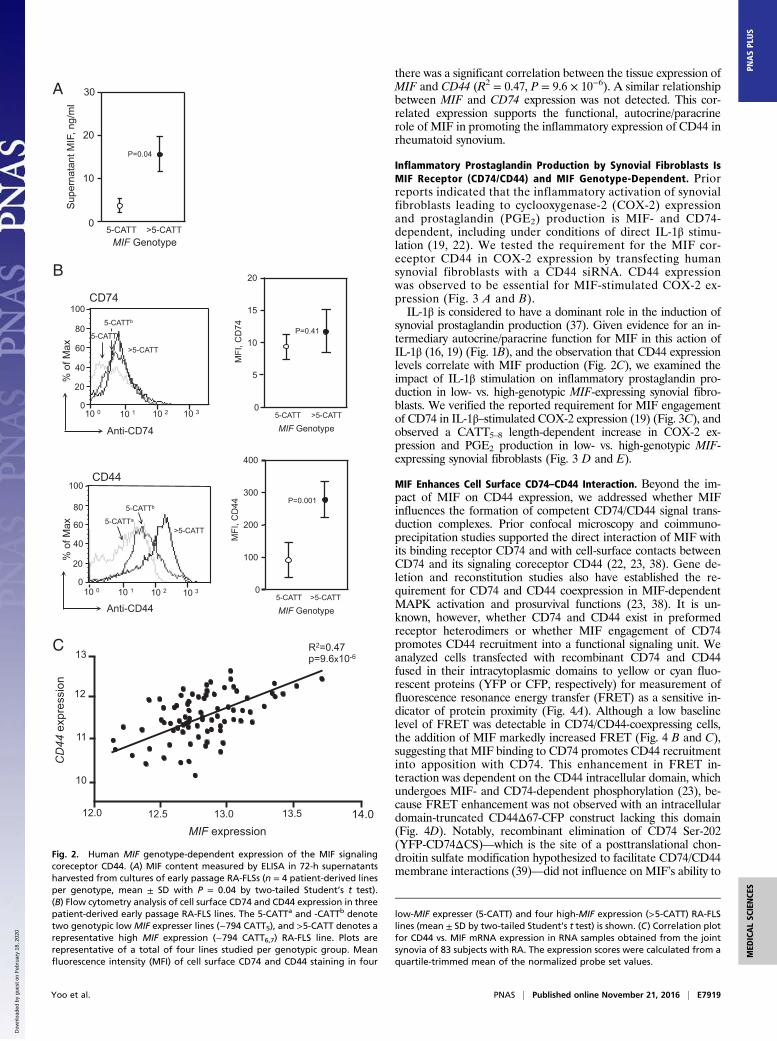
there was a significant correlation between the tissue expression ofMIF and CD44 (R2 = 0.47, P = 9.6 × 10⁻⁶). A similar relationshipbetween MIF and CD74 expression was not detected. This cor-related expression supports the functional, autocrine/paracrinerole of MIF in promoting the inflammatory expression of CD44 inrheumatoid synovium.
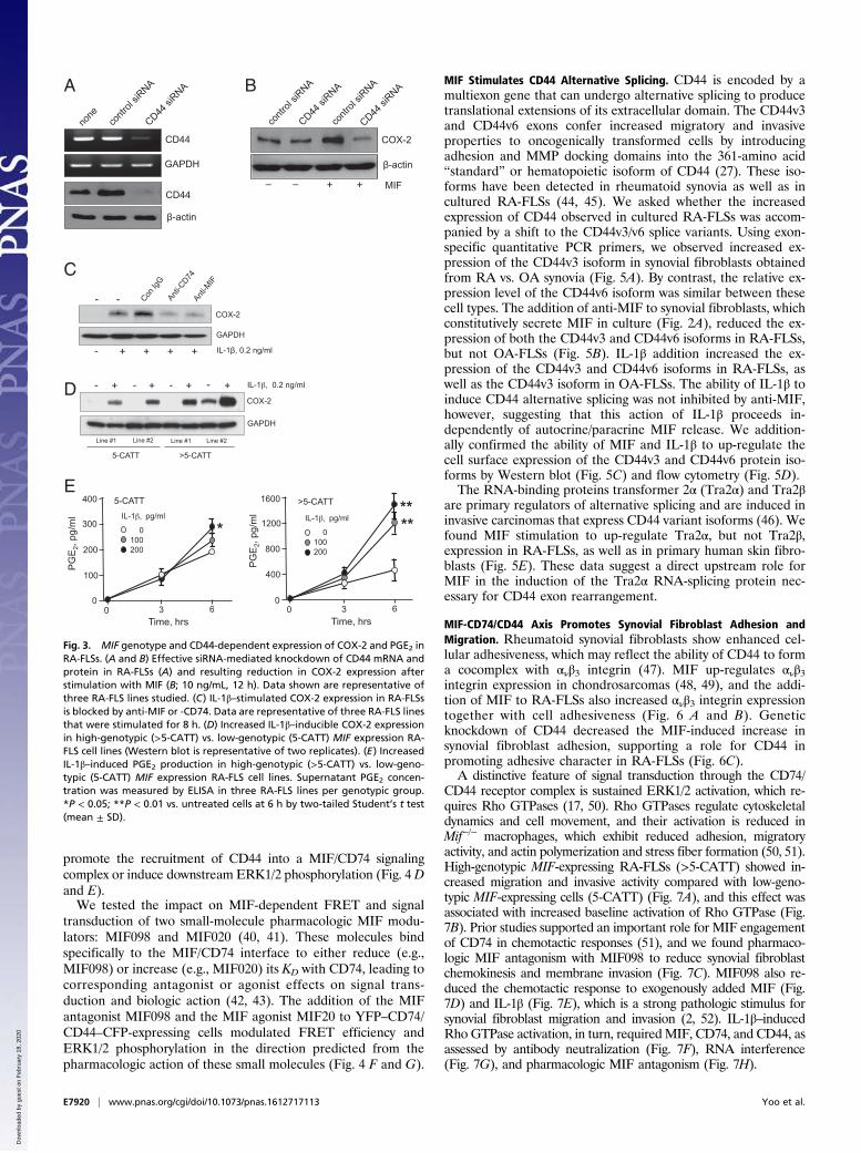
Inflammatory Prostaglandin Production by Synovial Fibroblasts IsMIF Receptor (CD74/CD44) and MIF Genotype-Dependent. Priorreports indicated that the inflammatory activation of synovialfibroblasts leading to cyclooxygenase-2 (COX-2) expressionand prostaglandin (PGE2) production is MIF- and CD74-dependent, including under conditions of direct IL-1β stimu-lation (19, 22). We tested the requirement for the MIF cor-eceptor CD44 in COX-2 expression by transfecting humansynovial fibroblasts with a CD44 siRNA. CD44 expressionwas observed to be essential for MIF-stimulated COX-2 ex-pression (Fig. 3 A and B).IL-1β is considered to have a dominant role in the induction of
synovial prostaglandin production (37). Given evidence for an in-termediary autocrine/paracrine function for MIF in this action ofIL-1β (16, 19) (Fig. 1B), and the observation that CD44 expressionlevels correlate with MIF production (Fig. 2C), we examined theimpact of IL-1β stimulation on inflammatory prostaglandin pro-duction in low- vs. high-genotypic MIF-expressing synovial fibro-blasts. We verified the reported requirement for MIF engagementof CD74 in IL-1β–stimulated COX-2 expression (19) (Fig. 3C), andobserved a CATT5–8 length-dependent increase in COX-2 ex-pression and PGE2 production in low- vs. high-genotypic MIF-expressing synovial fibroblasts (Fig. 3 D and E).
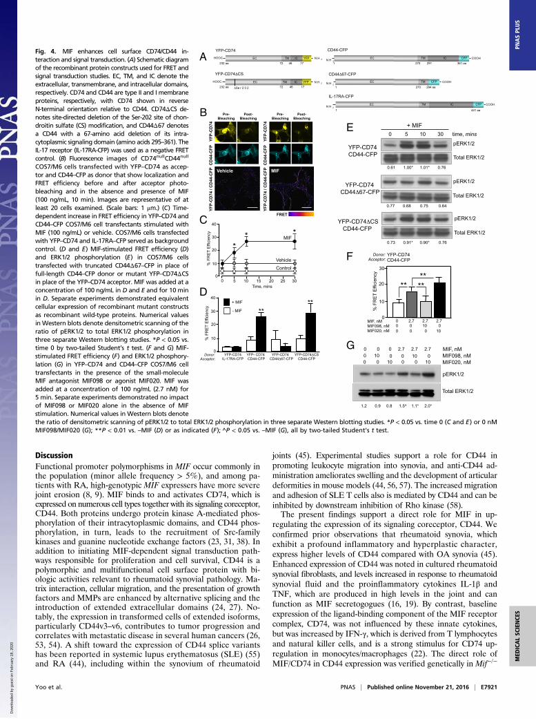
MIF Enhances Cell Surface CD74–CD44 Interaction. Beyond the im-pact of MIF on CD44 expression, we addressed whether MIFinfluences the formation of competent CD74/CD44 signal trans-duction complexes. Prior confocal microscopy and coimmuno-precipitation studies supported the direct interaction of MIF withits binding receptor CD74 and with cell-surface contacts betweenCD74 and its signaling coreceptor CD44 (22, 23, 38). Gene de-letion and reconstitution studies also have established the re-quirement for CD74 and CD44 coexpression in MIF-dependentMAPK activation and prosurvival functions (23, 38). It is un-known, however, whether CD74 and CD44 exist in preformedreceptor heterodimers or whether MIF engagement of CD74promotes CD44 recruitment into a functional signaling unit. Weanalyzed cells transfected with recombinant CD74 and CD44fused in their intracytoplasmic domains to yellow or cyan fluo-rescent proteins (YFP or CFP, respectively) for measurement offluorescence resonance energy transfer (FRET) as a sensitive in-dicator of protein proximity (Fig. 4A). Although a low baselinelevel of FRET was detectable in CD74/CD44-coexpressing cells,the addition of MIF markedly increased FRET (Fig. 4 B and C),suggesting that MIF binding to CD74 promotes CD44 recruitmentinto apposition with CD74. This enhancement in FRET in-teraction was dependent on the CD44 intracellular domain, whichundergoes MIF- and CD74-dependent phosphorylation (23), be-cause FRET enhancement was not observed with an intracellulardomain-truncated CD44Δ67-CFP construct lacking this domain(Fig. 4D). Notably, recombinant elimination of CD74 Ser-202(YFP-CD74ΔCS)—which is the site of a posttranslational chon-droitin sulfate modification hypothesized to facilitate CD74/CD44membrane interactions (39)—did not influence on MIF’s ability to
A
12.0 12.5 13.0 13.5
MIF expression
R2=0.47 p=9.6x10⁻⁶
10
12
13
CD
44 e
xpre
ssio
n
11
14.0
C
B
0
10
20
30
P=0.04
Sup
erna
tant
MIF
, ng/
ml
5-CATT >5-CATT MIF Genotype
10 3
Anti-CD74
20
40
60
80
100
0
% o
f Max
10 0 10 1 10 2
>5-CATT
5-CATTb
5-CATTa
MFI
, CD
74
5-CATT >5-CATT
P=0.41
MIF Genotype
0
5
10
15
20
CD74
Anti-CD44
20
40
60
80
100
0
% o
f Max
10 0 10 1 10 2 10 3
5-CATTa
5-CATTb
>5-CATT
MFI
, CD
44
5-CATT >5-CATT
P=0.001
MIF Genotype
0
100
200
300
400
CD44
Fig. 2. Human MIF genotype-dependent expression of the MIF signalingcoreceptor CD44. (A) MIF content measured by ELISA in 72-h supernatantsharvested from cultures of early passage RA-FLSs (n = 4 patient-derived linesper genotype, mean ± SD with P = 0.04 by two-tailed Student’s t test).(B) Flow cytometry analysis of cell surface CD74 and CD44 expression in threepatient-derived early passage RA-FLS lines. The 5-CATTa and -CATTb denotetwo genotypic low MIF expresser lines (−794 CATT5), and >5-CATT denotes arepresentative high MIF expression (−794 CATT6,7) RA-FLS line. Plots arerepresentative of a total of four lines studied per genotypic group. Meanfluorescence intensity (MFI) of cell surface CD74 and CD44 staining in four
low-MIF expresser (5-CATT) and four high-MIF expression (>5-CATT) RA-FLSlines (mean ± SD by two-tailed Student’s t test) is shown. (C) Correlation plotfor CD44 vs. MIF mRNA expression in RNA samples obtained from the jointsynovia of 83 subjects with RA. The expression scores were calculated from aquartile-trimmed mean of the normalized probe set values.
Yoo et al. PNAS | Published online November 21, 2016 | E7919
MED
ICALSC
IENCE
SPN
ASPL
US
Dow
nloa
ded
by g
uest
on
Feb
ruar
y 18
, 202
0
promote the recruitment of CD44 into a MIF/CD74 signalingcomplex or induce downstream ERK1/2 phosphorylation (Fig. 4 Dand E).We tested the impact on MIF-dependent FRET and signal
transduction of two small-molecule pharmacologic MIF modu-lators: MIF098 and MIF020 (40, 41). These molecules bindspecifically to the MIF/CD74 interface to either reduce (e.g.,MIF098) or increase (e.g., MIF020) its KD with CD74, leading tocorresponding antagonist or agonist effects on signal trans-duction and biologic action (42, 43). The addition of the MIFantagonist MIF098 and the MIF agonist MIF20 to YFP–CD74/CD44–CFP-expressing cells modulated FRET efficiency andERK1/2 phosphorylation in the direction predicted from thepharmacologic action of these small molecules (Fig. 4 F and G).
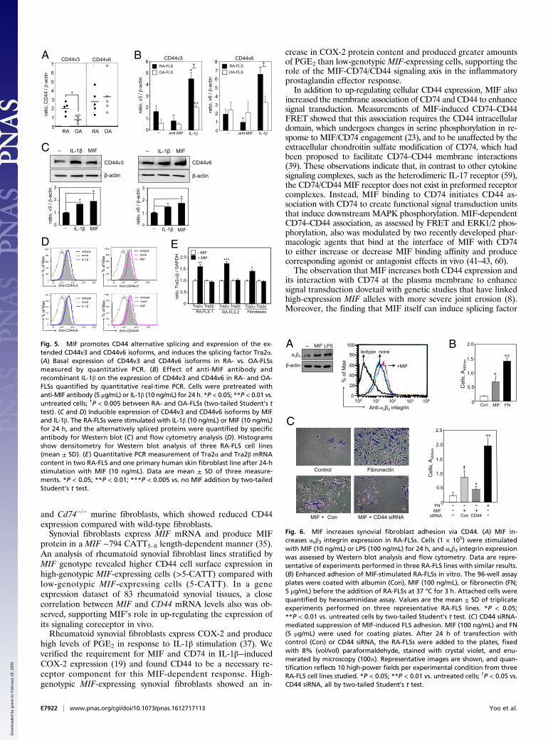
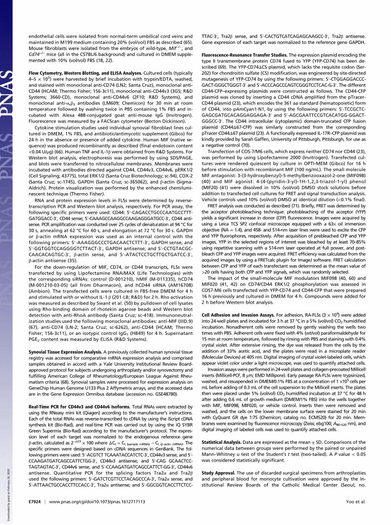
MIF Stimulates CD44 Alternative Splicing. CD44 is encoded by amultiexon gene that can undergo alternative splicing to producetranslational extensions of its extracellular domain. The CD44v3and CD44v6 exons confer increased migratory and invasiveproperties to oncogenically transformed cells by introducingadhesion and MMP docking domains into the 361-amino acid“standard” or hematopoietic isoform of CD44 (27). These iso-forms have been detected in rheumatoid synovia as well as incultured RA-FLSs (44, 45). We asked whether the increasedexpression of CD44 observed in cultured RA-FLSs was accom-panied by a shift to the CD44v3/v6 splice variants. Using exon-specific quantitative PCR primers, we observed increased ex-pression of the CD44v3 isoform in synovial fibroblasts obtainedfrom RA vs. OA synovia (Fig. 5A). By contrast, the relative ex-pression level of the CD44v6 isoform was similar between thesecell types. The addition of anti-MIF to synovial fibroblasts, whichconstitutively secrete MIF in culture (Fig. 2A), reduced the ex-pression of both the CD44v3 and CD44v6 isoforms in RA-FLSs,but not OA-FLSs (Fig. 5B). IL-1β addition increased the ex-pression of the CD44v3 and CD44v6 isoforms in RA-FLSs, aswell as the CD44v3 isoform in OA-FLSs. The ability of IL-1β toinduce CD44 alternative splicing was not inhibited by anti-MIF,however, suggesting that this action of IL-1β proceeds in-dependently of autocrine/paracrine MIF release. We addition-ally confirmed the ability of MIF and IL-1β to up-regulate thecell surface expression of the CD44v3 and CD44v6 protein iso-forms by Western blot (Fig. 5C) and flow cytometry (Fig. 5D).The RNA-binding proteins transformer 2α (Tra2α) and Tra2β
are primary regulators of alternative splicing and are induced ininvasive carcinomas that express CD44 variant isoforms (46). Wefound MIF stimulation to up-regulate Tra2α, but not Tra2β,expression in RA-FLSs, as well as in primary human skin fibro-blasts (Fig. 5E). These data suggest a direct upstream role forMIF in the induction of the Tra2α RNA-splicing protein nec-essary for CD44 exon rearrangement.
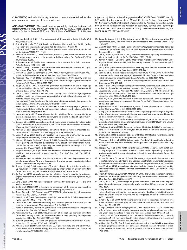
MIF-CD74/CD44 Axis Promotes Synovial Fibroblast Adhesion andMigration. Rheumatoid synovial fibroblasts show enhanced cel-lular adhesiveness, which may reflect the ability of CD44 to forma cocomplex with ανβ3 integrin (47). MIF up-regulates ανβ3integrin expression in chondrosarcomas (48, 49), and the addi-tion of MIF to RA-FLSs also increased ανβ3 integrin expressiontogether with cell adhesiveness (Fig. 6 A and B). Geneticknockdown of CD44 decreased the MIF-induced increase insynovial fibroblast adhesion, supporting a role for CD44 inpromoting adhesive character in RA-FLSs (Fig. 6C).A distinctive feature of signal transduction through the CD74/
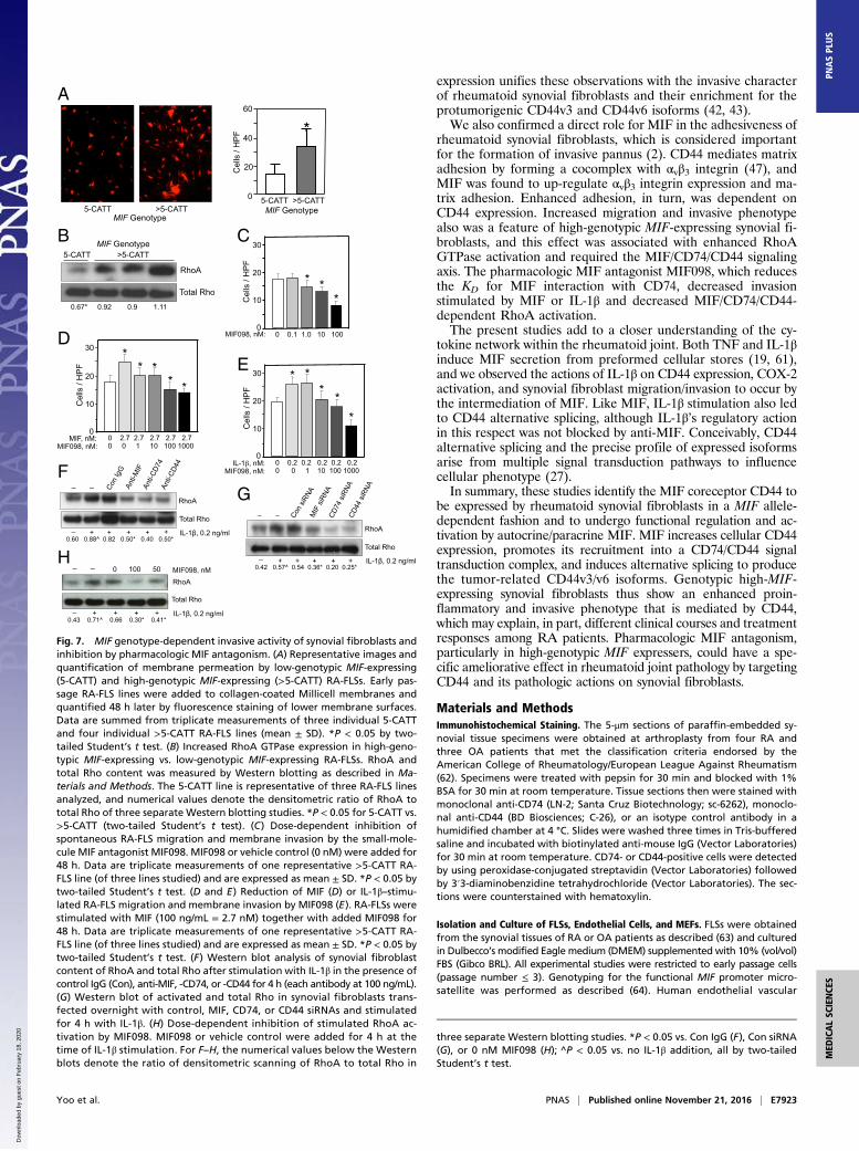
CD44 receptor complex is sustained ERK1/2 activation, which re-quires Rho GTPases (17, 50). Rho GTPases regulate cytoskeletaldynamics and cell movement, and their activation is reduced inMif−/− macrophages, which exhibit reduced adhesion, migratoryactivity, and actin polymerization and stress fiber formation (50, 51).High-genotypic MIF-expressing RA-FLSs (>5-CATT) showed in-creased migration and invasive activity compared with low-geno-typic MIF-expressing cells (5-CATT) (Fig. 7A), and this effect wasassociated with increased baseline activation of Rho GTPase (Fig.7B). Prior studies supported an important role for MIF engagementof CD74 in chemotactic responses (51), and we found pharmaco-logic MIF antagonism with MIF098 to reduce synovial fibroblastchemokinesis and membrane invasion (Fig. 7C). MIF098 also re-duced the chemotactic response to exogenously added MIF (Fig.7D) and IL-1β (Fig. 7E), which is a strong pathologic stimulus forsynovial fibroblast migration and invasion (2, 52). IL-1β–inducedRho GTPase activation, in turn, requiredMIF, CD74, and CD44, asassessed by antibody neutralization (Fig. 7F), RNA interference(Fig. 7G), and pharmacologic MIF antagonism (Fig. 7H).
C
A B
CD44
-actin
COX-2
-actin
MIF + + _ _
IL-1 , 0.2 ng/ml
COX-2
GAPDH
+ + + + -
- -
5-CATT
0 3 6 0
100
200
300
400
PG
E2,
pg/m
l
Time, hrs Time, hrs 0 3 6
0
400
800
1200
1600
5-CATT >5-CATT
- + - - - + + +
Line #1 Line #2 Line #2
IL-1 , 0.2 ng/ml
COX-2
GAPDH
Line #1
D
E >5-CATT
PG
E2,
pg/m
l IL-1 , pg/ml
0 100 200
IL-1 , pg/ml
0 100 200
* ** **
CD44
GAPDH
Fig. 3. MIF genotype and CD44-dependent expression of COX-2 and PGE2 inRA-FLSs. (A and B) Effective siRNA-mediated knockdown of CD44 mRNA andprotein in RA-FLSs (A) and resulting reduction in COX-2 expression afterstimulation with MIF (B; 10 ng/mL, 12 h). Data shown are representative ofthree RA-FLS lines studied. (C) IL-1β–stimulated COX-2 expression in RA-FLSsis blocked by anti-MIF or -CD74. Data are representative of three RA-FLS linesthat were stimulated for 8 h. (D) Increased IL-1β–inducible COX-2 expressionin high-genotypic (>5-CATT) vs. low-genotypic (5-CATT) MIF expression RA-FLS cell lines (Western blot is representative of two replicates). (E) IncreasedIL-1β–induced PGE2 production in high-genotypic (>5-CATT) vs. low-geno-typic (5-CATT) MIF expression RA-FLS cell lines. Supernatant PGE2 concen-tration was measured by ELISA in three RA-FLS lines per genotypic group.*P < 0.05; **P < 0.01 vs. untreated cells at 6 h by two-tailed Student’s t test(mean ± SD).
E7920 | www.pnas.org/cgi/doi/10.1073/pnas.1612717113 Yoo et al.
Dow
nloa
ded
by g
uest
on
Feb
ruar
y 18
, 202
0
DiscussionFunctional promoter polymorphisms in MIF occur commonly inthe population (minor allele frequency > 5%), and among pa-tients with RA, high-genotypic MIF expressers have more severejoint erosion (8, 9). MIF binds to and activates CD74, which isexpressed on numerous cell types together with its signaling coreceptor,CD44. Both proteins undergo protein kinase A-mediated phos-phorylation of their intracytoplasmic domains, and CD44 phos-phorylation, in turn, leads to the recruitment of Src-familykinases and guanine nucleotide exchange factors (23, 31, 38). Inaddition to initiating MIF-dependent signal transduction path-ways responsible for proliferation and cell survival, CD44 is apolymorphic and multifunctional cell surface protein with bi-ologic activities relevant to rheumatoid synovial pathology. Ma-trix interaction, cellular migration, and the presentation of growthfactors and MMPs are enhanced by alternative splicing and theintroduction of extended extracellular domains (24, 27). No-tably, the expression in transformed cells of extended isoforms,particularly CD44v3–v6, contributes to tumor progression andcorrelates with metastatic disease in several human cancers (26,53, 54). A shift toward the expression of CD44 splice variantshas been reported in systemic lupus erythematosus (SLE) (55)and RA (44), including within the synovium of rheumatoid
joints (45). Experimental studies support a role for CD44 inpromoting leukocyte migration into synovia, and anti-CD44 ad-ministration ameliorates swelling and the development of articulardeformities in mouse models (44, 56, 57). The increased migrationand adhesion of SLE T cells also is mediated by CD44 and can beinhibited by downstream inhibition of Rho kinase (58).The present findings support a direct role for MIF in up-
regulating the expression of its signaling coreceptor, CD44. Weconfirmed prior observations that rheumatoid synovia, whichexhibit a profound inflammatory and hyperplastic character,express higher levels of CD44 compared with OA synovia (45).Enhanced expression of CD44 was noted in cultured rheumatoidsynovial fibroblasts, and levels increased in response to rheumatoidsynovial fluid and the proinflammatory cytokines IL-1β andTNF, which are produced in high levels in the joint and canfunction as MIF secretogogues (16, 19). By contrast, baselineexpression of the ligand-binding component of the MIF receptorcomplex, CD74, was not influenced by these innate cytokines,but was increased by IFN-γ, which is derived from T lymphocytesand natural killer cells, and is a strong stimulus for CD74 up-regulation in monocytes/macrophages (22). The direct role ofMIF/CD74 in CD44 expression was verified genetically in Mif−/−
A
B
CD
44-C
FP
YFP-
CD
74
FRET
MIF
YFP-
CD
74 /
CD
44-C
FP
C C
D44
-CFP
Pre- Bleaching
Post- Bleaching
YFP-
CD
74YF
P-C
D74
/ C
D44
-CFP
Vehicle
Time, mins
40
%
FR
ET
Effi
cien
cy
0 5 10 15 20 25 30
MIF
*
0
30
20
Vehicle
* *
10
D
Control
Pre- Bleaching
Post- Bleaching
1 441 aa
COOH
IL-17RA-CFP
N H 2
COOH
CD44-CFP
1 270 291 361 aa
EC TM IC CFP
COOH
CD44 67-CFP
1 270 294 aa
N H 2
N H 2
YFP-CD74
HOOC EC TM IC YFP
232 aa 72 46 17
YFP-CD74 CS
EC TM IC YFP HOOC
N H 2
N H 2
232 aa 72 46 17
Se r 2 0 2
EC TM CFP
EC TM IC CFP
%
FR
ET
Effi
cien
cy
40
0
30
20
10
+ MIF
- MIF
Donor: Acceptor:
YFP-CD74 IL-17RA-CFP
YFP- CD74 CD44-CFP
**
YFP-CD74 CS CD44-CFP
YFP-CD74 CD44 67-CFP
**
YFP-CD74 CD44-CFP
0 5 10 30 time, mins
pERK1/2
Total ERK1/2
0.61 1.00* 1.01* 0.76
+ MIF
pERK1/2
Total ERK1/2
0.73 0.91* 0.90* 0.76
YFP-CD74 CS CD44-CFP
pERK1/2
Total ERK1/2
0.77 0.68 0.75 0.64
YFP-CD74 CD44 67-CFP
E
F
%
FR
ET
Effi
cien
cy
0
30
20
10
**
Donor: Acceptor:
YFP-CD74 CD44-CFP
** **
MIF, nM MIF098, nM MIF020, nM
0 2.7 2.7 2.7 0 0 10 0 0 0 0 10
G MIF, nM MIF098, nM MIF020, nM
0 0 0 2.7 2.7 2.7 0 10 0 0 10 0 0 10
1.2 0.9 0.8 1.5^ 1.1* 2.0*
Total ERK1/2
pERK1/2
0 0 10 0
Fig. 4. MIF enhances cell surface CD74/CD44 in-teraction and signal transduction. (A) Schematic diagramof the recombinant protein constructs used for FRET andsignal transduction studies. EC, TM, and IC denote theextracellular, transmembrane, and intracellular domains,respectively. CD74 and CD44 are type II and I membraneproteins, respectively, with CD74 shown in reverseN-terminal orientation relative to CD44. CD74ΔCS de-notes site-directed deletion of the Ser-202 site of chon-droitin sulfate (CS) modification, and CD44Δ67 denotesa CD44 with a 67-amino acid deletion of its intra-cytoplasmic signaling domain (amino acids 295–361). TheIL-17 receptor (IL-17RA-CFP) was used as a negative FRETcontrol. (B) Fluorescence images of CD74nullCD44null
COS7/M6 cells transfected with YFP–CD74 as accep-tor and CD44–CFP as donor that show localization andFRET efficiency before and after acceptor photo-bleaching and in the absence and presence of MIF(100 ng/mL, 10 min). Images are representative of atleast 20 cells examined. (Scale bars: 1 μm.) (C) Time-dependent increase in FRET efficiency in YFP–CD74 andCD44–CFP COS7/M6 cell transfectants stimulated withMIF (100 ng/mL) or vehicle. COS7/M6 cells transfectedwith YFP–CD74 and IL-17RA–CFP served as backgroundcontrol. (D and E) MIF-stimulated FRET efficiency (D)and ERK1/2 phosphorylation (E) in COS7/M6 cellstransfected with truncated CD44Δ67–CFP in place offull-length CD44–CFP donor or mutant YFP–CD74ΔCSin place of the YFP–CD74 acceptor. MIF was added at aconcentration of 100 ng/mL in D and E and for 10 minin D. Separate experiments demonstrated equivalentcellular expression of recombinant mutant constructsas recombinant wild-type proteins. Numerical valuesin Western blots denote densitometric scanning of theratio of pERK1/2 to total ERK1/2 phosphorylation inthree separate Western blotting studies. *P < 0.05 vs.time 0 by two-tailed Student’s t test. (F and G) MIF-stimulated FRET efficiency (F) and ERK1/2 phosphory-lation (G) in YFP–CD74 and CD44–CFP COS7/M6 celltransfectants in the presence of the small-moleculeMIF antagonist MIF098 or agonist MIF020. MIF wasadded at a concentration of 100 ng/mL (2.7 nM) for5 min. Separate experiments demonstrated no impactof MIF098 or MIF020 alone in the absence of MIFstimulation. Numerical values in Western blots denotethe ratio of densitometric scanning of pERK1/2 to total ERK1/2 phosphorylation in three separate Western blotting studies. *P < 0.05 vs. time 0 (C and E) or 0 nMMIF098/MIF020 (G); **P < 0.01 vs. –MIF (D) or as indicated (F); ^P < 0.05 vs. –MIF (G), all by two-tailed Student’s t test.
Yoo et al. PNAS | Published online November 21, 2016 | E7921
MED
ICALSC
IENCE
SPN
ASPL
US
Dow
nloa
ded
by g
uest
on
Feb
ruar
y 18
, 202
0
and Cd74−/− murine fibroblasts, which showed reduced CD44expression compared with wild-type fibroblasts.Synovial fibroblasts express MIF mRNA and produce MIF
protein in a MIF −794 CATT5–8 length-dependent manner (35).An analysis of rheumatoid synovial fibroblast lines stratified byMIF genotype revealed higher CD44 cell surface expression inhigh-genotypic MIF-expressing cells (>5-CATT) compared withlow-genotypic MIF-expressing cells (5-CATT). In a geneexpression dataset of 83 rheumatoid synovial tissues, a closecorrelation between MIF and CD44 mRNA levels also was ob-served, supporting MIF’s role in up-regulating the expression ofits signaling coreceptor in vivo.Rheumatoid synovial fibroblasts express COX-2 and produce
high levels of PGE2 in response to IL-1β stimulation (37). Weverified the requirement for MIF and CD74 in IL-1β−inducedCOX-2 expression (19) and found CD44 to be a necessary re-ceptor component for this MIF-dependent response. High-genotypic MIF-expressing synovial fibroblasts showed an in-
crease in COX-2 protein content and produced greater amountsof PGE2 than low-genotypic MIF-expressing cells, supporting therole of the MIF-CD74/CD44 signaling axis in the inflammatoryprostaglandin effector response.In addition to up-regulating cellular CD44 expression, MIF also
increased the membrane association of CD74 and CD44 to enhancesignal transduction. Measurements of MIF-induced CD74–CD44FRET showed that this association requires the CD44 intracellulardomain, which undergoes changes in serine phosphorylation in re-sponse to MIF/CD74 engagement (23), and to be unaffected by theextracellular chondroitin sulfate modification of CD74, which hadbeen proposed to facilitate CD74–CD44 membrane interactions(39). These observations indicate that, in contrast to other cytokinesignaling complexes, such as the heterodimeric IL-17 receptor (59),the CD74/CD44 MIF receptor does not exist in preformed receptorcomplexes. Instead, MIF binding to CD74 initiates CD44 as-sociation with CD74 to create functional signal transduction unitsthat induce downstream MAPK phosphorylation. MIF-dependentCD74–CD44 association, as assessed by FRET and ERK1/2 phos-phorylation, also was modulated by two recently developed phar-macologic agents that bind at the interface of MIF with CD74to either increase or decrease MIF binding affinity and producecorresponding agonist or antagonist effects in vivo (41–43, 60).The observation that MIF increases both CD44 expression and
its interaction with CD74 at the plasma membrane to enhancesignal transduction dovetail with genetic studies that have linkedhigh-expression MIF alleles with more severe joint erosion (8).Moreover, the finding that MIF itself can induce splicing factor
B
C
A
Cel
ls, A
450n
m
0.5
1.0
1.5
2.0
Con MIF FN
**
*
0
_ MIF LPS
-actin
v 3
Anti- v 3 integrin
none
101
% o
f Max
80
100
60
0
isotype
40
20
100 103 104 102
+MIF
Control Fibronectin
MIF + Con MIF + CD44 siRNA
Cel
ls, A
450n
m
0.5
1.0
1.5
2.0
+ + MIF FN
**
*
0
2.5
- -
siRNA
- Con CD44
- + -
- -
Fig. 6. MIF increases synovial fibroblast adhesion via CD44. (A) MIF in-creases αvβ3 integrin expression in RA-FLSs. Cells (1 × 10⁵) were stimulatedwith MIF (10 ng/mL) or LPS (100 ng/mL) for 24 h, and αvβ3 integrin expressionwas assessed by Western blot analysis and flow cytometry. Data are repre-sentative of experiments performed in three RA-FLS lines with similar results.(B) Enhanced adhesion of MIF-stimulated RA-FLSs in vitro. The 96-well assayplates were coated with albumin (Con), MIF (100 ng/mL), or fibronectin (FN;5 μg/mL) before the addition of RA-FLSs at 37 °C for 3 h. Attached cells werequantified by hexosaminidase assay. Values are the mean ± SD of triplicateexperiments performed on three representative RA-FLS lines. *P < 0.05;**P < 0.01 vs. untreated cells by two-tailed Student’s t test. (C) CD44 siRNA-mediated suppression of MIF-induced FLS adhesion. MIF (100 ng/mL) and FN(5 μg/mL) were used for coating plates. After 24 h of transfection withcontrol (Con) or CD44 siRNA, the RA-FLSs were added to the plates, fixedwith 8% (vol/vol) paraformaldehyde, stained with crystal violet, and enu-merated by microscopy (100×). Representative images are shown, and quan-tification reflects 10 high-power fields per experimental condition from threeRA-FLS cell lines studied. *P < 0.05; **P < 0.01 vs. untreated cells; †P < 0.05 vs.CD44 siRNA, all by two-tailed Student’s t test.
A B
0
1
2
3
4
5
6
7
*
CD44v3 CD44v6
ratio
, CD
44 /
-act
in
RA OA RA OA
E
C CD44v3
-actin
IL-1 _
ratio
,Tra
2/
/ G
AP
DH
0
0.5
1.0
1.5
2.0 - MIF+ MIF
Tra2 Tra2 Tra2 Tra2 Tra2 Tra2 RA-FLS 1 RA-FLS 2 Fibroblasts
** ***
*
MIF
OA-FLSRA-FLS
_ IL-1
0
1
2
3
4
5
6
*
*
anti-MIF
**
ratio
, v6
/ -a
ctin
CD44v3
*
*
_0
1
2
3
4
5
6
7
8
anti-MIF IL-1
CD44v6
OA-FLSRA-FLS
-
-
-
-
-
-
-
ratio
, v3
/ -a
ctin
0
1
2
3
ratio
, v3
/ -a
ctin
_
* *
MIF IL-1
isotypenoneIL-1
isotypenoneMIF
isotypenoneMIF
isotypenoneIL-1
% o
f Max
Anti-CD44v6
Anti-CD44v3
D
-actin
CD44v6
0
1
2
3
ratio
, v6
/ -a
ctin
_ MIF IL-1
* *
IL-1 _ MIF
Anti-CD44v6
% o
f Max
% o
f Max
% o
f Max
Anti-CD44v3
Fig. 5. MIF promotes CD44 alternative splicing and expression of the ex-tended CD44v3 and CD44v6 isoforms, and induces the splicing factor Tra2α.(A) Basal expression of CD44v3 and CD44v6 isoforms in RA- vs. OA-FLSsmeasured by quantitative PCR. (B) Effect of anti-MIF antibody andrecombinant IL-1β on the expression of CD44v3 and CD44v6 in RA- and OA-FLSs quantified by quantitative real-time PCR. Cells were pretreated withanti-MIF antibody (5 μg/mL) or IL-1β (10 ng/mL) for 24 h. *P < 0.05; **P < 0.01 vs.untreated cells; †P < 0.005 between RA- and OA-FLSs (two-tailed Student’s ttest). (C and D) Inducible expression of CD44v3 and CD44v6 isoforms by MIFand IL-1β. The RA-FLSs were stimulated with IL-1β (10 ng/mL) or MIF (10 ng/mL)for 24 h, and the alternatively spliced proteins were quantified by specificantibody for Western blot (C) and flow cytometry analysis (D). Histogramsshow densitometry for Western blot analysis of three RA-FLS cell lines(mean ± SD). (E) Quantitative PCR measurement of Tra2α and Tra2β mRNAcontent in two RA-FLS and one primary human skin fibroblast line after 24-hstimulation with MIF (10 ng/mL). Data are mean ± SD of three measure-ments. *P < 0.05; **P < 0.01; ***P < 0.005 vs. no MIF addition by two-tailedStudent’s t test.
E7922 | www.pnas.org/cgi/doi/10.1073/pnas.1612717113 Yoo et al.
Dow
nloa
ded
by g
uest
on
Feb
ruar
y 18
, 202
0
expression unifies these observations with the invasive characterof rheumatoid synovial fibroblasts and their enrichment for theprotumorigenic CD44v3 and CD44v6 isoforms (42, 43).We also confirmed a direct role for MIF in the adhesiveness of
rheumatoid synovial fibroblasts, which is considered importantfor the formation of invasive pannus (2). CD44 mediates matrixadhesion by forming a cocomplex with αvβ3 integrin (47), andMIF was found to up-regulate αvβ3 integrin expression and ma-trix adhesion. Enhanced adhesion, in turn, was dependent onCD44 expression. Increased migration and invasive phenotypealso was a feature of high-genotypic MIF-expressing synovial fi-broblasts, and this effect was associated with enhanced RhoAGTPase activation and required the MIF/CD74/CD44 signalingaxis. The pharmacologic MIF antagonist MIF098, which reducesthe KD for MIF interaction with CD74, decreased invasionstimulated by MIF or IL-1β and decreased MIF/CD74/CD44-dependent RhoA activation.The present studies add to a closer understanding of the cy-
tokine network within the rheumatoid joint. Both TNF and IL-1βinduce MIF secretion from preformed cellular stores (19, 61),and we observed the actions of IL-1β on CD44 expression, COX-2activation, and synovial fibroblast migration/invasion to occur bythe intermediation of MIF. Like MIF, IL-1β stimulation also ledto CD44 alternative splicing, although IL-1β’s regulatory actionin this respect was not blocked by anti-MIF. Conceivably, CD44alternative splicing and the precise profile of expressed isoformsarise from multiple signal transduction pathways to influencecellular phenotype (27).In summary, these studies identify the MIF coreceptor CD44 to
be expressed by rheumatoid synovial fibroblasts in a MIF allele-dependent fashion and to undergo functional regulation and ac-tivation by autocrine/paracrine MIF. MIF increases cellular CD44expression, promotes its recruitment into a CD74/CD44 signaltransduction complex, and induces alternative splicing to producethe tumor-related CD44v3/v6 isoforms. Genotypic high-MIF-expressing synovial fibroblasts thus show an enhanced proin-flammatory and invasive phenotype that is mediated by CD44,which may explain, in part, different clinical courses and treatmentresponses among RA patients. Pharmacologic MIF antagonism,particularly in high-genotypic MIF expressers, could have a spe-cific ameliorative effect in rheumatoid joint pathology by targetingCD44 and its pathologic actions on synovial fibroblasts.
Materials and MethodsImmunohistochemical Staining. The 5-μm sections of paraffin-embedded sy-novial tissue specimens were obtained at arthroplasty from four RA andthree OA patients that met the classification criteria endorsed by theAmerican College of Rheumatology/European League Against Rheumatism(62). Specimens were treated with pepsin for 30 min and blocked with 1%BSA for 30 min at room temperature. Tissue sections then were stained withmonoclonal anti-CD74 (LN-2; Santa Cruz Biotechnology; sc-6262), monoclo-nal anti-CD44 (BD Biosciences; C-26), or an isotype control antibody in ahumidified chamber at 4 °C. Slides were washed three times in Tris-bufferedsaline and incubated with biotinylated anti-mouse IgG (Vector Laboratories)for 30 min at room temperature. CD74- or CD44-positive cells were detectedby using peroxidase-conjugated streptavidin (Vector Laboratories) followedby 3′3-diaminobenzidine tetrahydrochloride (Vector Laboratories). The sec-tions were counterstained with hematoxylin.
Isolation and Culture of FLSs, Endothelial Cells, and MEFs. FLSs were obtainedfrom the synovial tissues of RA or OA patients as described (63) and culturedin Dulbecco’s modified Eagle medium (DMEM) supplemented with 10% (vol/vol)FBS (Gibco BRL). All experimental studies were restricted to early passage cells(passage number ≤ 3). Genotyping for the functional MIF promoter micro-satellite was performed as described (64). Human endothelial vascular
0
40
Cel
ls /
HP
F
5-CATT >5-CATT
20
60
*
5-CATT >5-CATT MIF Genotype
A
MIF Genotype
C C
ells
/ H
PF
10
20
30
0 MIF098, nM: 0 0.1 1.0 10 100
* * *
10
20
30
0 IL-1 , nM: MIF098, nM:
0 0.2 0.2 0.2 0.2 0.2 0 0 1 10 100 1000
*
* *
* *
E
Cel
ls /
HP
F
10
20
30
0 MIF, nM: MIF098, nM:
0 2.7 2.7 2.7 2.7 2.7 0 0 1 10 100 1000
* * *
* *
D
Cel
ls /
HP
F
Total Rho
RhoA
5-CATT >5-CATT MIF Genotype B
0.67* 0.92 0.9 1.11
IL-1 , 0.2 ng/ml_ + + + +
RhoA
Total Rho
F _ _
0.60 0.88^ 0.82 0.50* 0.40 0.50* +
IL-1 , 0.2 ng/ml_ + + + +
RhoA
Total Rho
G _ _
0.42 0.57^ 0.54 0.36* 0.20 0.25* +H
MIF098, nMRhoA
Total Rho
IL-1 , 0.2 ng/ml_ + + + +
0 100 50 _ _
0.43 0.71^ 0.66 0.30* 0.41*
Fig. 7. MIF genotype-dependent invasive activity of synovial fibroblasts andinhibition by pharmacologic MIF antagonism. (A) Representative images andquantification of membrane permeation by low-genotypic MIF-expressing(5-CATT) and high-genotypic MIF-expressing (>5-CATT) RA-FLSs. Early pas-sage RA-FLS lines were added to collagen-coated Millicell membranes andquantified 48 h later by fluorescence staining of lower membrane surfaces.Data are summed from triplicate measurements of three individual 5-CATTand four individual >5-CATT RA-FLS lines (mean ± SD). *P < 0.05 by two-tailed Student’s t test. (B) Increased RhoA GTPase expression in high-geno-typic MIF-expressing vs. low-genotypic MIF-expressing RA-FLSs. RhoA andtotal Rho content was measured by Western blotting as described in Ma-terials and Methods. The 5-CATT line is representative of three RA-FLS linesanalyzed, and numerical values denote the densitometric ratio of RhoA tototal Rho of three separate Western blotting studies. *P < 0.05 for 5-CATT vs.>5-CATT (two-tailed Student’s t test). (C) Dose-dependent inhibition ofspontaneous RA-FLS migration and membrane invasion by the small-mole-cule MIF antagonist MIF098. MIF098 or vehicle control (0 nM) were added for48 h. Data are triplicate measurements of one representative >5-CATT RA-FLS line (of three lines studied) and are expressed as mean ± SD. *P < 0.05 bytwo-tailed Student’s t test. (D and E) Reduction of MIF (D) or IL-1β–stimu-lated RA-FLS migration and membrane invasion by MIF098 (E). RA-FLSs werestimulated with MIF (100 ng/mL = 2.7 nM) together with added MIF098 for48 h. Data are triplicate measurements of one representative >5-CATT RA-FLS line (of three lines studied) and are expressed as mean ± SD. *P < 0.05 bytwo-tailed Student’s t test. (F) Western blot analysis of synovial fibroblastcontent of RhoA and total Rho after stimulation with IL-1β in the presence ofcontrol IgG (Con), anti-MIF, -CD74, or -CD44 for 4 h (each antibody at 100 ng/mL).(G) Western blot of activated and total Rho in synovial fibroblasts trans-fected overnight with control, MIF, CD74, or CD44 siRNAs and stimulatedfor 4 h with IL-1β. (H) Dose-dependent inhibition of stimulated RhoA ac-tivation by MIF098. MIF098 or vehicle control were added for 4 h at thetime of IL-1β stimulation. For F–H, the numerical values below the Westernblots denote the ratio of densitometric scanning of RhoA to total Rho in
three separate Western blotting studies. *P < 0.05 vs. Con IgG (F), Con siRNA(G), or 0 nM MIF098 (H); ^P < 0.05 vs. no IL-1β addition, all by two-tailedStudent’s t test.
Yoo et al. PNAS | Published online November 21, 2016 | E7923
MED
ICALSC
IENCE
SPN
ASPL
US
Dow
nloa
ded
by g
uest
on
Feb
ruar
y 18
, 202
0
endothelial cells were isolated from normal-term umbilical cord veins andmaintained in M199 medium containing 20% (vol/vol) FBS as described (65).Mouse fibroblasts were isolated from the embryos of wild-type, Mif−/−, andCd74−/− mice (all in the C57BL/6 background) and cultured in DMEM supple-mented with 10% (vol/vol) FBS (18, 22).
Flow Cytometry, Western Blotting, and ELISA Analyses. Cultured cells (typically4–5 × 10⁵) were harvested by brief incubation with trypsin/EDTA, washed,and stained with monoclonal anti-CD74 (LN2; Santa Cruz), monoclonal anti-CD44 (HCAM; Thermo Fisher; 156-3c11), monoclonal anti-CD44v3 (3G5; R&DSystems; 3660-CD), monoclonal anti-CD44v6 (2F10; R&D Systems), andmonoclonal anti-αvβ3 antibodies (LM609; Chemicon) for 30 min at roomtemperature followed by washing twice in PBS containing 1% FBS and in-cubated with Alexa 488-conjugated goat anti-mouse IgG (Invitrogen).Fluorescence was measured by a FACScan cytometer (Becton Dickinson).
Cytokine stimulation studies used individual synovial fibroblast lines cul-tured in DMEM, 1% FBS, and antibiotic/antimycotic supplement (Gibco) for24 h in the absence or presence of added cytokine. Human MIF (native se-quence) was produced recombinantly as described (final endotoxin content<0.04 U/μg) (66). Human TNF and IL-1βwere obtained from R&D Systems. ForWestern blot analysis, electrophoresis was performed by using SDS/PAGE,and blots were transferred to nitrocellulose membranes. Membranes wereincubated with antibodies directed against CD44, CD44v3, CD44v6, pERK1/2(Cell Signaling; 4377S), total ERK1/2 (Santa Cruz Biotechnology; sc-94), COX-2(Santa Cruz; sc-1745), GAPDH (Santa Cruz; sc-365062), and β-actin (Sigma-Aldrich). Protein visualization was performed by the enhanced chemilumi-nescent technique (Thermo Fisher).
RNA and protein expression levels in FLSs were determined by reverse-transcription PCR and Western blot analysis, respectively. For PCR assay, thefollowing specific primers were used: CD44: 5′-CAGACCTGCCCAATGCCTTT-GATGGACC-3′, CD44 sense; 5′-CAAAGCCAAGGCCAAGAGGGATGCC-3′, CD44 anti-sense. PCR amplification was conducted over 25 cycles of denaturation at 94 °C for30 s, annealing at 62 °C for 60 s, and elongation at 72 °C for 30 s. GAPDHor β-actin mRNA expression was used as an internal control with thefollowing primers: 5′-AAAGGGCCCTGACAACTCTTT-3′, GAPDH sense, and5′-GGTGGTCCAGGGGTCTTACT-3′, GAPDH antisense; and 5′-CCTGTACGC-CAACACAGTGC-3′, β-actin sense, and 5′-ATACTCCTGCTTGCTGATCC-3′,β-actin antisense (35).
For the down-regulation of MIF, CD74, or CD44 transcripts, FLSs weretransfected by using Lipofectamine RNAiMAX (Life Technologies) withthe corresponding siRNAs: control (D-001210), hMIF (M-011335), hCD74(M-001210-03-05) (all from Dharmacon), and hCD44 siRNA (AM16708)(Ambion). The transfected cells were cultured in FBS-free DMEM for 4 hand stimulated with or without IL-1 β (201-LB; R&D) for 2 h. Rho activationwas measured as described by Swant et al. (50) by pulldown of cell lysatesusing Rho-binding domain of rhotekin agarose beads and Western blotdetection with anti-RhoA antibody (Santa Cruz; sc-418). Immunoneutral-ization studies used the following monoclonal antibodies: anti-MIF (IIID.9)(67), anti-CD74 (LN-2; Santa Cruz; sc-6262), anti-CD44 (HCAM; ThermoFisher; 156-3c11), or an isotypic control IgG1 (HB49) for 4 h. SupernatantPGE2 content was measured by ELISA (R&D Systems).
Synovial Tissue Expression Analysis. A previously collected human synovial tissueregistry was accessed for comparative mRNA expression analysis and comprisedsamples obtained in accord with a Yale University Institutional Review Board-approved protocol for subjects undergoing arthroplasty and/or synovectomy andfulfilling American College of Rheumatology/European League Against Rheu-matism criteria (68). Synovial samples were processed for expression analysis onGeneChip Human Genome U133 Plus 2 Affymetrix arrays, and the accessed dataare in the Gene Expression Omnibus database (accession no. GSE48780).
Real-Time PCR for CD44v3 and CD44v6 Isoforms. Total RNAs were extracted byusing the RNeasy mini kit (Qiagen) according to the manufacturer’s instructions.Each of the total RNAs was reverse-transcribed to cDNA by using the iScript cDNAsynthesis kit (Bio-Rad), and real-time PCR was carried out by using the iQ SYBRGreen Supermix (Bio-Rad) according to the manufacturer’s protocol. The expres-sion level of each target was normalized to the endogenous reference geneβ-actin, calculated as 2−ΔCt × 100 where ΔCt = Ct (sample mRNA) − Ct (β-acitn mRNA). Thespecific primers were designed based on cDNA sequences in GenBank. The fol-lowing primers were used: 5′-ACGTCT TCAAATACCATCTC-3′, CD44v3 sense, and 5′-CCAAGATGATCAGCCATTCTGG-3′, CD44v3 antisense; and 5′-CAG GCAACTCC-TAGTAGTAC-3′, CD44v6 sense, and 5′-CCAAGATGATCAGCCATTCT-GG-3′, CD44v6antisense. Quantitative PCR for the splicing factors Tra2α and Tra2βused the following primers: 5′-GATCTCGTTCCTACAGCCCA-3′, Tra2α sense, and5′-ATTAACTGCCACCTTCCACC-3′, Tra2α antisense; and 5′-GGCGGTCACCTTCTCC-
TTAC-3′, Tra2β sense, and 5′-CACTGTCATCAGAGCAAGCC-3′, Tra2β antisense.Gene expression of each target was normalized to the reference gene GAPDH.
Fluorescence-Resonance Transfer Studies. The expression plasmid encoding thetype II transmembrane protein CD74 fused to YFP (YFP-CD74) has been de-scribed (69). The YFP-CD74ΔCS plasmid, which lacks the requisite codon (Ser-202) for chondroitin sulfate (CS) modification, was engineered by site-directedmutagenesis of YFP-CD74 by using the following primers: 5′-CTGGAGGACCC-GACT-GGGCTGGGT-3′ and 5′-ACCCAGCCCAGTCGGGTCCTCAC-G-3′. The differentCD44-CFP–expressing plasmids were constructed as follows. The CD44-CFPplasmid was cloned by inserting a CD44 cDNA amplified from the pTracer-CD44 plasmid (23), which encodes the 361 aa standard (hematopoietic) formof CD44, into pAmCyan1-N1, by using the following primers: 5′-TCCGCTC-GAGCGATGCACAGGAGGAGA-3′ and 5′-AGCGAATTCCGTCACATGG-GGACT-GGGCC-3′. The CD44 intracellular (cytoplasmic) domain-truncated CFP fusionplasmid (CD44Δ67-CFP) was similarly constructed from the correspondingpTracer-CD44Δ67 plasmid (23). A functionally expressed IL-17R–CFP plasmid waskindly provided by Sarah Gaffen, University of Pittsburgh, Pittsburgh, for use asa negative control (70).
Transfection of COS-7/M6 cells, which express neither CD74 nor CD44 (23),was performed by using Lipofectamine 2000 (Invitrogen). Transfected cul-tures were rendered quiescent by culture in OPTI-MEM (Gibco) for 16 hbefore stimulation with recombinant MIF (100 ng/mL). The small moleculeMIF antagonist: 3-(3-hydroxybenzyl)-5-methylbenzooxazol-2-one (MIF098)(40) and MIF agonist: 4-(4-(pyridin-3-yl)-1H-1,2,3-triazol-1-yl) phenol(MIF20) (41) were dissolved in 10% (vol/vol) DMSO stock solutions beforeaddition to transfected cell cultures for FRET and signal transduction analysis.Vehicle controls used 10% (vol/vol) DMSO at identical dilution (<0.1% final).
FRET analysis was conducted as described (71). Briefly, FRET was determined bythe acceptor photobleaching technique: photobleaching of the acceptor (YFP)yields a significant increase in donor (CFP) fluorescence. Images were acquired byusing a Leica TCS SP2 confocal microscope equipped with a 63× oil immersionobjective (NA = 1.4), and 458- and 514-nm laser lines were used to excite the CFPand YFP fluorophores, respectively. After acquisition of prebleached CFP and YFPimages, YFP in the selected regions of interest was bleached by at least 70–85%using repetitive scanning with a 514-nm laser operated at full power, and post-bleach CFP and YFP images were acquired. FRET efficiency was calculated from theacquired images by using a FRETcalc plugin for ImageJ software. FRET calculationbetween CFP and YFP of each transfectant was determined as the mean value of>20 cells having both CFP and YFP signals, which was randomly selected.
The impact of the small-molecule MIF modulators MIF098 (40, 60) andMIF020 (41, 42) on CD74/CD44 ERK1/2 phosphorylation was assessed inCOS7-M6 cells transfected with YFP-CD74 and CD44-CFP that were prepared16 h previously and cultured in DMEM for 4 h. Compounds were added for2 h before Western blot analysis.
Cell Adhesion and Invasion Assays. For adhesion, RA-FLSs (3 × 10⁴) were addedinto 24-well plates and incubated for 3 h at 37 °C in a 5% (vol/vol) CO2 humidifiedincubation. Nonadherent cells were removed by gently washing the wells twotimes with PBS. Adherent cells were fixed with 4% (wt/vol) paraformaldehyde for15min at room temperature, followed by rinsingwith PBS and stainingwith 0.4%crystal violet. After extensive rinsing, the dye was released from the cells by theaddition of 33% acetic acid, and the plates were read in a microplate reader(Molecular Devices) at 405 nm. Digital imaging of crystal violet-labeled cells, whichappear violet color under a light microscope, was used to quantify attached cells.
Invasionassayswereperformed in24-well plates and collagen-precoatedMillicellinserts (Millicell-PCF, 8 μm; EMDMillipore). Early passage RA-FLSs were trypsinized,washed, and resuspended in DMEM/0.1% FBS at a concentration of 1 ×10⁵ cells permL before adding of 0.3 mL of the cell suspension to theMillicell inserts. The platesthen were placed under 5% (vol/vol) CO2 humidified incubation at 37 °C for 48 hafter adding 0.6 mL of growth medium (DMEM/1% FBS) into the wells togetherwith MIF, MIF098, MIF020, or vehicle control. Inserts then were removed andwashed, and the cells on the lower membrane surface were stained for 20 minwith CyQuant GR dye 1:75 (Chemicon; catalog no. ECM520) for 20 min. Mem-branes were examined by fluorescence microscopy (Zeiss; ebg100; A80–520 nm), anddigital imaging of labeled cells was used to quantify attached cells.
Statistical Analysis. Data are expressed as the mean ± SD. Comparisons of thenumerical data between groups were performed by the paired or unpairedMann–Whitney u test of the Student’s t test (two-tailed). A P value < 0.05was considered statistically significant.
Study Approval. The use of discarded surgical specimens from arthroplastiesand peripheral blood for monocyte cultivation were approved by the In-stitutional Review Boards of the Catholic Medical Center (Seoul; no.
E7924 | www.pnas.org/cgi/doi/10.1073/pnas.1612717113 Yoo et al.
Dow
nloa
ded
by g
uest
on
Feb
ruar
y 18
, 202
0
CUMC09U034) and Yale University. Informed consent was obtained for theprocurement and analysis of these specimens.
ACKNOWLEDGMENTS. This work was supported by National Institutes ofHealth Grants AR049610 and AR050498 (to R.B.) and T32 HL007778 (to M.S.); theAlliance for Lupus Research (R.B.); and FAMRI Grant CIA82384 (to P.L.). J.B. was
supported by Deutsche Forschungsgemeinschaft (DFG) Grant SFB1123 and byDFG within the framework of the Munich Cluster for Systems Neurology (EXC1010 SyNergy). Additional support was provided by National Research Founda-tion of Korea (funded by the Ministry of Education, Science and Technology)Grants 2014R1A6A3A04054066 (to S.-A.Y.), 2014R1A2A1A11049812, and2015R1A3A2032927 (to W.-U.K.).
1. McInnes IB, Schett G (2011) The pathogenesis of rheumatoid arthritis. N Engl J Med365(23):2205–2219.
2. Bottini N, Firestein GS (2013) Duality of fibroblast-like synoviocytes in RA: Passiveresponders and imprinted aggressors. Nat Rev Rheumatol 9(1):24–33.
3. Lefèvre S, et al. (2009) Synovial fibroblasts spread rheumatoid arthritis to unaffectedjoints. Nat Med 15(12):1414–1420.
4. Firestein GS, Echeverri F, Yeo M, Zvaifler NJ, Green DR (1997) Somatic mutations in thep53 tumor suppressor gene in rheumatoid arthritis synovium. Proc Natl Acad Sci USA94(20):10895–10900.
5. Roivainen A, et al. (1997) H-ras oncogene point mutations in arthritic synovium.Arthritis Rheum 40(9):1636–1643.
6. Greven D, Leng L, Bucala R (2010) Autoimmune diseases: MIF as a therapeutic target.Expert Opin Ther Targets 14(3):253–264.
7. Morand EF, Leech M, Bernhagen J (2006) MIF: A new cytokine link between rheu-matoid arthritis and atherosclerosis. Nat Rev Drug Discov 5(5):399–410.
8. Radstake TRDJ, et al. (2005) Correlation of rheumatoid arthritis severity with thegenetic functional variants and circulating levels of macrophage migration inhibitoryfactor. Arthritis Rheum 52(10):3020–3029.
9. Baugh JA, et al. (2002) A functional promoter polymorphism in the macrophagemigration inhibitory factor (MIF) gene associated with disease severity in rheumatoidarthritis. Genes Immun 3(3):170–176.
10. Leech M, Metz C, Bucala R, Morand EF (2000) Regulation of macrophage migrationinhibitory factor by endogenous glucocorticoids in rat adjuvant-induced arthritis.Arthritis Rheum 43(4):827–833.
11. Leech M, et al. (2003) Regulation of p53 by macrophage migration inhibitory factor ininflammatory arthritis. Arthritis Rheum 48(7):1881–1889.
12. Mikulowska A, Metz CN, Bucala R, Holmdahl R (1997) Macrophage migration in-hibitory factor is involved in the pathogenesis of collagen type II-induced arthritis inmice. J Immunol 158(11):5514–5517.
13. Herrero LJ, et al. (2013) Macrophage migration inhibitory factor receptor CD74 me-diates alphavirus-induced arthritis and myositis in murine models of alphavirus in-fection. Arthritis Rheum 65(10):2724–2736.
14. Singh A, et al. (2013) Macrophage-derived, macrophage migration inhibitory factor(MIF) is necessary to induce disease in the K/BxN serum-induced model of arthritis.Rheumatol Int 33(9):2301–2308.
15. Morand EF, et al. (2002) Macrophage migration inhibitory factor in rheumatoid ar-thritis: Clinical correlations. Rheumatology (Oxford) 41(5):558–562.
16. Lacey D, et al. (2003) Control of fibroblast-like synoviocyte proliferation by macro-phage migration inhibitory factor. Arthritis Rheum 48(1):103–109.
17. Mitchell RA, Metz CN, Peng T, Bucala R (1999) Sustained mitogen-activated proteinkinase (MAPK) and cytoplasmic phospholipase A2 activation by macrophage migra-tion inhibitory factor (MIF). Regulatory role in cell proliferation and glucocorticoidaction. J Biol Chem 274(25):18100–18106.
18. Fingerle-Rowson G, et al. (2003) The p53-dependent effects of macrophage migrationinhibitory factor revealed by gene targeting. Proc Natl Acad Sci USA 100(16):9354–9359.
19. Sampey AV, Hall PH, Mitchell RA, Metz CN, Morand EF (2001) Regulation of syno-viocyte phospholipase A2 and cyclooxygenase 2 by macrophage migration inhibitoryfactor. Arthritis Rheum 44(6):1273–1280.
20. Schurigt U, et al. (2008) Interactions of T helper cells with fibroblast-like synoviocytes:Up-regulation of matrix metalloproteinases by macrophage migration inhibitoryfactor from both Th1 and Th2 cells. Arthritis Rheum 58(10):3030–3040.
21. Gu R, et al. (2015) Macrophage migration inhibitory factor is essential for osteoclas-togenic mechanisms in vitro and in vivo mouse model of arthritis. Cytokine 72(2):135–145.
22. Leng L, et al. (2003) MIF signal transduction initiated by binding to CD74. J Exp Med197(11):1467–1476.
23. Shi X, et al. (2006) CD44 is the signaling component of the macrophage migrationinhibitory factor-CD74 receptor complex. Immunity 25(4):595–606.
24. Turley EA, Noble PW, Bourguignon LYW (2002) Signaling properties of hyaluronanreceptors. J Biol Chem 277(7):4589–4592.
25. Jiang D, et al. (2005) Regulation of lung injury and repair by Toll-like receptors andhyaluronan. Nat Med 11(11):1173–1179.
26. Godar S, et al. (2008) Growth-inhibitory and tumor-suppressive functions of p53 de-pend on its repression of CD44 expression. Cell 134(1):62–73.
27. Ponta H, Sherman L, Herrlich PA (2003) CD44: From adhesion molecules to signallingregulators. Nat Rev Mol Cell Biol 4(1):33–45.
28. Kerschbaumer RJ, et al. (2012) Neutralization of macrophage migration inhibitoryfactor (MIF) by fully human antibodies correlates with their specificity for the β-sheetstructure of MIF. J Biol Chem 287(10):7446–7455.
29. Bucala R (2013) MIF, MIF alleles, and prospects for therapeutic intervention in auto-immunity. J Clin Immunol 33(Suppl 1):S72–S78.
30. Alinari L, et al. (2011) Combination anti-CD74 (milatuzumab) and anti-CD20 (ritux-imab) monoclonal antibody therapy has in vitro and in vivo activity in mantle celllymphoma. Blood 117(17):4530–4541.
31. Bucala R, Shachar I (2014) The integral role of CD74 in antigen presentation, MIFsignal transduction, and B cell survival and homeostasis. Mini Rev Med Chem 14(14):1132–1138.
32. Leech M, et al. (1999) Macrophage migration inhibitory factor in rheumatoid arthritis:Evidence of proinflammatory function and regulation by glucocorticoids. ArthritisRheum 42(8):1601–1608.
33. Liao H, Bucala R, Mitchell RA (2003) Adhesion-dependent signaling by macrophagemigration inhibitory factor (MIF). J Biol Chem 278(1):76–81.
34. Renner P, Roger T, Calandra T (2005) Macrophage migration inhibitory factor: Genepolymorphisms and susceptibility to inflammatory diseases. Clin Infect Dis 41(Suppl 7):S513–S519.
35. Yao J, et al. (2016) Transcription factor ICBP90 regulates the MIF promoter and im-mune susceptibility locus. J Clin Invest 126(2):732–744.
36. Donn R, et al.; British Paediatric Rheumatology Study Group (2004) A functionalpromoter haplotype of macrophage migration inhibitory factor is linked and asso-ciated with juvenile idiopathic arthritis. Arthritis Rheum 50(5):1604–1610.
37. McInnes IB, Schett G (2007) Cytokines in the pathogenesis of rheumatoid arthritis. NatRev Immunol 7(6):429–442.
38. Gore Y, et al. (2008) Macrophage migration inhibitory factor induces B cell survival byactivation of a CD74-CD44 receptor complex. J Biol Chem 283(5):2784–2792.
39. Naujokas MF, Morin M, Anderson MS, Peterson M, Miller J (1993) The chondroitinsulfate form of invariant chain can enhance stimulation of T cell responses throughinteraction with CD44. Cell 74(2):257–268.
40. Hare AA, et al. (2010) Optimization of N-benzyl-benzoxazol-2-ones as receptor an-tagonists of macrophage migration inhibitory factor (MIF). Bioorg Med Chem Lett20(19):5811–5814.
41. Jorgensen WL, et al. (2010) Receptor agonists of macrophage migration inhibitoryfactor. Bioorg Med Chem Lett 20(23):7033–7036.
42. Wang J, et al. (2013) Limiting cardiac ischemic injury by pharmacological augmen-tation of macrophage migration inhibitory factor-AMP-activated protein kinase sig-nal transduction. Circulation 128(3):225–236.
43. Leng L, et al. (2011) A small-molecule macrophage migration inhibitory factor an-tagonist protects against glomerulonephritis in lupus-prone NZB/NZW F1 and MRL/lprmice. J Immunol 186(1):527–538.
44. Wibulswas A, et al. (2002) Influence of epitopes CD44v3 and CD44v6 in the invasivebehavior of fibroblast-like synoviocytes derived from rheumatoid arthritic joints.Arthritis Rheum 46(8):2059–2064.
45. Grisar J, et al. (2012) Expression patterns of CD44 and CD44 splice variants in patientswith rheumatoid arthritis. Clin Exp Rheumatol 30(1):64–72.
46. Watermann DO, et al. (2006) Splicing factor Tra2-beta1 is specifically induced inbreast cancer and regulates alternative splicing of the CD44 gene. Cancer Res 66(9):4774–4780.
47. Katagiri YU, et al. (1999) CD44 variants but not CD44s cooperate with beta1-con-taining integrins to permit cells to bind to osteopontin independently of arginine-glycine-aspartic acid, thereby stimulating cell motility and chemotaxis. Cancer Res59(1):219–226.
48. Bondza PK, Metz CN, Akoum A (2008) Macrophage migration inhibitory factor up-regulates alpha(v)beta(3) integrin and vascular endothelial growth factor expressionin endometrial adenocarcinoma cell line Ishikawa. J Reprod Immunol 77(2):142–151.
49. Lee CY, et al. (2012) Macrophage migration inhibitory factor increases cell motilityand up-regulates αvβ3 integrin in human chondrosarcoma cells. J Cell Biochem 113(5):1590–1598.
50. Swant JD, Rendon BE, Symons M, Mitchell RA (2005) Rho GTPase-dependent signalingis required for macrophage migration inhibitory factor-mediated expression of cyclinD1. J Biol Chem 280(24):23066–23072.
51. Fan H, et al. (2011) Macrophage migration inhibitory factor and CD74 regulatemacrophage chemotactic responses via MAPK and Rho GTPase. J Immunol 186(8):4915–4924.
52. Wang AZ, Wang JC, Fisher GW, Diamond HS (1997) Interleukin-1beta-stimulated in-vasion of articular cartilage by rheumatoid synovial fibroblasts is inhibited by anti-bodies to specific integrin receptors and by collagenase inhibitors. Arthritis Rheum40(7):1298–1307.
53. Klingbeil P, et al. (2009) CD44 variant isoforms promote metastasis formation by atumor cell-matrix cross-talk that supports adhesion and apoptosis resistance. MolCancer Res 7(2):168–179.
54. Wang SJ, Wreesmann VB, Bourguignon LY (2007) Association of CD44 V3-containingisoforms with tumor cell growth, migration, matrix metalloproteinase expression,and lymph node metastasis in head and neck cancer. Head Neck 29(6):550–558.
55. Crispín JC, et al. (2010) Expression of CD44 variant isoforms CD44v3 and CD44v6 isincreased on T cells from patients with systemic lupus erythematosus and is correlatedwith disease activity. Arthritis Rheum 62(5):1431–1437.
56. Neidhart M, Gay RE, Gay S (2000) Anti-interleukin-1 and anti-CD44 interventionsproducing significant inhibition of cartilage destruction in an in vitro model of car-tilage invasion by rheumatoid arthritis synovial fibroblasts. Arthritis Rheum 43(8):1719–1728.
Yoo et al. PNAS | Published online November 21, 2016 | E7925
MED
ICALSC
IENCE
SPN
ASPL
US
Dow
nloa
ded
by g
uest
on
Feb
ruar
y 18
, 202
0

57. Mikecz K, Brennan FR, Kim JH, Glant TT (1995) Anti-CD44 treatment abrogates tissue
oedema and leukocyte infiltration in murine arthritis. Nat Med 1(6):558–563.58. Li Y, et al. (2007) Phosphorylated ERM is responsible for increased T cell polarization,
adhesion, and migration in patients with systemic lupus erythematosus. J Immunol
178(3):1938–1947.59. Gaffen S (2016) IL-17 receptor composition. Nat Rev Immunol 16(1):4.60. Weiser JN, et al. (2015) Macrophage migration inhibitory factor (MIF) is detrimental in
pneumococcal pneumonia and a target for therapeutic immunomodulation. J Infect
Dis 212(10):1677–1682.61. Merk M, et al. (2009) The Golgi-associated protein p115 mediates the secretion of
macrophage migration inhibitory factor. J Immunol 182(11):6896–6906.62. Aletaha D, et al. (2010) 2010 Rheumatoid arthritis classification criteria: An American
College of Rheumatology/European League Against Rheumatism collaborative ini-
tiative. Arthritis Rheum 62(9):2569–2581.63. Bucala R, Ritchlin C, Winchester R, Cerami A (1991) Constitutive production of in-
flammatory and mitogenic cytokines by rheumatoid synovial fibroblasts. J Exp Med
173(3):569–574.
64. Sreih A, et al. (2011) Dual effect of the macrophage migration inhibitory factor geneon the development and severity of human systemic lupus erythematosus. ArthritisRheum 63(12):3942–3951.
65. Kim WU, et al. (2007) Soluble Fas ligand inhibits angiogenesis in rheumatoid arthritis.Arthritis Res Ther 9(2):R42.
66. Bernhagen J, et al. (1994) Purification, bioactivity, and secondary structure analysis ofmouse and human macrophage migration inhibitory factor (MIF). Biochemistry33(47):14144–14155.
67. Donnelly SC, et al. (1997) Regulatory role for macrophage migration inhibitory factorin acute respiratory distress syndrome. Nat Med 3(3):320–323.
68. Sun Y, et al. (2014) PILRα negatively regulates mouse inflammatory arthritis.J Immunol 193(2):860–870.
69. Schwartz V, et al. (2009) A functional heteromeric MIF receptor formed by CD74 andCXCR4. FEBS Lett 583(17):2749–2757.
70. Kramer JM, et al. (2006) Evidence for ligand-independent multimerization of the IL-17 receptor. J Immunol 176(2):711–715.
71. Stepensky D (2007) FRETcalc plugin for calculation of FRET in non-continuous in-tracellular compartments. Biochem Biophys Res Commun 359(3):752–758.
E7926 | www.pnas.org/cgi/doi/10.1073/pnas.1612717113 Yoo et al.
Dow
nloa
ded
by g
uest
on
Feb
ruar
y 18
, 202
0