Embed Size (px)
Citation preview
MitoTracker® and MitoFluor� Mitochondrion-Selective ProbesMP 07510
Revised: 10�December�2002
Product Information
MitoTracker® and MitoFluor� Mitochondrion-Selective Probes
IntroductionMitoTracker Dyes � Fixable Mitochondrion-Selective Probes
Although conventional fluorescent stains for mitochondria,such as tetramethylrosamine and rhodamine 123, are readilysequestered by functioning mitochondria, these stains are easilywashed out of cells once the mitochondria experience a loss inmembrane potential. This characteristic limits the use of suchconventional stains in experiments that require cells to be treatedwith aldehyde fixatives or with other agents that affect the ener-getic state of the mitochondria. To overcome this limitation,Molecular Probes has developed the MitoTracker® probes — aseries of patented mitochondrion-selective stains that are concen-trated by active mitochondria and well retained during cell fixation.1
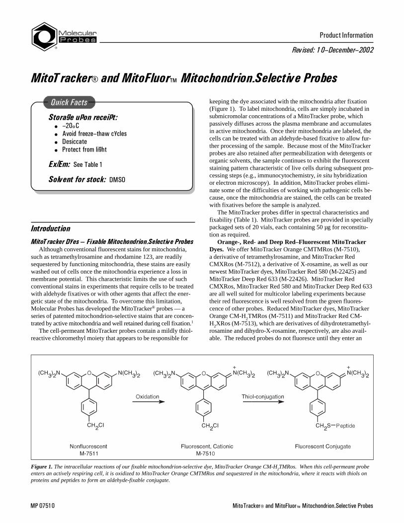
The cell-permeant MitoTracker probes contain a mildly thiol-reactive chloromethyl moiety that appears to be responsible for
Storage upon receipt:� �20°C� Avoid freeze�thaw cycles� Desiccate� Protect from light
Ex/Em: See Table 1
Solvent for stock: DMSO
keeping the dye associated with the mitochondria after fixation(Figure 1). To label mitochondria, cells are simply incubated insubmicromolar concentrations of a MitoTracker probe, whichpassively diffuses across the plasma membrane and accumulatesin active mitochondria. Once their mitochondria are labeled, thecells can be treated with an aldehyde-based fixative to allow fur-ther processing of the sample. Because most of the MitoTrackerprobes are also retained after permeabilization with detergents ororganic solvents, the sample continues to exhibit the fluorescentstaining pattern characteristic of live cells during subsequent pro-cessing steps (e.g., immunocytochemistry, in situ hybridizationor electron microscopy). In addition, MitoTracker probes elimi-nate some of the difficulties of working with pathogenic cells be-cause, once the mitochondria are stained, the cells can be treatedwith fixatives before the sample is analyzed.
The MitoTracker probes differ in spectral characteristics andfixability (Table 1). MitoTracker probes are provided in speciallypackaged sets of 20 vials, each containing 50 µg for reconstitu-tion as required.
Orange-, Red- and Deep Red–Fluorescent MitoTrackerDyes. We offer MitoTracker Orange CMTMRos (M-7510),a derivative of tetramethylrosamine, and MitoTracker RedCMXRos (M-7512), a derivative of X-rosamine, as well as ournewest MitoTracker dyes, MitoTracker Red 580 (M-22425) andMitoTracker Deep Red 633 (M-22426). MitoTracker RedCMXRos, MitoTracker Red 580 and MitoTracker Deep Red 633are all well suited for multicolor labeling experiments becausetheir red fluorescence is well resolved from the green fluores-cence of other probes. Reduced MitoTracker dyes, MitoTrackerOrange CM-H2TMRos (M-7511) and MitoTracker Red CM-H2XRos (M-7513), which are derivatives of dihydrotetramethyl-rosamine and dihydro-X-rosamine, respectively, are also avail-able. The reduced probes do not fluoresce until they enter an
Figure 1. The intracellular reactions of our fixable mitochondrion-selective dye, MitoTracker Orange CM-H2TMRos. When this cell-permeant probeenters an actively respiring cell, it is oxidized to MitoTracker Orange CMTMRos and sequestered in the mitochondria, where it reacts with thiols onproteins and peptides to form an aldehyde-fixable conjugate.
MitoTracker® and MitoFluor� Mitochondrion-Selective Probes2
actively respiring cell, where they are oxidized to the correspond-ing fluorescent mitochondrion-selective probe and then seques-tered in the mitochondria. All of these MitoTracker probes arewell retained after fixation and permeabilization.
Green-Fluorescent MitoTracker Green FM. Mitochondriain cells stained with nanomolar concentrations of MitoTrackerGreen FM dye (M-7514) exhibit bright green, fluorescein-likefluorescence.2 MitoTracker Green FM dye has the added advan-tage that it is essentially nonfluorescent in aqueous solutions,only becoming fluorescent once it accumulates in the lipid envi-ronment of mitochondria. Hence, background fluorescence isnegligible, enabling researchers to clearly visualize mitochondriain live cells immediately following addition of the stain, andwithout a wash step. Furthermore, MitoTracker Green FM dye issubstantially more photostable than the widely used fluorescentdye rhodamine 123 and produces a brighter, more mitochon-drion-selective signal at lower concentrations. Because its emis-sion maxima is blue-shifted approximately 10 nm relative to theemission maximum of rhodamine 123, MitoTracker Green FMdye produces a fluorescent staining pattern that should be betterresolved from that of red-fluorescent probes in double-labelingexperiments. Although MitoTracker Green FM dye can selec-tively stain mitochondria both in live cells and in cells that havebeen fixed, the dye is not well retained after cellpermeabilization.
MitoFluor Dyes � Nonfixable Mitochondrion-Selective ProbesMitoFluor Red 594 and MitoFluor Far Red 680 Dyes. We
offer two mitochondrial membrane potential–sensing dyes thathave long-wavelength fluorescence emission: MitoFluor™ Red594 dye (M-22422) and MitoFluor Far Red 680 dye (M-22423).MitoFluor Red 594 dye was designed for optimal excitation bythe 594 spectral line of the He–Ne laser. This long-wavelength
probe provides a clear spectral window below 600 nm for the si-multaneous detection of green-fluorescent labels, other site-selec-tive probes and green-fluorescent protein chimeras. MitoFluorFar Red 680 dye, also known as rhodamine 800, is a mitochon-drial potential sensor with absorption and fluorescence emissionin the near-infrared spectral region. Accumulation of MitoFluorFar Red 680 dye by active mitochondria produces a slight red-shift in its absorption and fluorescence emission peaks that isaccompanied by a marked decrease in fluorescence intensity.3
Although its fluorescence is not directly visible and must be cap-tured using an infrared light–sensitive detector such as a CCDcamera, MitoFluor Far Red 680 dye offers advantages whenworking with tissue, blood and other biological fluids prone tohigh absorbance or autofluorescence at shorter wavelengths.4
The MitoFluor Red 594 and MitoFluor Far Red 680 dyes canonly be used to stain live cells and are not retained after fixation.
MitoFluor Red 589 Dye. MitoFluor Red 589 dye (M-22424)appears to accumulate in mitochondria regardless of mitochon-drial membrane potential, making it a useful stain in both liveand fixed cells. The probe has absorption and emission peaks at588 nm and 622 nm, respectively, and can be viewed with com-mon filter sets appropriate for the Texas Red dye. Like our otherMitoFluor Red dyes, MitoFluor Red 589 dye provides a clearspectral window below 600 nm for double-labeling with green-fluorescent probes.
MitoFluor Green Dye. As a companion to MitoTrackerGreen FM dye, we offer MitoFluor Green dye 2,5 (M-7502).MitoFluor Green dye has a structure similar to MitoTrackerGreen FM dye, but lacks its reactive chloromethyl moieties, isnot as well retained following aldehyde fixation, and does notsurvive cell permeabilization. Like MitoTracker Green FM dye,it can selectively stain mitochondria both in live cells and in cellsthat have been fixed. MitoFluor Green dye is also substantiallymore photostable than rhodamine 123, producing a brighter,more mitochondrion-selective signal at lower concentrations.Because its emission maxima is blue-shifted relative to the emis-sion maximum of rhodamine 123, MitoFluor Green dye producesa fluorescent staining pattern that should be better resolved fromthat of red-fluorescent probes in double-labeling experiments.
Storage and HandlingThe MitoTracker probes are provided in specially packaged
sets of 20 separate vials, each containing 50 µg of lyophilizedsolid for reconstitution as required. The MitoFluor Green,MitoFluor Red 589 and MitoFluor Red 594 probes are providedin unit sizes of 1 mg. The MitoFluor Far Red 680 probe is sup-plied in a unit size of 10 mg. Upon receipt, the lyophilized solidsshould be stored desiccated at –20°C until required for use.When stored as solids, these reagents are stable for at least sixmonths. AVOID REPEATED FREEZING AND THAWING.
Before opening a vial, allow the product to warm to roomtemperature. To prepare a stock solution, dissolve the lyo-philized product in high-quality, anhydrous dimethylsulfoxide(DMSO) to a final concentration of 1 mM; the molecular weight(MW) is indicated on the product label. The reduced rosamineMitoTracker probes (M-7511, M-7513) are quite sensitive to oxi-dation, especially in solution, and must be stored under argon ornitrogen, at –20°C and protected from light. It is preferable touse solutions of the dihydro derivatives on the day that they areprepared. All other solutions of the MitoTracker and MitoFluordyes can be stored frozen at –20°C and protected from light.
Table 1. Spectral characteristics of MitoTracker and MitoFluor probes.
Product Catalog Number
Ex (nm) *
Em (nm) *
Fixable?
MitoTracker Green FM M-7514 † 490 516 No
MitoTracker Orange CMTMRos
M-7510 554 576 Yes
MitoTracker Orange CM-H2TMRos
M-7511 ‡ 554 576 Yes
MitoTracker Red CMXRos
M-7512 579 599 Yes
MitoTracker Red CM-H2XRos
M-7513 ‡ 579 599 Yes
MitoTracker Red 580 M-22425 581 644 Yes
MitoTracker Deep Red 633
M-22426 644 665 Yes
MitoFluor Green M-7502 † 490 516 No
MitoFluor Red 589 M-22424 588 622 No
MitoFluor Red 594 M-22422 598 630 No
MitoFluor Far Red 680 M-22423 680 >700 No
* Fluorescence excitation and emission maxima determined in methanol; values may vary somewhat in cellular environments. † Nonfluorescent in aqueous solution. ‡ Nonfluorescent until oxidized.
MitoTracker® and MitoFluor� Mitochondrion-Selective Probes3
ProtocolCell Preparation and Staining1.1 Preparing staining solutions. The concentration of probefor optimal staining will vary by application. The initial condi-tions suggested here are guidelines that may need to be modifiedbased on the particular cell type or on other factors, such as thepermeability of the cells or tissues to the probe. In general, wehave found that the reduced rosamine MitoTracker probes(M-7511, M-7513) should be loaded at three- to fivefold higherconcentrations than other MitoTracker and MitoFluor probes.Dilute the 1 mM MitoTracker or MitoFluor stock solution (seeStorage and Handling for preparation) to the final working con-centration in growth medium, e.g., Dulbecco’s modified Eaglemedium (D-MEM), with or without serum to match the mediumthat the cells were grown in. For live-cell staining, we recom-mend working concentrations of 25–500 nM. For staining cellsthat are to be fixed and permeabilized (see Fixation andPermeabilization after Staining), we suggest using a workingconcentration of 100–500 nM. To reduce potential artifacts fromoverloading, the concentration of dye should be kept as low aspossible. For the MitoTracker Green FM and MitoFluor Greenprobes, we suggest using a slightly lower concentration (20–200 nM). At higher concentrations, these probes tend to stainother cellular structures.
1.2 Staining adherent cells. Grow cells on coverslips inside aPetri dish filled with the appropriate culture medium. Whencells have reached the desired confluence, remove the mediumfrom the dish and add the prewarmed (37°C) growth mediumcontaining the MitoTracker or MitoFluor probe (prepared in step1.1). Incubate the cells for 15–45 minutes under growth condi-tions appropriate for the particular cell type. Then replace theloading solution with fresh prewarmed medium and observe thecells using a fluorescence microscope fitted with the correct filterset (see Table 1). If the cells do not appear to be sufficientlystained, we recommend either increasing the labeling concentra-tion or increasing the time allowed for the dye to accumulate inthe mitochondria once the cells have been transferred to freshmedium. If the cells are to be fixed and permeabilized, continueto Fixation and Permeabilization after Staining. The MitoFluorprobes are not well retained after fixation and permeabilization.
1.3 Staining suspension cells. Centrifuge to obtain a cellpellet and aspirate the supernatant. Resuspend the cells gentlyin prewarmed (37°C) medium containing the MitoTracker orMitoFluor probe (prepared in step 1.1). Incubate the cells for15–45 minutes under growth conditions that are appropriate forthe particular cell type. Re-pellet the cells by centrifugation andresuspend in fresh prewarmed medium. Again, if the cells arenot sufficiently stained, we recommend increasing the labelingconcentration or increasing the time allowed for the dye to accu-mulate in the mitochondria once the cells have been transferredto fresh medium. Alternatively, suspension cells may be attachedto coverslips that have been treated with BD Cell-Tak™ Cell andTissue Adhesive (BD Bioscience, Bedford, MA); in this case, seestep 1.2. If the cells are to be fixed and permeabilized, continue
to Fixation and Permeabilization after Staining. The MitoFluorprobes are not well retained after fixation and permeabilization.
1.4 Staining fixed cells. The MitoTracker Green FM, MitoFluorGreen, MitoFluor Red 589, MitoTracker Red 580 andMitoTracker Deep Red 633 probes may be used to stain cellsfixed in formaldehyde (see step 2.2, below). Following fixation,the cells should be rinsed in PBS. Incubate the fixed cells (onslides or in suspension) for 10–20 minutes in PBS containing10–200 nM of probe. Note that the loading concentration islower and staining time is shorter than in the procedure for stain-ing live cells. After the incubation, wash the cells at least oncewith fresh PBS.
Fixation and Permeabilization after Staining withMitoTracker Dyes
After staining live cells with one of the MitoTracker dyes,it is often convenient to fix the cells in formaldehyde and topermeabilize them with Triton® X-100. For example, fixationand permeabilization makes it possible to probe for otherintracellular structures by immunocytochemistry. Most of theMitoTracker dyes are well-retained following fixation andpermeabilization using the protocol described below. However,MitoTracker Green FM and the MitoFluor dyes are not retainedwell. Note: Mitotracker Red 580 is not compatible with TritonX-100 detergent permeabilization.
2.1 Washing the cells. After staining, wash the cells in fresh,prewarmed growth medium. This step is especially important ifthe cells are attached to a BD Cell-Tak Adhesive–coated cover-slip or another amine-containing surface.
2.2 Fixing the cells. Carefully remove the growth medium cov-ering the cells, and replace it with freshly prepared, prewarmedgrowth medium containing 3.7% formaldehyde. Note, if thegrowth medium contains serum, then the formaldehyde solutionshould be prepared in growth medium containing serum. Incu-bate at 37°C for 15 minutes.
2.3 Rinsing the cells. After fixation, rinse the cells several timesin PBS.
2.4 Permeabilization. When the cells are going to be subse-quently labeled with an antibody, a permeabilization step is usu-ally required to enhance the antigen’s accessibility. Incubate thefixed cells in PBS containing 0.2% Triton X-100 at room tem-perature for 5 minutes. Following permeabilization, rinse thecells in PBS. Alternatively, the cells may be permeablilized byincubating in ice-cold acetone for 5 minutes, and then washed inPBS. Even when cells are not going to be labeled with an anti-body, this acetone-permeabilization step may be useful because itappears to improve signal retention.
Fluorescence MicroscopySpectral characteristics for the MitoTracker and MitoFluor
probes are summarized in Table 1.
References1. J Histochem Cytochem 44, 1363 (1996); 2. Cytometry 39, 203 (2000); 3. J Biochem (Tokyo) 121, 29 (1997); 4. Anal Biochem 279, 142 (2000); 5. Mol CellBiochem 172, 171 (1997).
MitoTracker® and MitoFluor� Mitochondrion-Selective Probes4
Contact InformationFurther information on Molecular Probes' products, including product bibliographies, is available from your local distributor or directly from Molecular
Probes. Customers in Europe, Africa and the Middle East should contact our office in Leiden, the Netherlands. All others should contact our Technical Assis-tance Department in Eugene, Oregon.
Please visit our Web site � www.probes.com � for the most up-to-date information
Molecular Probes, Inc.29851 Willow Creek Rd., Eugene, OR 97402Phone: (541) 465-8300 � Fax: (541) 344-6504
Customer Service: 6:00 am to 4:30 pm (Pacific Time)Phone: (541) 465-8338 � Fax: (541) 344-6504 � [email protected]
Toll-Free Ordering for USA and Canada:Order Phone: (800) 438-2209 � Order Fax: (800) 438-0228
Technical Assistance: 8:00 am to 4:00 pm (Pacific Time)Phone: (541) 465-8353 � Fax: (541) 465-4593 � [email protected]
Molecular Probes Europe BVPoortGebouw, Rijnsburgerweg 102333 AA Leiden, The NetherlandsPhone: +31-71-5233378 � Fax: +31-71-5233419
Customer Service: 9:00 to 16:30 (Central European Time)Phone: +31-71-5236850 � Fax: [email protected]
Technical Assistance: 9:00 to 16:30 (Central European Time)Phone: +31-71-5233431 � Fax: [email protected]
Molecular Probes� products are high-quality reagents and materials intended for research purposes only. These products must be used by, or directlyunder the supervision of, a technically qualified individual experienced in handling potentially hazardous chemicals. Please read the Material Safety Data Sheetprovided for each product; other regulatory considerations may apply.
Several of Molecular Probes� products and product applications are covered by U.S. and foreign patents and patents pending. Our products are notavailable for resale or other commercial uses without a specific agreement from Molecular Probes, Inc. We welcome inquiries about licensing the use of ourdyes, trademarks or technologies. Please submit inquiries by e-mail to [email protected]. All names containing the designation ® are registered with theU.S. Patent and Trademark Office.
Copyright 2002, Molecular Probes, Inc. All rights reserved. This information is subject to change without notice.
Product List Current prices may be obtained from our Web site or from our Customer Service Department.
Cat # Product Name Unit Size
M-22423 MitoFluor� Far Red 680 ........................................................................................................................................................... 10 mgM-7502 MitoFluor� Green ..................................................................................................................................................................... 1 mgM-22424 MitoFluor� Red 589 .................................................................................................................................................................. 1 mgM-22422 MitoFluor� Red 594 .................................................................................................................................................................. 1 mgM-22426 MitoTracker® Deep Red 633 *special packaging* ..................................................................................................................... 20 x 50 µgM-7514 MitoTracker® Green FM *special packaging* ........................................................................................................................... 20 x 50 µgM-7511 MitoTracker® Orange CM-H2TMRos *special packaging* ........................................................................................................ 20 x 50 µgM-7510 MitoTracker® Orange CMTMRos *special packaging* ............................................................................................................. 20 x 50 µgM-22425 MitoTracker® Red 580 *special packaging* .............................................................................................................................. 20 x 50 µgM-7513 MitoTracker® Red CM-H2XRos *special packaging* ................................................................................................................ 20 x 50 µgM-7512 MitoTracker® Red CMXRos *special packaging* ..................................................................................................................... 20 x 50 µg