Embed Size (px)
Citation preview
NZ
DA
NE
WS
| V
ol. 1
95 |
Oct
ober
201
9
Molar Hypomineralisation
& Chalky Teeth a translational science
perspective
The MH problem from a paediatric perspective
Orthodontic Considerations in the management of
compromised first molars
con t
inu i
ng p ro f e s s i ona l development verifiable issue
Volume 195 October 2019 35
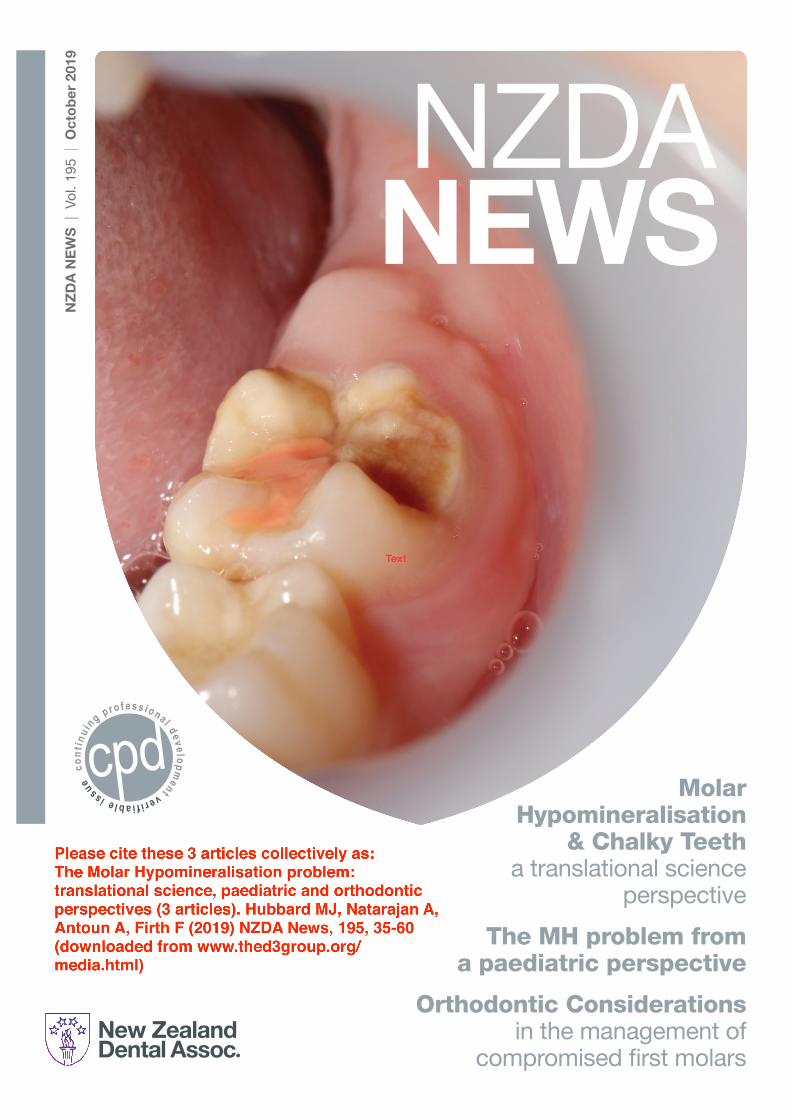
Perspectives on Molar Hypomineralisation (MH) and chalky teeth are heterogeneous and variably informed. Yet it's overwhelmingly obvious that major rewards beckon should the profession join a concerted effort to alleviate this global scourge.
Clinically, MH is a "regular pain in the backside" for those who know about it and take care to manage it well, whereas for others it's a foggy thing they didn't get taught at school and haven't yet mastered through CPD. For many practitioners, a patient's claim about having "chalky teeth" is nothing but misplaced excuse for poor oral hygiene. Multiple complexities surround MH and its differential diagnoses, leading many to simply badge anything odd as "hypoplastic". Largely ignored are other considerations ranging from science that could lead to prevention, through to massive burdens on society that should hasten funding of such research.
Meanwhile, MH as a major contributor to childhood tooth decay is not fully re-spected by systems put in place to develop good policy, to educate well, to do key research, and to make ideal products. On the rosy side, New Zealand has great history in this field and now together with Australia leads a new healthcare movement focused on MH. This and the following two articles offer a variety of well-informed perspectives, seeking to provide an overview that's both educa-tional and thought-provoking. We hope that by covering obstacles and challeng-es, as well as what's known (or should be) and what's not, national conversation will be generated leading to better understanding and care of New Zealanders with chalky teeth.
What is science translation?Science translation is essentially a modernised retake of the "bench to bed-
side" paradigm. The problem being addressed is discontinuity between those who do the science and those who need it (e.g. practitioners, industry, policy-makers) whilst aiming to improve "scientific bang for the buck" to taxpayers. The D3 Group for Developmental Dental Defects (D3G) grew out of a translational research initiative inspired by two Melbourne practitioners (OMFS Andrew Heggie and orthodontist Paul Schneider) and funded by a major benefaction intended to draw medicine, dentistry and science together for the good of children.
Developmental dental defects (DDDs, or "D3s") clearly fitted this bill and none more so than MH, being the dental outcome of medical problems experienced earlier in childhood. What beckoned translationally was (and still is) the exciting prospect of a medical intervention that could prevent MH and so wipe out a major chunk of childhood tooth decay and allied damage to society.>> read more about translation and D3G's genesis at: rch.org.au/mrufd
Feature article
Molar Hypomineralisation& Chalky Teeth [A TRANSLATIONAL SCIENCE PERSPECTIVE]
t
by Prof. Mike Hubbard BDS, PhD
NZDA NEWS
What is the "Molar Hypomin problem"?If MH is to be prevented, medico-dental research is needed to identify its causes
followed by industrial development of appropriate medical interventions that work at population level. Clearly the world will benefit from a concerted effort to tackle this problem by addressing all three of its nominal tiers—that is, through better clinical practice, education and investigation. But rather than attack MH in isola-tion, the field should look to leverage decades of progress made around the three "classical D3s"—being amelogenesis imperfecta (AI), dental fluorosis and enamel hypoplasia. For instance, AI often has massive impact on individual sufferers and their families yet through its rarity imposes modest cost on society, whereas the opposite holds for (highly prevalent) MH.
>> read more in two free academic articles available at: thed3group.org/media
‘THE MOLAR HYPOMIN PROBLEM’
Lack of MH research
Ignorance about MH
Molar Hypomineralisation (MH)
On the shoulders of a Kiwi giant
BO
X 1
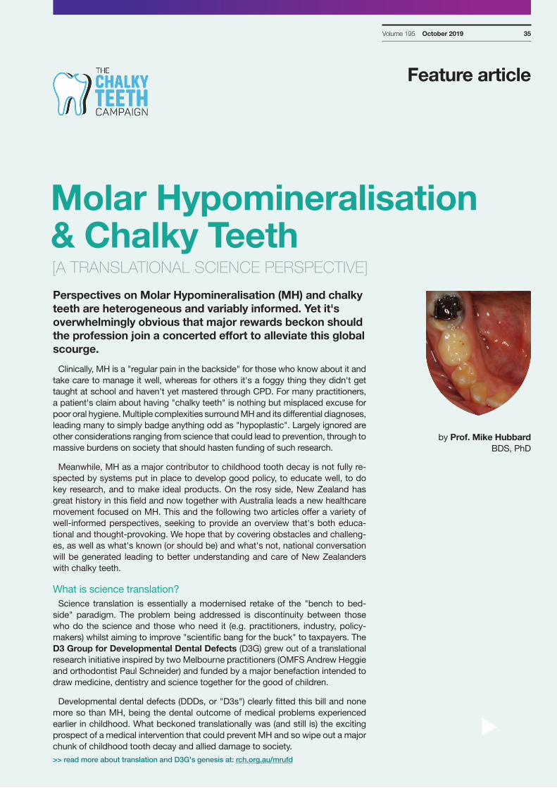
New Zealand dentist and researcher Grace Suckling (1922-2015) has influenced the D3 field more than anyone. Yet, due to failings in research translation, this overly modest woman never received the breadth of fame she deserved – something D3G is working on! What did get translated (through to medical practice and social good no less) was her 1976 report on tetracycline-stained enamel, which triggered immediate deletion of paediatric tetracyclines from the NZ drug tariff. And still famous around the dental epidemiol-ogy world is Grace's DDE Index – a survey tool for developmental defects of enamel (DDEs) that emanated from her unprecedented clinico-scientific analyses of Kiwi kids and lambs. Translation failed however with the obvious follow-up step of integrating DDEs into measures of tooth decay (e.g. DMFT Index) – we can only wonder how much better oral health would be today if this had happened 30 years ago. On becoming D3G's inaugural patron in 2013, Grace was thrilled the Chalky Teeth Campaign seeks to bridge this translational gap. Her research not only prompted a quantum leap in diagnosis, but also established two other D3 watersheds. Regards causation, she showed that DDEs reflected the timing, type and severity of insult to the enamel-forming machinery. And clinico-pathologically, it was her work that established what's taken for granted today about MH diagnosis and the underlying role of developmental timing (i.e. hypomineralisation and hypoplasia being linked to the maturation and secretory stages of enamel formation, respectively).
>> read more about Grace at: thed3group.org/grace-suckling
Clinico-scientific aspects of MHIf the MH problem is to be shared with scientists and translated across the sec-
tor into social good, there's need to develop communication strategies and criti-cal thinking that's both accessible and relevant to all parties. In this light, MH (or "chalky molars") can be defined simply as various manifestations of a particular enamel defect (demarcated opacity, or "chalky spot") that is distinguished from those lesions associated with fluorosis (diffuse opacity) and early decay (white spot lesion). To be MH, at least one molar must be affected, whether it be in the primary or adult dentition (i.e. "two-year", "six-year", "twelve-year" molars, alone or in com-bination), and unattributable to a known cause such as local trauma (i.e. idiopathic). This definition focusses on molars, being the teeth at highest risk of decay, and em-braces the fact that all other teeth (not just adult incisors) are prone to demarcated opacities. As elaborated in the accompanying articles, key features of MH are:
1. its sporadic presentation (affecting from one to all four molars of any type); 2. the tendency for post-eruptive changes including rapid breakdown and decay
(often creating orthodontic need and diagnostic confusion with hypoplasia, or rampant caries);
3. common association with dental pain (compromising oral hygiene and aggravating decay); and
4. frequent co-involvement of other teeth, including adult incisors (causing cosmetic and psychosocial issues).
Widespread misunderstandings relate to "MH" being a case descriptor only (i.e. it is incorrect to say MH molars or MH enamel). Perennial confusion also exists over the differences between hypomineralisation and hypoplasia, which carry sci-entific and clinical significance.
>> read more about the clinico-scientific aspects at: thed3group.org/practitioner
Volume 195 October 2019 37
Thinking beyond "MIH"Introduction of the "Molar-Incisor Hypomineralisation" (MIH) designation
eighteen years ago has undoubtedly advanced the field by providing a focal point that clinicians can easily relate to. Scientifically and translationally however, this clinical term is non-ideal (can we say flawed?) and so is better replaced with the MH concept outlined above. For instance, being restricted to the commonest presentation (i.e. involving the six-year molars, and sometimes the co-developed incisors), MIH by definition excludes equivalent phenotypes in the two-year and twelve-year molars that together make equal if not greater contributions to total prevalence of hypomineralisation (and allied decay). Scientifically, such "tunnel vision" on MIH risks missing important aetiological clues associated with hypomineralisation of teeth that develop before and after the six-year molars, and the inclusion of incisors complicates discrimination between idiopathic (systemic) and traumatic (local) origins.
>> see how MH literature has grown in the "MIH era" at: thed3group.org/d3-bibliography.html
Population aspects of MHWhat's devastating yet empowering about MH is its vastly underappreciated
prevalence. When launching the Chalky Teeth Campaign in 2013, D3G used the metaphor of MH being "a silent epidemic affecting one-in-six schoolchildren worldwide". Today, when the two-year and twelve-year molars are included, that prevalence rises to well above one-in-five children/adolescents. The problems surrounding such high prevalence are recognised, but remain poorly quantified. For instance, difficulties experienced at levels of individual sufferer (toothache, rapid decay, appearance, dental fear) and dental practitioner (prognosis, analge-sia, failed restorations) may carry a variety of cost impositions for affected fami-lies, dental providers, taxpayers and society. Unfortunately, the frequent public health claim that "tooth decay is preventable", doesn't hold much water for the multitude of children with severe MH – where, in absence of dental intervention, the standard preventive triad (hygiene, sugar, fluoride) is usually insufficient to avoid tooth breakdown and loss. Also disturbing is that, by failing to identify MH's major contribution to DMFT-based survey data, the conclusions attributed to den-tal caries and community water fluoridation may be grossly confounded. This has significant implications for government policies and funding.
>> read more about public health issues at: http://www.thed3group.org/prevalence.html
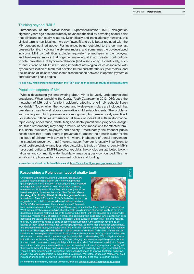
tOverlapping with Grace Suckling's wonderful legacy, New Zealand holds a second slice of D3 history that provides great opportunity for translation to social good. First reported amongst East Coast Māori in 1956, what's now generally referred to as "Polynesian AI" (or Poly-AI for short) has since been investigated by researchers from New Zealand (Grace Suckling, John Rodda, Alistair Smillie, Margeurite Crooks), Australia and French Polynesia. Today's limited understanding suggests an AI mutation happened historically somewhere in the Tahiti/Marquesas region, then spread across Polynesia to New Zealand where it's found throughout the country in a subset of Māori and other Polynesians. What makes it Polynesia's own type of chalky teeth is a distinctive phenotype involving severely discoloured opacities restricted largely to posterior adult teeth, with the anteriors and primary den-tition usually being mildly affected or normal. This contrasts with classical AI where all teeth in both dentitions are usually affected to a similar degree. Being curiously similar to MH in some regards, the Poly-AI phenotype raises all sorts of aetiological questions. Although much remains to be learned at tooth (biochemistry), case (phenotype, genetics, quality of life), population (prevalence), and socioeconomic levels, it's obvious that "Poly-AI kids" deserve better recognition and manage-ment today. Pleasingly, Michelle Martin – senior dentist at Northland DHB – has commenced an MoH-funded investigation aimed at elucidating clinical phenotype and kids' quality-of-life burdens with a view to betterments in dentalcare, policy, and public understanding. With thirty-five affected Māori kids under her wing, Michelle says Poly-AI is largely unknown amongst the general popula-tion and health professions, many dental practitioners included. Children (and adults) with Poly-AI face unique challenges in receiving the complex restorative treatment they require and coping with the impacts these teeth have on their life – particularly tooth sensitivity and psycho-social distress. There's a clear requirement to understand their needs better and to improve treatment pathways. Already involving collaborators from Northland, Auckland, Wellington, Otago and Melbourne, excit-ing opportunities exist to grow this investigation into a national if not pan-Polynesian project.
>> For more information, contact Michelle Martin at: [email protected]
Researching a Polynesian type of chalky teeth
BO
X 2
NZDA NEWS
Feature article
Causation and prevention of MHIf the dream of a paediatric medical intervention for preventing MH (and thereby
much tooth decay) is to come true, what likely will be multiple causes of MH need to be discovered – and although the answer is currently unclear, things are looking good for this being a researchable, and ultimately solvable, problem. Boding well for prevention, isolated demarcated opacities have long been regarded as "ac-quired" (i.e. resulting from local or systemic insults) – as opposed to being genetic in origin like AI. And much overlooked is the fact that iconic New Zealand research-er, Grace Suckling (see Box 1), was able to reproduce demarcated opacities in Kiwi sheep using systemic infection (with parasites, offset by drench) as well as lo-cal trauma. She also looked at New Zealand schoolchildren expecting to find links with "gastro" or other paediatric infections but, apart from a general association with illness, no specific cause stuck out – a situation that persists for MH today.
>> read more about the medical origins of MH at: thed3group.org/medicos.html
Research into MHSurprisingly to many, MH research can be traced back to the first description of
idiopathic demarcated opacities (comprising acid-sensitive "chalk-like enamel") by an American researcher in 1949. Today's research landscape is dominated by MH epidemiology plus scattered investigations into myriad other aspects rang-ing from biophysical characterisation of chalky enamel through to quality-of-life impacts. D3G researchers in Melbourne have made breakthrough insights to MH pathology and "late onset MH" (twelve-year molars, wisdom teeth), thereby open-ing an exciting path towards understanding the causes of MH. It's hoped that New Zealand can remain a forefront contributor to D3 research, not least by pur-suing the wonderful opportunities associated with an indigenous type of chalky teeth (see Box 2).
>> read more about MH research at: thed3group.org/researcher-directory.html
A D3 perspective on oral healthcare provision in NZ
BO
X 3
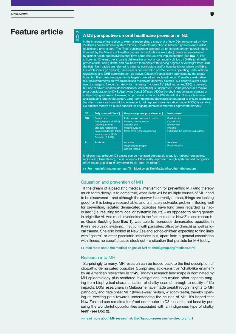
In the interests of translation to external readership, a snapshot of how D3s are covered by New Zealand's oral healthcare system follows. Residents may choose between government-funded (public) and private care. The "free" public system operates up to 18 years under national regula-tions set by the Ministry of Health (specialist orthodontics is excluded). Services are delivered by district health boards (DHBs) that have some latitude over implementation (see Box 7). For children (≤ 12 years), basic care is delivered in school or community clinics by OHPs (oral health professionals, being dental and oral health therapists) with varying degrees of oversight from DHB dentists. Non-basics are referred to external contractors and/or hospital clinics where available. For adolescents (≤18 years), basic care is contracted to private dentists operating under national regulations and DHB administration, as above. D3s aren't specifically addressed by the regula-tions, but their basic management is largely covered as tabulated below. Premature restorative failures/retreatments on hypomineralised molars are generally covered, but policy is silent about use of amalgam. A recent strategy for managing "hypomin Es" (Hall technique/SSC) is covered, but use of silver fluorides (desensitisation, cariostasis) is unapproved. Some procedures require prior consideration by DHB Approving Dental Officers (ADOs) thereby introducing an element of subjectivity (grey areas). However, no provision is made for D3-related difficulties such as slow analgesia and lengthy education. Long-term treatment planning is encouraged to ensure seamless transfer of services from child to adolescent, but regional implementation (under ADOs) is variable. D3 patients receive no public support for ongoing dentalcare after their eighteenth birthday.
D3 Fully covered ("free") Grey area (pre-approval needed) Not covered
MH Basic examRadiographs (incl. OPG)Cleaning, sealingStandard restorationsBasic endodontics (RCT)Interim crowns (SSC)Extraction (LA/GA)
Full coverage permanent crownsVeneers, microabrasionSedation (RA)Imaging (CBCT)Minor ortho (space maintainer)
PaedodontistOrthodontistEndodontistImplantsExtra time (LA, chairside education)
AI As above As abovePsychological supportGenetic testing
As aboveProsthodontist
It follows that, although D3 basics can be managed adequately today (cf. national regulations, regional implementation), the situation could be vastly improved through systematised recognition of D3 issues (e.g. Box 7: "Hypomin Track" and "D3 clinics")
>> For more information, contact Tim Mackay at: [email protected]
Volume 195 October 2019 39
t
D3 challenges for OHPs
BO
X 4
Being the first face of dentistry for most New Zealand children, community OHPs (oral health professionals) probably see more D3s than anyone – and not just severe MH meriting immediate specialist attention, but also the "mild" cases (many of which have hypersensitivity) that require ongoing preventive care and family education. Yet despite this "D3-coalface" role, the OHP workforce faces patchy support from the public system. New Zealand Dental and Oral Health Therapist Association president, Arish Naresh, has been pushing to improve D3 education amongst the OHP community, not least by having D3G members lecture at their CPD events. Arish says besides workforce deficiencies in education and training, he suspects there's considerable variation in D3 management quality across different clinics and DHBs. Regards MH, particular concerns surround early detection and subsequent management of moderate/severe cases, where OHPs often struggle to get appropriate support from dentists, paedodontists and orthodontists. For instance, exams could be better scheduled to coincide with newly erupted molars, and recalls then set depending on what's found and done. Patient and guardian education is another aspect needing attention, with most clinics lacking D3G's family-friendly chairside aids (storybook, reward stickers, website referral cards). Arish recognises particular issues in this regard surrounding "Poly-nesian AI" (Box 2) through his personal experiences working with East Coast Māori.
>> For more information, contact Arish Naresh at: [email protected]
D3 challenges for GDPs
BO
X 5
When it comes to D3-savviness, there is diversity among general dental practitioners (GDPs) – although much less so in NZ than many other countries. Unsurprisingly, this reflects heterogeneity not only in dental education and CPD, but also in exposure to children with newly erupted teeth. Rural Taranaki's MaryAnne Costelloe sees more children than most, having a postgraduate diploma in paediatric dentistry and a busy CPD schedule to match. With D3 expertise not an is-sue, the main difficulties hampering MaryAnne's own daily toils relate to the time-consuming and often costly nature of MH management. She says proper treatment planning is paramount, both in the short and long terms. For instance, at her practice, visits are minimised to avoid repeatedly exposing MH children to local analgesia difficulties, and also to limit work absences for parents/caregivers (many of whom are in "Struggle Street"). Countering this is the extra time MaryAnne takes explaining D3s to families – she's aware that for many children this is their first confrontation with invasive dentistry and so a child-friendly approach is paramount. For "hypomin 6s", unless early extraction is anticipated, composites are generally favoured having observed many SSCs being lost to follow-up – and in extraction cases, it pays to screen parents for congenitally-absent 8s. Looking more broadly across her referral base, MaryAnne sees common difficulties detect-ing MH early and often wonders whether GDPs realise cast overlays/onlays are covered under contract. Regards grey funding areas (Box 3), lack of recognition for extra time spent on paediat-ric treatments and D3 education is troubling – for example, SSCs on (hypomin) 6s typically take double the normal time for Es. MaryAnne also considers RA/sedation a must-have for anxious children, and rues that the regulations default to GA, which is often less appropriate for MH. With AI children, the above challenges are only amplified, but the system functions better when good justification (including photos/records) is provided to a D3-savvy ADO.
>> For more information, contact MaryAnne Costelloe at: [email protected]
D3 challenges for ADOs
BO
X 6
Becoming an Approving Dental Officer (ADO) reflects a variety of accomplishments and skills, but being fully on top of "everything D3" isn't necessarily included. No such concerns with Tim Mack-ay, ADO for the Southern region and leader of a major investigation addressing links between D3s, fluoridation and decay. Tim reflects that, with "hypomin 6s" having been a constant throughout his career, it seems odd that today he can only rarely help patients identify a tentative cause. From his experiences, long-term treatment planning is pivotal, but often complex given the varied presenta-tions of MH – certainly there's no room for "one size fits all" thinking. Severity grading and progno-sis should be considered foremost, balancing immediate concerns (e.g. sensitivity, decay) against long-term risk of overtreatment burdens, including dental fear. Often early extraction of severely affected molars seems the best option, and Tim always aims to get specialist orthodontic advice before finalising plans (e.g. driftodontics vs orthodontics) – he acknowledges general inacces-sibility of orthodontists within the public system is problematic. With moderate cases, long-term restorative solutions are generally favoured, often using SSCs to buy time before extraction is ruled out. Wearing his ADO hat, Tim is sympathetic to issues faced by service providers and suggests several things can be done to get the best out of current regulations. With children, MH manage-ment should start with strong teamwork between OHPs and supervising dentists, and by including others (hospital, specialists) where practicable. ADOs should be involved early in complex cases facing long-term maintenance. Moreover, the importance of OHPs in early detection and preven-tive follow-up shouldn't be underestimated. These considerations extend to adolescents, whether they be late presenters ("hypomin 7s") or carryovers from earlier treatment. Regards full crowns, the approval guidelines should be followed as usual, but it helps to spell out that MH (rather than "normal" decay) is the underlying problem. The same holds for hypomineralised anteriors where aesthetic concerns might be addressed by microabrasion or composite veneers. In AI cases requiring major rehabilitation, special dispensation might be sought to involve specialist prostho-dontic input.
>> Access Tim's study report at: www.ncbi.nlm.nih.gov/pubmed/16011308 >> For more information, contact Tim Mackay at: [email protected]
NZDA NEWS
Feature article
Patching gaps in the public health system
BO
X 7
The following examples show that deficiencies in the public system may well be patched over with a wee bit of Kiwi DIY, prompting questions as to whether such advances should be nationalised?
Christchurch's "Hypomin Clinic"About 15 years ago, some concerned practitioners took it upon themselves to implement a "Hypomin Clinic" at Christchurch hospital, targeting 6- to 8-year-olds for whom timely extraction might hold long-term benefit. This free referral service provides panoramic radiographs (OPGs) before consultation with a D3-savvy dentist, enabling a forward-looking management plan to be offered – including specialist orthodontic input (at own cost) as appropriate. Depending on needs and choice, management is then handled by the hospital dentistry department (liaison between specialist paedodontists and orthodontists), family dentist and/or referring OHP. Public health den-tist Tule Misa who has run the service for 10 years says that, while it's heavily used, unfortunately not all deserving MH cases get to the clinic. Consequently, to improve awareness and resourc-ing, Tule is working with children's dentist Joanna Pedlow (diplomate in paediatric dentistry and co-founder of the clinic) to capture MH prevalence data from across their service area. A similar initiative (named "D3 Clinic", to embrace AI etc.) has recently commenced in Wellington, led by public health dentists Kathy Fuge and Charlotte Hurst.
Auckland's "Hypomin Management Plan"Realising the need to intervene early, public health dentist Satha Kanagaratnam has formulated a "Hypomineralised Teeth Management Plan" for OHPs and dentists in Auckland's Community Den-tal Services. Adopted in 2018, the plan covers early screening for MH and puts affected children on a "Hypomin Track" to foster best care through to 18 years. Included are the needs to address all molars (primary, permanent), to consider severity grade (preventive vs invasive treatment), to seek orthodontic opinion, to monitor regularly, and to educate about appropriate homecare. Con-sequently, children are screened from birth and 2-year molars (Es) are checked once fully erupted. Other important aspects covered are patient anxiety (MH-related pain and dentistry), timing of extractions (orthodontic input), and choice of restorative material (no amalgam!). An associated "Hypomineralisation Fact Sheet" for patients/families outlines the basics and refers to D3G for further information. While such plans are a great start, effective implementation is another matter. Satha says that current hurdles include clunky access to OPGs (requires referral to contracting dentist), a lack of government-funded orthodontist, and no integrated "D3 home" along Christch-urch and Wellington lines. Limitations aside, when it comes to MH, it's apparent that Auckland children are now being served better.
>> For more information, please contact Tule and Satha at: [email protected]
Attacking MH translationallyIf it's accepted that the MH problem warrants a translationally-directed attack,
questioning must turn to today's widespread lack of knowledge and scientific un-derstanding, and suboptimal clinical results for children. While nuggets of aware-ness (and action) can easily be found amongst its various elements, the dental profession as a whole seems inadequately positioned regards MH. An outline of New Zealand's regulatory framework for oral healthcare provision, and various D3 perspectives from across the profession, are given in Boxes 3-10. These consid-erations point to several aspects that should be improved upon, prompting the question how might this be accomplished?
The D3 Group and its Chalky Teeth CampaignFormed across Australia and New Zealand a decade ago, D3G is a cross-sector
translational network dedicated to the better understanding and care of people with chalky teeth/D3s. Internationalisation of D3G was commenced in 2017 and mem-bership today extends across thirty-seven countries with representation from the dental and medical professions, science, industry, government and the public. Re-flecting growing status, D3G's inaugural International Symposium on MH and Chalky Teeth will be held in Canada (22-24 October 2020) hosted by Toronto's cel-ebrated University of Toronto and SickKids Hospital. D3G's public-facing arm, the Chalky Teeth Campaign, aims to educate about MH and other D3s, the links with tooth decay, the need for research, and the important advocacy roles the public can play. Some key achievements of D3G are outlined in Boxes 8-10. Given the heritage from Grace Suckling and its Kiwi founder, D3G stands to shine light on New Zea-land's capacity to innovate in an overlooked yet highly important area of healthcare.
>> read more about D3G's educational and media contributions at: thed3group.org/media
Volume 195 October 2019 41
t
Sharing a storybook about "chalky molars"
BO
X 8
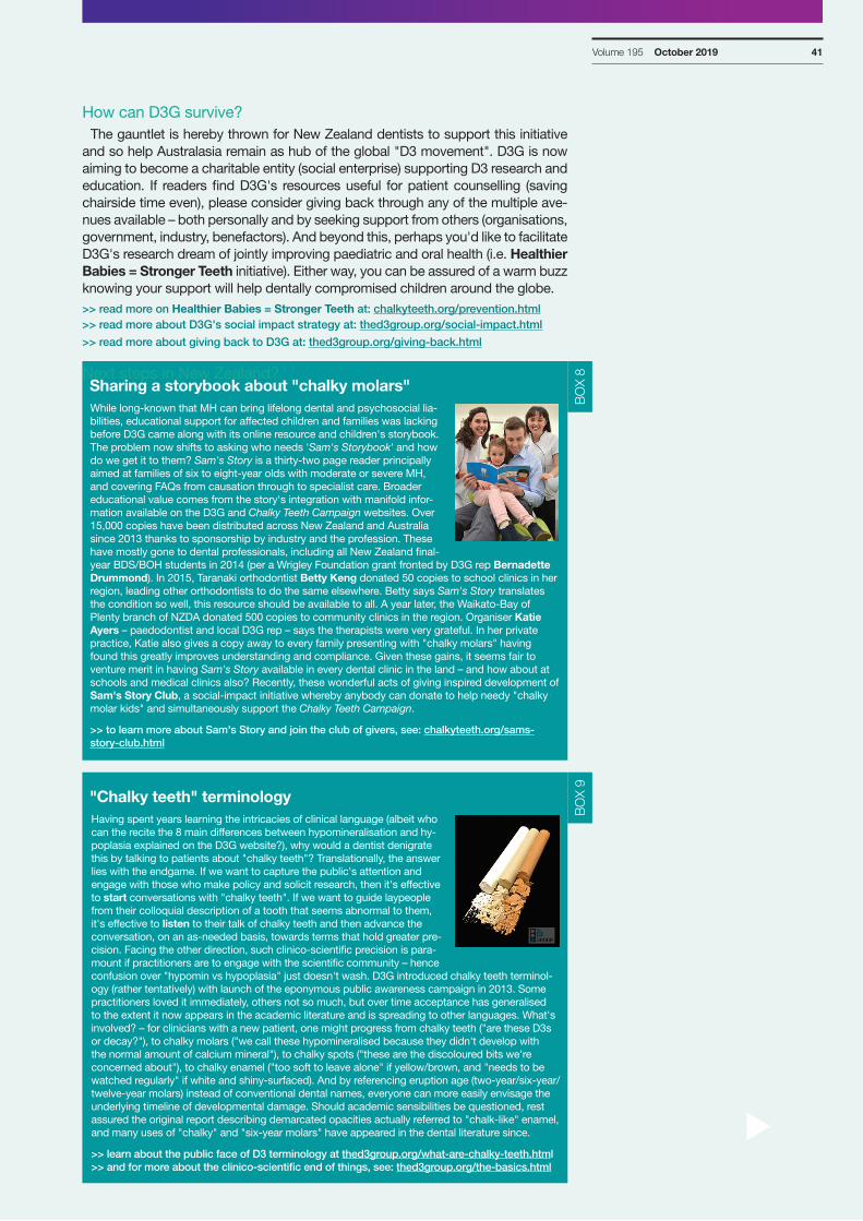
While long-known that MH can bring lifelong dental and psychosocial lia-bilities, educational support for affected children and families was lacking before D3G came along with its online resource and children's storybook. The problem now shifts to asking who needs 'Sam's Storybook' and how do we get it to them? Sam's Story is a thirty-two page reader principally aimed at families of six to eight-year olds with moderate or severe MH, and covering FAQs from causation through to specialist care. Broader educational value comes from the story's integration with manifold infor-mation available on the D3G and Chalky Teeth Campaign websites. Over 15,000 copies have been distributed across New Zealand and Australia since 2013 thanks to sponsorship by industry and the profession. These have mostly gone to dental professionals, including all New Zealand final-year BDS/BOH students in 2014 (per a Wrigley Foundation grant fronted by D3G rep Bernadette Drummond). In 2015, Taranaki orthodontist Betty Keng donated 50 copies to school clinics in her region, leading other orthodontists to do the same elsewhere. Betty says Sam's Story translates the condition so well, this resource should be available to all. A year later, the Waikato-Bay of Plenty branch of NZDA donated 500 copies to community clinics in the region. Organiser Katie Ayers – paedodontist and local D3G rep – says the therapists were very grateful. In her private practice, Katie also gives a copy away to every family presenting with "chalky molars" having found this greatly improves understanding and compliance. Given these gains, it seems fair to venture merit in having Sam's Story available in every dental clinic in the land – and how about at schools and medical clinics also? Recently, these wonderful acts of giving inspired development of Sam's Story Club, a social-impact initiative whereby anybody can donate to help needy "chalky molar kids" and simultaneously support the Chalky Teeth Campaign.
>> to learn more about Sam's Story and join the club of givers, see: chalkyteeth.org/sams-story-club.html
How can D3G survive?The gauntlet is hereby thrown for New Zealand dentists to support this initiative
and so help Australasia remain as hub of the global "D3 movement". D3G is now aiming to become a charitable entity (social enterprise) supporting D3 research and education. If readers find D3G's resources useful for patient counselling (saving chairside time even), please consider giving back through any of the multiple ave-nues available – both personally and by seeking support from others (organisations, government, industry, benefactors). And beyond this, perhaps you'd like to facilitate D3G's research dream of jointly improving paediatric and oral health (i.e. Healthier Babies = Stronger Teeth initiative). Either way, you can be assured of a warm buzz knowing your support will help dentally compromised children around the globe. >> read more on Healthier Babies = Stronger Teeth at: chalkyteeth.org/prevention.html>> read more about D3G's social impact strategy at: thed3group.org/social-impact.html>> read more about giving back to D3G at: thed3group.org/giving-back.html
Next steps in New Zealand?
"Chalky teeth" terminologyHaving spent years learning the intricacies of clinical language (albeit who can the recite the 8 main differences between hypomineralisation and hy-poplasia explained on the D3G website?), why would a dentist denigrate this by talking to patients about "chalky teeth"? Translationally, the answer lies with the endgame. If we want to capture the public's attention and engage with those who make policy and solicit research, then it's effective to start conversations with "chalky teeth". If we want to guide laypeople from their colloquial description of a tooth that seems abnormal to them, it's effective to listen to their talk of chalky teeth and then advance the conversation, on an as-needed basis, towards terms that hold greater pre-cision. Facing the other direction, such clinico-scientific precision is para-mount if practitioners are to engage with the scientific community – hence confusion over "hypomin vs hypoplasia" just doesn't wash. D3G introduced chalky teeth terminol-ogy (rather tentatively) with launch of the eponymous public awareness campaign in 2013. Some practitioners loved it immediately, others not so much, but over time acceptance has generalised to the extent it now appears in the academic literature and is spreading to other languages. What's involved? – for clinicians with a new patient, one might progress from chalky teeth ("are these D3s or decay?"), to chalky molars ("we call these hypomineralised because they didn't develop with the normal amount of calcium mineral"), to chalky spots ("these are the discoloured bits we're concerned about"), to chalky enamel ("too soft to leave alone" if yellow/brown, and "needs to be watched regularly" if white and shiny-surfaced). And by referencing eruption age (two-year/six-year/twelve-year molars) instead of conventional dental names, everyone can more easily envisage the underlying timeline of developmental damage. Should academic sensibilities be questioned, rest assured the original report describing demarcated opacities actually referred to "chalk-like" enamel, and many uses of "chalky" and "six-year molars" have appeared in the dental literature since.
>> learn about the public face of D3 terminology at thed3group.org/what-are-chalky-teeth.html >> and for more about the clinico-scientific end of things, see: thed3group.org/the-basics.html
BO
X 9
NZDA NEWS
Feature articleA network of Chalky Teeth Specialists
BO
X 10
Support from the profession is pivotal to D3G’s innovations and activities, so what could be better than a group of specialist den-tists proclaiming We Fight Chalky Teeth? This emerging network venture (dubbed WFCT) is already shaping as a 4-way win. For the public, having a practice-based face to the Chalky Teeth Campaign adds a more personal touch to existing endorsements from professional organisations and industry. Within the field, WFCT has much potential to serve as expert opinion, informer for education, contributor to research, and testbed for industry. Network members stand to benefit from associations with each other and across D3G more broadly. And, in addition to all the above, D3G benefits from allied revenue to support research and education. Nina Vasan, pioneering paedodontist and WFCT member, says she backed the idea from day one wanting to both support D3G and visibly connect her practice with the D3 movement. D3s have consumed much of her time since going private 20 years ago and MH today accounts for up to 40% of her patients. Keep-ing up-to-date with "all things D3" helps in providing top quality advice and management, and the chalky teeth/D3 workflow has become a natural part of daily business for Nina and her team. For example, it's been refreshing to provide "chalky teeth check-ups" and use Sam's Story to explain background information and clinical steps. Parents are directed to the D3G website for more detailed information and the feedback has always been positive. Over the years, Nina has enjoyed working together with orthodontists to produce joint treatment plans that optimise outcomes and reduce the long-term restorative burden – she expects these liaisons will only get better with WFCT. And from New Zealand's first WFCT orthodontic practice, Marguerite Crooks and John Perry add they're enjoying being able to talk simply about chalky molars and interact with the or-thodontics pages in Sam's Storybook, whilst also supporting D3G's mission to foster research and education. Chalky molars can be an emotional issue for parents who think (or may've been told) they're to blame for what's been misconstrued as regular decay – so it’s great having D3G's family-friendly tools available to put minds at ease. They encourage other orthodontic practices to get on board not only to utilise all the resources, but also to support MH research, saying "who knows, maybe a vaccine or supplement can be developed to nip this disturbing problem in the bud – much like taking folate to prevent neural tube defects". While WFCT is presently restricted to specialist practices, plans are afoot to develop partner initiatives for other branches of the D3 family.
>> learn more about the WFCT network at chalkyteeth.org/we-fight-chalky-teeth-practices.html
BIOGRAPHY
Professor Mike Hubbard BDS, PhDMike Hubbard is a Professorial Fellow in Oral and Facial Sciences at the University of Melbourne, Australia. After his BDS at Otago, Mike did a PhD in biochemistry leading to a biomedical research career. He later moved to Melbourne to establish a "translational" network of dentists, doctors and scientists addressing medico-dental problems in children. A major outcome was The D3 Group, an international initiative focussed on Molar Hypomineralisation and other D3s (developmental dental defects). To help "translate" research advances into social good, Mike has introduced a public awareness campaign (chalkyteeth.org), authored a children's storybook about Molar Hypomineralisation and developed a world-first online education resource for D3s (thed3group.org).
Is it time for a formalised national conversation about New Zealand’s children with chalky teeth? The foregoing suggests there is a lot to be spoken about by many, plus consensus that today's dental management could be improved on quite read-ily. Add to that New Zealand's ongoing leadership role in the D3 movement plus the global health opportunities surrounding medical prevention of MH, then the case for a translationally-directed forum/hui seems compelling. Meanwhile, inter-ested parties are encouraged to engage with their local D3G representative.
>> for D3G contacts in NZ, see: thed3group.org/contact-d3g.html
More informationFor more information and enquiries, please contact Mike Hubbard at: mike.
Volume 195 October 2019 45
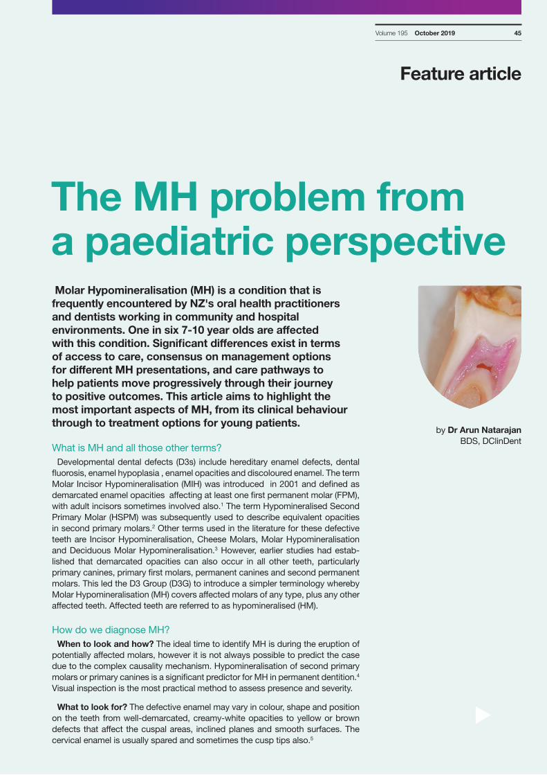
Molar Hypomineralisation (MH) is a condition that is frequently encountered by NZ's oral health practitioners and dentists working in community and hospital environments. One in six 7-10 year olds are affected with this condition. Significant differences exist in terms of access to care, consensus on management options for different MH presentations, and care pathways to help patients move progressively through their journey to positive outcomes. This article aims to highlight the most important aspects of MH, from its clinical behaviour through to treatment options for young patients.
What is MH and all those other terms?Developmental dental defects (D3s) include hereditary enamel defects, dental
fluorosis, enamel hypoplasia , enamel opacities and discoloured enamel. The term Molar Incisor Hypomineralisation (MIH) was introduced in 2001 and defined as demarcated enamel opacities affecting at least one first permanent molar (FPM), with adult incisors sometimes involved also.1 The term Hypomineralised Second Primary Molar (HSPM) was subsequently used to describe equivalent opacities in second primary molars.2 Other terms used in the literature for these defective teeth are Incisor Hypomineralisation, Cheese Molars, Molar Hypomineralisation and Deciduous Molar Hypomineralisation.3 However, earlier studies had estab-lished that demarcated opacities can also occur in all other teeth, particularly primary canines, primary first molars, permanent canines and second permanent molars. This led the D3 Group (D3G) to introduce a simpler terminology whereby Molar Hypomineralisation (MH) covers affected molars of any type, plus any other affected teeth. Affected teeth are referred to as hypomineralised (HM).
How do we diagnose MH? When to look and how? The ideal time to identify MH is during the eruption of
potentially affected molars, however it is not always possible to predict the case due to the complex causality mechanism. Hypomineralisation of second primary molars or primary canines is a significant predictor for MH in permanent dentition.4 Visual inspection is the most practical method to assess presence and severity.
What to look for? The defective enamel may vary in colour, shape and position on the teeth from well-demarcated, creamy-white opacities to yellow or brown defects that affect the cuspal areas, inclined planes and smooth surfaces. The cervical enamel is usually spared and sometimes the cusp tips also.5
Feature article
The MH problem from a paediatric perspective
t
by Dr Arun Natarajan BDS, DClinDent
Member only access
NZDA NEWS
Feature article The clinical presentations of MH are:
n Change in translucency of enameln Sharp demarcation between affected and sound enameln Discoloured patches – white-cream, yellow-brownn Dull (porous) or shiny surface appearancen Porous, brittle enamel n Post-eruptive enamel breakdown (PEB)n Atypical caries (i.e. located in areas not usually associated with plaque
accumulation)
Once identified with these clinical signs, questions should be asked about any illness that occurred in the prenatal, perinatal or postnatal periods, or the first three of years of life, to support the diagnosis.
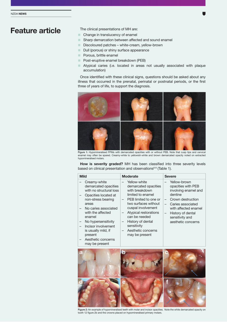
Figure 1: Hypomineralised FPMs with demarcated opacities with or without PEB. Note that cusp tips and cervical enamel may often be spared. Creamy-white to yellowish-white and brown demarcated opacity noted on extracted hypomineralised molars.
How is severity graded? MH has been classified into three severity levels based on clinical presentation and observations6-9 (Table 1).
Mild Moderate Severe– Creamy-white
demarcated opacities with no structural loss
– Opacities located at non-stress bearing areas
– No caries associated with the affected enamel
– No hypersensitivity – Incisor involvement
is usually mild, if present
– Aesthetic concerns may be present
– Yellow-white demarcated opacities with breakdown limited to enamel
– PEB limited to one or two surfaces without cuspal involvement
– Atypical restorations can be needed
– History of dental sensitivity
– Aesthetic concerns may be present
– Yellow-brown opacities with PEB involving enamel and dentine
– Crown destruction – Caries associated
with affected enamel – History of dental
sensitivity and aesthetic concerns
Figure 2: An example of hypomineralised teeth with molar and incisor opacities. Note the white demarcated opacity on tooth 12 figure 2b and the crowns placed on hypomineralised primary molars.
a b c
d e f
Volume 195 October 2019 47
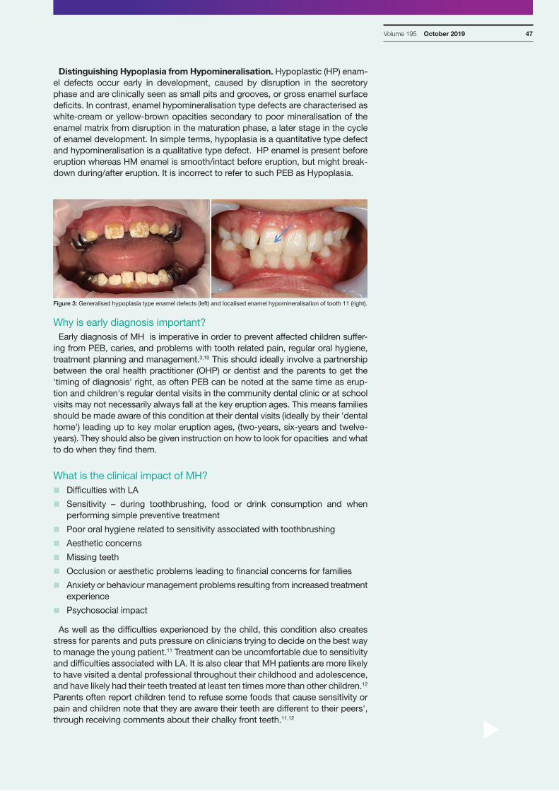
Distinguishing Hypoplasia from Hypomineralisation. Hypoplastic (HP) enam-el defects occur early in development, caused by disruption in the secretory phase and are clinically seen as small pits and grooves, or gross enamel surface deficits. In contrast, enamel hypomineralisation type defects are characterised as white-cream or yellow-brown opacities secondary to poor mineralisation of the enamel matrix from disruption in the maturation phase, a later stage in the cycle of enamel development. In simple terms, hypoplasia is a quantitative type defect and hypomineralisation is a qualitative type defect. HP enamel is present before eruption whereas HM enamel is smooth/intact before eruption, but might break-down during/after eruption. It is incorrect to refer to such PEB as Hypoplasia.
Why is early diagnosis important?Early diagnosis of MH is imperative in order to prevent affected children suffer-
ing from PEB, caries, and problems with tooth related pain, regular oral hygiene, treatment planning and management.3,10 This should ideally involve a partnership between the oral health practitioner (OHP) or dentist and the parents to get the 'timing of diagnosis' right, as often PEB can be noted at the same time as erup-tion and children's regular dental visits in the community dental clinic or at school visits may not necessarily always fall at the key eruption ages. This means families should be made aware of this condition at their dental visits (ideally by their 'dental home') leading up to key molar eruption ages, (two-years, six-years and twelve-years). They should also be given instruction on how to look for opacities and what to do when they find them.
What is the clinical impact of MH? n Difficulties with LA
n Sensitivity – during toothbrushing, food or drink consumption and when performing simple preventive treatment
n Poor oral hygiene related to sensitivity associated with toothbrushing
n Aesthetic concerns
n Missing teeth
n Occlusion or aesthetic problems leading to financial concerns for families
n Anxiety or behaviour management problems resulting from increased treatment experience
n Psychosocial impact
As well as the difficulties experienced by the child, this condition also creates stress for parents and puts pressure on clinicians trying to decide on the best way to manage the young patient.11 Treatment can be uncomfortable due to sensitivity and difficulties associated with LA. It is also clear that MH patients are more likely to have visited a dental professional throughout their childhood and adolescence, and have likely had their teeth treated at least ten times more than other children.12 Parents often report children tend to refuse some foods that cause sensitivity or pain and children note that they are aware their teeth are different to their peers', through receiving comments about their chalky front teeth.11,12
Figure 3: Generalised hypoplasia type enamel defects (left) and localised enamel hypomineralisation of tooth 11 (right).
t
NZDA NEWS
What makes hypomineralised enamel abnormal?The structural, mechanical and chemical properties of HM enamel have been
studied extensively, all of which has shown differences in mineral quantity and quality, increased porosity and organic content along with reduced hardness and elasticity. Jalevik et al13 compared HM enamel to that of normal enamel using sec-ondary ion mass spectrometry and X-ray microanalysis to determine the mineral composition. They found that the median Ca/P ratios in the cervical hypomineral-ised areas were significantly lower (1.4) when compared to those in the adjacent normal cervical enamel (1.8). However, the Ca levels were higher for both HM and normal enamel at the outermost surface layer of enamel, which suggests that further mineralisation occurred during post-eruptive maturation.
Mahoney et al8 investigated the mechanical and morphological properties of HM enamel using several methods. Scanning electron microscopy (SEM) revealed HM enamel was more porous and had disorganized enamel rods of varying widths which lacked distinct boundaries between them. The hardness and modulus of elasticity were also found to be statistically significantly lower (50-75% reduction).
Farah et al,6 used X-ray microtomography (XMT) to study the mineral density (MD) of HM enamel samples with varying severity. They found that the mean MD was significantly lower for HM enamel (19% lower) when compared to normal enamel. MD values dropped from the CEJ to the enamel surface, but were in-creased in the cusp tips. In contrast, a steady increase in MD from the CEJ to the enamel surface and the cusp tips was observed in normal enamel.
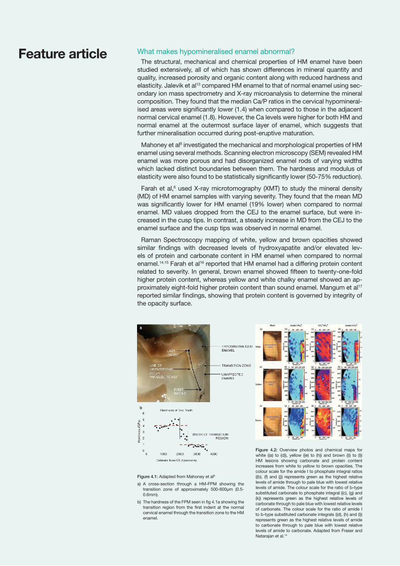
Raman Spectroscopy mapping of white, yellow and brown opacities showed similar findings with decreased levels of hydroxyapatite and/or elevated lev-els of protein and carbonate content in HM enamel when compared to normal enamel.14,15 Farah et al16 reported that HM enamel had a differing protein content related to severity. In general, brown enamel showed fifteen to twenty-one-fold higher protein content, whereas yellow and white chalky enamel showed an ap-proximately eight-fold higher protein content than sound enamel. Mangum et al17 reported similar findings, showing that protein content is governed by integrity of the opacity surface.
Feature article
Figure 4.2: Overview photos and chemical maps for white ((a) to (d)), yellow ((e) to (h)) and brown ((i) to (l)) HM lesions showing carbonate and protein content increases from white to yellow to brown opacities. The colour scale for the amide I to phosphate integral ratios ((b), (f) and (j)) represents green as the highest relative levels of amide through to pale blue with lowest relative levels of amide. The colour scale for the ratio of b-type substituted carbonate to phosphate integral ((c), (g) and (k)) represents green as the highest relative levels of carbonate through to pale blue with lowest relative levels of carbonate. The colour scale for the ratio of amide I to b-type substituted carbonate integrals ((d), (h) and (l)) represents green as the highest relative levels of amide to carbonate through to pale blue with lowest relative levels of amide to carbonate. Adapted from Fraser and Natarajan et al.14
Figure 4.1: Adapted from Mahoney et al8
a) A cross-section through a HM-FPM showing the transition zone of approximately 500-600µm (0.5-0.6mm).
b) The hardness of the FPM seen in fig 4.1a showing the transition region from the first indent at the normal cervical enamel through the transition zone to the HM enamel.
Volume 195 October 2019 49
Why are affected molars hypersensitive and how to manage? HM molars can be very sensitive to air, cold and/or heat and mechanical stimuli
including toothbrushing.3 It is thought this may result from repeated small painful stimuli resulting in mild inflammation of the pulp.19,20 Rodd et al20 reported that some noncarious HM molars have an underlying pulpal inflammation demonstrat-ed by increased vascularity, pulpal innervation and immune cell accumulation.20 Porous HM enamel plus PEB along with wide dentinal tubules in young perma-nent teeth may act as an easy pathway for external stimuli to reach the dentinal tubules resulting in increased sensitivity despite administration of LA. The hydro-dynamic theory of fluid movement within dentine is likely the mechanism behind short sharp pain experienced by some children and this likely depends on the severity of the affected HM enamel and the underlying pulpal inflammation.20 All of these pathophysiological changes are thought to contribute to failure of pulpal analgesia following administration of LA in some MH children.
Recommendations to manage sensitivity in HM molars are based upon empiri-cal evidence and long-term clinical studies are underway to support the practice-based evidence. Hypersensitivity with MH can manifest as problems with main-taining good oral hygiene, refusal of eating or drinking specific foods and drinks, through to difficulty in performing simple 'triaging' procedures such as glass iono-mer sealants or resin based restorations.
Reports suggest the use of topical fluorides and remineralising agents such as CPP-ACP can help control hypersensitivity, as well as further mineralise the HM enamel.10,21,22 Toothpastes prescribed to manage dentine hypersensitivity in adults are sometimes seen as a cost-effective alternative to CPP-ACP based products when managing everyday sensitivity with MH. Fissure sealants and adhesive ma-terials appear to prevent sensitivity and further breakdown23 and may be done as an interim triage measure until a more definitive management plan is made. Rela-tive analgesia (RA) or nitrous oxide/oxygen inhalational sedation combined with single administration of pre-emptive analagesics (Paracetamol 30mg/kg one hour prior or Ibuprofen 15-20mg/kg thirty mins prior to treatment) may help children cope with restorative treatments. LA with 4% Articaine is reported to be more ef-fective than standard lignocaines.
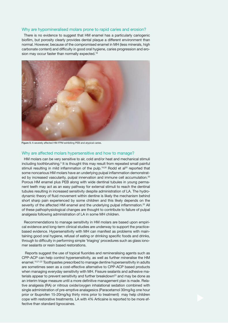
Figure 5: A severely affected HM-FPM exhibiting PEB and atypical caries.
t
Why are hypomineralised molars prone to rapid caries and erosion?There is no evidence to suggest that HM enamel has a particularly cariogenic
biofilm, but porosity clearly provides dental plaque a different environment than normal. However, because of the compromised enamel in MH (less minerals, high carbonate content) and difficulty in good oral hygiene, caries progression and ero-sion may occur faster than normally expected.18
NZDA NEWS
Why are the borders of demarcated opacities hard to define? – what does this mean for my cavity margins?
It has been recommended that the clinical success of a resin composite resto-ration could be improved in HM enamel by removing all discoloured enamel and placing the cavity margins on apparently normal enamel as visible to the naked eye.21 As shown in figure 4.1, the presence of a transition zone between opaque and visibly normal enamel (approximately 0.6mm) has been documented8,28 and studies have shown prism sheaths in the transition zone and the affected enam-el are much weaker than those unaffected enamel, due to loosely packed hy-droxyapatite crystals surrounded by weaker prism sheaths.28 What appears to be sound enamel under the naked eye has been proven to exhibit poor mechanical and chemical properties similar to that of demarcated defects seen in HM enam-el. Hence the clinical recommendation that placing cavity margins on apparently sound enamel when restoring HM teeth could well be an invasive, less practical approach and might not necessarily prove to improve the clinical success of resin composite restorations, given most defects in HM enamel appear to be full length defects (from surface layer to DEJ).
Feature article Why is my etching and bonding abnormal in hypomineralised enamel? It is now well known that the etching process with HM enamel is disrupted by
abnormal mineral composition and protein content, leading to ineffective bond-ing. The poor bond strength of resin adhesives to HM enamel is well documented in the dental literature. Organic matter has poor acid solubility and its presence in increased amounts in the HM enamel24 may well inhibit the creation of adequate etch pattern, which in turn will compromise the adhesion between resin based restorative materials and the defective enamel.25 The chemical composition of HM enamel along with the soft, and porous nature of the enamel matrix with de-graded prism sheaths makes resin restoration of PEB difficult to manage.8,26 An SEM study found that etching HM enamel using 35% phosphoric acid resulted in minimal preferential dissolution of interrod enamel, providing less surface area for bonding adhesives.25 Jalevik and Dietz et al19 suggested that the poor acid solubility could be due to both the higher protein content and also the higher car-bonate content leading to greater acid solubility of enamel mineral.27 This has led some authors to suggest longer etching time when treating HM enamel, although there is little evidence to show this is useful.
The enamel-adhesive interface has been investigated in MH and shown to be po-rous with enamel cracks lacking a consistent hybrid layer. A high frequency of co-hesive failures reported in HM enamel indicates its inherent weakness and the poor etching response that does not allow for the desired resin tag formation and micro-mechanical retention.25 This is seen clinically as gaps between the restoration and the remaining tooth that often occurs from rupture of remaining affected enamel.
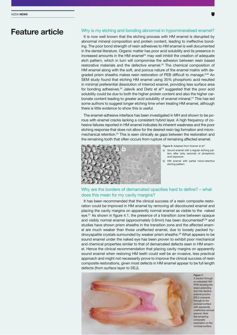
Figure 6: Adapted from Kramer et al47
a) Sound enamel with a regular etching pat-tern after sixty seconds of phosphoric acid exposure.
b) HM enamel with partial micro-retentive etching pattern.
Figure 7: A section through an extracted HM-FPM showing the lesion extending from the dentine-enamel junction (DEJ) outwards through to the occlusal surface with apparently unaffected cervical enamel. Note the remaining composite restoration on the occlusal surface.
Volume 195 October 2019 51
Other problems faced when restoring hypomineralised molarsDifficult restorative problems have been reported to occur when cusps of HM
molars are involved.22,25 Kotsanos et al29, retrospectively reviewed treatment re-cords of seventy-two children and found a significantly higher mean DMFS in the MH group (n=36) compared with the unaffected group (n=36). At the 4.5-year follow up, MH children had required more restorative retreatments, with 61% of amalgam restorations requiring retreatment compared with 25% of composite res-torations and no molar treated with a stainless steel crown needed re-intervention. Hence, amalgam restorations are not recommended on HM molars.
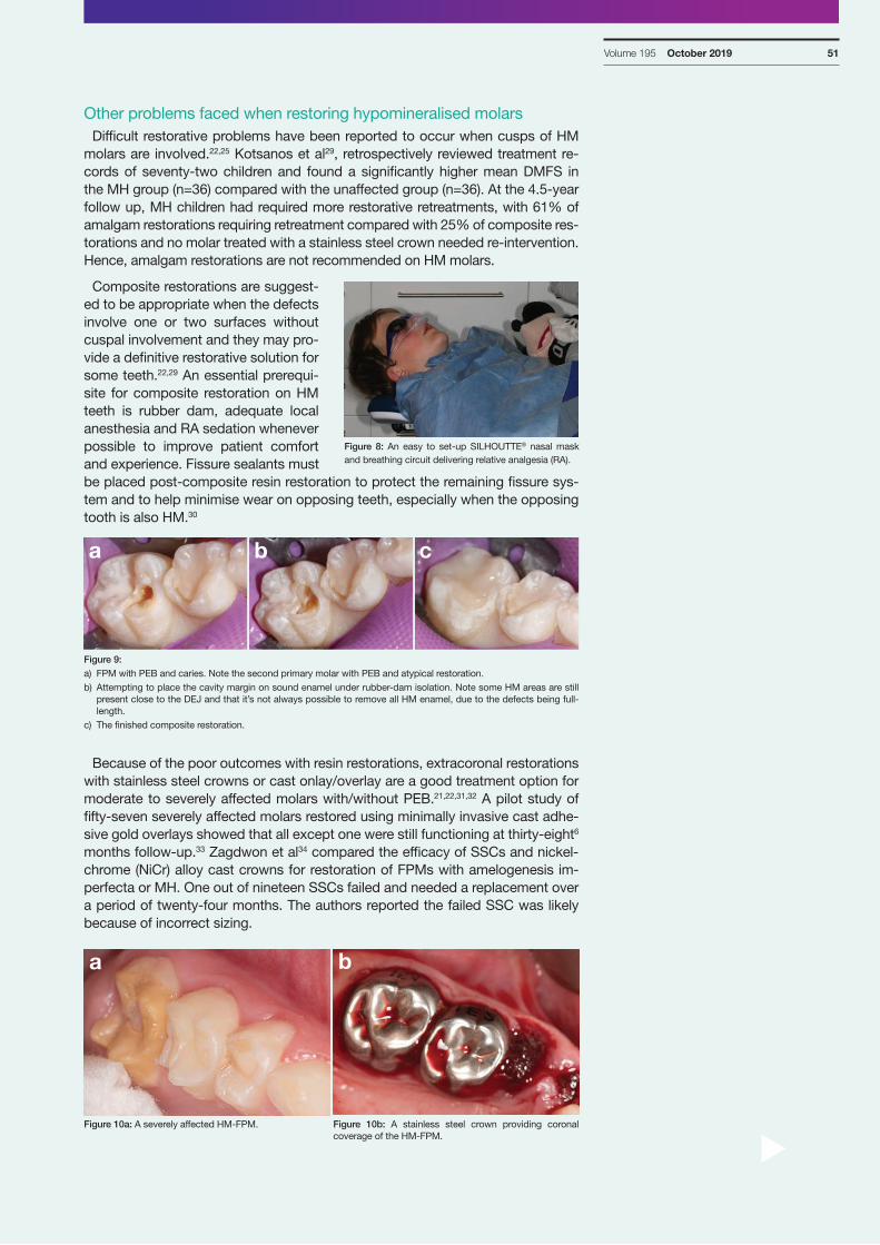
Composite restorations are suggest-ed to be appropriate when the defects involve one or two surfaces without cuspal involvement and they may pro-vide a definitive restorative solution for some teeth.22,29 An essential prerequi-site for composite restoration on HM teeth is rubber dam, adequate local anesthesia and RA sedation whenever possible to improve patient comfort and experience. Fissure sealants must be placed post-composite resin restoration to protect the remaining fissure sys-tem and to help minimise wear on opposing teeth, especially when the opposing tooth is also HM.30
t
a b c
a b
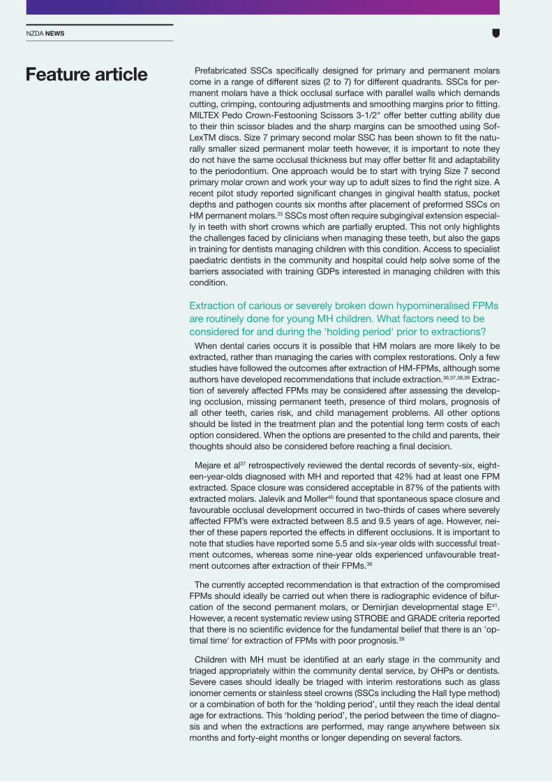
Because of the poor outcomes with resin restorations, extracoronal restorations with stainless steel crowns or cast onlay/overlay are a good treatment option for moderate to severely affected molars with/without PEB.21,22,31,32 A pilot study of fifty-seven severely affected molars restored using minimally invasive cast adhe-sive gold overlays showed that all except one were still functioning at thirty-eight6 months follow-up.33 Zagdwon et al34 compared the efficacy of SSCs and nickel-chrome (NiCr) alloy cast crowns for restoration of FPMs with amelogenesis im-perfecta or MH. One out of nineteen SSCs failed and needed a replacement over a period of twenty-four months. The authors reported the failed SSC was likely because of incorrect sizing.
Figure 9: a) FPM with PEB and caries. Note the second primary molar with PEB and atypical restoration.b) Attempting to place the cavity margin on sound enamel under rubber-dam isolation. Note some HM areas are still
present close to the DEJ and that it’s not always possible to remove all HM enamel, due to the defects being full-length.
c) The finished composite restoration.
Figure 10a: A severely affected HM-FPM. Figure 10b: A stainless steel crown providing coronal coverage of the HM-FPM.
Figure 8: An easy to set-up SILHOUTTE® nasal mask and breathing circuit delivering relative analgesia (RA).
NZDA NEWS
Prefabricated SSCs specifically designed for primary and permanent molars come in a range of different sizes (2 to 7) for different quadrants. SSCs for per-manent molars have a thick occlusal surface with parallel walls which demands cutting, crimping, contouring adjustments and smoothing margins prior to fitting. MILTEX Pedo Crown-Festooning Scissors 3-1/2" offer better cutting ability due to their thin scissor blades and the sharp margins can be smoothed using Sof-LexTM discs. Size 7 primary second molar SSC has been shown to fit the natu-rally smaller sized permanent molar teeth however, it is important to note they do not have the same occlusal thickness but may offer better fit and adaptability to the periodontium. One approach would be to start with trying Size 7 second primary molar crown and work your way up to adult sizes to find the right size. A recent pilot study reported significant changes in gingival health status, pocket depths and pathogen counts six months after placement of preformed SSCs on HM permanent molars.35 SSCs most often require subgingival extension especial-ly in teeth with short crowns which are partially erupted. This not only highlights the challenges faced by clinicians when managing these teeth, but also the gaps in training for dentists managing children with this condition. Access to specialist paediatric dentists in the community and hospital could help solve some of the barriers associated with training GDPs interested in managing children with this condition.
Extraction of carious or severely broken down hypomineralised FPMs are routinely done for young MH children. What factors need to be considered for and during the 'holding period' prior to extractions?
When dental caries occurs it is possible that HM molars are more likely to be extracted, rather than managing the caries with complex restorations. Only a few studies have followed the outcomes after extraction of HM-FPMs, although some authors have developed recommendations that include extraction.36,37,38,39 Extrac-tion of severely affected FPMs may be considered after assessing the develop-ing occlusion, missing permanent teeth, presence of third molars, prognosis of all other teeth, caries risk, and child management problems. All other options should be listed in the treatment plan and the potential long term costs of each option considered. When the options are presented to the child and parents, their thoughts should also be considered before reaching a final decision.
Mejare et al37 retrospectively reviewed the dental records of seventy-six, eight-een-year-olds diagnosed with MH and reported that 42% had at least one FPM extracted. Space closure was considered acceptable in 87% of the patients with extracted molars. Jalevik and Moller40 found that spontaneous space closure and favourable occlusal development occurred in two-thirds of cases where severely affected FPM’s were extracted between 8.5 and 9.5 years of age. However, nei-ther of these papers reported the effects in different occlusions. It is important to note that studies have reported some 5.5 and six-year olds with successful treat-ment outcomes, whereas some nine-year olds experienced unfavourable treat-ment outcomes after extraction of their FPMs.36
The currently accepted recommendation is that extraction of the compromised FPMs should ideally be carried out when there is radiographic evidence of bifur-cation of the second permanent molars, or Demirjian developmental stage E41. However, a recent systematic review using STROBE and GRADE criteria reported that there is no scientific evidence for the fundamental belief that there is an 'op-timal time' for extraction of FPMs with poor prognosis.39
Children with MH must be identified at an early stage in the community and triaged appropriately within the community dental service, by OHPs or dentists. Severe cases should ideally be triaged with interim restorations such as glass ionomer cements or stainless steel crowns (SSCs including the Hall type method) or a combination of both for the ‘holding period’, until they reach the ideal dental age for extractions. This ‘holding period’, the period between the time of diagno-sis and when the extractions are performed, may range anywhere between six months and forty-eight months or longer depending on several factors.
Feature article
Volume 195 October 2019 53
How does all this affect treatment planning for MH children?As clinicians we are often faced with a child who has HM teeth and it can be
challenging to plan treatment for many reasons. The management of teeth with HM involves several aspects including prevention of PEB, prevention of dental caries, management of hypersensitivity, management of required restorations, and/or management of the growth and development with extraction when teeth cannot be restored for the long term.11 Ambiguity is common in health decision making and MH is not an exception. The decision as to which of these manage-ment options is suitable needs to be made individually considering the severity of the lesions, symptoms, growth and development, patient’s dental age, malocclu-sion, psychosocial factors and individual patient needs and expectations.11
The decision to treat or to refer to a specialist paediatric dentist or senior dentist experienced in managing children depends on access to specialist care and facili-ties, operator skillsets and training, child's anxiety level or behavioural issues and parental preferences.
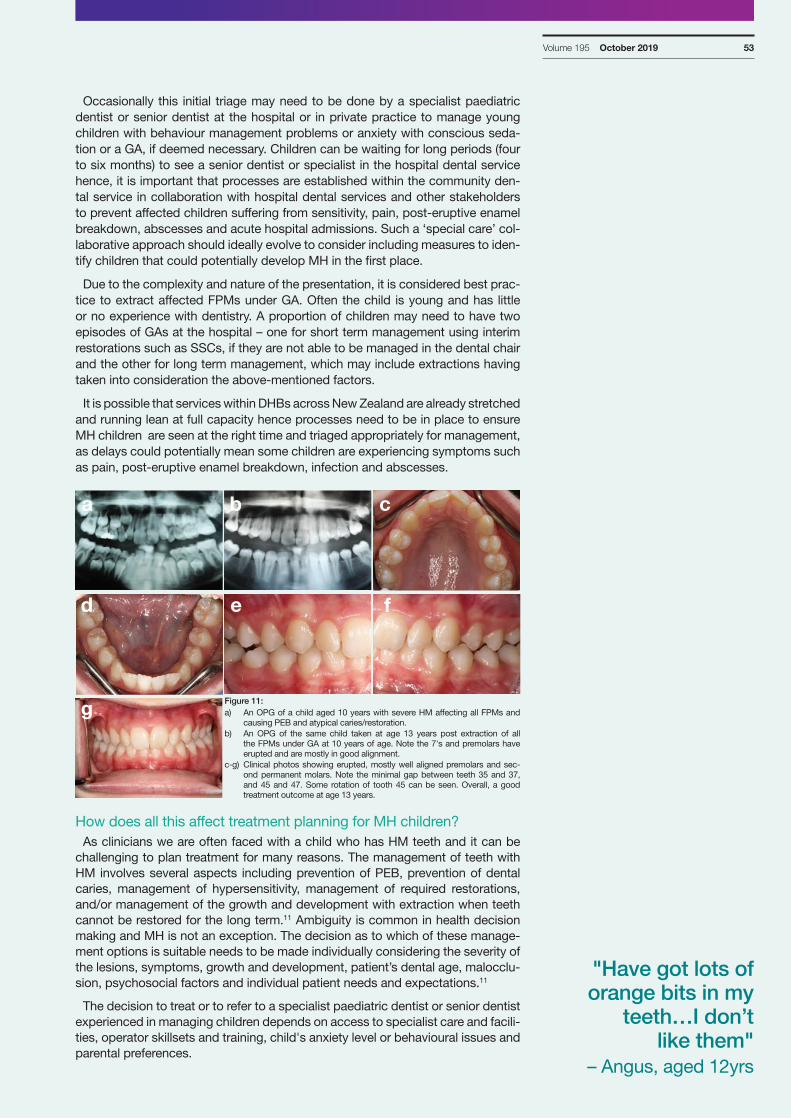
Figure 11: a) An OPG of a child aged 10 years with severe HM affecting all FPMs and
causing PEB and atypical caries/restoration.b) An OPG of the same child taken at age 13 years post extraction of all
the FPMs under GA at 10 years of age. Note the 7's and premolars have erupted and are mostly in good alignment.
c-g) Clinical photos showing erupted, mostly well aligned premolars and sec-ond permanent molars. Note the minimal gap between teeth 35 and 37, and 45 and 47. Some rotation of tooth 45 can be seen. Overall, a good treatment outcome at age 13 years.
Occasionally this initial triage may need to be done by a specialist paediatric dentist or senior dentist at the hospital or in private practice to manage young children with behaviour management problems or anxiety with conscious seda-tion or a GA, if deemed necessary. Children can be waiting for long periods (four to six months) to see a senior dentist or specialist in the hospital dental service hence, it is important that processes are established within the community den-tal service in collaboration with hospital dental services and other stakeholders to prevent affected children suffering from sensitivity, pain, post-eruptive enamel breakdown, abscesses and acute hospital admissions. Such a ‘special care’ col-laborative approach should ideally evolve to consider including measures to iden-tify children that could potentially develop MH in the first place.
Due to the complexity and nature of the presentation, it is considered best prac-tice to extract affected FPMs under GA. Often the child is young and has little or no experience with dentistry. A proportion of children may need to have two episodes of GAs at the hospital – one for short term management using interim restorations such as SSCs, if they are not able to be managed in the dental chair and the other for long term management, which may include extractions having taken into consideration the above-mentioned factors.
It is possible that services within DHBs across New Zealand are already stretched and running lean at full capacity hence processes need to be in place to ensure MH children are seen at the right time and triaged appropriately for management, as delays could potentially mean some children are experiencing symptoms such as pain, post-eruptive enamel breakdown, infection and abscesses.
a b c
d e f
g
"Have got lots of orange bits in my
teeth…I don’t like them"
– Angus, aged 12yrs
NZDA NEWS
Children and families affected with MH suffer from burden of treatment, and treatment should not be attempted without taking into consideration the above mentioned factors, history and planning.
Early identification of at-risk patients and follow up may lead to early diagnosis that could potentially reduce the depth of clinical impact that can occur with MH children and families. This also means more conservative treatment could be per-formed at an early stage to prevent enamel breakdown or caries and potentially reduce the reportedly ten-fold higher risk of developing caries when severely hy-pomineralised. Treatment should address short- and long-term issues depending on severity of MH and specific subjective symptoms. Although not completely evidence-based, the currently available treatment modalities extend from use of CPP-ACP and/or fluoride for minimising sensitivity in mild to moderate cases to restoring and/or extraction of some teeth with or without orthodontic manage-ment in some severe cases.12,32,37
How can we help children manage their anxiety or behavioural issues better?
Studies have shown that children with severe MH received more than nine times more dental treatment for their FPMs than the children without MH. Affected chil-dren are said to experience more anxiety (44%) compared with children without MH (2%).12,31 Not surprisingly, the study also reported that almost every defective tooth was treated at least twice, reflecting bonding issues. The same children were examined again at eighteen years-of-age to assess the long-term outcomes of restorative treatment, dental anxiety, and patient satisfaction with respect to MH.12 The young adults with MH had significantly higher DMFT scores and had received treatment for their HM molars 4.2 times more than the unaffected young adults.
Given that children with severe MH will experience significant problems includ-ing pain and discomfort, dental anxiety and sensitivity, routine use of triplex or air blowing HM teeth should be avoided at the time of dental examination or when performing simple procedures such as sealants. However, poor isolation may com-promise the already compromised enamel leading to poor restorative outcomes. A range of alternatives such as a toothbrush or bonding applicator brush, or cotton pellets all wet (using warm water), can be used when cleaning or etching sensitive teeth and using dry cotton pellets combined with low vacuum suction around the affected tooth can be used when drying the teeth post-etching. Use of RA with or without pre-emptive analgesics has been previously recommended when per-forming simple procedures such as sealants on sensitive HM teeth.
Use of extra-short needles are recommended for infiltration when administer-ing standard LA procedure. The computer controlled local anaesthetic delivery system (CCLAD) has been shown to reduce patient pain during LA.42 The Wand® Single tooth Anaesthesia System (Milestone Scientific, USA) is well known for its ease of use due to its light weight and a circumference that is about half that of traditional anaesthetic syringes. The dental team must remain flexible and adapt to the behaviours of the child in the chair and help children better manage their anxiety and other associated behavioural problems. The decision to treat or refer to a specialist paediatric dentist or senior dentist experienced in managing chil-dren depends on the factors listed earlier in this article. Even simple dental treat-ment requires careful management because of the significant problems posed by MH.
Causation – Why my child, am I to blame?As clinicians, we often hear parents of MH children say – “We brush his teeth
both morning and night, he has a good diet overall and I am not sure why this happened to him?” Often the answer is ambiguous, however it is important that families and children are fully informed about the nature of this condition, includ-ing the ambiguity of the causality. It means that parents can be freed from any guilt and liberated from feeling responsibility for the state of their child’s teeth.
Feature article
"I get told to brush them every day even though they are sore, but they break away anyway" – Emma, aged 9yrs
"I was told by my teacher at school to brush my teeth morning and night, even though I do that everyday" - Madison, aged 8yrs
Volume 195 October 2019 55
t
There have been numerous studies and four systematic reviews conducted on causation of MH, with no consensus being achieved other than that multiple fac-tors seem to be involved. Given the specific distribution of the D3s associated with MH, the timing of any disruption can be hypothesised to be between the 18th week of pregnancy and around 3-5 years of age for the FPMs to be affected.
Several pre-, perinatal or early life illnesses or events have been implicated how-ever, the evidence surrounding these specific factors remains equivocal. Recent aetiological studies have suggested that a distinct genetic predisposition does ex-ist among other proven systemic and environmental factors.43,44,45 Environmental factors like antibiotics in first three years of life, dioxins in breast milk, nutritional conditions during the first three years of life and medical factors like premature birth, cyanosis, prolonged delivery and/or neonatal hypocalcaemia have been speculated to act individually or synergistically in genetically predisposed individu-als to cause MH.44,45 A number of biological mechanisms have been suggested including changes induced by systemic illness to ameloblast function in their sur-rounding environment. However, how such factors lead to specific changes in ameloblasts that then result in HM lesions is still unclear.
Translational perspective1. It is incorrect to refer to HM with PEB as hypoplasia. We now understand well
the differences between these two different entities however, a translational change among clinicians including (but not limited to) OHPs and dentists is yet to happen, especially when referring children with this condition. Specifically, it is not uncommon to come across referral letters that still refer to MH children as having 'hypoplasia' or 'hypoplastic teeth'. This can create confusion for some parents’ full understanding of the nature of the condition.
2. New Zealand children affected with D3s deserve a ‘special care’ approach to management that can be achieved through integration between the community, hospital and private practice. For example, initiatives within NZ are: ‘Hypomin clinic’ and ‘Inherited Dental Anomalies Clinic’ in Canterbury and the new ‘D3 clinic’ in Wellington.
3. There is a need for consistent use of service codes in base charting in Titanium across DHBs in NZ to record D3s. A ‘Collective Impact’ case needs to be put forward for the Ministry of Health’s Age 5 and Year 8 oral health data to include reporting of D3s.
4. Access to specialist care including specialist paediatric dentists, orthodontists and prosthodontists must be included within Ministry of Health's Tier Two Hospital Dental Services Nationwide Service Specifications for the purpose of management of children and young adults affected with D3s.
5. A shared learning approach between services across DHBs in New Zealand may facilitate a nationally consistent approach towards early diagnosis and management of D3 children in the community. A national D3 symposium would generate solutions to bring about the transformative change needed to achieve a NZ-wide consistent approach and greater oral health sector accountability when managing children affected with D3s. DHBs and associations such as NZDA, NZDOHTA, SPDNZ and NZAO should take the lead on this.
6. Further clinical research within NZ should focus on reporting long term outcomes after extraction of HM-FPM's, and studying the prevalence of HM second permanent molars to better inform patients and reduce ambiguity with decision making when extracting HM-FPM's.
More informationFor more information or queries, please contact Arun at: Arun.Natarajan@cdhb.
health.nz
"Lived in a 100 year old mouldy
weatherboard house and he was sick
continuously until 3 years of age…much
better since we moved out"
– Mother of child, aged 10yrs
"We want these teeth to be gone
as it has given her too many problems
and she has been to see five different
dentists so far"– Mother of
child, aged 8yrs
NZDA NEWS
REFERENCES:1. Weerheijm K, Jälevik B, Alaluusua S. 2001.
Molar–incisor hypomineralisation. Caries research. 35(5):390-391.
2. Elfrink M, Schuller A, Weerheijm K, Veerkamp J. 2008. Prevalence of deciduous molar hy-pomineralisation in 5-year-old dutch children. Deciduous Molar Hypomineralisation, its nature and nurture. 42:23.
3. Weerheijm K. 2004. Molar incisor hypominer-alization (mih): Clinical presentation, aetiology and management. DENTAL UPDATE-LON-DON-. 31(1):9-12.
4. Garot E, Denis A, Delbos Y, Manton D, Silva M, Rouas P. 2018. Are hypomineralised lesions on second primary molars (hspm) a predictive sign of molar incisor hypomineralisation (mih)? A systematic review and a meta-analysis. Journal of dentistry. 72:8-13.
5. Farah R, Drummond B, Swain M, Williams S. 2008. Relationship between laser fluorescence and enamel hypomineralisation. Journal of dentistry. 36(11):915-921.
6. Farah R, Swain M, Drummond B, Cook R, Atieh M. 2010a. Mineral density of hypominer-alised enamel. Journal of dentistry. 38(1):50-58.
7. Jälevik B, Klingberg G, Barregård L, Norén JG. 2001d. The prevalence of demarcated opacities in permanent first molars in a group of swedish children. Acta Odontologica. 59(5):255-260.
8. Mahoney EK, Rohanizadeh R, Ismail F, Kilpat-rick N, Swain M. 2004. Mechanical properties and microstructure of hypomineralised enamel of permanent teeth. Biomaterials. 25(20):5091-5100.
9. Mathu-Muju K, Wright JT. 2006. Diagnosis and treatment of molar incisor hypomineraliza-tion. Compendium of continuing education in dentistry (Jamesburg, NJ: 1995). 27(11):604.
10. Lygidakis N, Wong F, Jälevik B, Vierrou A, Alaluusua S, Espelid I. 2010a. Best clini-cal practice guidance for clinicians dealing with children presenting with molar-incisor-hypomineralisation (mih): An eapd policy document. European archives of paediatric dentistry: official journal of the European Acad-emy of Paediatric Dentistry. 11(2):75.
11. Drummond BK, Kilpatrick N. 2016. Planning and care for children and adolescents with dental enamel defects. Springer.
12. Jalevik B, Klingberg G. 2012. Treatment outcomes and dental anxiety in 18‐year‐olds with mih, comparisons with healthy controls–a longitudinal study. International Journal of Paediatric Dentistry. 22(2):85-91.
13. Jälevik B, Odelius H, Dietz W, Norén J. 2001c. Secondary ion mass spectrometry and x-ray microanalysis of hypomineralized enamel in human permanent first molars. Archives of Oral Biology. 46(3):239-247.
14. Fraser SJ, Natarajan AK, Clark AS, Drummond BK, Gordon KC. 2015. A raman spectroscopic study of teeth affected with molar–incisor hy-pomineralisation. Journal of Raman Spectros-copy. 46(2):202-210.
15. Natarajan A. 2013. Characterisation of enamel and effect of resin infiltration in molar-incisor hypomineralisation. University of otago. Thesis, Doctor of Clinical Dentistry.
16. Farah RA, Monk BC, Swain MV, Drummond BK. 2010c. Protein content of molar–incisor hypomineralisation enamel. Journal of den-tistry. 38(7):591-596.
BIOGRAPHYDr Arun Natarajan, BDS, DClinDent
Arun Natarajan obtained his BDS from India and his Doctorate in Clinical Dentistry (Paediatric Dentistry) from the University of Otago, New Zealand. He then joined the Hospital Dental Service at Christchurch Hospital as a specialist paediatric dentist before moving to Melbourne to work as a senior registrar trainee for six months at The Royal Children's Hospital, Melbourne. Currently, Arun works as Consultant Paediatric Dentist at the Hospital Dental Service, Christchurch. Arun has presented in several conferences and meetings both at national and international level and his research looked at the effect of resin infiltration in hypomineralised enamel using Raman Spectroscopy. Arun has recently been elected as president-elect for the NZ branch Australia and New Zealand Society of Paediatric Dentistry (ANZSPD).
17. Mangum J, Crombie F, Kilpatrick N, Manton D, Hubbard M. 2010. Surface integrity governs the proteome of hypomineralized enamel. Journal of dental research. 89(10):1160-1165.
18. Natarajan AK, Fraser SJ, Swain MV, Drum-mond BK, Gordon KC. 2015. Raman spec-troscopic characterisation of resin-infiltrated hypomineralised enamel. Analytical and bioanalytical chemistry. 407(19):5661-5671.
19. Jälevik B, Dietz W, Norén J. 2005. Scanning electron micrograph analysis of hypomin-eralized enamel in permanent first molars. International Journal of Paediatric Dentistry. 15(4):233-240.
20. Rodd HD, Boissonade FM, Day PF. 2007. Pulpal status of hypomineralized permanent molars. Pediatric Dentistry. 29(6):514-520.
21. William V, Messer LB, Burrow MF. 2006. Molar incisor hypomineralization: Review and recom-mendations for clinical management. Pediatric dentistry. 28(3):224-232.
22. Willmott N, Bryan R, Duggal M. 2008. Molar-incisor-hypomineralisation: A literature review. Eur Arch Paediatr Dent. 9(4):172-179.
23. Fearne J, Anderson P, Davis G. 2004. 3d x-ray microscopic study of the extent of variations in enamel density in first permanent molars with idiopathic enamel hypomineralisation. British dental journal. 196(10):634-638.
24. Xie Z, Kilpatrick NM, Swain MV, Munroe PR, Hoffman M. 2008. Transmission electron mi-croscope characterisation of molar-incisor-hy-pomineralisation. Journal of Materials Science: Materials in Medicine. 19(10):3187-3192.
25. William V, Burrow MF, Palamara JE, Messer LB. 2006a. Microshear bond strength of resin composite to teeth affected by molar hypomin-eralization using 2 adhesive systems. Pediatric Dentistry. 28(3):233-241.
26. Jälevik B. 2001b. Enamel hypomineralization in permanent first molars. A clinical, histo-morphological and biochemical study. Swedish dental journal Supplement. (149):1.
27. Weatherell J. 1975. Composition of dental enamel. British medical bulletin. 31(2):115-119.
28. Chan Y, Ngan A, King N. 2010. Degraded prism sheaths in the transition region of hypomineral-ized teeth. Journal of dentistry. 38(3):237-244.
29. Kotsanos N, Kaklamanos E, Arapostathis K. 2005. Treatment management of first permanent molars in children with molar-incisor hypomineralisation. European journal of paediatric dentistry: official journal of European Academy of Paediatric Dentistry. 6(4):179.
30. Fayle S. 2003. Molar incisor hypomineralisa-tion: Restorative management. European Journal of Paediatric Dentistry. 4:121-126.
31. Jälevik B, Klingberg G. 2002. Dental treatment, dental fear and behaviour management prob-lems in children with severe enamel hypomin-eralization of their permanent first molars. International Journal of Paediatric Dentistry. 12(1):24-32.
32. Lygidakis N. 2010b. Treatment modalities in children with teeth affected by molar-incisor enamel hypomineralisation (mih): A system-atic review. European Archives of Paediatric Dentistry. 11(2):65.
33. Gaardmand E, Poulsen S, Haubek D. 2013. Pilot study of minimally invasive cast adhesive copings for early restoration of hypomineral-ised first permanent molars with post-eruptive breakdown. European Archives of Paediatric Dentistry.1-5.
34. Zagdwon A, Fayle S, Pollard M. 2003. A prospective clinical trial comparing preformed metal crowns and cast restorations for defec-tive first permanent molars. European Journal of Paediatric Dentistry. 4:138-142.
35. Koleventi A, Sakellari D, Arapostathis KN, Kotsanos N. 2018. Periodontal impact of preformed metal crowns on permanent molars of children and adolescents: A pilot study. Pediatric dentistry. 40(2):117-121.
36. Cobourne M, Williams A, Harrison M. 2014. National clinical guidelines for the extraction of first permanent molars in children. British dental journal. 217(11):643.
37. Mejàre I, Bergman E, Grindefjord M. 2005. Hy-pomineralized molars and incisors of unknown origin: Treatment outcome at age 18 years. International Journal of Paediatric Dentistry. 15(1):20-28.
38. Williams J, Gowans A. 2003. Hypomineralised first permanent molars and the orthodontist. European journal of paediatric dentistry: official journal of European Academy of Paediatric Dentistry. 4(3):129.
39. Wu M, Chen L, Bawole E, Anthonappa R, King N. 2017. Is there sufficient evidence to support an optimum time for the extraction of first permanent molars? European Archives of Paediatric Dentistry. 18(3):155-161.
40. Jalevik B, Moller M. 2007. Evaluation of spontaneous space closure and develop-ment of permanent dentition after extraction of hypomineralized permanent first molars. International Journal of Paediatric Dentistry. 17(5):328-335.
41. Demirjian A, Goldstein H, Tanner J. 1973. A new system of dental age assessment. Human biology.211-227.
42. Kwak E-J, Pang N-S, Cho J-H, Jung B-Y, Kim K-D, Park W. 2016. Computer-controlled local anesthetic delivery for painless anesthesia: A literature review. Journal of dental anesthesia and pain medicine. 16(2):81-88.
43. Bussaneli DG, Restrepo M, Fragelli CMB, Santos-Pinto L, Jeremias F, Cordeiro RdCL, Bezamat M, Vieira AR, Scarel-Caminaga RM. 2019. Genes regulating immune response and amelogenesis interact in increasing the sus-ceptibility to molar-incisor hypomineralization. Caries research. 53(2):217-227.
44. Fatturi AL, Wambier LM, Chibinski AC, As-sunção LRdS, Brancher JA, Reis A, Souza JF. 2019. A systematic review and meta‐analysis of systemic exposure associated with molar in-cisor hypomineralization. Community dentistry and oral epidemiology.
45. Lygidakis N, Dimou G, Marinou D. 2008b. Molar-incisor-hypomineralisation (mih). A retrospective clinical study in greek children. Ii. Possible medical aetiological factors. Eur Arch Paediatr Dent. 9(4):207-217.
46. Silva MJ, Scurrah KJ, Craig JM, Manton DJ, Kilpatrick N. 2016. Etiology of molar incisor hypomineralization–a systematic review. Community dentistry and oral epidemiology. 44(4):342-353.
47. Krämer N, Bui Khac NN, Lücker S, Stach-niss V, Frankenberger R Dent Mater. 2018 Feb;34(2):331-340
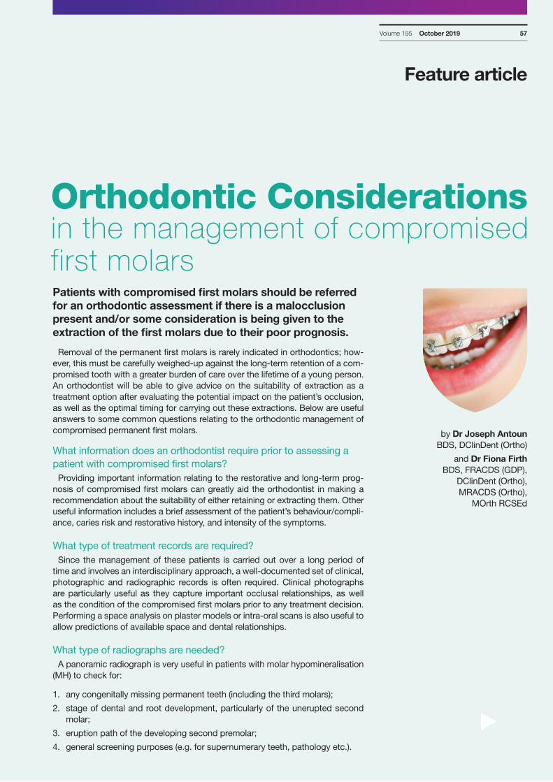
Volume 195 October 2019 57
Patients with compromised first molars should be referred for an orthodontic assessment if there is a malocclusion present and/or some consideration is being given to the extraction of the first molars due to their poor prognosis.
Removal of the permanent first molars is rarely indicated in orthodontics; how-ever, this must be carefully weighed-up against the long-term retention of a com-promised tooth with a greater burden of care over the lifetime of a young person. An orthodontist will be able to give advice on the suitability of extraction as a treatment option after evaluating the potential impact on the patient’s occlusion, as well as the optimal timing for carrying out these extractions. Below are useful answers to some common questions relating to the orthodontic management of compromised permanent first molars.
What information does an orthodontist require prior to assessing a patient with compromised first molars?
Providing important information relating to the restorative and long-term prog-nosis of compromised first molars can greatly aid the orthodontist in making a recommendation about the suitability of either retaining or extracting them. Other useful information includes a brief assessment of the patient’s behaviour/compli-ance, caries risk and restorative history, and intensity of the symptoms.
What type of treatment records are required? Since the management of these patients is carried out over a long period of
time and involves an interdisciplinary approach, a well-documented set of clinical, photographic and radiographic records is often required. Clinical photographs are particularly useful as they capture important occlusal relationships, as well as the condition of the compromised first molars prior to any treatment decision. Performing a space analysis on plaster models or intra-oral scans is also useful to allow predictions of available space and dental relationships.
What type of radiographs are needed? A panoramic radiograph is very useful in patients with molar hypomineralisation
(MH) to check for:
1. any congenitally missing permanent teeth (including the third molars);
2. stage of dental and root development, particularly of the unerupted second molar;
3. eruption path of the developing second premolar;
4. general screening purposes (e.g. for supernumerary teeth, pathology etc.).
Feature article
Orthodontic Considerations in the management of compromised first molars
t
by Dr Joseph Antoun BDS, DClinDent (Ortho)
and Dr Fiona Firth BDS, FRACDS (GDP),
DClinDent (Ortho), MRACDS (Ortho),
MOrth RCSEd
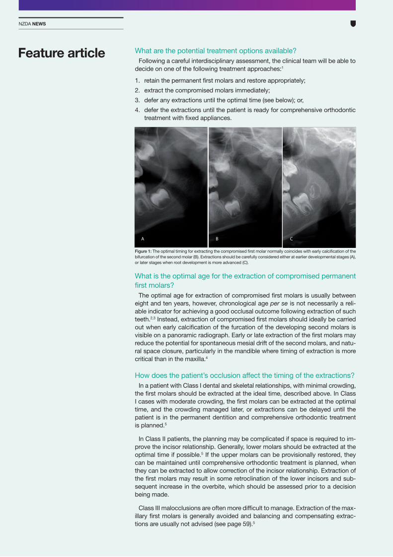
NZDA NEWS
What are the potential treatment options available? Following a careful interdisciplinary assessment, the clinical team will be able to
decide on one of the following treatment approaches:1
1. retain the permanent first molars and restore appropriately;
2. extract the compromised molars immediately;
3. defer any extractions until the optimal time (see below); or,
4. defer the extractions until the patient is ready for comprehensive orthodontic treatment with fixed appliances.
Feature article
BA C
Figure 1: The optimal timing for extracting the compromised first molar normally coincides with early calcification of the bifurcation of the second molar (B). Extractions should be carefully considered either at earlier developmental stages (A), or later stages when root development is more advanced (C).
What is the optimal age for the extraction of compromised permanent first molars?
The optimal age for extraction of compromised first molars is usually between eight and ten years, however, chronological age per se is not necessarily a reli-able indicator for achieving a good occlusal outcome following extraction of such teeth.2,3 Instead, extraction of compromised first molars should ideally be carried out when early calcification of the furcation of the developing second molars is visible on a panoramic radiograph. Early or late extraction of the first molars may reduce the potential for spontaneous mesial drift of the second molars, and natu-ral space closure, particularly in the mandible where timing of extraction is more critical than in the maxilla.4
How does the patient’s occlusion affect the timing of the extractions? In a patient with Class I dental and skeletal relationships, with minimal crowding,
the first molars should be extracted at the ideal time, described above. In Class I cases with moderate crowding, the first molars can be extracted at the optimal time, and the crowding managed later, or extractions can be delayed until the patient is in the permanent dentition and comprehensive orthodontic treatment is planned.5
In Class II patients, the planning may be complicated if space is required to im-prove the incisor relationship. Generally, lower molars should be extracted at the optimal time if possible.5 If the upper molars can be provisionally restored, they can be maintained until comprehensive orthodontic treatment is planned, when they can be extracted to allow correction of the incisor relationship. Extraction of the first molars may result in some retroclination of the lower incisors and sub-sequent increase in the overbite, which should be assessed prior to a decision being made.
Class III malocclusions are often more difficult to manage. Extraction of the max-illary first molars is generally avoided and balancing and compensating extrac-tions are usually not advised (see page 59).5
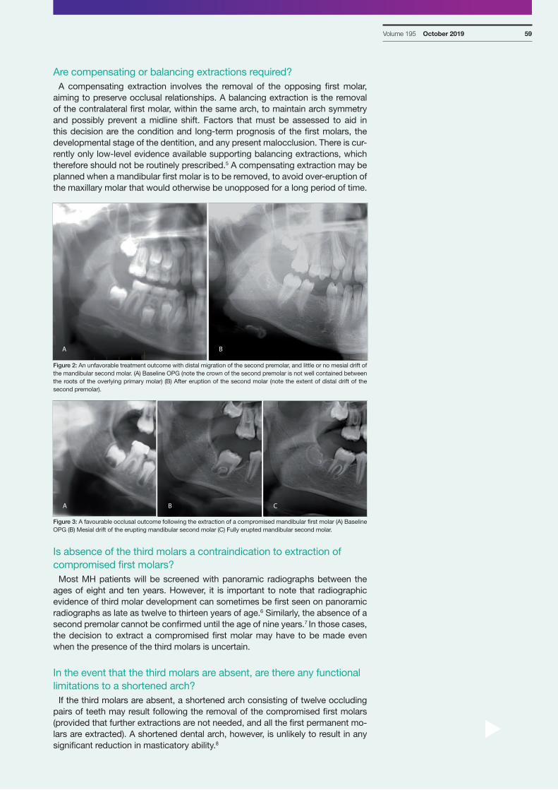
Volume 195 October 2019 59
t
Are compensating or balancing extractions required? A compensating extraction involves the removal of the opposing first molar,
aiming to preserve occlusal relationships. A balancing extraction is the removal of the contralateral first molar, within the same arch, to maintain arch symmetry and possibly prevent a midline shift. Factors that must be assessed to aid in this decision are the condition and long-term prognosis of the first molars, the developmental stage of the dentition, and any present malocclusion. There is cur-rently only low-level evidence available supporting balancing extractions, which therefore should not be routinely prescribed.5 A compensating extraction may be planned when a mandibular first molar is to be removed, to avoid over-eruption of the maxillary molar that would otherwise be unopposed for a long period of time.
BA
Figure 2: An unfavorable treatment outcome with distal migration of the second premolar, and little or no mesial drift of the mandibular second molar. (A) Baseline OPG (note the crown of the second premolar is not well contained between the roots of the overlying primary molar) (B) After eruption of the second molar (note the extent of distal drift of the second premolar).
BA C
Figure 3: A favourable occlusal outcome following the extraction of a compromised mandibular first molar (A) Baseline OPG (B) Mesial drift of the erupting mandibular second molar (C) Fully erupted mandibular second molar.
Is absence of the third molars a contraindication to extraction of compromised first molars?
Most MH patients will be screened with panoramic radiographs between the ages of eight and ten years. However, it is important to note that radiographic evidence of third molar development can sometimes be first seen on panoramic radiographs as late as twelve to thirteen years of age.6 Similarly, the absence of a second premolar cannot be confirmed until the age of nine years.7 In those cases, the decision to extract a compromised first molar may have to be made even when the presence of the third molars is uncertain.
In the event that the third molars are absent, are there any functional limitations to a shortened arch?
If the third molars are absent, a shortened arch consisting of twelve occluding pairs of teeth may result following the removal of the compromised first molars (provided that further extractions are not needed, and all the first permanent mo-lars are extracted). A shortened dental arch, however, is unlikely to result in any significant reduction in masticatory ability.8
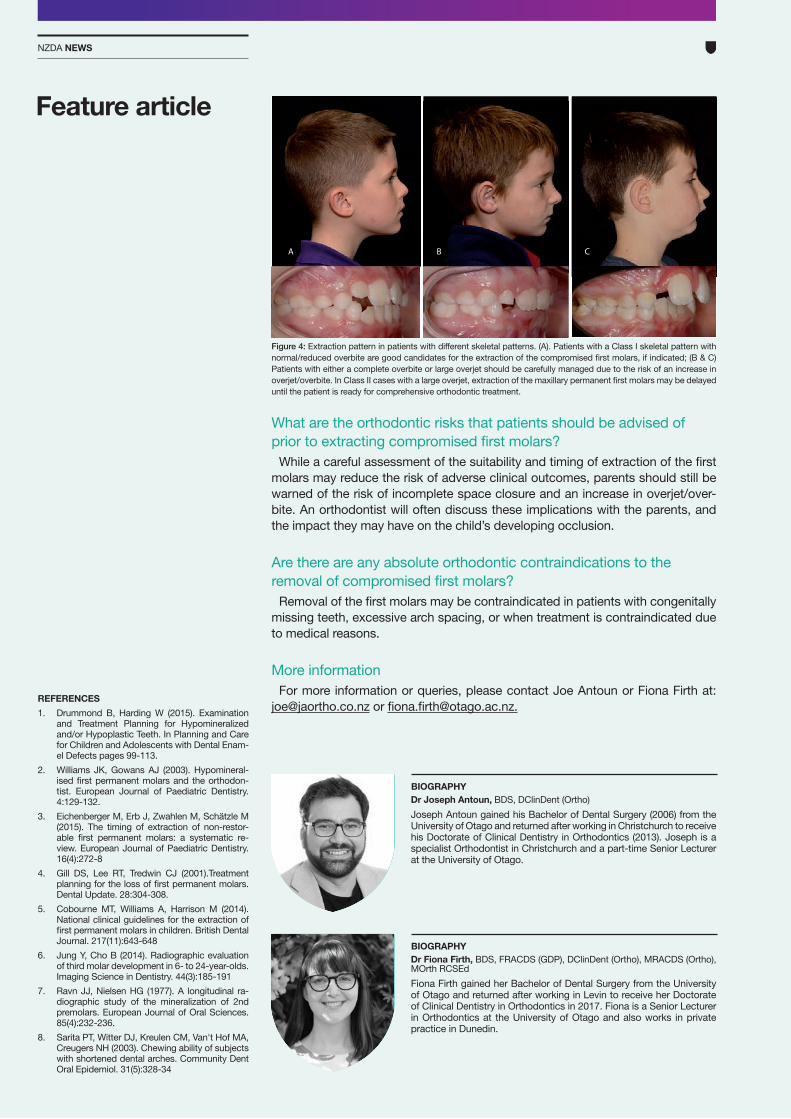
NZDA NEWS
Feature article
REFERENCES1. Drummond B, Harding W (2015). Examination
and Treatment Planning for Hypomineralized and/or Hypoplastic Teeth. In Planning and Care for Children and Adolescents with Dental Enam-el Defects pages 99-113.
2. Williams JK, Gowans AJ (2003). Hypomineral-ised first permanent molars and the orthodon-tist. European Journal of Paediatric Dentistry. 4:129-132.
3. Eichenberger M, Erb J, Zwahlen M, Schätzle M (2015). The timing of extraction of non-restor-able first permanent molars: a systematic re-view. European Journal of Paediatric Dentistry. 16(4):272-8
4. Gill DS, Lee RT, Tredwin CJ (2001).Treatment planning for the loss of first permanent molars. Dental Update. 28:304-308.
5. Cobourne MT, Williams A, Harrison M (2014). National clinical guidelines for the extraction of first permanent molars in children. British Dental Journal. 217(11):643-648
6. Jung Y, Cho B (2014). Radiographic evaluation of third molar development in 6- to 24-year-olds. Imaging Science in Dentistry. 44(3):185-191
7. Ravn JJ, Nielsen HG (1977). A longitudinal ra-diographic study of the mineralization of 2nd premolars. European Journal of Oral Sciences. 85(4):232-236.
8. Sarita PT, Witter DJ, Kreulen CM, Van't Hof MA, Creugers NH (2003). Chewing ability of subjects with shortened dental arches. Community Dent Oral Epidemiol. 31(5):328-34
AA B C
Figure 4: Extraction pattern in patients with different skeletal patterns. (A). Patients with a Class I skeletal pattern with normal/reduced overbite are good candidates for the extraction of the compromised first molars, if indicated; (B & C) Patients with either a complete overbite or large overjet should be carefully managed due to the risk of an increase in overjet/overbite. In Class II cases with a large overjet, extraction of the maxillary permanent first molars may be delayed until the patient is ready for comprehensive orthodontic treatment.
What are the orthodontic risks that patients should be advised of prior to extracting compromised first molars?
While a careful assessment of the suitability and timing of extraction of the first molars may reduce the risk of adverse clinical outcomes, parents should still be warned of the risk of incomplete space closure and an increase in overjet/over-bite. An orthodontist will often discuss these implications with the parents, and the impact they may have on the child’s developing occlusion.
Are there are any absolute orthodontic contraindications to the removal of compromised first molars?
Removal of the first molars may be contraindicated in patients with congenitally missing teeth, excessive arch spacing, or when treatment is contraindicated due to medical reasons.
More informationFor more information or queries, please contact Joe Antoun or Fiona Firth at:
[email protected] or [email protected].
BIOGRAPHYDr Fiona Firth, BDS, FRACDS (GDP), DClinDent (Ortho), MRACDS (Ortho), MOrth RCSEd
Fiona Firth gained her Bachelor of Dental Surgery from the University of Otago and returned after working in Levin to receive her Doctorate of Clinical Dentistry in Orthodontics in 2017. Fiona is a Senior Lecturer in Orthodontics at the University of Otago and also works in private practice in Dunedin.
BIOGRAPHYDr Joseph Antoun, BDS, DClinDent (Ortho)
Joseph Antoun gained his Bachelor of Dental Surgery (2006) from the University of Otago and returned after working in Christchurch to receive his Doctorate of Clinical Dentistry in Orthodontics (2013). Joseph is a specialist Orthodontist in Christchurch and a part-time Senior Lecturer at the University of Otago.