Embed Size (px)
Citation preview
MOLECULAR GENETICS OF TIBIAL MUSCULAR
DYSTROPHY (TMD) AND A NOVEL DISTAL MYOPATHY
Henna Haravuori
Department of Molecular Medicine, National Public Health Institute and Department of Medical Genetics, University of Helsinki, Helsinki, Finland and
Department of Neurology, Helsinki University Central Hospital, Helsinki, Finland
Academic Dissertation
To be publicly discussed with permission of the Medical Faculty of the University of Helsinki, in the small lecture hall of the Haartman Institute,
Haartmaninkatu 3, Helsinki, on December 5th, at 12 o’clock noon.
Helsinki 2003
2
Supervised by Professor Leena Peltonen-Palotie and Docent Hannu Somer National Public Health Institute Department of Neurology and University of Helsinki Helsinki University Central Hospital Helsinki, Finland Helsinki, Finland Reviewed by Professor Nigel G. Laing and Docent Katarina Pelin Centre for Neuromuscular and Department of Biosciences Neurological Disorders Division of Genetics University of Western Australia University of Helsinki Australian Neuromuscular Helsinki, Finland Research Institute Nedlands, Australia To be publicly discussed with Professor Kate Bushby Institute of Human Genetics International Centre for Life Newcastle upon Tyne, the United Kingdom Publications of the National Public Health Insitute KTL A24 / 2003 Copyright National Public Health Institute Julkaisija – Utgivare – Publisher Kansanterveyslaitos Folhälsoinstitutet National Public Health Institute Mannerheimintie 166 Mannerheimvägen 166 Mannerheimintie 166 00300 Helsinki 00300 Helsingfors FIN-00300 Helsinki, Finland puh. 09-47441 tel. 09-47441 telephone +358-9-47441 ISBN 951-740-402-6 ISSN 0359-3584 ISBN 951-740-403-4 (PDF version) ISSN 1458-6290 (PDF version) http://ethesis.helsinki.fi Cosmoprint Oy Helsinki 2003

3

4
5
CONTENTS LIST OF ORIGINAL PUBLICATIONS .........................................................7 ABBREVIATIONS ............................................................................................8 SUMMARY ......................................................................................................10 REVIEW OF THE LITERATURE ...............................................................12 Introduction ...............................................................................................12 Distal myopathies ......................................................................................12 Classification .......................................................................................12 Tibial muscular dystrophy (TMD) .....................................................16 Clinical picture .....................................................................................16 Morphological findings .........................................................................17 Genetic studies ......................................................................................18 TMD outside Finland ............................................................................19 Late-onset distal myopathy (LODM, Markesbery-Griggs) ............19 Limb-girdle muscular dystrophies (LGMD) ...........................................20 Classification .......................................................................................20 Clinical picture and laboratory findings ...........................................20 Proximal and distal phenotypes in the same family .........................22 Molecular background of muscular dystrophies and myopathies ........23 Sarcolemmopathies .............................................................................24 Sarcomere protein defects ..................................................................27 Calpain 3 and myonuclear apoptosis ................................................29 Glycosylation defects ..........................................................................30 Titin (TTN) .................................................................................................31 Molecular structure of titin ................................................................32 Functions of titin and interactions with sarcomeric proteins .........33 Z-disc and sarcomere assembly ............................................................33 I-band titin, elastic spring .....................................................................34 I-band titin, regulatory functions ..........................................................34 A-band titin, scaffold for thick filament assembly .................................34 M-line titin ............................................................................................35 Novel titin interacting molecules ..........................................................37 Identification of disease genes ..................................................................38 The Human Genome Project .............................................................39 Genetic mapping .................................................................................40
6
AIMS OF THE PRESENT STUDY ...............................................................42 SUBJECTS AND METHODS ........................................................................43 RESULTS AND DISCUSSION ......................................................................51 Tibial muscular dystrophy (TMD) ..........................................................51 Assignment of the TMD locus to chromosome 2q31 (I) ..................51 Haplotype analysis reveals a common core haplotype .........................52 Haplotype analysis of 150 Finnish TMD patients ................................54 TMD in a French family .......................................................................55 Linkage studies with a late-onset distal myopathy family(LODM) .......56 Search for TMD causing gene and mutations (II, III) .....................57 TMD physical region – revised .............................................................57 TTN mutation screening ........................................................................58 TMD is caused by Mex6 mutations in TTN ...........................................58 SSCP may be utilized in TMD diagnostics ...........................................60 Homozygous TMDFin mutation results in a novel form of recessive LGMD (LGMD2J) .................................................................60 Consequences of TMDFin mutation (II, III) ......................................60 Both mutant and wild type alleles are expressed ..................................60 Loss of epitope recognition by M8/9 antibody in TMDFin homozygote..61 The TMDFin mutation in TTN results in a secondary calpain 3 deficiency ..............................................................................62 Apoptosis and a disturbed IκBα/NF-κB pathway similar to LGMD2A................................................................................................63 MDM mouse – a possible model for TMD/ LGMD2J (II) ...............63 General discussion on the molecular pathogenesis in titinopathies 64 A new distal myopathy (MPD3) ...............................................................68 Clinical characterization (IV) ............................................................68 Linkage studies (IV, V) .......................................................................72 CONCLUDING REMARKS ..........................................................................77 ACKNOWLEDGMENTS ...............................................................................79 ELECTRONIC DATABASE INFORMATION ...........................................81 REFERENCES ................................................................................................82
7
LIST OF ORIGINAL PUBLICATIONS This thesis is based on the following original articles, which are referred to in the text by their Roman numerals. In addition, some unpublished data are presented. I Haravuori H, Mäkelä-Bengs P, Udd B, Partanen J, Pulkkinen L, Somer H and Peltonen L. Assignment of the tibial muscular dystrophy locus to chromosome 2q31. Am J Hum Genet (1998) 62:620-6. II Haravuori H*, Vihola A*, Straub V, Auranen M, Richard I, Marchand S, Voit T, Labeit S, Somer H, Peltonen L, Beckmann JS and Udd B. Secondary calpain3 deficiency in 2q-linked muscular dystrophy – Titin is the candidate gene. Neurology (2001) 56:869-77. III Hackman P, Vihola A*, Haravuori H*, Marchand S, Sarparanta J, de Seze J, Labeit S, Witt C, Peltonen L, Richard I and Udd B. Tibial muscular dystrophy is a titinopathy caused by mutations in TTN, the gene encoding the giant skeletal-muscle protein titin. Am J Hum Genet (2002) 71:492- 500. IV Mahjneh I, Haravuori H, Paetau A, Anderson LVB, Saarinen A, Udd B and Somer H. A distinct phenotype of distal myopathy in a large Finnish family. Neurology (2003) 61:87-92. V Haravuori H, Siitonen HA, Mahjneh I, Hackman P, Lahti L, Somer H, Peltonen L, Kestilä M, and Udd B. Linkage to two separate loci in a family with a novel distal myopathy phenotype (MPD3). Submitted. * These authors contributed equally to the respective works.
8
ABBREVIATIONS AD autosomal dominant inheritance ANK1 ankyrin 1 gene AR autosomal recessive inheritance ATP adenosine triphosphate BAC bacterial artificial chromosome BLAST basic local alignment search tool bp base pair cDNA complementary DNA CK creatine kinase cM centiMorgan CT computed tomography DAB 3,3’-diaminobenzidine DCM dilated cardiomyopathy DGC dystrophin glycoprotein complex DMD Duchenne muscular dystrophy DMRV distal myopathy with rimmed vacuoles, Nonaka myopathy DNA deoxyribonucleic acid DRAL LIM-domain protein down-regulated in rhabdomyosarcoma EM electron microscopy EMG electromyography ENMG electroneuromyography EST expressed sequence tag FHL four and a half LIM-only protein FISH Fluorescence in situ hybridization FITC fluorescein isothiocyanate FN3 fibronectin type III HCM hypertrophic cardiomyopathy HGP Human Genome Project HIBM hereditary inclusion body myopathy (h-IBM) Ig immunoglobulin C2 IκBα inhibitory protein κBα kb kilobase kD kiloDalton LGMD limb-girdle muscular dystrophy LOD logarithm of odds LODM late-onset distal myopathy Mb megabase
9
MD megaDalton MDM muscular dystrophy with myositis in mouse MM Miyoshi myopathy MPD distal myopathy MLC1SA myosin light chain 1 slow-twitch muscle A MLCK myosin light chain kinase MRI magnetic resonance imaging mRNA messenger RNA MURF muscle-specific ring finger MyBP myosin-binding protein MYH7 β-cardiac myosin heavy chain NCBI National Center for Biotechnology Information NF-κB nuclear factor κB NM nemaline myopathy nNOS neuronal nitric oxide synthase OMIM Online Mendelian Inheritance in Man PAC P1 derived artificial chromosome PAGE polyacrylamide gel electrophoresis PCR polymerase chain reaction RH radiation hybrid RNA ribonucleic acid RT reverse transcription SG sarcoglycan SGC sarcoglycan complex s-IBM sporadic inclusion body myositis SNP single nucleotide polymorphism SSCP single strand conformational polymorphism STS sequence tagged site TA tibialis anterior TMD tibial muscular dystrophy TTN titin gene TUNEL deoxynucleotidyl transferase-mediated dUTP nick end labeling Θ recombination fraction WDM Welander distal myopathy YAC yeast artificial chromosome In addition, the standard abbreviations of nucleotides and amino acids are used.
10
SUMMARY Distal myopathies are hereditary disorders that cause progressive distal muscle weakness and atrophy without clinically significant involvement of proximal muscles at the early stages of the disease. One representative of this group is autosomal dominant tibial muscular dystrophy (TMD). TMD was originally described in Finland and the disease prevalence according to the known patients is currently 7:105. The first symptoms, weakness of ankle dorsiflexion and inability to walk on the heels, occur after 35 years of age. Tibialis anterior muscles are particularly involved and muscle weakness and atrophy usually remain confined to the anterior compartment of the lower legs. The objective of this study was to clarify the molecular genetic background of TMD. The TMD locus was assigned to chromosome 2q31 by performing a genome wide scan and linkage analysis on one Finnish family. The locus finding was confirmed in three other families. Haplotype analysis revealed a founder haplotype shared by TMD chromosomes of the affected individuals. The critical region was restricted to approximately one cM spanning the region between markers D2S2173 and D2S2310. Genetic homogeneity of TMD was supported by linkage to the 2q31 locus in a French family. Further, late-onset distal myopathy (LODM) with phenotypic resemblance was also linked to chromosome 2q31. The critical region included one major candidate gene encoding the giant sarcomeric protein titin. Sequencing of the gene identified TMD causing mutations in the 363rd and the last exon (Mex6), which encodes an Ig-domain located in the M-line region of the sarcomere. The identified mutations were an 11-bp deletion/insertion changing four amino acids in Finnish patients and a leucine to proline change in French patients. The alterations possibly disrupt the Ig-domain structure. Three Finnish patients with severe limb-girdle muscular dystrophy phenotype were homozygous for the 11-bp mutation. Muscle biopsy of a homozygous patient showed specific loss of antibody recognition of the carboxy-terminal titin epitopes located in the M-line, while the sarcomere structure remains intact. Further, the mutated Ig-domain resides in the vicinity of the calpain 3 binding site. Mutations in calpain 3 cause LGMD2A. The amount of calpain 3 varied in the muscle samples from
11
heterozygous TMD patients and there was a severe loss of calpain 3 in the muscle of the homozygous patient. Myonuclear apoptosis and perturbation of the IκBα/NF-κB pathway similar to LGMD2A were observed in both heterozygous and homozygous patients. Identification of the TMD causing mutations in titin and the preliminary results of the consequences are the basis for future studies on molecular mechanisms involved in the muscle degeneration in TMD and other myopathies. During the TMD study, a new phenotype of autosomal dominant distal myopathy was discovered in a Finnish family and the aims of this thesis became broader to include the characterization of this phenotype. The first symptoms are clumsiness with the hands and stumbling around the age of 30. The thenar and hypothenar muscles of the palms are involved at onset. The disease progresses to other hand muscles, to the lower legs, the forearm muscles, and later to the proximal muscles. This phenotype is distinct from the previously described ones and was termed MPD3. Molecular genetic studies were initiated. Unexpectedly, a genome wide scan provided significant evidence for linkage to two chromosomal regions, 8p22-q11 and 12q13-q22. Statistical and haplotype analyses showed identical results for both loci making any distinction impossible. In the future, both loci may be tested with unclassified distal myopathy patients and with families reported by others to confirm the locus assignment.
12
REVIEW OF THE LITERATURE Introduction During the preceding two decades, there has been immense progress in resolving the molecular genetic background of the inherited muscular dystrophies and myopathies. Most of the classified entities have been assigned to a chromosomal locus or the causative gene and mutations are known. This has resulted in reclassification of these disorders based on molecular genetics. Pathogenetic mechanisms and details of the muscle cell structure and function have been studied both at the molecular and cellular level. However, many questions remain to be answered. One of the most intriguing is the molecular background of the selective pattern of muscle involvement in the different muscular dystrophies and myopathies. Distal myopathies Classification Distal myopathies are hereditary disorders that cause progressive distal muscle weakness and atrophy without clinically significant involvement of proximal muscles at the early stages of the disease. Serum CK activity ranges from normal or slightly elevated levels up to 150 times the upper normal limit in certain types of distal myopathy. Electrophysiological findings are compatible with myopathy without myotonic discharges or altered nerve conduction velocities. Morphological studies of muscle tissue show myopathic changes: variation in fibre size, central nuclei and increased connective tissue are seen in muscle biopsy. Fibre necrosis and regeneration are a less frequent findings in most entities. At later stages, the muscle tissue may be totally replaced by fat and fibrous connective tissue. There are no inflammatory cell infiltrates. Rimmed vacuolated fibres are often present in the biopsy but are not a specific finding for distal myopathy. Central cores, nemaline bodies, inclusion bodies, desmin storage and accumulation of glycogen or lipid are associated with other diagnoses, although the clinical presentation may show a distal phenotype. (Barohn 1993, Somer 1995, Somer 1997, Barohn et al. 1998).
13
During the early nineties four clinical entities were proposed for classification of the adult onset distal myopathies, two autosomal dominant and two autosomal recessive disorders: late adult onset myopathy with onset in the hands (Welander distal myopathy) and late adult onset myopathy with onset in the legs (Late-onset distal myopathy, tibial muscular dystrophy), early adult onset myopathy in the posterior compartment of the lower legs (Miyoshi myopathy) and early adult onset myopathy in the anterior compartment of the lower legs (Nonaka myopathy) (Barohn 1993). Because the morphological findings in many distal myopathies have similarities to those found in sporadic inclusion body myositis (s-IBM), the term hereditary inclusion body myopathy (h-IBM) has also been used for its classification (Askanas and Engel 1995, Griggs et al. 1995). With the identification of the molecular genetic cause for the different distal myopathies, the classification suggested by Barohn (1993) has become obsolete and progress in molecular studies has set a completely new approach for the diagnostics. However, the genotype-phenotype correlation may not be straightforward. Many known genes responsible for distal myopathies may also cause proximal phenotypes or cardiomyopathy making the category of distal myopathies somewhat arbitrary. Nevertheless, the term distal myopathy is still useful for clinical practise when the causative mutation is not known. (Udd and Griggs 2001). Classification of the distal myopathy entities is reviewed in table 1. In addition, there are several muscle diseases that present distal muscle weakness predominantly or occasionally. They have specific features or may be less well defined (table 2 and table 3).
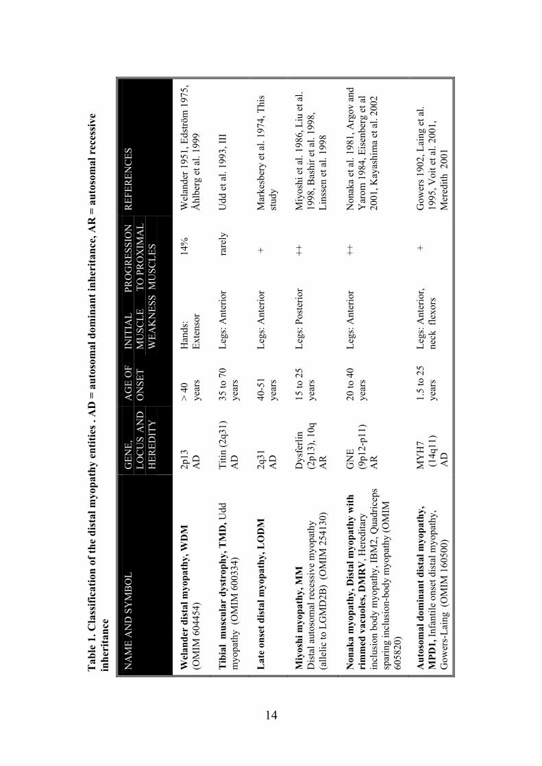
14
Tab
le 1
. Cla
ssifi
catio
n of
the
dist
al m
yopa
thy
entit
ies .
AD
= a
utos
omal
dom
inan
t inh
erita
nce,
AR
= a
utos
omal
rec
essi
ve
inhe
rita
nce
REF
EREN
CES
Wel
ande
r 195
1, E
dströ
m 1
975,
Å
hlbe
rg e
t al.
1999
Udd
et a
l. 19
93, I
II
Mar
kesb
ery
et a
l. 19
74, T
his
stud
y
Miy
oshi
et a
l. 19
86, L
iu e
t al.
1998
, Bas
hir e
t al.
1998
, Li
nsse
n et
al.
1998
Non
aka
et a
l. 19
81, A
rgov
and
Y
arom
198
4, E
isen
berg
et a
l 20
01, K
ayas
him
a et
al.
2002
Gow
ers 1
902,
Lai
ng e
t al.
1995
, Voi
t et a
l. 20
01,
Mer
edith
200
1
INIT
IAL
PR
OG
RES
SIO
N
MU
SCLE
TO
PR
OX
IMA
L W
EAK
NES
S M
USC
LES
Han
ds:
14%
Ex
tens
or
Legs
: Ant
erio
r
ra
rely
Legs
: Ant
erio
r
+
Legs
: Pos
terio
r
++
Legs
: Ant
erio
r
++
Legs
: Ant
erio
r,
+
ne
ck f
lexo
rs
AG
E O
F O
NSE
T
> 40
ye
ars
35 to
70
year
s
40-5
1 ye
ars
15 to
25
year
s
20 to
40
year
s
1.5
to 2
5 ye
ars
GEN
E,
LOC
US
AN
D
HER
EDIT
Y
2p13
A
D
Titin
(2q3
1)
AD
2q31
A
D
Dys
ferli
n (2
p13)
, 10q
A
R
GN
E (9
p12-
p11)
A
R
MY
H7
(14q
11)
AD
NA
ME
AN
D S
YM
BO
L
Wel
ande
r di
stal
myo
path
y, W
DM
(O
MIM
604
454)
Tib
ial
mus
cula
r dy
stro
phy,
TM
D, U
dd
myo
path
y (O
MIM
600
334)
Lat
e on
set d
ista
l myo
path
y, L
OD
M
Miy
oshi
myo
path
y, M
M
Dis
tal a
utos
omal
rece
ssiv
e m
yopa
thy
(a
llelic
to L
GM
D2B
) (O
MIM
254
130)
Non
aka
myo
path
y, D
ista
l myo
path
y w
ith
rim
med
vac
uole
s, D
MR
V, H
ered
itary
in
clus
ion
body
myo
path
y, IB
M2,
Qua
dric
eps
spar
ing
incl
usio
n-bo
dy m
yopa
thy
(OM
IM
6058
20)
Aut
osom
al d
omin
ant d
ista
l myo
path
y,
MPD
1, In
fant
ile o
nset
dis
tal m
yopa
thy,
G
ower
s-La
ing
(OM
IM 1
6050
0)
15
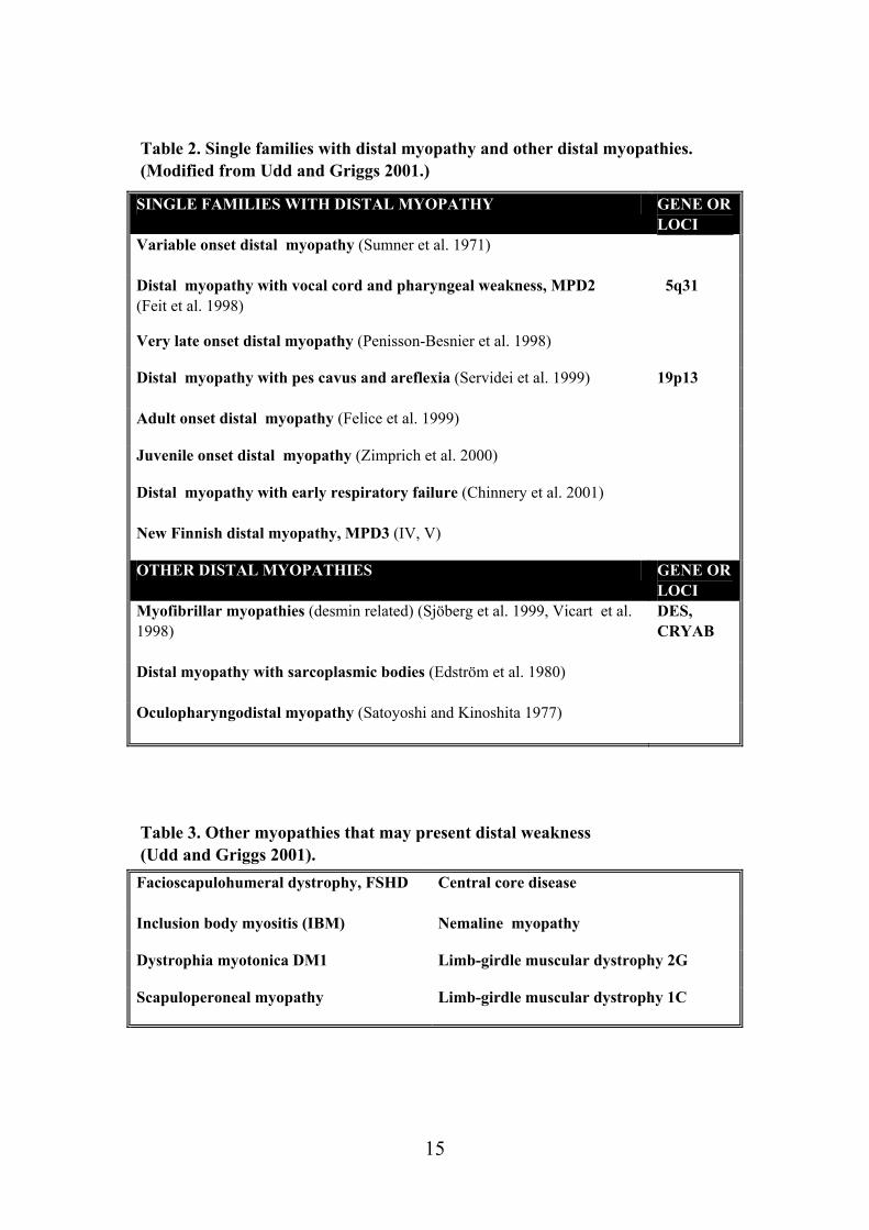
Table 2. Single families with distal myopathy and other distal myopathies. (Modified from Udd and Griggs 2001.)
Table 3. Other myopathies that may present distal weakness (Udd and Griggs 2001).
SINGLE FAMILIES WITH DISTAL MYOPATHY GENE OR LOCI
Variable onset distal myopathy (Sumner et al. 1971)
Distal myopathy with vocal cord and pharyngeal weakness, MPD2 (Feit et al. 1998)
5q31
Very late onset distal myopathy (Penisson-Besnier et al. 1998)
Distal myopathy with pes cavus and areflexia (Servidei et al. 1999)
19p13
Adult onset distal myopathy (Felice et al. 1999)
Juvenile onset distal myopathy (Zimprich et al. 2000)
Distal myopathy with early respiratory failure (Chinnery et al. 2001)
New Finnish distal myopathy, MPD3 (IV, V)
OTHER DISTAL MYOPATHIES GENE OR LOCI
Myofibrillar myopathies (desmin related) (Sjöberg et al. 1999, Vicart et al. 1998)
DES, CRYAB
Distal myopathy with sarcoplasmic bodies (Edström et al. 1980)
Oculopharyngodistal myopathy (Satoyoshi and Kinoshita 1977)
Facioscapulohumeral dystrophy, FSHD
Central core disease
Inclusion body myositis (IBM) Nemaline myopathy
Dystrophia myotonica DM1 Limb-girdle muscular dystrophy 2G
Scapuloperoneal myopathy Limb-girdle muscular dystrophy 1C
16
Tibial muscular dystrophy (TMD) TMD was originally described in two different patient groups in Finland and it was not well understood whether the two groups represented the same disease (Laulumaa et al. 1989, Partanen et al. 1990, Udd et al. 1990). Classification of this new disorder was complicated due to distal and proximal phenotypes observed in the original large consanguineous kindred originating from the Larsmo Islands on the West coast of Finland (Udd et al. 1991a, Udd et al. 1992). Further, rimmed vacuoles were observed only in muscle biopsies of patients originating from the Savo-Karelia region (Partanen et al 1994, Udd et al. 1992). A nation-wide survey revealed 66 patients with uniform clinical presentation and the term tibial muscular dystrophy was proposed (Udd et al. 1993). To date there are more than 200 patients examined and they have more than 150 known symptomatic relatives leading to a point prevalence of 7/100 000 TMD patients in Finland (Hackman et al. 2002).
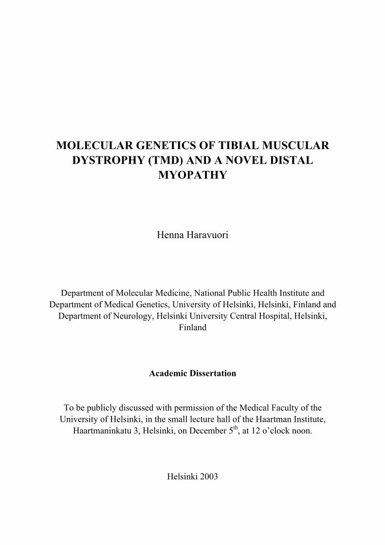
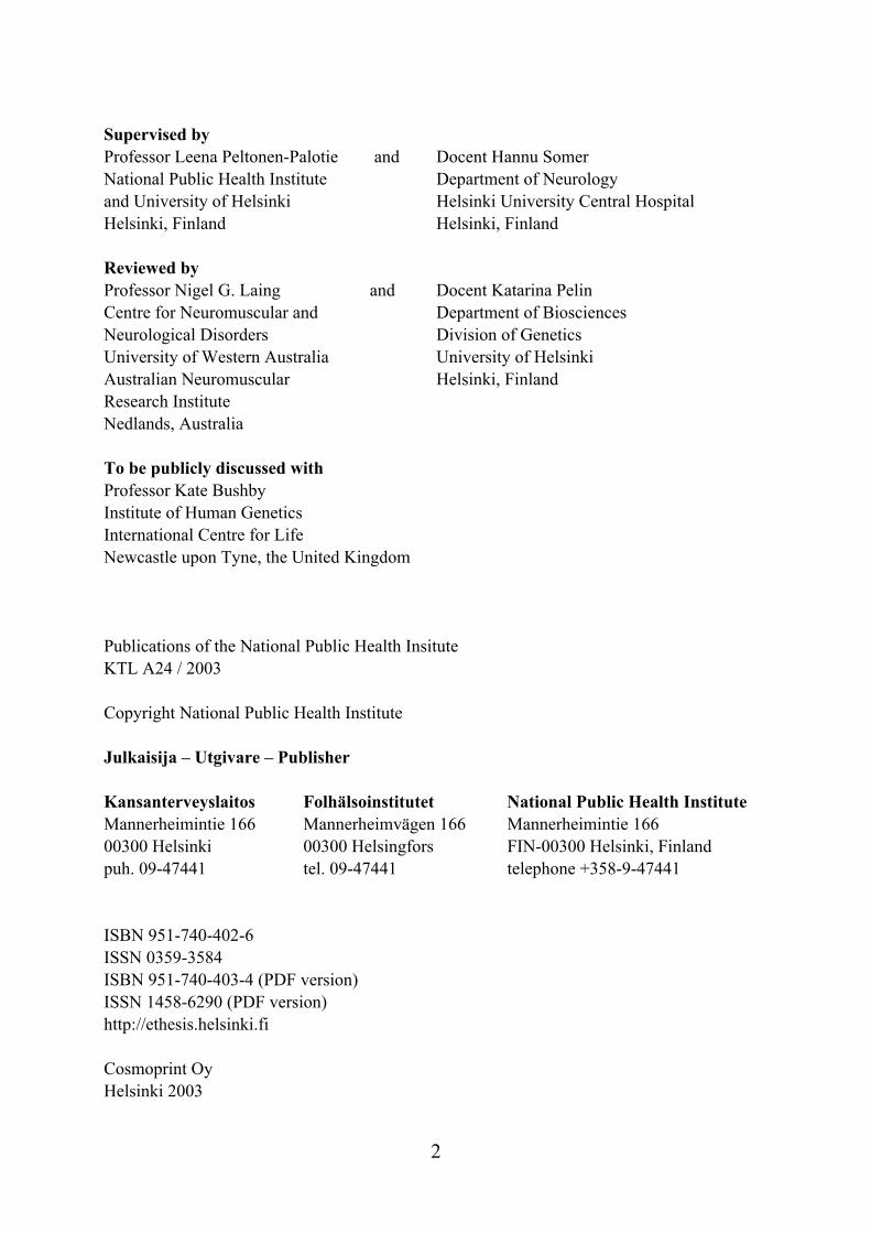
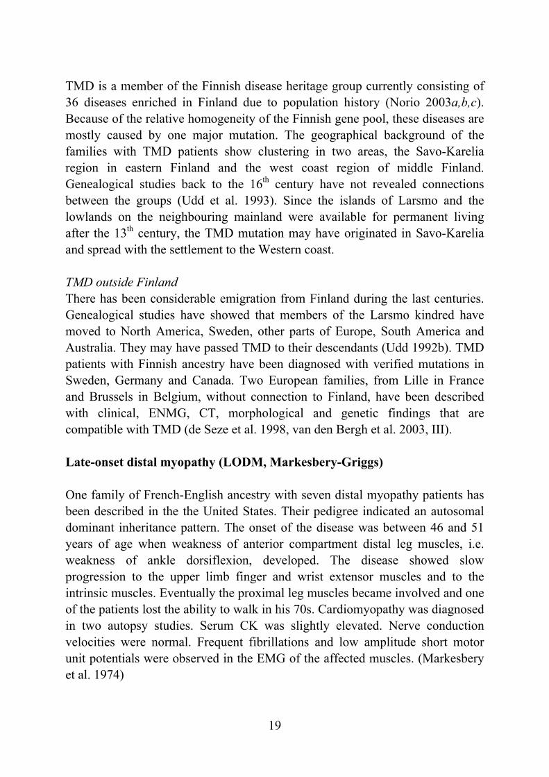
Clinical picture The first symptoms, weakness of ankle dorsiflexion and an inability to walk on the heels, occur after 35 years of age. On inspection, the tibial anterior (TA) muscles are found to be atrophic (fig. 1). The short toe extensor muscles remain intact. There is no sensory loss and the tendon reflexes remain preserved. The disease is very slowly progressive and weakness usually stays confined to the anterior compartment muscles of the lower legs. After 10-20 years, the long toe extensors become clinically involved leading to foot drop and clumsy walking. In some rare cases patients have developed weakness in their proximal leg muscles early in the course of the disease. At the age of 75 years one third of the patients show mild to moderate walking impairment due to proximal leg muscle weakness. Neither the upper limb muscles or facial muscles are not involved and cardiomyopathy has not been diagnosed in TMD patients. The overall clinical symptoms are mild: patients remain ambulatory throughout their lifetime and in the mildest form may not be aware of their condition at all. (Udd et al. 1992, Udd et al. 1993, Partanen et al. 1994). Serum creatine kinase (CK) levels are normal or slightly elevated. Normal nerve conduction velocities are observed in neurophysiological studies whereas EMG shows myopathic changes especially in the TA muscles: a reduced number of very low and short polyphasic motor unit potentials and frequent fibrillation activity (Udd et al. 1993, Partanen et al. 1994, Udd et al. 1998). Computed
17
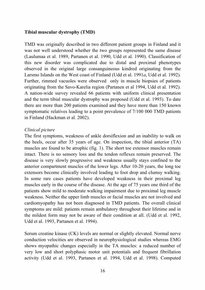
Figure 1. Lower legs of a 58-year-old male TMD patient. Ventral edges of the tibial bone are prominent due to the atrophy of the TA muscles. Symptoms of reduced ankle dorsiflexion had existed for 15 years.
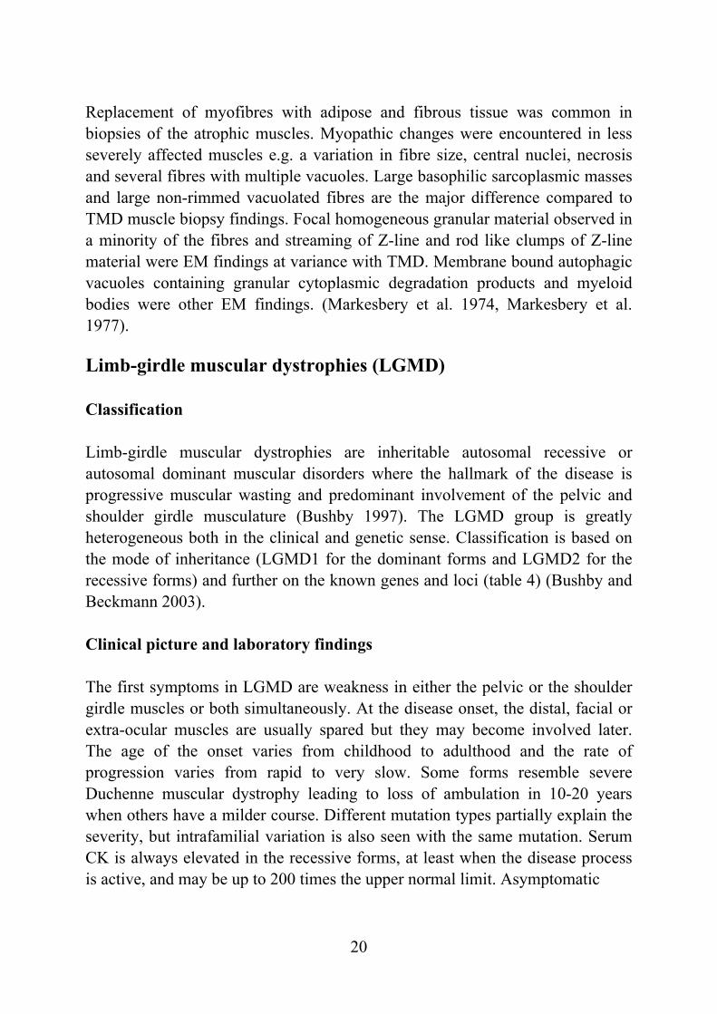
Figure 2. CT scan of the leg muscles of a 64-year-old female patient with 8-10 years of symptoms and inability to walk on her heels. Selective fatty degeneration of the TA muscles is shown on both sides with an early degeneration of the right extensor hallucis longus muscle (arrowhead).
tomography or MRI show selective involvement of TA muscles that are replaced by adipose tissue. In the late stages, the long toe extensors are also involved and there are patchy lesions seen in clinically unaffected muscles (fig. 2) (Udd et al. 1991b, Partanen et al. 1994).
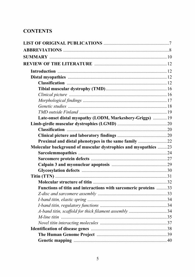
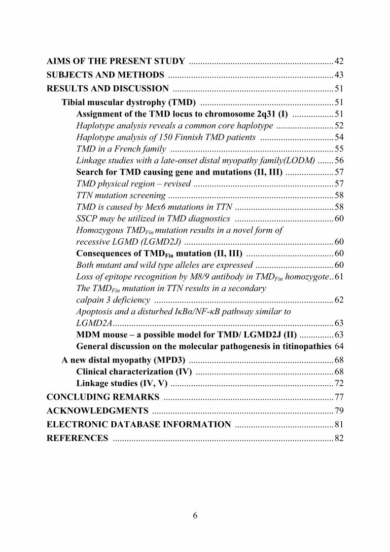
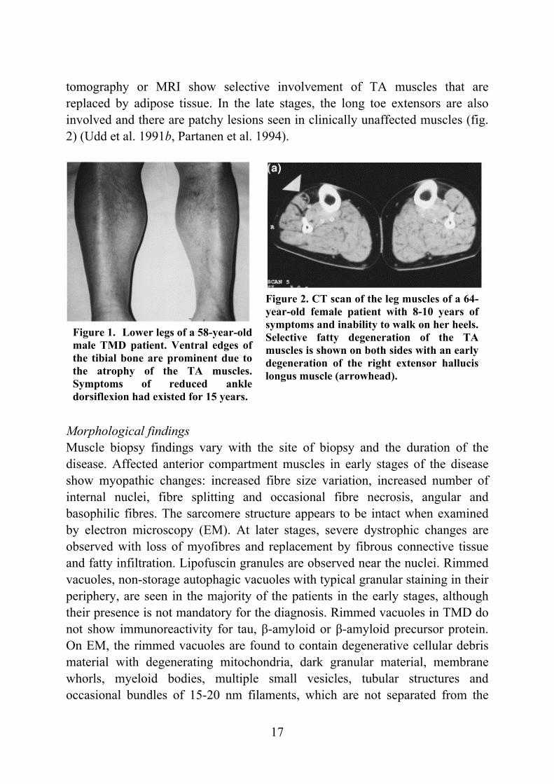
Morphological findings Muscle biopsy findings vary with the site of biopsy and the duration of the disease. Affected anterior compartment muscles in early stages of the disease show myopathic changes: increased fibre size variation, increased number of internal nuclei, fibre splitting and occasional fibre necrosis, angular and basophilic fibres. The sarcomere structure appears to be intact when examined by electron microscopy (EM). At later stages, severe dystrophic changes are observed with loss of myofibres and replacement by fibrous connective tissue and fatty infiltration. Lipofuscin granules are observed near the nuclei. Rimmed vacuoles, non-storage autophagic vacuoles with typical granular staining in their periphery, are seen in the majority of the patients in the early stages, although their presence is not mandatory for the diagnosis. Rimmed vacuoles in TMD do not show immunoreactivity for tau, β-amyloid or β-amyloid precursor protein. On EM, the rimmed vacuoles are found to contain degenerative cellular debris material with degenerating mitochondria, dark granular material, membrane whorls, myeloid bodies, multiple small vesicles, tubular structures and occasional bundles of 15-20 nm filaments, which are not separated from the
18
Figure 3. Muscle biopsy from the TA muscle of a patient with 1-2 years of clinical symptoms showing early changes: increased variation of fibre size, increased number of internal nuclei, angular atrophic fibres, hypertrophic fibres, one atrophic fibre with rimmed vacuolar change (arrow) and one split fibre. (H&E 400x).
Figure 4. Electron micrograph of a TA muscle biopsy showing the edge of a rimmed vacuole containing cellular debris, i.e. myeloid figures (arrow), small vesicular structures, amorphous material and a 15-20 nm filamentous inclusion (F). Bar 1 µm.
sarcoplasm by a membrane (fig. 4) Clinically non-affected muscles show normal morphology or minor myopathic changes. (Udd et al. 1992, Udd et al. 1993, Partanen et al. 1994, Udd et al. 1998). Genetic studies The pedigree findings in TMD are consistent with autosomal dominant inheritance as there are affected family members in all generations in about 0.5 proportion and there are male-to-male transmissions excluding X-chromosomal inheritance (Partanen et al.1990, Udd 1992a). There is no significant difference in disease severity between genders. In the highly consanguineous Larsmo kindred eight patients were described to suffer from severe proximal LGMD-type of muscle weakness and atrophy, but the age of onset, the progression rate and severity varied. Three patients had good distal strength and remained ambulatory at an advanced age, whereas the others had also severe distal weakness and were wheelchair bound in early adulthood (Udd et al. 1991a). Limb-girdle type muscular dystrophy was found in a proportion of 0.246 in this pedigree and was suggested to result from a homozygous manifestation of the dominant gene (Udd 1992a). A genome-wide locus search was initiated in this pedigree with multiple inheritance models but it resulted in no significant linkage findings (Nokelainen et al. 1996).
19
TMD is a member of the Finnish disease heritage group currently consisting of 36 diseases enriched in Finland due to population history (Norio 2003a,b,c). Because of the relative homogeneity of the Finnish gene pool, these diseases are mostly caused by one major mutation. The geographical background of the families with TMD patients show clustering in two areas, the Savo-Karelia region in eastern Finland and the west coast region of middle Finland. Genealogical studies back to the 16th century have not revealed connections between the groups (Udd et al. 1993). Since the islands of Larsmo and the lowlands on the neighbouring mainland were available for permanent living after the 13th century, the TMD mutation may have originated in Savo-Karelia and spread with the settlement to the Western coast. TMD outside Finland There has been considerable emigration from Finland during the last centuries. Genealogical studies have showed that members of the Larsmo kindred have moved to North America, Sweden, other parts of Europe, South America and Australia. They may have passed TMD to their descendants (Udd 1992b). TMD patients with Finnish ancestry have been diagnosed with verified mutations in Sweden, Germany and Canada. Two European families, from Lille in France and Brussels in Belgium, without connection to Finland, have been described with clinical, ENMG, CT, morphological and genetic findings that are compatible with TMD (de Seze et al. 1998, van den Bergh et al. 2003, III).
Late-onset distal myopathy (LODM, Markesbery-Griggs) One family of French-English ancestry with seven distal myopathy patients has been described in the the United States. Their pedigree indicated an autosomal dominant inheritance pattern. The onset of the disease was between 46 and 51 years of age when weakness of anterior compartment distal leg muscles, i.e. weakness of ankle dorsiflexion, developed. The disease showed slow progression to the upper limb finger and wrist extensor muscles and to the intrinsic muscles. Eventually the proximal leg muscles became involved and one of the patients lost the ability to walk in his 70s. Cardiomyopathy was diagnosed in two autopsy studies. Serum CK was slightly elevated. Nerve conduction velocities were normal. Frequent fibrillations and low amplitude short motor unit potentials were observed in the EMG of the affected muscles. (Markesbery et al. 1974)
20
Replacement of myofibres with adipose and fibrous tissue was common in biopsies of the atrophic muscles. Myopathic changes were encountered in less severely affected muscles e.g. a variation in fibre size, central nuclei, necrosis and several fibres with multiple vacuoles. Large basophilic sarcoplasmic masses and large non-rimmed vacuolated fibres are the major difference compared to TMD muscle biopsy findings. Focal homogeneous granular material observed in a minority of the fibres and streaming of Z-line and rod like clumps of Z-line material were EM findings at variance with TMD. Membrane bound autophagic vacuoles containing granular cytoplasmic degradation products and myeloid bodies were other EM findings. (Markesbery et al. 1974, Markesbery et al. 1977).
Limb-girdle muscular dystrophies (LGMD) Classification Limb-girdle muscular dystrophies are inheritable autosomal recessive or autosomal dominant muscular disorders where the hallmark of the disease is progressive muscular wasting and predominant involvement of the pelvic and shoulder girdle musculature (Bushby 1997). The LGMD group is greatly heterogeneous both in the clinical and genetic sense. Classification is based on the mode of inheritance (LGMD1 for the dominant forms and LGMD2 for the recessive forms) and further on the known genes and loci (table 4) (Bushby and Beckmann 2003). Clinical picture and laboratory findings The first symptoms in LGMD are weakness in either the pelvic or the shoulder girdle muscles or both simultaneously. At the disease onset, the distal, facial or extra-ocular muscles are usually spared but they may become involved later. The age of the onset varies from childhood to adulthood and the rate of progression varies from rapid to very slow. Some forms resemble severe Duchenne muscular dystrophy leading to loss of ambulation in 10-20 years when others have a milder course. Different mutation types partially explain the severity, but intrafamilial variation is also seen with the same mutation. Serum CK is always elevated in the recessive forms, at least when the disease process is active, and may be up to 200 times the upper normal limit. Asymptomatic
21
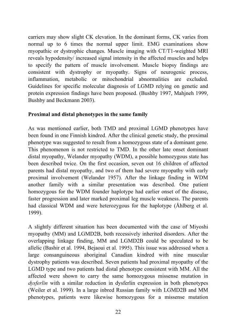
Table 4. Current classification of LGMD. http://www.neuro.wustl.edu/neuromuscular/musdist/lg.html
NAME GENE PRODUCT LOCUS REFERENCES
LGMD 1A myotilin (MYOT) 5q22-q34 Speer et al. 1992, Hauser et al. 2000
LGMD 1B lamin A/C (LMNA) (also mutated in AD Emery-Dreifuss MD and partial lipodystrophy)
1q11-q21 van der Kooi et al. 1997, Muchir et al. 2000
LGMD 1C caveolin-3 (CAV3) (also mutated in rippling muscle disease)
3p25 Minetti et al. 1998, McNally et al. 1998
LGMD 1D
(= dilated cardiomyopathy CMD1F)
6q23 Messina et al. 1997
LGMD 1E
- 7q Speer et al. 1999
LGMD 1F - 7q32 Palenzuela et al. 2003
LGMD 2A calpain 3 (CAPN3) 15q15-q21 Beckmann et al. 1991, Richard et al. 1995
LGMD 2B dysferlin (DYSF) (allelic to Miyoshi myopathy)
2p13 Bashir et al. 1994, Bashir et al. 1998, Liu et al. 1998
LGMD 2C γ-sarcoglycan (SGCG) 13q12 Ben Othmane et al. 1992, Noguchi et al. 1995, McNally et al. 1996
LGMD 2D α-sarcoglycan (SGCA)
17q12-q21 Roberds et al. 1994, Piccolo et al. 1995
LGMD 2E β-sarcoglycan (SGCB)
4q12 Lim et al. 1995, Bonnemann et al. 1995
LGMD 2F δ-sarcoglycan (SGCD) 5q33-q34 Passos-Bueno et al. 1996, Nigro et al. 1996
LGMD 2G telethonin (TCAP) 17q11-q12 Moreira et al. 1997, Moreira et al. 2000
LGMD 2H TRIM32 (putative E3 ubiquitin ligase)
9q31-q34 Weiler et al.1998, Frosk et al. 2002
LGMD 2I fukutin related protein (FKRP) (also mutated in MDC1C)
19q13 Driss et al. 2000, Brockington et al. 2001b
LGMD 2J titin (TTN) 2q31 III
22
carriers may show slight CK elevation. In the dominant forms, CK varies from normal up to 6 times the normal upper limit. EMG examinations show myopathic or dystrophic changes. Muscle imaging with CT/T1-weighted MRI reveals hypodensity/ increased signal intensity in the affected muscles and helps to specify the pattern of muscle involvement. Muscle biopsy findings are consistent with dystrophy or myopathy. Signs of neurogenic process, inflammation, metabolic or mitochondrial abnormalities are excluded. Guidelines for specific molecular diagnosis of LGMD relying on genetic and protein expression findings have been proposed. (Bushby 1997, Mahjneh 1999, Bushby and Beckmann 2003). Proximal and distal phenotypes in the same family As was mentioned earlier, both TMD and proximal LGMD phenotypes have been found in one Finnish kindred. After the clinical genetic study, the proximal phenotype was suggested to result from a homozygous state of a dominant gene. This phenomenon is not restricted to TMD. In the other late onset dominant distal myopathy, Welander myopathy (WDM), a possible homozygous state has been described twice. On the first occasion, seven out 16 children of affected parents had distal myopathy, and two of them had severe myopathy with early proximal involvement (Welander 1957). After the linkage finding in WDM another family with a similar presentation was described. One patient homozygous for the WDM founder haplotype had earlier onset of the disease, faster progression and later marked proximal leg muscle weakness. The parents had classical WDM and were heterozygous for the haplotype (Åhlberg et al. 1999). A slightly different situation has been documented with the case of Miyoshi myopathy (MM) and LGMD2B, both recessively inherited disorders. After the overlapping linkage finding, MM and LGMD2B could be speculated to be allelic (Bashir et al. 1994, Bejaoui et al. 1995). This issue was addressed when a large consanguineous aboriginal Canadian kindred with nine muscular dystrophy patients was described. Seven patients had proximal myopathy of the LGMD type and two patients had distal phenotype consistent with MM. All the affected were shown to carry the same homozygous missense mutation in dysferlin with a similar reduction in dysferlin expression in both phenotypes (Weiler et al. 1999). In a large inbred Russian family with LGMD2B and MM phenotypes, patients were likewise homozygous for a missense mutation
23
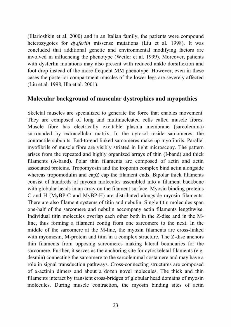
(Illarioshkin et al. 2000) and in an Italian family, the patients were compound heterozygotes for dysferlin missense mutations (Liu et al. 1998). It was concluded that additional genetic and environmental modifying factors are involved in influencing the phenotype (Weiler et al. 1999). Moreover, patients with dysferlin mutations may also present with reduced ankle dorsiflexion and foot drop instead of the more frequent MM phenotype. However, even in these cases the posterior compartment muscles of the lower legs are severely affected (Liu et al. 1998, Illa et al. 2001). Molecular background of muscular dystrophies and myopathies Skeletal muscles are specialized to generate the force that enables movement. They are composed of long and multinucleated cells called muscle fibres. Muscle fibre has electrically excitable plasma membrane (sarcolemma) surrounded by extracellular matrix. In the cytosol reside sarcomeres, the contractile subunits. End-to-end linked sarcomeres make up myofibrils. Parallel myofibrils of muscle fibre are visibly striated in light microscopy. The pattern arises from the repeated and highly organized arrays of thin (I-band) and thick filaments (A-band). Polar thin filaments are composed of actin and actin associated proteins. Tropomyosin and the troponin complex bind actin alongside whereas tropomodulin and capZ cap the filament ends. Bipolar thick filaments consist of hundreds of myosin molecules assembled into a filament backbone with globular heads in an array on the filament surface. Myosin binding proteins C and H (MyBP-C and MyBP-H) are distributed alongside myosin filaments. There are also filament systems of titin and nebulin. Single titin molecules span one-half of the sarcomere and nebulin accompany actin filaments lengthwise. Individual titin molecules overlap each other both in the Z-disc and in the M-line, thus forming a filament contig from one sarcomere to the next. In the middle of the sarcomere at the M-line, the myosin filaments are cross-linked with myomesin, M-protein and titin in a complex structure. The Z-disc anchors thin filaments from opposing sarcomeres making lateral boundaries for the sarcomere. Further, it serves as the anchoring site for cytoskeletal filaments (e.g. desmin) connecting the sarcomere to the sarcolemmal costamere and may have a role in signal transduction pathways. Cross-connecting structures are composed of α-actinin dimers and about a dozen novel molecules. The thick and thin filaments interact by transient cross-bridges of globular head domains of myosin molecules. During muscle contraction, the myosin binding sites of actin
24
filaments are exposed and hydrolysis of ATP drives the head domain conformational change causing the thin and thick filaments to slide past each other in series. (Stryer 1995, Squire 1997, Stromer 1998, Clark et al. 2002).
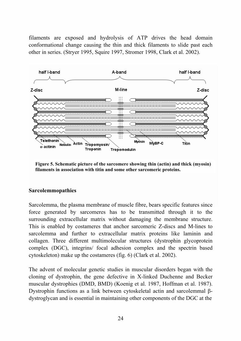
Sarcolemmopathies Sarcolemma, the plasma membrane of muscle fibre, bears specific features since force generated by sarcomeres has to be transmitted through it to the surrounding extracellular matrix without damaging the membrane structure. This is enabled by costameres that anchor sarcomeric Z-discs and M-lines to sarcolemma and further to extracellular matrix proteins like laminin and collagen. Three different multimolecular structures (dystrophin glycoprotein complex (DGC), integrins/ focal adhesion complex and the spectrin based cytoskeleton) make up the costameres (fig. 6) (Clark et al. 2002). The advent of molecular genetic studies in muscular disorders began with the cloning of dystrophin, the gene defective in X-linked Duchenne and Becker muscular dystrophies (DMD, BMD) (Koenig et al. 1987, Hoffman et al. 1987). Dystrophin functions as a link between cytoskeletal actin and sarcolemmal β-dystroglycan and is essential in maintaining other components of the DGC at the
Figure 5. Schematic picture of the sarcomere showing thin (actin) and thick (myosin) filaments in association with titin and some other sarcomeric proteins.
25
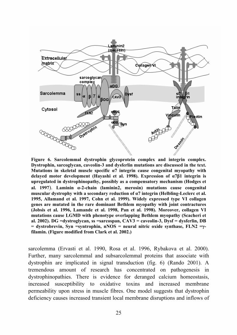
Figure 6. Sarcolemmal dystrophin glycoprotein complex and integrin complex. Dystrophin, sarcoglycan, caveolin-3 and dysferlin mutations are discussed in the text. Mutations in skeletal muscle specific α7 integrin cause congenital myopathy with delayed motor development (Hayashi et al. 1998). Expression of α7β1 integrin is upregulated in dystrophinopathy, possibly as a compensatory mechanism (Hodges et al. 1997). Laminin α-2-chain (laminin2, merosin) mutations cause congenital muscular dystrophy with a secondary reduction of α7 integrin (Helbling-Leclerc et al. 1995, Allamand et al. 1997, Cohn et al. 1999). Widely expressed type VI collagen genes are mutated in the rare dominant Bethlem myopathy with joint contractures (Jobsis et al. 1996, Lamande et al. 1998, Pan et al. 1998). Moreover, collagen VImutations cause LGMD with phenotype overlapping Bethlem myopathy (Scacheri et al. 2002). DG =dystroglycan, ss =sarcospan, CAV3 = caveolin-3, Dysf = dysferlin, DB = dystrobrevin, Syn =syntrophin, nNOS = neural nitric oxide synthase, FLN2 =γ-filamin. (Figure modified from Clark et al. 2002.)
sarcolemma (Ervasti et al. 1990, Rosa et al. 1996, Rybakova et al. 2000). Further, many sarcolemmal and subsarcolemmal proteins that associate with dystrophin are implicated in signal transduction (fig. 6) (Rando 2001). A tremendous amount of research has concentrated on pathogenesis in dystrophinopathies. There is evidence for deranged calcium homeostasis, increased susceptibility to oxidative toxins and increased membrane permeability upon stress in muscle fibres. One model suggests that dystrophin deficiency causes increased transient local membrane disruptions and inflows of
26
calcium. Calcium activates intracellular proteases and leads to a vicious circle with altered calcium channel activity and further inflows of calcium. Disturbed calcium homeostasis causes apoptosis and/or necrosis of muscle fibre. (Blake et al. 2002, Alderton and Steinhardt 2000). Studies on dystrophin resulted in purification of the sarcoglycan complex (SGC). SGC in skeletal muscle is composed of membrane spanning α-, β-, γ- and δ- SG and sarcospan (Ozawa et al. 1998, Crosbie et al. 1997). Subsequently, α-, β-, γ- and δ-sarcoglycans were found to be mutated in LGMD2D, 2E, 2C and 2F respectively (table 4). The SGC is assembled en bloc to the sarcolemma while mutation in any one of the sarcoglycans may prevent targeting of the complex to the plasma membrane (Holt and Campbell 1998). Thus, loss of one SG or dystrophin often results in the absence or reduction of the whole complex including sarcospan (Ohlendieck et al. 1993, Crosbie et al. 1999). One function of the sarcoglycan complex may be to strengthen the dystrophin-dystroglycan axis connecting cytoskeletal actin to the extracellular matrix (Ohlendieck 1996, Blake et al. 2002). Many studies support functions in signalling as well (Yoshida et al. 1998, Hack et al. 1999, Betto et al. 1999, Crosbie et al. 2002, Thompson et al. 2000). Molecular genetic studies have identified other sarcolemmal DGC associated proteins, caveolin-3 and dysferlin, also to be mutated in LGMD. Caveolins are structural proteins involved in the formation of caveolae (small membrane invaginations) which participate in signalling and cellular transformation (Lisanti et al. 1995, Parton 1996). Muscle specific caveolin-3 mutations cause clinically variable dominant muscular phenotypes including LGMD1C, hyperCKaemia, distal myopathy and rippling muscle disease (Minetti et al. 1998, McNally et al. 1998, Carbone et al. 2000, Betz et al. 2001). Caveolin-3 binds to the same β-dystroglycan domain as dystrophin and competitive binding may regulate this interaction (Sotgia et al. 2000). Further, caveolin-3 interacts with subsarcolemmal nNOS and the developing T-tubule system (Parton et al. 1997), and seems to interact with dysferlin according to reported reciprocal secondary defects (Matsuda et al 2001). Caveolin-3 mutant myopathic mice show increased activity of nNOS as well as the changes in DGC distribution and T-tubule abnormalities that are also observed in LGMD1C patients (Sunada et al. 2001, Galbiati et al. 2001, Minetti et al. 2002).
27
Dysferlin mutations cause LGMD2B, Miyoshi myopathy (MM) and distal myopathy with anterior tibial onset (Bashir et al. 1998, Liu et al. 1998). Thus, modifying environmental and genetic factors are suggested to be involved in the pathogenesis because identical mutations cause these phenotypes (Weiler et al. 1999). Dysferlin is expressed at early embryonic stages and although dysferlin is not vital for myofiber assembly, altered dysferlin levels at a critical stage are hypothesized to contribute to the phenotypic variability (Anderson et al. 1999). Dysferlinopathy patients lack dysferlin at the plasma membrane and have intact caveolin-3 whereas in caveolinopathy the expression of both may be disturbed (Matsuda et al. 2001). Some secondary reduction in calpain 3 expression has been noted in LGMD2B patients but a potential dysferlin-calpain interaction has not been verified (Anderson et al. 2000, Vainzof et al. 2001). Recently, dysferlin was demonstrated to have a role in membrane repair. Dysferlin deficiency prevents Ca2+-dependent resealing of the sarcolemma after injury potentially leading to muscle degeneration (Bansal et al. 2003, Lennon et al. 2003). Sarcomere protein defects The contractile subunits, sarcomeres, make up the bulk of the striated muscle fibre. As a natural consequence mutations in sarcomeric molecules cause variable muscular phenotypes and cardiomyopathy. Thin filament protein defects are associated with nemaline myopathies (NM), a subtype of congenital myopathies. NM present typically generalized muscle weakness of variable severity ranging from neonatal death to late onset slowly progressive disease. The characteristic findings on muscle biopsy are dark red or purple nemaline rod clusters under sarcolemma or around the nuclei with Gomori trichrome staining (Sanoudou and Beggs 2001). On electron microscopy, the nemaline rods appear to be extensions of Z-discs, and in fact consist of actin, α-actinin and other Z-disc proteins (Sanoudou and Beggs 2001). Autosomal recessive NM is caused by mutations in nebulin, skeletal muscle α-actin, α-tropomyosin, troponin T and dominant forms by mutations in α-tropomyosin, β-tropomyosin and α-actin (Laing et al. 1995b, Pelin et al. 1999, Nowak et al. 1999, Tan et al. 1999, Johnston et al. 2000, Ilkovski et al. 2001, Donner et al. 2002). Autosomal dominant distal myopathy of Gowers-Laing type (MPD1) has been genetically linked to a large region on chromosome 14 (Laing et al. 1995a, Voit et al. 2001, Mastaglia et al. 2002). Muscle biopsy shows myopathic changes and in some cases autophagic vacuoles and intranuclear inclusions of 15-20-nm
28
filaments. Sequencing of the β-cardiac myosin MYH7 gene, has revealed a missense mutation A1663P encoding the tail region of the myosin molecule in an Australian family (Meredith 2001). Myosin is thought not to be able to form its α-helical coiled-coil structure due to introduction of a proline (Meredith 2001). Further, binding sites for myomesin and titin are located in the disturbed tail region. A nearby L1617 deletion of MYH7 has been associated with the disease in a German family (Udd et al. 2002). In addition, mutations in the MYH7 gene are the most common cause for the inherited hypertrophic cardiomyopathy. Especially missense mutations in the myosin head-neck region are known to alter the motor function (Seidman and Seidman 2001). The MYH2 gene encoding the fast myosin heavy chain is mutated in AD childhood onset hereditary inclusion body myopathy (HIBM3) with joint contractures and external ophthalmoplegia (Martinsson et al. 2000). The myosin motor domain is altered by an E706K missense mutation. Breakdown of the sarcomeric proteins was suggested to have a role in the pathogenesis of rimmed vacuoles (Tajsharghi et al. 2002) Dominantly inherited LGMD1A is caused by missense mutations in myotilin, a novel protein identified through interaction with α-actinin (Salmikangas et al. 1999, Hauser et al. 2000, Hauser et al. 2002). Myotilin interacts also with γ-filamin (FLN2) and has direct actin filament cross-linking and stabilizing properties (van der Ven et al. 2000, Salmikangas et al. 2003). These myotilin interactions and correct timing of expression seem to be essential for myofibril assembly. It has been suggested that myotilin stabilizes the Z-disc structure (Salmikangas et al. 2003). This is supported by the streaming of the Z-lines observed in patient muscle biopsies. The identified mutations locate outside the α-actinin and γ-filamin interaction sites. Further, the mutant molecules are expressed and myotilin is correctly localized to Z-discs while α-actinin binding remains normal. This indicates that yet unknown interactions are disrupted (Hauser et al. 2000, Hauser et al. 2002). Telethonin (T-CAP) mutations cause recessive LGMD2G (Moreira et al. 2000). In addition to proximal weakness, some LGMD2G patients have early weakness in distal muscles, a few patients have cardiomyopathy. Rimmed vacuoles are found in their muscle biopsies while the sarcomeric ultrastructure remains intact (Moreira et al. 1997). The known mutations result in a premature stop codon and patient muscle biopsies show a deficiency of telethonin protein (Moreira et al. 2000). Telethonin specifically binds titin Ig-domains Z1 and Z2 in the Z-disc. A
29
ternary complex of titin, telethonin and sarcoplasmic reticulum membrane protein, small splice variant of ankyrin-1, is implicated in organizing sarcoplasmic reticulum around the Z-disc (Gregorio et al. 1998, Mues et al. 1998, Zou et al. 2003, Kontrogianni-Konstantopoulos and Bloch 2003). When telethonin was identified, the protein was found to localize to the A-band with myosin (Valle et al.1997). This observation remains unclear. Telethonin interacts with many signalling associated molecules and the muscle LIM protein (MLP) telethonin complex is suggested to be a component of the stretch sensor machinery in cardiomyocytes (Faulkner et al. 2000, Frey and Olson 2002, Furukawa et al. 2001, Nicholas et al. 2002, Knöll et al. 2002). Defects in MLP lead to dilated cardiomyopathy. Calpain 3 and myonuclear apoptosis Structural proteins are reasonable targets for mutation in muscular dystrophy. In this context it was surprising that LGMD2A is caused by defective calpain 3 (Richard et al. 1995). Calpains are calcium-dependent cytosolic nonlysosomal cysteine proteases. Calpain 3, encoded by CAPN3, is the muscle specific member of this group (Sorimachi et al. 1989). In addition to four homologous domains found in ubiquitous calpains, it has three unique domains named NS, IS1 and IS2 (Sorimachi et al. 1989). Calpain 3 is subject to alternative splicing and the full-length protein is the major isoform found in mature muscle (Herasse et al. 1999). The IS2 domain contains a nuclear translocation signal and a titin binding site (Sorimachi et al. 1989, Sorimachi et al. 1995). Binding to titin may regulate calpain 3 activity by preventing autolysis (Sorimachi et al. 1995). Calpain 3 has been suggested to have a role in myofibrillar integrity and in muscle maturation and it is not expressed in the mature heart muscle (Spencer et al. 2002). In in vitro function studies, calpain 3 proteins with the LGMD2A mutations lack proteolytic activity (Ono et al. 1998, Jia et al. 2001). Necrosis is thought to account for myofibre destruction in most of the muscular dystrophies. However, at least LGMD2A seems to be an exception. Patients and capn3-defective mouse exhibit TUNEL-positive apoptotic myonuclei in muscle fibres, and possibly in satellite cells, reducing the regenerative capacity of the muscles (Baghdiguian et al. 1999, Richard et al. 2000). Alterations in the IκBα/NF-κB signalling pathway have also been demonstrated. NF-κB is a transcription factor promoting expression of cell survival genes (Baghdiguian et al. 2001). IκBα masks the NF-κB nuclear localization signal and the inactive
30
IκBα/NF-κB complexes reside in the cytosol. Upon activation, IκBα is degraded allowing NF-κB to enter the nucleus. NF-κB induces, among others, expression of IκBα, which then transfers NF-κB back to the cytosol (Baldwin 1996). Calpain 3 is able to hydrolyse IκBα and possibly controls IκBα turnover in vivo. TUNEL-positive myonuclei in LGMD2A show IκBα labelling and aberrant NF-κB subsarcolemmal localization surrounding myonuclei (Baghdiguian et al. 1999). It has been postulated that accumulation of IκBα in calpain 3 deficiency prevents NF-κB dependent expression of survival genes and makes myofibres vulnerable to death signals (Baghdiguian et al. 2001). The protective role of NF-κB is supported by the finding that the NF-κB pathway is activated in inflammatory myopathies and to some extent in DMD (Monici et al. 2003). Some components of the ubiquitin-proteasome system are down regulated in the gastrocnemius muscle from calpain 3 deficient mice (Combaret et al. 2003). Ubiquination targets proteins to proteasome degradation in cells (Joazeiro and Weissman 2000). These findings suggest defective ubiquitination of specific targets and accumulation of altered proteins and/or dysregulation of the NFkB/IkBa pathway. Interestingly TRIM32, a putative E3-ubiquitin-ligase gene of the tripartite-motif family, is mutated in LGMD2H (Frosk et al. 2002). Glycosylation defects Post-translational modification of cell surface proteins with oligosaccharides is essential for their function in cell adhesion and signal transduction. Disruption in these processes has recently been recognized to cause muscular pathology. A deficiency of fukutin in Fukuyama congenital muscular dystrophy (FCMD) leads to hypoglycosylation of α-dystroglycan (Michele et al. 2002). α- and β-dystroglycan, encoded by a single gene, are expressed in various tissues but different glycosylation isoforms exist (fig. 6) (Ibraghimov-Beskrovnaya et al. 1993). Hypoglycosylation abolishes α-dystroglycan ligand binding, including binding to laminin (Michele et al. 2002).
Fukutin-related protein gene (FKRP) was identified by a homology search with the fukutin sequence, and it was found to be mutated in one form of congenital muscular dystrophy (MDC1C). Sequence analysis predicted a similarity to glycosyltransferases and both fukutin and FKRP have been localized to the Golgi apparatus (Esapa et al. 2002). MDC1C patient muscle biopsies show reduction in α-dystroglycan immunostaining similar to FCMD and secondary deficiency of laminin α-2 suggests pathogenetic similarity to FCMD. FKRP was
31
also found to be mutated in LGMD2I. Variable reduction of α –dystroglycan expression was observed in muscle biopsy of these patients accompanied by deficiency of laminin α-2 in some cases. (Brockington et al. 2001a, Brockington et al. 2001b)
GNE (UDP-N-Acetylglucosamine 2-Epimerase/N-Acetylmannosamine kinase) is the rate-limiting enzyme in the sialic acid biosynthesis pathway, one of the sugar residues found in glycoproteins (Keppler et al. 1999). GNE is mutated in h-IBM and Nonaka distal myopathy (DMRV) in addition to sialuria (Eisenberg et al. 2001, Kayashima et al. 2002). H-IBM and DMRV phenotypes overlap and they probably represent the same entity. The variability in phenotypes may be due to mutations in different domains of GNE or other modifying factors. Titin (TTN) It was noted early on that the thin and thick filament characteristics cannot explain all the features observed in muscle fibres. Subsequently, titin was identified independently as the elastic protein of the sarcomere and as the high molecular weight protein found in striated muscles (Maruyama et al 1977, Wang et al. 1979, Maruyama et al. 1981). Physiological roles for titin were established as titin degradation was shown to result in decreased passive tension of muscle fibres and immunoelectron microscopy studies revealed titin as an extendable filamentous protein spanning one-half of the sarcomere from Z-disc to M-line (Horowits et al. 1986, Fürst et al. 1988). This molecular giant (3700 kD in the soleus skeletal muscle) has been identified as the third filament system in the sarcomere besides the thick and thin filaments. Titin amino-termini are embedded in the Z-disc where titin filaments from opposing sarcomeres overlap (Gregorio et al. 1998). Central I-band titin constitutes the elastic part of the molecule and is composed of tandem Ig-repeats. The C-terminal region contributes to M-line structures where titin filaments from both sides of the sarcomere again overlap (Fürst et al. 1999). Thus, titin molecules provide a filament system extending the length of the myofibrils (fig. 5). Recent studies suggest a stoichometry of six titin filaments per one half myosin filament (Liversage et al. 2001, Knupp et al. 2002). Titin is associated with multiple roles: a scaffold in sarcomere assembly and molecular ruler of the thick filament length, an elastic spring that keeps contractile elements in place and finally a signal transducer (Trinick 1994, Labeit and Kolmerer 1995, Squire 1997, Gregorio et al. 1999, Machado and Andrew 2000, Clark et al. 2002).
32
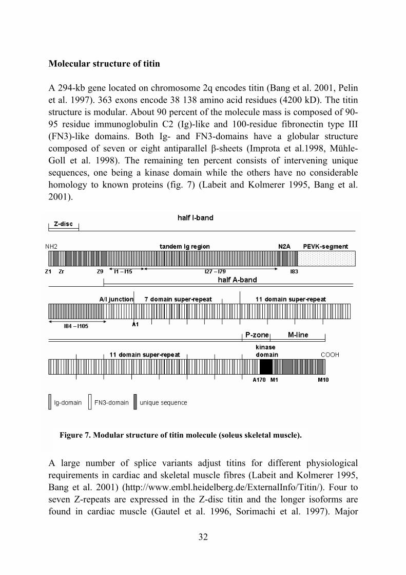
Figure 7. Modular structure of titin molecule (soleus skeletal muscle).
Molecular structure of titin A 294-kb gene located on chromosome 2q encodes titin (Bang et al. 2001, Pelin et al. 1997). 363 exons encode 38 138 amino acid residues (4200 kD). The titin structure is modular. About 90 percent of the molecule mass is composed of 90-95 residue immunoglobulin C2 (Ig)-like and 100-residue fibronectin type III (FN3)-like domains. Both Ig- and FN3-domains have a globular structure composed of seven or eight antiparallel β-sheets (Improta et al.1998, Mühle-Goll et al. 1998). The remaining ten percent consists of intervening unique sequences, one being a kinase domain while the others have no considerable homology to known proteins (fig. 7) (Labeit and Kolmerer 1995, Bang et al. 2001). A large number of splice variants adjust titins for different physiological requirements in cardiac and skeletal muscle fibres (Labeit and Kolmerer 1995, Bang et al. 2001) (http://www.embl.heidelberg.de/ExternalInfo/Titin/). Four to seven Z-repeats are expressed in the Z-disc titin and the longer isoforms are found in cardiac muscle (Gautel et al. 1996, Sorimachi et al. 1997). Major
33
splicing differences in the I-band titin explain size differences observed between skeletal (3350-3700 kD) and cardiac muscle (2970-3300 kD) (Freiburg et al. 2000). Skeletal muscles express larger isoforms of the tandem Ig-repeat segments and a PEVK-region with the N2A domain (fig. 7). Cardiac isoforms have shorter Ig-repeat segments with reduced number of the Ig-domains and shorter PEVK domains. Further, the N2A domain is replaced by the N2B or N2BA domain in cardiac muscle (Freiburg et al. 2000, Trombitas et al. 2001). The A-band region has a conserved structure in all muscle types whereas the M-line region of titin is expressed in two splice isoforms either containing or lacking the Mex5 exon, designated as Mex5 + and Mex5 – isoforms (Kolmerer et al. 1996). A C-terminally truncated 700 kD isoform having an alternative C-terminus, novex-3, is expressed in both skeletal and cardiac muscle but is less abundant than the other isoforms (Bang et al. 2001). Functions of titin and interactions with sarcomeric proteins Z-disc and sarcomere assembly Titin is expressed amongst the first sarcomeric proteins during myofibrillogenesis and is observed in dot-like aggregates with α-actinin that further organize to precursory Z-discs coordinating filament assembly (Lin et al. 1994, van der Loop et al. 1996, Gautel et al. 1999, Sanger et al. 2002). The aminoterminal 80 kD of the titin molecule spans the Z-disc (Gregorio et al. 1998, Young et al. 1998). The first two Ig-domains (Z1 and Z2) bind telethonin in a conformation-dependent manner suggesting regulation of this interaction by phosphorylation (Gregorio et al. 1998, Mues et al. 1998). One function of telethonin may be linking titin filaments in the Z-disc (Zou et al. 2003). In addition, telethonin is a potential molecule in the stretch sensor machinery with muscle LIM protein (Knöll et al. 2002) Variably expressed 45-residue Z-repeats provide multiple binding sites for α-actinin C-termini (Gautel et al. 1996, Sorimachi et al. 1997). The adjacent sequence insertion (Zq) has a single binding site for the spectrin like repeats of the α-actinin molecule (Young et al. 1998). Titin-α-actinin interaction is regulated by phosphatidylinositol 4,5 bisphosphate (Young and Gautel 2000). Titin, together with nebulin, are suggested to regulate the diversity of Z-disc widths observed in different striated muscle types, although the correlation is not straightforward (Millevoi et al. 1998, Luther and Squire 2002).
34
I-band titin, elastic spring Titin exhibits important mechano-physiological functions. The elastic properties of titin determine the passive tension of both skeletal and cardiac muscle. Titin helps to set the resting (slack) length of the muscle and further restores sarcomere length, and positions thick filaments at the centre of the sarcomere after stretching or contraction. Maintenance of the symmetrical thick and thin filament overlap is necessary for the sequential contractions by the sarcomeres (Horowits 1999). The I-band region is composed of tandem Ig-domain repeats, insertion of a PEVK domain (rich in proline, glutamine, valine and lysine residues) and a N2A domain in skeletal or the N2B/N2BA domain in cardiac muscle (fig. 7) (Labeit and Kolmerer 1995, Freiburg et al. 2000). The elasticity of I-band titin is determined by varying properties of these modules. Upon stretching with a low force these Ig-domains elongate without producing significant tension. The PEVK region unfolds at higher forces with a linear increase in passive tension (Linke et al. 1999, Li et al. 2001, Granzier and Labeit 2002). In cardiac muscle the N2B region is capable of extending after the PEVK domain at increasing levels of stretch (Linke et al. 1999, Trombitas et al. 1999). Different muscles have varying elastic properties that can be explained by alternative splicing events in the I-band titin (Freiburg et al. 2000, Cazorla et al. 2000). Stiffer cardiac muscle has the shortest isoforms of the I-band titins. I-band titin, regulatory functions Titin has been found to posses many unique regulatory mechanisms affecting the elastic and contractile properties of the muscle, such as the interaction with actin and nebulin in the PEVK region and modification of the N2B domain in cardiac muscle (Linke et al. 2002, Kulke et al. 2001, Yamasaki et al. 2001, Yamasaki et al. 2002, Neagoe et al. 2002, Golenhofen et al. 2002). The N2A domain binds the skeletal muscle specific protease calpain 3. However, the functional role of the titin calpain 3 interaction in the I-band is unknown. Implications for pathology exist as shown by the deletion of the N2A domain in the spontaneous MDM mutant mouse (Sorimachi et al. 1995, Garvey et al. 2002). The PEVK domain interaction with nebulin may have a role in myofibrillogenesis (Ma and Wang 2002). A-band titin, scaffold for thick filament assembly Titin is thought to serve as a ruler for myosin filament assembly (Trinick 1994). This is supported by the regular super repeat structure of FN3- and Ig- domains of the A-band titin matching the myosin helix repeats (fig. 7) (Labeit et al.
35
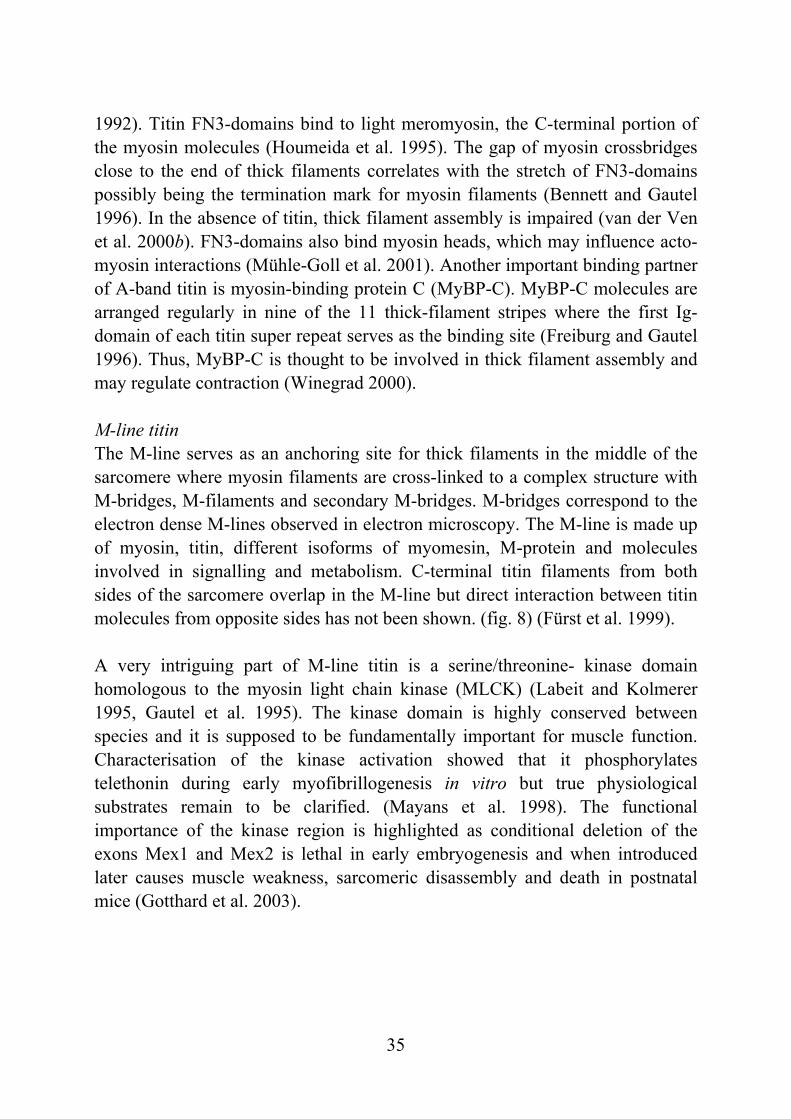
1992). Titin FN3-domains bind to light meromyosin, the C-terminal portion of the myosin molecules (Houmeida et al. 1995). The gap of myosin crossbridges close to the end of thick filaments correlates with the stretch of FN3-domains possibly being the termination mark for myosin filaments (Bennett and Gautel 1996). In the absence of titin, thick filament assembly is impaired (van der Ven et al. 2000b). FN3-domains also bind myosin heads, which may influence acto-myosin interactions (Mühle-Goll et al. 2001). Another important binding partner of A-band titin is myosin-binding protein C (MyBP-C). MyBP-C molecules are arranged regularly in nine of the 11 thick-filament stripes where the first Ig-domain of each titin super repeat serves as the binding site (Freiburg and Gautel 1996). Thus, MyBP-C is thought to be involved in thick filament assembly and may regulate contraction (Winegrad 2000). M-line titin The M-line serves as an anchoring site for thick filaments in the middle of the sarcomere where myosin filaments are cross-linked to a complex structure with M-bridges, M-filaments and secondary M-bridges. M-bridges correspond to the electron dense M-lines observed in electron microscopy. The M-line is made up of myosin, titin, different isoforms of myomesin, M-protein and molecules involved in signalling and metabolism. C-terminal titin filaments from both sides of the sarcomere overlap in the M-line but direct interaction between titin molecules from opposite sides has not been shown. (fig. 8) (Fürst et al. 1999). A very intriguing part of M-line titin is a serine/threonine- kinase domain homologous to the myosin light chain kinase (MLCK) (Labeit and Kolmerer 1995, Gautel et al. 1995). The kinase domain is highly conserved between species and it is supposed to be fundamentally important for muscle function. Characterisation of the kinase activation showed that it phosphorylates telethonin during early myofibrillogenesis in vitro but true physiological substrates remain to be clarified. (Mayans et al. 1998). The functional importance of the kinase region is highlighted as conditional deletion of the exons Mex1 and Mex2 is lethal in early embryogenesis and when introduced later causes muscle weakness, sarcomeric disassembly and death in postnatal mice (Gotthard et al. 2003).
36
Figure 8. Schematic representation of the molecular structure of M-line titin and associated proteins (modified from Fürst et al. 1999). Is7 encoded by the Mex5 exon is subject to alternative splicing and may be left out. Myomesin, M-protein and obscurin have modular structures similar to titin. Myomesin binding to titin is regulated by phosphorylation. M-protein has not been demonstrated to bind titin so far and its expression is restricted to cardiac and fast-twitch muscles. Binding sites of the signalling molecules calpain 3, MURF-1 and FHL2-DRAL are indicated. DRAL has been suggested to link metabolic enzymes like CK to M-line. The localization of obscurin is obscure. Binding sites of M-line titin antibodies are indicated with arrows. (Oberman et al. 1997, Fürst et al. 1999, Auerbach et al. 1999, Ehler et al. 1999, Lange et al. 2002).
The second last titin exon (Mex5) codes for a unique sequence insertion (is7) and is alternatively spliced (Kolmerer et al. 1996). Heart muscle expresses Mex5+ isoforms only. Fast-twitch muscles express Mex5- as the major isoform while slow-twitch muscles express both isoforms (Kolmerer et al. 1996). This variability may correlate with the electron microscopic differences seen in M-lines of different muscle types (Kolmerer et al. 1996). Other interesting features of M-line titin are the existence of a putative phosphorylation site in the unique sequence insertion is4, the interaction and binding of creatine kinase, the
37
regulatory FHL2-DRAL and myomesin in the centre of the M-line (Labeit and Kolmerer 1995, Oberman et al. 1997, Lange et al. 2002). As mentioned above, titin contains at least two binding sites for the muscle-specific protease calpain 3, one in the N2A domain and the second in the M-line. Interaction with Z-disc titin has also been suggested. The M-line binding site has been mapped to the is7 encoded by Mex5. The two different titin-calpain 3 interactions are not equal when it comes to binding affinities and the binding region of calpain 3 itself. Mature cardiac muscle does not express calpain 3 and both titin binding sites are subject to alternative splicing. Thus, binding to titin may regulate calpain 3 activity in a muscle specific manner. M-line titin may also provide a scaffold for other molecules interacting with calpain 3. (Sorimachi et al. 1995, Kinbara et al. 1997). Novel titin interacting molecules Obscurin is a recently identified 800 kD striated muscle protein which has a modular structure similar to titin. However, it is expressed in low levels in skeletal and cardiac muscle (Young et al. 2001, Russell et al. 2002). During myofibrillogenesis, obscurin interacts with titin Z-disc Z9-Z10 Ig-domains and transfers to the M-line region later in differentiation (Young et al. 2001). The small Novex-3 titin isoform was found to interact with obscurin - providing a second binding site to obscurin and implicating obscurin-titin complexes with elastic properties (Bang et al. 2001). Obscurin has two kinase domains homologous to myosin light chain kinases (MLCK), a calmodulin-binding IQ motif and a Rho guanine nucleotide exchange factor domain, indicating a role in calcium dependent and G-protein coupled signalling (Young et al. 2001, Russell et al. 2002). Obscurin interactions with ankyrin-1 isoforms and titin are suggested to organize sarcoplasmic reticulum components in the Z-disc and M-line (Zhou et al. 1997, Bagnato et al. 2003 Kontrogianni-Konstantopoulos and Bloch 2003, Kontrogianni-Konstantopoulos et al. 2003) Yeast two-hybrid screening with titin baits resulted in the discovery of a muscle-specific RING finger protein-1 (MURF-1) (Centner et al. 2001, Dai and Liew et al. 2001). Subsequent yeast two-hybrid screens with MURF-1 as a bait led to the discovery of MURF-2 and MURF-3 (Centner et al. 2001). MURFs belong to the RING-B-box-coiled-coil proteins implicated in signalling, ubiquitination and transcription. MURF-1 binds to titin A168-A169 domains adjacent to the kinase domain, which has led to a suggestion that it may regulate kinase activity
38
(Centner et al. 2001). Some expression of MURF-1 has been observed in Z-discs and nuclei (Centner et al. 2001, McElhinny et al. 2002). M-line and thick filament structure are maintained by MURF-1 interaction with titin (McElhinny et al. 2002). Interestingly, MURF-1 has ubiquitin ligase activity and is upregulated in skeletal muscle atrophy. In fact, MURF-1 knockout mice are resistant to muscle atrophy and MURF-1 may be a key molecule in regulating protein degradation (Bodine et al. 2001). During myofibrillogenesis, MURF-2 associates in sequential order with microtubules, myosin and titin. In mature cardiac muscle, it locates to the M-line and nuclei (Pizon et al. 2002). MURF-3 locates to Z-discs, interacts with MURF-1 and MURF-2 and is involved in microtubule maintenance (Spencer et al. 2000). DRAL/FHL-2 (cardiac muscle four and a half LIM-only protein) binds titin in the I-band N2B-domain and the is2 domain in the M-line and may couple the metabolic enzymes creatine kinase, adenylate kinase and phosphofructokinase to the sarcomeric structures (Lange et al. 2002). Homologous FHL-1 and FHL-3 proteins are expressed specifically in skeletal muscle but their binding partners have not been defined (Morgan and Madgwick 1999). Identification of disease genes Several approaches to identify disease-causing genes exist, once a disorder with Mendelian inheritance (monogenic) is characterized. The four main paths are functional cloning, the candidate gene approach, positional cloning and the positional candidate approach (Collins 1995). Which path is used is dependent on the level of prior knowledge of the functional defect in the disease and the patient/family material available. - Functional cloning is based on existing information of the biochemical defect or protein product. The amino acid sequence is used to design oligonucleotides that are further used to identify cDNA e.g. by screening transcript libraries. This requires no information on the chromosomal map position of the gene and used to be the conventional method in disease gene identification (Gitschier et al. 1984, Ikonen et al. 1991). - The candidate gene approach is also based on prior knowledge of the pathogenesis of the disease. On the grounds of a hypothesis, probable defective genes are directly screened for mutations or families are used for linkage analysis to candidate gene loci.
39
- Positional cloning of a disease gene is based solely on chromosomal location of the disease gene (Rommens et al. 1989, Hästbacka et al. 1994). Pure positional cloning used to be a laborious method: After locus finding and refinement, a genomic clone contig (physical map) was created and identified transcripts were searched for mutations. The advancements brought by the Human Genome Project (HGP) has facilitated this method by providing genetic and physical maps of the human genome and most recently the complete human genome sequence (International Human Genome Sequencing Consortium 2001, Venter et al. 2001). Identification of genes within the linked region can be performed in silico and the physical mapping step is no longer needed. - The positional candidate approach has gained dominance over the above methods due to the HGP (Collins et al. 1995). Once the locus assignment is established with sufficient family material, pathogenetically likely genes, predicted genes or expressed sequence tags (EST) from the linked region are sequenced (Aittomäki et al. 1995). The Human Genome Project The Human Genome Project (HGP) was launched in the late 1980s when the National Human Genome Research Institute (NHGRI) was founded by the US Department of Energy (DOE) and the National Institutes of Health (NIH) in the United States. The goals for the HGP were to create genetic and physical maps and to sequence the human genome and the genomes of key model organisms in parallel, to develop technology supporting these objectives and to study ethical, legal and social aspects of human genome research (Collins and Galas 1993, http://www.genome.gov/). Other countries launched their own genome projects simultaneously. The International Human Genome Organization (HUGO) was created to coordinate the national projects (McKusick 1989). It may be stated that the HGP has reached all its goals. During the 1990s dense genetic maps based on polymorphic DNA markers were released (Gyapay et al. 1994, Dib et al. 1996, Broman et al. 1998) as well as sequence-tagged site (STS) -anchored and gene based physical YAC/RH maps available from the public databases (Cohen et al. 1993, Hudson et al. 1995, Stewart et al. 1997, Deloukas et al. 1998). In February 2001, the International Human Genome Sequencing Consortium published the draft sequence of the human genome representing about 94 percent of the 3200 Mb human genome. Their strategy “hierarchical shotgun sequencing” was based on sequencing physically mapped BAC clones
40
(International Human Genome Sequencing Consortium 2001). Assembled sequences were released daily and made available in public databases. Simultaneously, the commercial organization Celera published another version of the human genome sequence that has not been freely available (Venter et al. 2001). Their strategy was “whole genome shotgun sequencing” although they could use freely accessible mapping and sequence data produced by the HGP. In April 2003, the International Human Genome Sequencing Consortium declared that the HGP was successfully completed and the human genome has been 99 percent sequenced with outstanding accuracy (http://www.genome.gov/). New goals have been set for the scientific community. The raw sequence has to be interpreted, how the genome functions as a whole and virtually all the molecular pathways it encodes have to be determined. Further, information of the human genome and other organisms has to be applied in practice in a manner that will benefit the health and well-being of all individuals. (Collins et al. 2003). Genetic mapping The initial step towards disease gene identification with no functional evidence is to define the genetic locus for the disease gene. A genome wide scan and family-based linkage analysis is the conventional and straightforward method. The requirement is a well-characterized phenotype, because misdiagnoses potentially jeopardize the locus assignment. DNA samples from both affected and nonaffected family members are screened for genetic markers segregating with the disease phenotype. Two loci are linked if they are inherited together more often than would be expected by chance. This is based on the biology of meiosis. Alleles in the same chromosome are more likely to be separated by a recombination event the further they are apart. The genetic distance unit is a centiMorgan (cM), which is defined as the probability of the recombination event in meiosis. A one percent probability corresponds to one cM. The human genetic map is on average 3690 cM but the recombination frequency varies with the chromosomal region and sex (Gyapay et al. 1994, International Human Genome Sequencing Consortium 2001). Genetic and physical distances are comparable to some extent and roughly long chromosome arms have a recombination rate of 1cM per one Mb and short arms 2 cM per 1 Mb (International Human Genome Sequencing Consortium 2001).
41
The aim of statistical linkage analysis is to approximate the recombination fraction (Θ) between the disease and the marker locus and further determine the significance level. In the conventional likelihood (L) based method the odds ratio for or against linkage is calculated and the result is often expressed as a LOD (logarithm of odds, Z) score, which is the log10 of the likelihood ratio (Ott 1991, Terwilliger and Ott 1994): Z(Θ) = log10 L(linkage) = log10 L(Θ < 0.5) L( no linkage) L(Θ = 0.5) For monogenic diseases a LOD score >3 (odds ratio 1000:1) is considered significant evidence for linkage, while a LOD score < -2 (odds ratio 1:100) excludes the linkage (Conneally et al. 1985). The most probable distance of the disease locus from a tested marker is the recombination fraction that gives the highest LOD score (Ott 1991). Specific computer software is utilized to perform linkage calculations. For example the LINKAGE package programs may be used to calculate LOD scores using information from one marker (pair-wise analysis) or several markers (multipoint analysis) (Lathrop and Lalouel 1984, Terwilliger and Ott 1994, Cottingham et al.1993). The VITESSE algorithm has been developed to enable faster multipoint analyses (O’Connell and Weeks 1995). These are parametric analyses i.e. they require prior knowledge of the mode of inheritance, disease gene frequency and penetrance. There are also non-parametric analysis methods like GENEHUNTER to circumvent these problems (Kruglyak et al. 1996). After locus assignment by linkage analysis, the linked region may still be too large and contain too many genes for gene identification attempts. The use of isolated populations, like the Finns, may be beneficial as many diseases in such a population are caused by one major founder mutation (Peltonen et al. 1999, Norio 2003c). Linkage disequilibrium (LD), the non-random association of marker alleles in the linked loci, may help to restrict the critical region (Jorde 1995). Further, haplotyping markers close to the vicinity of the disease locus may reveal a shared haplotype among affected family members and, on the other hand, indicate ancestral recombination events restricting the critical chromosomal region. This method has been used successfully to identify disease genes of the Finnish disease heritage (Peltonen et al. 1999).
42
AIMS OF THE PRESENT STUDY Prior to this study, genes and mutations underlying several inherited muscle disorders had been identified. However, for the clinically identified distal myopathies, no genes had been identified and only linkage assignment had been established for Miyoshi myopathy. A genomewide search for the TMD locus had been initiated with the original complex Larsmo pedigree without success. Meanwhile, diligent clinicians in Finland had identified more TMD families on clinical grounds and the available material for a linkage approach had become considerably larger and clinically well characterized. The aim of this study was to clarify the molecular genetic background of TMD and the specific aims of this study were: - To establish the genetic locus for TMD, - To restrict the critical chromosomal region, - To elucidate the relation of TMD to other described distal myopathies, - To identify the causative gene by the positional candidate approach, and - To study functional consequences of the putative mutation(s). During the TMD study, a novel distal myopathy with a distinct phenotype was discovered in a Finnish family. This triggered further aims of this study: - To clinically characterize the new form of distal myopathy, and - To begin molecular genetic studies of this novel entity.
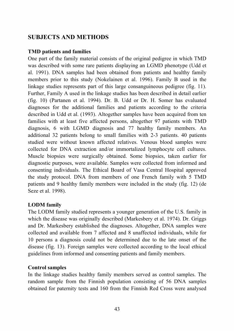
43
SUBJECTS AND METHODS TMD patients and families One part of the family material consists of the original pedigree in which TMD was described with some rare patients displaying an LGMD phenotype (Udd et al. 1991). DNA samples had been obtained from patients and healthy family members prior to this study (Nokelainen et al. 1996). Family B used in the linkage studies represents part of this large consanguineous pedigree (fig. 11). Further, Family A used in the linkage studies has been described in detail earlier (fig. 10) (Partanen et al. 1994). Dr. B. Udd or Dr. H. Somer has evaluated diagnoses for the additional families and patients according to the criteria described in Udd et al. (1993). Altogether samples have been acquired from ten families with at least five affected persons, altogether 97 patients with TMD diagnosis, 6 with LGMD diagnosis and 77 healthy family members. An additional 32 patients belong to small families with 2-3 patients. 40 patients studied were without known affected relatives. Venous blood samples were collected for DNA extraction and/or immortalized lymphocyte cell cultures. Muscle biopsies were surgically obtained. Some biopsies, taken earlier for diagnostic purposes, were available. Samples were collected from informed and consenting individuals. The Ethical Board of Vasa Central Hospital approved the study protocol. DNA from members of one French family with 5 TMD patients and 9 healthy family members were included in the study (fig. 12) (de Seze et al. 1998). LODM family The LODM family studied represents a younger generation of the U.S. family in which the disease was originally described (Markesbery et al. 1974). Dr. Griggs and Dr. Markesbery established the diagnoses. Altogether, DNA samples were collected and available from 7 affected and 8 unaffected individuals, while for 10 persons a diagnosis could not be determined due to the late onset of the disease (fig. 13). Foreign samples were collected according to the local ethical guidelines from informed and consenting patients and family members. Control samples In the linkage studies healthy family members served as control samples. The random sample from the Finnish population consisting of 56 DNA samples obtained for paternity tests and 160 from the Finnish Red Cross were analysed
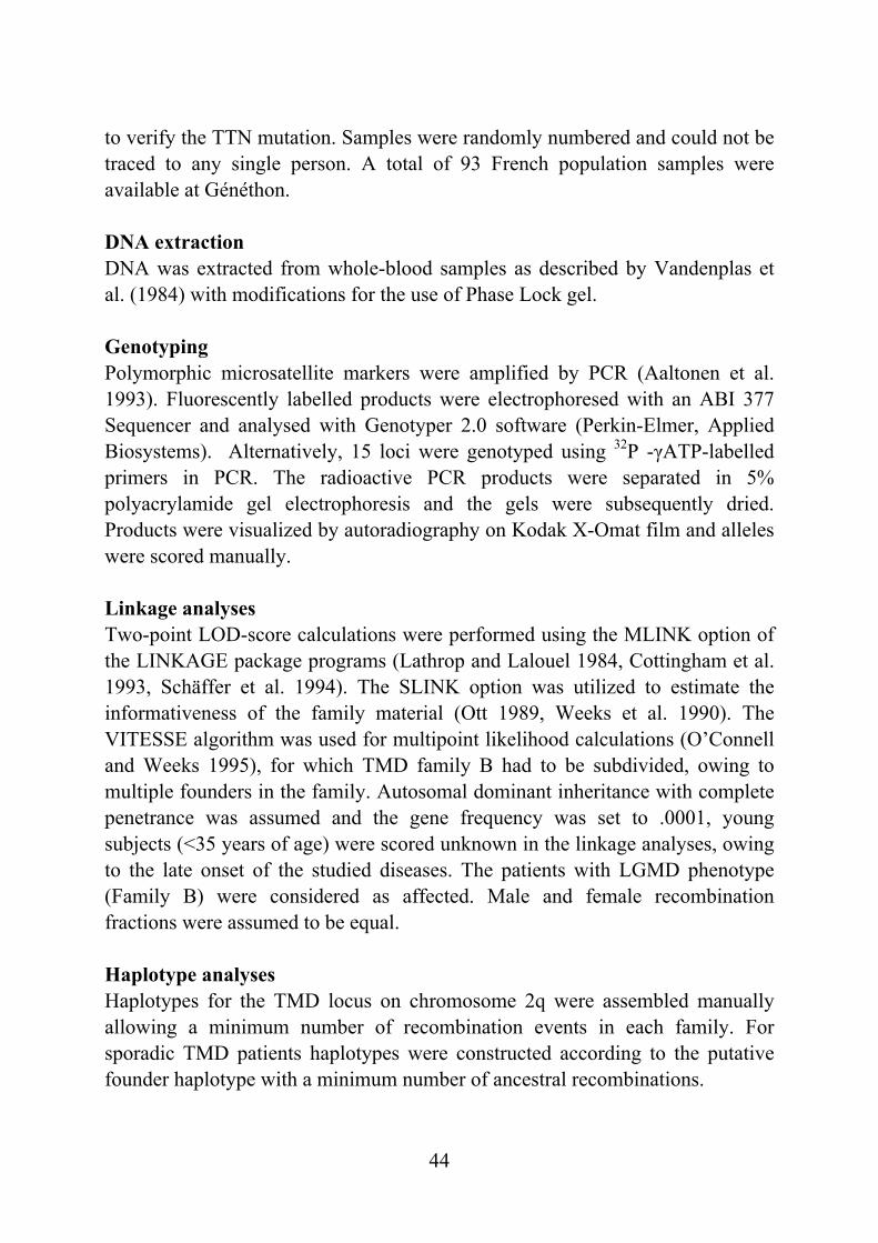
44
to verify the TTN mutation. Samples were randomly numbered and could not be traced to any single person. A total of 93 French population samples were available at Généthon. DNA extraction DNA was extracted from whole-blood samples as described by Vandenplas et al. (1984) with modifications for the use of Phase Lock gel. Genotyping Polymorphic microsatellite markers were amplified by PCR (Aaltonen et al. 1993). Fluorescently labelled products were electrophoresed with an ABI 377 Sequencer and analysed with Genotyper 2.0 software (Perkin-Elmer, Applied Biosystems). Alternatively, 15 loci were genotyped using 32P -γATP-labelled primers in PCR. The radioactive PCR products were separated in 5% polyacrylamide gel electrophoresis and the gels were subsequently dried. Products were visualized by autoradiography on Kodak X-Omat film and alleles were scored manually. Linkage analyses Two-point LOD-score calculations were performed using the MLINK option of the LINKAGE package programs (Lathrop and Lalouel 1984, Cottingham et al. 1993, Schäffer et al. 1994). The SLINK option was utilized to estimate the informativeness of the family material (Ott 1989, Weeks et al. 1990). The VITESSE algorithm was used for multipoint likelihood calculations (O’Connell and Weeks 1995), for which TMD family B had to be subdivided, owing to multiple founders in the family. Autosomal dominant inheritance with complete penetrance was assumed and the gene frequency was set to .0001, young subjects (<35 years of age) were scored unknown in the linkage analyses, owing to the late onset of the studied diseases. The patients with LGMD phenotype (Family B) were considered as affected. Male and female recombination fractions were assumed to be equal. Haplotype analyses Haplotypes for the TMD locus on chromosome 2q were assembled manually allowing a minimum number of recombination events in each family. For sporadic TMD patients haplotypes were constructed according to the putative founder haplotype with a minimum number of ancestral recombinations.
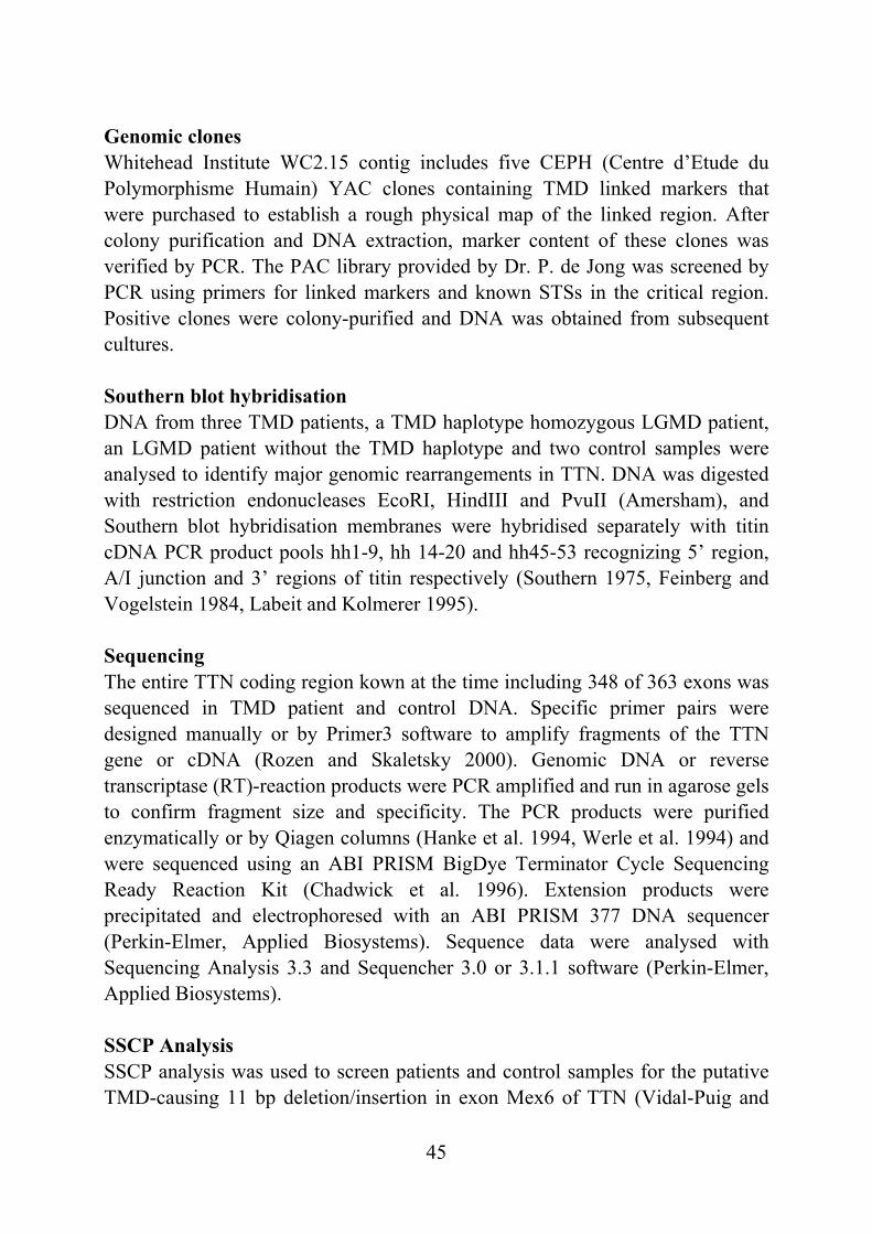
45
Genomic clones Whitehead Institute WC2.15 contig includes five CEPH (Centre d’Etude du Polymorphisme Humain) YAC clones containing TMD linked markers that were purchased to establish a rough physical map of the linked region. After colony purification and DNA extraction, marker content of these clones was verified by PCR. The PAC library provided by Dr. P. de Jong was screened by PCR using primers for linked markers and known STSs in the critical region. Positive clones were colony-purified and DNA was obtained from subsequent cultures. Southern blot hybridisation DNA from three TMD patients, a TMD haplotype homozygous LGMD patient, an LGMD patient without the TMD haplotype and two control samples were analysed to identify major genomic rearrangements in TTN. DNA was digested with restriction endonucleases EcoRI, HindIII and PvuII (Amersham), and Southern blot hybridisation membranes were hybridised separately with titin cDNA PCR product pools hh1-9, hh 14-20 and hh45-53 recognizing 5’ region, A/I junction and 3’ regions of titin respectively (Southern 1975, Feinberg and Vogelstein 1984, Labeit and Kolmerer 1995). Sequencing The entire TTN coding region kown at the time including 348 of 363 exons was sequenced in TMD patient and control DNA. Specific primer pairs were designed manually or by Primer3 software to amplify fragments of the TTN gene or cDNA (Rozen and Skaletsky 2000). Genomic DNA or reverse transcriptase (RT)-reaction products were PCR amplified and run in agarose gels to confirm fragment size and specificity. The PCR products were purified enzymatically or by Qiagen columns (Hanke et al. 1994, Werle et al. 1994) and were sequenced using an ABI PRISM BigDye Terminator Cycle Sequencing Ready Reaction Kit (Chadwick et al. 1996). Extension products were precipitated and electrophoresed with an ABI PRISM 377 DNA sequencer (Perkin-Elmer, Applied Biosystems). Sequence data were analysed with Sequencing Analysis 3.3 and Sequencher 3.0 or 3.1.1 software (Perkin-Elmer, Applied Biosystems). SSCP Analysis SSCP analysis was used to screen patients and control samples for the putative TMD-causing 11 bp deletion/insertion in exon Mex6 of TTN (Vidal-Puig and
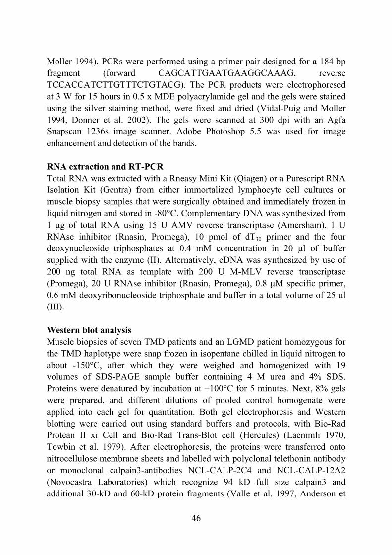
46
Moller 1994). PCRs were performed using a primer pair designed for a 184 bp fragment (forward CAGCATTGAATGAAGGCAAAG, reverse TCCACCATCTTGTTTCTGTACG). The PCR products were electrophoresed at 3 W for 15 hours in 0.5 x MDE polyacrylamide gel and the gels were stained using the silver staining method, were fixed and dried (Vidal-Puig and Moller 1994, Donner et al. 2002). The gels were scanned at 300 dpi with an Agfa Snapscan 1236s image scanner. Adobe Photoshop 5.5 was used for image enhancement and detection of the bands. RNA extraction and RT-PCR Total RNA was extracted with a Rneasy Mini Kit (Qiagen) or a Purescript RNA Isolation Kit (Gentra) from either immortalized lymphocyte cell cultures or muscle biopsy samples that were surgically obtained and immediately frozen in liquid nitrogen and stored in -80°C. Complementary DNA was synthesized from 1 µg of total RNA using 15 U AMV reverse transcriptase (Amersham), 1 U RNAse inhibitor (Rnasin, Promega), 10 pmol of dT30 primer and the four deoxynucleoside triphosphates at 0.4 mM concentration in 20 µl of buffer supplied with the enzyme (II). Alternatively, cDNA was synthesized by use of 200 ng total RNA as template with 200 U M-MLV reverse transcriptase (Promega), 20 U RNAse inhibitor (Rnasin, Promega), 0.8 µM specific primer, 0.6 mM deoxyribonucleoside triphosphate and buffer in a total volume of 25 ul (III). Western blot analysis Muscle biopsies of seven TMD patients and an LGMD patient homozygous for the TMD haplotype were snap frozen in isopentane chilled in liquid nitrogen to about -150°C, after which they were weighed and homogenized with 19 volumes of SDS-PAGE sample buffer containing 4 M urea and 4% SDS. Proteins were denatured by incubation at +100°C for 5 minutes. Next, 8% gels were prepared, and different dilutions of pooled control homogenate were applied into each gel for quantitation. Both gel electrophoresis and Western blotting were carried out using standard buffers and protocols, with Bio-Rad Protean II xi Cell and Bio-Rad Trans-Blot cell (Hercules) (Laemmli 1970, Towbin et al. 1979). After electrophoresis, the proteins were transferred onto nitrocellulose membrane sheets and labelled with polyclonal telethonin antibody or monoclonal calpain3-antibodies NCL-CALP-2C4 and NCL-CALP-12A2 (Novocastra Laboratories) which recognize 94 kD full size calpain3 and additional 30-kD and 60-kD protein fragments (Valle et al. 1997, Anderson et
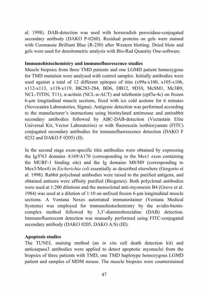
47
al. 1998). DAB-detection was used with horseradish peroxidase-conjugated secondary antibody (DAKO P-0260). Residual proteins on gels were stained with Coomassie Brilliant Blue (R-250) after Western blotting. Dried blots and gels were used for densitometric analysis with Bio-Rad Quantity One-software. Immunohistochemistry and immunofluorescence studies Muscle biopsies from three TMD patients and one LGMD patient homozygous for TMD mutation were analysed with control samples. Initially antibodies were used against a total of 12 different epitopes of titin (x99a-x100, x105-x106, x112-x113, x118-x119, BK283-284, BD6, DB12, 9D10, McSM1, Mc3B9, NCL-TITIN, T11), α-actinin (NCL-α-ACT) and telethonin (cpf3a-4c) on frozen 6-µm longitudinal muscle sections, fixed with ice cold acetone for 6 minutes (Novocastra Laboratories, Sigma). Antigenic detection was performed according to the manufacturer’s instructions using biotinylated antimouse and antirabbit secondary antibodies followed by ABC-DAB-detection (Vectastain Elite Universal Kit, Vector Laboratories) or with fluorescein isothiocyanate (FITC) conjugated secondary antibodies for immunofluorescence detection (DAKO F 0232 and DAKO F 0205) (II). In the second stage exon-specific titin antibodies were obtained by expressing the Ig/FN3 domains A169/A170 (corresponding to the Mex1 exon containing the MURF-1 binding site) and the Ig domains M8/M9 (corresponding to Mex3/Mex4) in Escherichia coli essentially as described elsewhere (Gregorio et al. 1998). Rabbit polyclonal antibodies were raised to the purified antigens, and obtained antisera were affinity purified (Biogenes). Both polyclonal antibodies were used at 1:200 dilutions and the monoclonal anti-myomesin B4 (Grove et al. 1984) was used at a dilution of 1:10 on unfixed frozen 6-µm longitudinal muscle sections. A Ventana Nexes automated immunostainer (Ventana Medical Systems) was employed for immunohistochemistry by the avidin-biotin-complex method followed by 3,3’-diaminobenzidine (DAB) detection. Immunofluorescent detection was manually performed using FITC-conjugated secondary antibody (DAKO 0205, DAKO A/S) (III). Apoptosis studies The TUNEL staining method (an in situ cell death detection kit) and anticaspase3 antibodies were applied to detect apoptotic myonuclei from the biopsies of three patients with TMD, one TMD haplotype homozygous LGMD patient and samples of MDM mouse. The muscle biopsies were counterstained
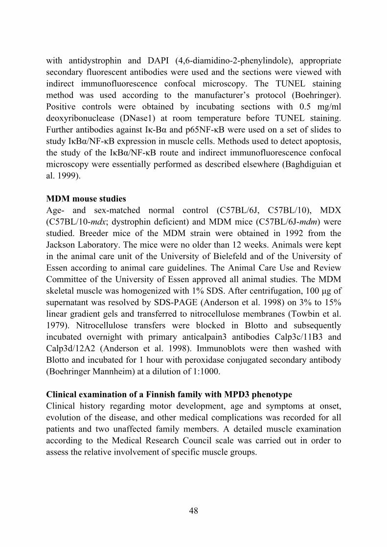
48
with antidystrophin and DAPI (4,6-diamidino-2-phenylindole), appropriate secondary fluorescent antibodies were used and the sections were viewed with indirect immunofluorescence confocal microscopy. The TUNEL staining method was used according to the manufacturer’s protocol (Boehringer). Positive controls were obtained by incubating sections with 0.5 mg/ml deoxyribonuclease (DNase1) at room temperature before TUNEL staining. Further antibodies against Iκ-Bα and p65NF-κB were used on a set of slides to study IκBα/NF-κB expression in muscle cells. Methods used to detect apoptosis, the study of the IκBα/NF-κB route and indirect immunofluorescence confocal microscopy were essentially performed as described elsewhere (Baghdiguian et al. 1999). MDM mouse studies Age- and sex-matched normal control (C57BL/6J, C57BL/10), MDX (C57BL/10-mdx; dystrophin deficient) and MDM mice (C57BL/6J-mdm) were studied. Breeder mice of the MDM strain were obtained in 1992 from the Jackson Laboratory. The mice were no older than 12 weeks. Animals were kept in the animal care unit of the University of Bielefeld and of the University of Essen according to animal care guidelines. The Animal Care Use and Review Committee of the University of Essen approved all animal studies. The MDM skeletal muscle was homogenized with 1% SDS. After centrifugation, 100 µg of supernatant was resolved by SDS-PAGE (Anderson et al. 1998) on 3% to 15% linear gradient gels and transferred to nitrocellulose membranes (Towbin et al. 1979). Nitrocellulose transfers were blocked in Blotto and subsequently incubated overnight with primary anticalpain3 antibodies Calp3c/11B3 and Calp3d/12A2 (Anderson et al. 1998). Immunoblots were then washed with Blotto and incubated for 1 hour with peroxidase conjugated secondary antibody (Boehringer Mannheim) at a dilution of 1:1000. Clinical examination of a Finnish family with MPD3 phenotype Clinical history regarding motor development, age and symptoms at onset, evolution of the disease, and other medical complications was recorded for all patients and two unaffected family members. A detailed muscle examination according to the Medical Research Council scale was carried out in order to assess the relative involvement of specific muscle groups.
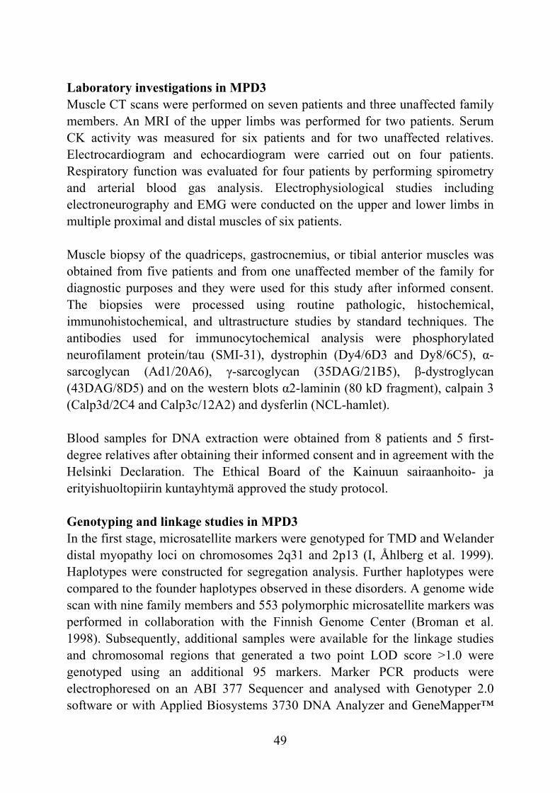
49
Laboratory investigations in MPD3 Muscle CT scans were performed on seven patients and three unaffected family members. An MRI of the upper limbs was performed for two patients. Serum CK activity was measured for six patients and for two unaffected relatives. Electrocardiogram and echocardiogram were carried out on four patients. Respiratory function was evaluated for four patients by performing spirometry and arterial blood gas analysis. Electrophysiological studies including electroneurography and EMG were conducted on the upper and lower limbs in multiple proximal and distal muscles of six patients. Muscle biopsy of the quadriceps, gastrocnemius, or tibial anterior muscles was obtained from five patients and from one unaffected member of the family for diagnostic purposes and they were used for this study after informed consent. The biopsies were processed using routine pathologic, histochemical, immunohistochemical, and ultrastructure studies by standard techniques. The antibodies used for immunocytochemical analysis were phosphorylated neurofilament protein/tau (SMI-31), dystrophin (Dy4/6D3 and Dy8/6C5), α-sarcoglycan (Ad1/20A6), γ-sarcoglycan (35DAG/21B5), β-dystroglycan (43DAG/8D5) and on the western blots α2-laminin (80 kD fragment), calpain 3 (Calp3d/2C4 and Calp3c/12A2) and dysferlin (NCL-hamlet). Blood samples for DNA extraction were obtained from 8 patients and 5 first-degree relatives after obtaining their informed consent and in agreement with the Helsinki Declaration. The Ethical Board of the Kainuun sairaanhoito- ja erityishuoltopiirin kuntayhtymä approved the study protocol. Genotyping and linkage studies in MPD3 In the first stage, microsatellite markers were genotyped for TMD and Welander distal myopathy loci on chromosomes 2q31 and 2p13 (I, Åhlberg et al. 1999). Haplotypes were constructed for segregation analysis. Further haplotypes were compared to the founder haplotypes observed in these disorders. A genome wide scan with nine family members and 553 polymorphic microsatellite markers was performed in collaboration with the Finnish Genome Center (Broman et al. 1998). Subsequently, additional samples were available for the linkage studies and chromosomal regions that generated a two point LOD score >1.0 were genotyped using an additional 95 markers. Marker PCR products were electrophoresed on an ABI 377 Sequencer and analysed with Genotyper 2.0 software or with Applied Biosystems 3730 DNA Analyzer and GeneMapper™
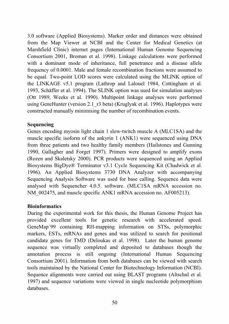
50
3.0 software (Applied Biosystems). Marker order and distances were obtained from the Map Viewer at NCBI and the Center for Medical Genetics (at Marshfield Clinic) internet pages (International Human Genome Sequencing Consortium 2001, Broman et al. 1998). Linkage calculations were performed with a dominant mode of inheritance, full penetrance and a disease allele frequency of 0.0001. Male and female recombination fractions were assumed to be equal. Two-point LOD scores were calculated using the MLINK option of the LINKAGE v5.1 program (Lathrop and Lalouel 1984, Cottingham et al. 1993, Schäffer et al. 1994). The SLINK option was used for simulation analyses (Ott 1989, Weeks et al. 1990). Multipoint linkage analyses were performed using GeneHunter (version 2.1_r3 beta) (Kruglyak et al. 1996). Haplotypes were constructed manually minimising the number of recombination events. Sequencing Genes encoding myosin light chain 1 slow-twitch muscle A (MLC1SA) and the muscle specific isoform of the ankyrin 1 (ANK1) were sequenced using DNA from three patients and two healthy family members (Hailstones and Gunning 1990, Gallagher and Forget 1997). Primers were designed to amplify exons (Rozen and Skaletsky 2000). PCR products were sequenced using an Applied Biosystems BigDye® Terminator v3.1 Cycle Sequencing Kit (Chadwick et al. 1996). An Applied Biosystems 3730 DNA Analyzer with accompanying Sequencing Analysis Software was used for base calling. Sequence data were analysed with Sequencher 4.0.5. software. (MLC1SA mRNA accession no. NM_002475, and muscle specific ANK1 mRNA accession no. AF005213). Bioinformatics During the experimental work for this thesis, the Human Genome Project has provided excellent tools for genetic research with accelerated speed. GeneMap´99 containing RH-mapping information on STSs, polymorphic markers, ESTs, mRNAs and genes and was utilized to search for positional candidate genes for TMD (Deloukas et al. 1998). Later the human genome sequence was virtually completed and deposited to databases though the annotation process is still ongoing (International Human Sequencing Consortium 2001). Information from both databases can be viewed with search tools maintained by the National Center for Biotechnology Information (NCBI). Sequence alignments were carried out using BLAST programs (Altschul et al. 1997) and sequence variations were viewed in single nucleotide polymorphism databases.
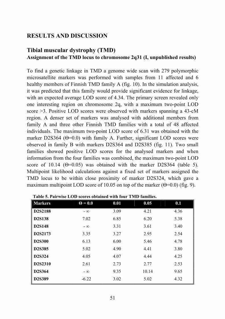
51
Table 5. Pairwise LOD scores obtained with four TMD families.
RESULTS AND DISCUSSION Tibial muscular dystrophy (TMD) Assignment of the TMD locus to chromosome 2q31 (I, unpublished results) To find a genetic linkage in TMD a genome wide scan with 279 polymorphic microsatellite markers was performed with samples from 11 affected and 6 healthy members of Finnish TMD family A (fig. 10). In the simulation analysis, it was predicted that this family would provide significant evidence for linkage, with an expected average LOD score of 4.34. The primary screen revealed only one interesting region on chromosome 2q, with a maximum two-point LOD score >3. Positive LOD scores were observed with markers spanning a 43-cM region. A denser set of markers was analysed with additional members from family A and three other Finnish TMD families with a total of 48 affected individuals. The maximum two-point LOD score of 6.31 was obtained with the marker D2S364 (Θ=0.0) with family A. Further, significant LOD scores were observed in family B with markers D2S364 and D2S385 (fig. 11). Two small families showed positive LOD scores for the analysed markers and when information from the four families was combined, the maximum two-point LOD score of 10.14 (Θ=0.05) was obtained with the marker D2S364 (table 5). Multipoint likelihood calculations against a fixed set of markers assigned the TMD locus to be within close proximity of marker D2S324, which gave a maximum multipoint LOD score of 10.05 on top of the marker (Θ=0.0) (fig. 9).
Markers Θ = 0.0 0.01 0.05 0.1
D2S2188 - ∞ 3.09 4.21 4.36
D2S138 7.02 6.85 6.20 5.38
D2S148 - ∞ 3.31 3.61 3.40
D2S2173 3.35 3.27 2.95 2.54
D2S300 6.13 6.00 5.46 4.78
D2S385 5.02 4.90 4.41 3.80
D2S324 4.05 4.07 4.44 4.25
D2S2310 2.61 2.73 2.77 2.53
D2S364 - ∞ 9.35 10.14 9.65
D2S389 -6.22 3.02 5.02 4.32
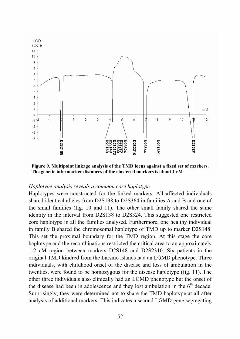
52
Figure 9. Multipoint linkage analysis of the TMD locus against a fixed set of markers. The genetic intermarker distances of the clustered markers is about 1 cM
Haplotype analysis reveals a common core haplotype Haplotypes were constructed for the linked markers. All affected individuals shared identical alleles from D2S138 to D2S364 in families A and B and one of the small families (fig. 10 and 11). The other small family shared the same identity in the interval from D2S138 to D2S324. This suggested one restricted core haplotype in all the families analysed. Furthermore, one healthy individual in family B shared the chromosomal haplotype of TMD up to marker D2S148. This set the proximal boundary for the TMD region. At this stage the core haplotype and the recombinations restricted the critical area to an approximately 1-2 cM region between markers D2S148 and D2S2310. Six patients in the original TMD kindred from the Larsmo islands had an LGMD phenotype. Three individuals, with childhood onset of the disease and loss of ambulation in the twenties, were found to be homozygous for the disease haplotype (fig. 11). The other three individuals also clinically had an LGMD phenotype but the onset of the disease had been in adolescence and they lost ambulation in the 6th decade. Surprisingly, they were determined not to share the TMD haplotype at all after analysis of additional markers. This indicates a second LGMD gene segregating
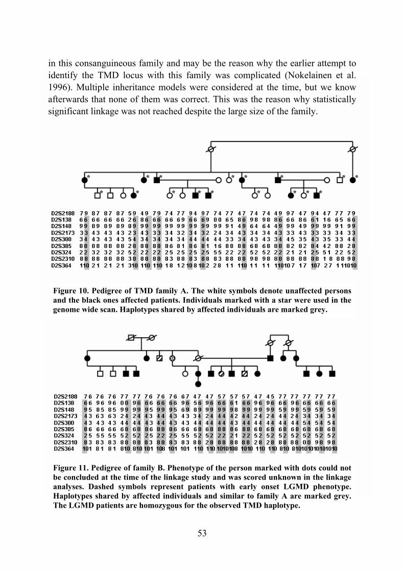
53
Figure 10. Pedigree of TMD family A. The white symbols denote unaffected persons and the black ones affected patients. Individuals marked with a star were used in the genome wide scan. Haplotypes shared by affected individuals are marked grey.
Figure 11. Pedigree of family B. Phenotype of the person marked with dots could not be concluded at the time of the linkage study and was scored unknown in the linkage analyses. Dashed symbols represent patients with early onset LGMD phenotype. Haplotypes shared by affected individuals and similar to family A are marked grey. The LGMD patients are homozygous for the observed TMD haplotype.
in this consanguineous family and may be the reason why the earlier attempt to identify the TMD locus with this family was complicated (Nokelainen et al. 1996). Multiple inheritance models were considered at the time, but we know afterwards that none of them was correct. This was the reason why statistically significant linkage was not reached despite the large size of the family.
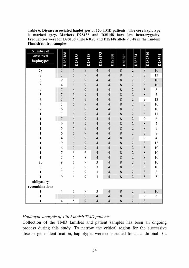
54
Table 6. Disease associated haplotypes of 150 TMD patients. The core haplotype is marked grey. Markers D2S138 and D2S148 have low heterozygosity, Frequencies were for D2S138 allele 6 0.27 and D2S148 allele 9 0.48 in the random Finnish control samples.
Haplotype analysis of 150 Finnish TMD patients Collection of the TMD families and patient samples has been an ongoing process during this study. To narrow the critical region for the successive disease gene identification, haplotypes were constructed for an additional 102
Number of observed
haplotypes D
2S21
88
D2S
138
D2S
148
D2S
2173
D2S
300
D2S
385
D2S
324
D2S
2310
D2S
364
78 7 6 9 4 4 8 2 8 10 8 7 6 9 4 4 8 2 8 13 5 9 6 9 4 4 8 2 8 10 5 4 6 9 4 4 8 2 8 10 4 7 6 9 4 4 8 2 8 8 3 7 6 9 4 4 8 2 8 5 3 7 6 9 4 4 8 2 9 13 4 5 6 9 4 4 8 2 8 10 2 6 6 9 4 4 8 2 8 10 1 7 6 9 4 4 8 2 8 11 1 7 6 9 4 4 8 2 9 6 1 9 6 9 4 4 8 2 8 7 1 6 6 9 4 4 8 2 8 9 1 6 6 9 4 4 8 2 8 8 1 4 6 9 4 4 8 2 9 4 1 9 6 9 4 4 8 2 8 13 1 6 9 9 4 4 8 2 8 10 1 7 6 6 4 4 8 2 8 10 1 7 6 8 4 4 8 2 8 10 20 9 6 9 3 4 8 2 8 10 3 7 6 9 3 4 8 2 8 10 1 7 6 9 3 4 8 2 8 8 1 9 6 9 3 4 8 2 8 5
obligatory recombinations
1 4 6 9 3 4 8 2 8 10 1 7 6 9 4 4 8 2 9 3 1 4 5 9 4 4 8 2 8
55
Figure 12. The French TMD family. Haplotyes shared by affected individuals are marked grey. Two-point LOD scores for the markers at Θ = 0.0 are displayed next to the haplotypes.
Finnish TMD patients adding up to 150 analysed cases. Haplotype analyses revealed the most common haplotype 9-4-4-8-2-8 for the TMD patients. A total of 116 out of 150 disease chromosomes showed this haplotype. Some allelic variation was seen with the centromeric markers D2S148 and D2S2173 and telomeric marker D2S2310. All 150 disease chromosomes carried a core haplotype of 4-8-2. The core haplotype and three obligatory recombinations restricted the disease haplotype between markers D2S2173 and D2S2310 consistent with the linkage analyses (table 6). TMD families show an uneven geographical distribution in Finland with clusters of cases on the western coastline and in the eastern Savo-Karelia region. The same core haplotype was found from the disease alleles in both clusters as further evidence for one founder mutation. To some extent, these haplotypes have diagnostic value when symptomatic patients are studied. TMD in a French family After assignment of the TMD locus to chromosome 2q, one French family was analysed. Five patients in the family showed a TMD phenotype clinically identical to the Finnish patients. Linked markers were genotyped with samples from the five affected patients and five healthy relatives. The LOD scores did not reach significant level as expected by the simulation analysis; however, they were positive except for marker D2S300. Haplotypes were constructed and all the affected were found to share the same haplotype which was not present in their healthy relatives (fig. 12). This haplotype is dissimilar to the Finnish one. Thus, an independent TMD-causing mutation was thought to have arisen in this family at the same chromosome 2q locus. Moreover, one of the patients was recombinant for the centromeric markers and combined with the earlier data the critical TMD region spans between markers D2S300 and D2S2310. (de Seze et al. 1998).
56
Figure 13. LODM family pedigree and haplotypes. Two-point LOD scores for the markers at Θ = 0.0 are displayed next to the haplotypes.
Linkage studies with a late-onset distal myopathy family (LODM) When TMD was described, it was noted that it resembles LODM clinically (Markesbery et al. 1974). Both diseases have late onset and their symptoms begin from the anterior tibial muscles. However, LODM progresses to proximal and hand muscles and eventually leads to loss of ambulation. The original pedigree, in which LODM was described, was studied for linkage with the TMD associated markers. Diagnosis could be confirmed for six patients and six healthy relatives. Pairwise LOD scores did not reach significant level: the highest LOD score of 2.24 was found with marker D2S2310 (Θ=0.0). Multipoint analysis however settled the locus to be in close proximity of markers D2S385 and D2S324 with a maximum multipoint LOD score of 3.38. All the affected individuals share the same haplotype providing further evidence for the linkage (fig. 13). TMD and LODM are linked to the same markers on chromosome 2q and could be considered as allelic disorders. Some families representing distal myopathy have been tested for the TMD linked markers without evidence for linkage (Felice et al. 1999, Chinnery et al. 2001). Linkage in one family could not be excluded or verified (Penisson-Besnier et al. 1998, unpublished data). The heterogeneity is not surprising and is not restricted to the distal myopathies, since e.g. an LGMD phenotype may be caused by mutations in at least fifteen different genes (table 4). On the other hand, late-onset autosomal dominant myopathy with proximal muscle weakness, early respiratory failure and characteristic cytoplasmic bodies found in muscle biopsy was linked to chromosome 2q24-q31 overlapping the TMD locus (Edström et al. 1990, Nicolao et al. 1999). The phenotype associated with chromosome 2q31 was expanded further to dilated cardiomyopathy (Siu et al. 1999).
57
Figure 14. Markers blotted against genomic sequence. The genetic distances between markers are as obtained from the Center for Medical Genetics (Marshfield) map. WI-9271 is a STS marker located in TTN 3’ end while markers D2S324 and D2S385 are both found in 5’ introns of the gene 30 kb apart.
Search for TMD causing gene and mutations (II, III) TMD physical region – revised After the locus assignment had been confirmed, the physical mapping databases were explored for the marker order and genes in the candidate region. The Whitehead Institute STS based physical YAC map was available at the time. Clones 935-E-10, 930-H-10 and 788-A-10 containing the linked markers were obtained and the marker content was confirmed. The 3’ region of the TTN gene (STS marker WI-9271) was within the region of interest. Additional primers designed for other parts of the gene confirmed that the entire coding region was present in the YAC clone 930-H-10. RH-assay had demonstrated that the orientation of the TTN gene is 3’ to 5’ from the centromere (Pelin et al. 1997). At this point TTN became the major positional and functional candidate gene for obvious reasons: It is expressed in skeletal and heart muscle in different isoforms and has major structural and physiological functions in the sarcomere. No other muscle specific transcripts were within the region. Even now, when the genomic sequence is available, there are no such genes among the 30 annotated or predicted genes in the critical area. There is some inconsistency between the genomic sequence and genetic distances over the region. Different maps give values 0-1 cM between markers D2S324 and D2S2310, which is not present in the RH-mapping panels. However, the physical distance between them is about 2.5 Mb in the finished sequence (fig. 14). Yet, fiber-FISH experiments with BAC clones No. 14104 and No. 23155 used for TTN genomic sequencing (kindly provided by Dr. Labeit) and PACs screened with the linked markers approximate the distance to be between 0.55 to 0.8 Mb (S. Kilpinen, unpublished data).
58
TTN mutation screening The size of the candidate gene TTN was discouraging. The TTN gene, although compact, encodes mRNAs up to 100 kb. Full-length cDNA had been published as well as the M-line genomic sequence. Additional genomic sequences were available to us prior to publication (kindly provided by Dr. Labeit). Exploration for possible disease causing alterations was made step by step. Southern blot hybridisations were performed with TTN cDNA PCR product pools to detect gross rearrangements. The fragment patterns produced by multiple restriction enzymes did not show any differences between patients and controls. Further, several titin antibodies showed normal labelling in muscle biopsies giving evidence against major rearrangements (II). TMD does not affect heart muscle. For this reason skeletal muscle expressed tandem Ig-repeat exons were sequenced. In addition, N2A and Mex5 calpain3 binding sites and the Z1-Z2 telethonin binding site were sequenced. Several polymorphisms were detected but none segregated with the disease. Finally, the attempt to sequence the whole gene was undertaken in collaboration with Dr. Richards’s group at Généthon and 348 of the 363 exons were sequenced. TMD is caused by Mex6 mutations in TTN In the Finnish TMD samples, a unique alteration was identified in Mex6, the 363rd and the last exon of TTN. Mex6 of TTN encodes an Ig-domain that is localized at the periphery of the M-line lattice next to calpain3 binding Mex5/ Is7 (Kinbara et al. 1997). The alteration consists of an 11-bp change, AAGTAACATGG→ TGAAAGAAAAA, at position 293,269-293,279 in the TTN sequence and changes four amino acids Glu-Val-Thr-Trp→Val-Lys-Glu-Lys but does not cause a frame shift or a premature stop codon. Each of the four residues is changed to an amino acid of another charge, and the overall charge is changed from acidic to basic. This sequence variation had not been reported earlier. Further, the altered segment is relatively well conserved between humans, mice, chicken and pigs. The 11-bp change was easily detected by SSCP analysis. SSCP studies showed that this mutation did not occur in the 216 Finnish unaffected population control individuals tested. The change showed cosegregation with TMD in 12 unrelated pedigrees, in agreement with full penetrance after the age of 35. One family was an exception: three offspring aged 50-55 years did show the mutation without clinically manifesting the disease. However, in this family, the transmitting affected parents in the older generation had very late onset of symptoms at 65 years. These three individuals are followed up for detection of exceptionally late manifestation. This 11-bp
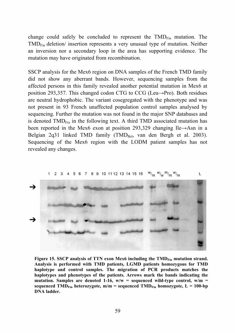
59
Figure 15. SSCP analysis of TTN exon Mex6 including the TMDFin mutation strand.Analysis is performed with TMD patients, LGMD patients homozygous for TMD haplotype and control samples. The migration of PCR products matches the haplotypes and phenotypes of the patients. Arrows mark the bands indicating the mutation. Samples are denoted 1-16, w/w = sequenced wild-type control, w/m = sequenced TMDFin heterozygote, m/m = sequenced TMDFin homozygote, L = 100-bp DNA ladder.
change could safely be concluded to represent the TMDFin mutation. The TMDFin deletion/ insertion represents a very unusual type of mutation. Neither an inversion nor a secondary loop in the area has supporting evidence. The mutation may have originated from recombination. SSCP analysis for the Mex6 region on DNA samples of the French TMD family did not show any aberrant bands. However, sequencing samples from the affected persons in this family revealed another potential mutation in Mex6 at position 293,357. This changed codon CTG to CCG (Leu→Pro). Both residues are neutral hydrophobic. The variant cosegregated with the phenotype and was not present in 93 French unaffected population control samples analysed by sequencing. Further the mutation was not found in the major SNP databases and is denoted TMDFra in the following text. A third TMD associated mutation has been reported in the Mex6 exon at position 293,329 changing Ile→Asn in a Belgian 2q31 linked TMD family (TMDBel, van den Bergh et al. 2003). Sequencing of the Mex6 region with the LODM patient samples has not revealed any changes.
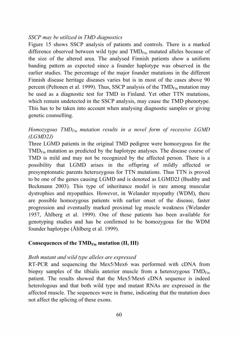
60
SSCP may be utilized in TMD diagnostics Figure 15 shows SSCP analysis of patients and controls. There is a marked difference observed between wild type and TMDFin mutated alleles because of the size of the altered area. The analysed Finnish patients show a uniform banding pattern as expected since a founder haplotype was observed in the earlier studies. The percentage of the major founder mutations in the different Finnish disease heritage diseases varies but is in most of the cases above 90 percent (Peltonen et al. 1999). Thus, SSCP analysis of the TMDFin mutation may be used as a diagnostic test for TMD in Finland. Yet other TTN mutations, which remain undetected in the SSCP analysis, may cause the TMD phenotype. This has to be taken into account when analysing diagnostic samples or giving genetic counselling. Homozygous TMDFin mutation results in a novel form of recessive LGMD (LGMD2J) Three LGMD patients in the original TMD pedigree were homozygous for the TMDFin mutation as predicted by the haplotype analyses. The disease course of TMD is mild and may not be recognized by the affected person. There is a possibility that LGMD arises in the offspring of mildly affected or presymptomatic parents heterozygous for TTN mutations. Thus TTN is proved to be one of the genes causing LGMD and is denoted as LGMD2J (Bushby and Beckmann 2003). This type of inheritance model is rare among muscular dystrophies and myopathies. However, in Welander myopathy (WDM), there are possible homozygous patients with earlier onset of the disease, faster progression and eventually marked proximal leg muscle weakness (Welander 1957, Åhlberg et al. 1999). One of these patients has been available for genotyping studies and has be confirmed to be homozygous for the WDM founder haplotype (Åhlberg et al. 1999). Consequences of the TMDFin mutation (II, III) Both mutant and wild type alleles are expressed RT-PCR and sequencing the Mex5/Mex6 was performed with cDNA from biopsy samples of the tibialis anterior muscle from a heterozygous TMDFin patient. The results showed that the Mex5/Mex6 cDNA sequence is indeed heterologous and that both wild type and mutant RNAs are expressed in the affected muscle. The sequences were in frame, indicating that the mutation does not affect the splicing of these exons.
61
Figure 16. Immunostaining of muscle sections with titin M8/M9 antibody (A-C) and A169/170 antibody (D-F) detected by FITC. Unaffected control (A) and TMDFin heterozygous patient (B) have normal labelling with M8/M9 antibody, which shows M-line striated labelling pattern. TMDFin homozygous LGMD patient(C) is missing M8/M9 labelling altogether. A169/A170 labelling in control (D), TMDFin heterozygous patient (E) and TMDFin homozygous LGMD patient muscle sections (F) are identical.
Loss of epitope recognition by M8/9 antibody in TMDFin homozygote Screening of titin with twelve antibodies had showed normal striated labelling of patient muscle in immunohistochemical analyses (II). Specific antibodies against M8/M9 epitopes encoded by Mex3/Mex4 exons and A169/A170 epitopes preceding the titin kinase domain were tested in the second phase (III). Immunohistochemical analyses of muscle biopsy samples of a TMDFin homozygous LGMD patient indicated a loss of the M8/M9 titin epitopes when either DAB or FITC detection methods were used. Three TMDFin heterozygous patients and a control individual showed normal cross-striated sarcomeric M-line-specific labelling (fig. 16).
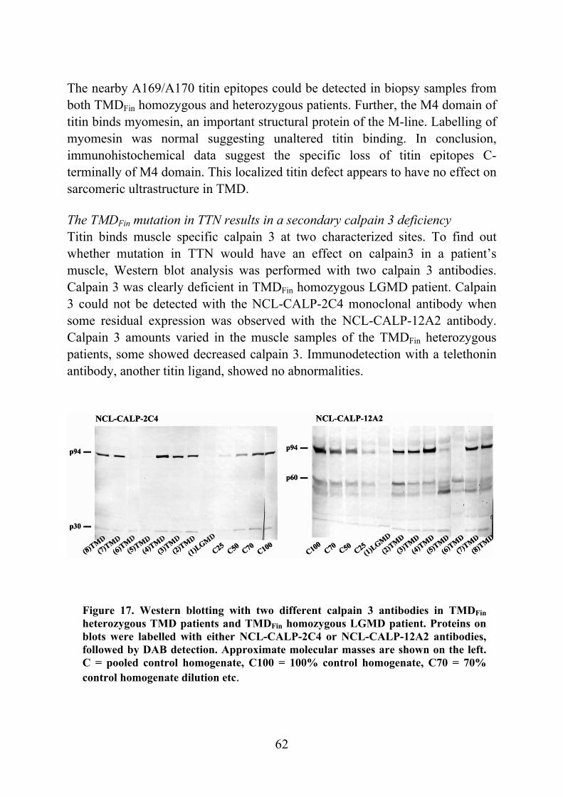
62
Figure 17. Western blotting with two different calpain 3 antibodies in TMDFinheterozygous TMD patients and TMDFin homozygous LGMD patient. Proteins on blots were labelled with either NCL-CALP-2C4 or NCL-CALP-12A2 antibodies, followed by DAB detection. Approximate molecular masses are shown on the left. C = pooled control homogenate, C100 = 100% control homogenate, C70 = 70%control homogenate dilution etc.
The nearby A169/A170 titin epitopes could be detected in biopsy samples from both TMDFin homozygous and heterozygous patients. Further, the M4 domain of titin binds myomesin, an important structural protein of the M-line. Labelling of myomesin was normal suggesting unaltered titin binding. In conclusion, immunohistochemical data suggest the specific loss of titin epitopes C-terminally of M4 domain. This localized titin defect appears to have no effect on sarcomeric ultrastructure in TMD. The TMDFin mutation in TTN results in a secondary calpain 3 deficiency Titin binds muscle specific calpain 3 at two characterized sites. To find out whether mutation in TTN would have an effect on calpain3 in a patient’s muscle, Western blot analysis was performed with two calpain 3 antibodies. Calpain 3 was clearly deficient in TMDFin homozygous LGMD patient. Calpain 3 could not be detected with the NCL-CALP-2C4 monoclonal antibody when some residual expression was observed with the NCL-CALP-12A2 antibody. Calpain 3 amounts varied in the muscle samples of the TMDFin heterozygous patients, some showed decreased calpain 3. Immunodetection with a telethonin antibody, another titin ligand, showed no abnormalities.
63
Apoptosis and a disturbed IκBα/NF-κB pathway similar to LGMD2A Calpain 3 deficiency, caused by primary calpain 3 mutations, has been associated with myonuclear apoptosis and perturbation of the IκBα/NF-κB signalling pathway (Baghdiguian et al. 1999). To find out whether secondary calpain 3 deficiency could cause similar alterations, the TUNEL staining method detecting DNA fragmentation and anticaspase3 immunolabelling were used on patient muscle biopsies. Apoptotic myonuclei were observed in the muscles of the TMDFin heterozygous patient and a greater frequency of them were seen in the TMDFin homozygous LGMD patient. Staining was detected in centralized myonuclei and in small clusters. All the TUNEL-positive myonuclei were anticaspase3 positive, but anticaspase3 staining positive fibres were more widely distributed. Normally both IκBα and NF-κB are evenly distributed in the cytoplasm, a situation which was seen in the control sections. In the samples from both the heterozygous and homozygous patients IκBα was found to be aberrantly relocated in the TUNEL-positive myonuclei. Further, expression of NF-κB was subsarcolemmal. These findings are similar to those observed in the primary calpainopathy in LGMD2A and may be involved in the pathogenesis of TMD/ LGMD2J. MDM mouse – a possible model for TMD/ LGMD2J (II) A spontaneous recessive mutation in the mouse causes severe progressive skeletal muscle wasting, muscular dystrophy with myositis (MDM) (Lane 1985). MDM mice are reduced in weight and have shortened lifespans (Heimann et al. 1996). Muscles postnatally degenerate and regenerate and there is notable overproduction of satellite cells (Heimann et al. 1996). The MDM mutation has been linked to the proximal part of chromosome 2, syntenic to human chromosome 2q31 region with the genes for nebulin, titin and nicotinic acetylcholine receptor α1-subunit (Müller-Seitz et al. 1993). Based on this and the finding of a secondary calpain 3 deficiency in the TMDFin homozygous LGMD patient, we analysed MDM skeletal muscle for expression of calpain 3. MDM muscles showed a severe reduction of the 94 kD band of calpain 3 with antibodies calp3c/11B3 and Calp3d/12A2 (II). Further, TUNEL positive myonuclei were found in the muscle sections and alterations in IκBα and NF-κB labelling similar to patients were observed, although less pronounced. Subsequently a Ttn mutation causing MDM has been identified (Garvey et al. 2002). This complex mutation with a deletion and a LINE insertion results in an in-frame deletion in the calpain 3 interacting N2A domain (Garvey et al. 2002).
64
Mutant molecules are expressed in two splice isoforms, a major 249 bp deleted isoform and a minor isoform with a LINE insertion (Garvey et al. 2002). A decrease of calpain 3 in the muscle of the MDM mouse was confirmed but the decrease was in the range of 50-60% compared to almost total loss observed by us. This may reflect different muscle groups analysed or different ages of the mice. However, calpain 3 deficiency does not explain the observed phenotype alone because mice with targeted calpain 3 mutation have a less severe phenotype than the MDM mice (Richard et al. 2000). This is also the case with the TMDFin homozygous patients with small amounts of calpain 3 left in their muscles and phenotype as severe as LGMD2A. There are no studies on the phenotype of the MDM heterozygote mice. The heterozygotes having a myopathy restricted to one muscle group, comparable to human TMD, may have gone unnoticed. General discussion on the molecular pathogenesis in titinopathies The gigantic titin molecules have multiple functions in muscle cells. They provide a filament system extending the length of the myofibrils and contribute to sarcomere assembly and probably determine the thick filament length. Titin molecules function as elastic springs that keep contractile elements in place. Further, titin has a kinase domain and may be involved in the signalling regulating muscle contraction and sarcomere protein turnover. Thus, mutations in different titin domains are likely to cause varying phenotypes. M-line mutations cause the skeletal muscle phenotype as shown by the present study while there are several titin mutations observed that cause cardiomyopathy (fig.18). The TMDFin mutation changed four amino acids within the conserved hydrophobic core domain of the M10 Ig-domain of M-line titin. One of the four amino acids, the tryptophan, is mutated into charged lysine. The tryptophan residue is generally conserved in all titin Ig-repeats and is known to be crucially important for stabilization of the hydrophobic core of the Ig β-barrel fold (Improta et al. 1996). The TMDFra mutation at position 293,357 changed leucine to proline. Both amino acids are neutral hydrophobic. Since proline differs from leucine because of its circular structure, it may introduce a kink and cause significant conformational change in the M-line titin structure. The TMDBel mutation may have similar effects as a non-polar hydrophobic isoleucine is changed to a polar uncharged asparagine. The mutant titin molecules are
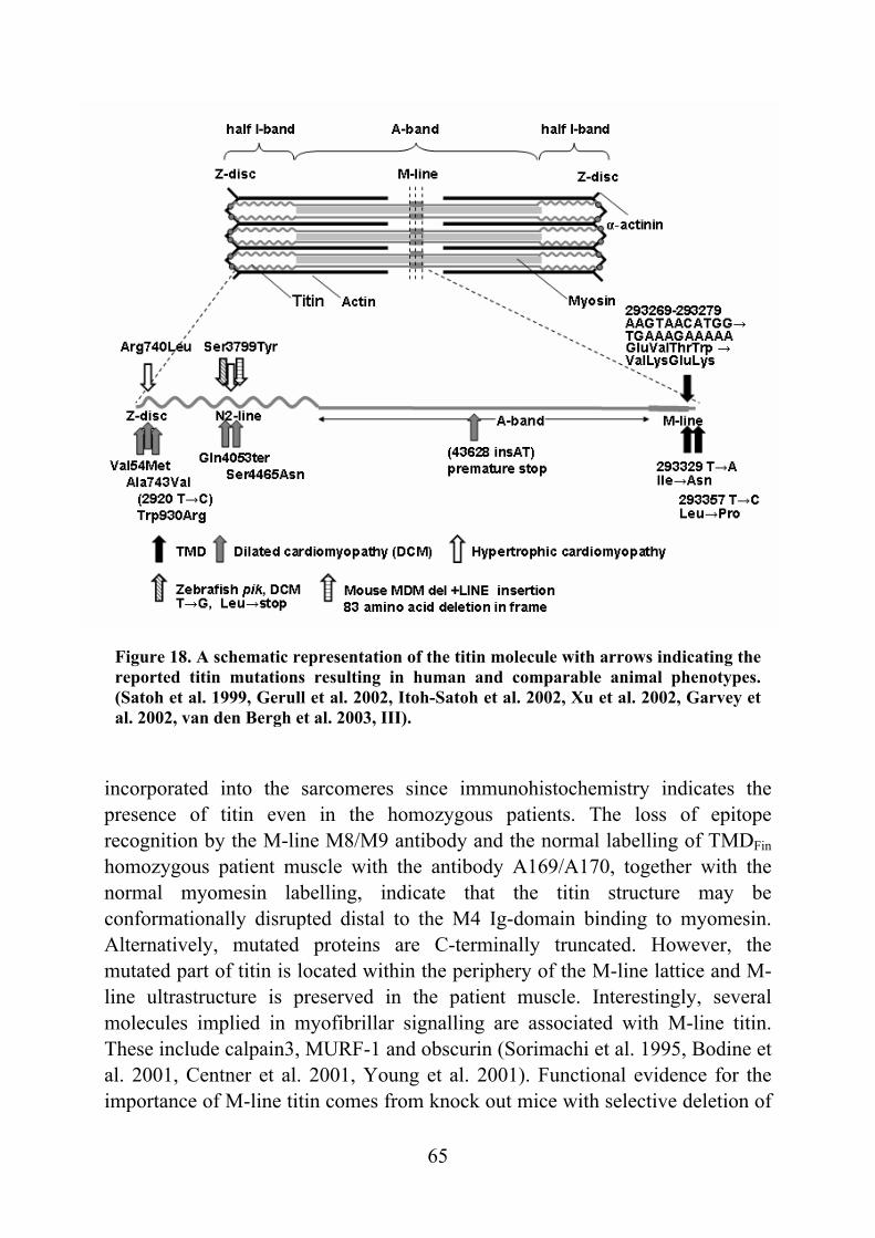
65
Figure 18. A schematic representation of the titin molecule with arrows indicating the reported titin mutations resulting in human and comparable animal phenotypes. (Satoh et al. 1999, Gerull et al. 2002, Itoh-Satoh et al. 2002, Xu et al. 2002, Garvey et al. 2002, van den Bergh et al. 2003, III).
incorporated into the sarcomeres since immunohistochemistry indicates the presence of titin even in the homozygous patients. The loss of epitope recognition by the M-line M8/M9 antibody and the normal labelling of TMDFin homozygous patient muscle with the antibody A169/A170, together with the normal myomesin labelling, indicate that the titin structure may be conformationally disrupted distal to the M4 Ig-domain binding to myomesin. Alternatively, mutated proteins are C-terminally truncated. However, the mutated part of titin is located within the periphery of the M-line lattice and M-line ultrastructure is preserved in the patient muscle. Interestingly, several molecules implied in myofibrillar signalling are associated with M-line titin. These include calpain3, MURF-1 and obscurin (Sorimachi et al. 1995, Bodine et al. 2001, Centner et al. 2001, Young et al. 2001). Functional evidence for the importance of M-line titin comes from knock out mice with selective deletion of
66
the kinase region of M-line ttn (Gotthard et al. 2003). The knock out mice are not viable. When M-line excision is introduced in late embryonic development, the mice have a progressive myopathy leading to an early death. Ultrastructural analysis suggested that the mutant titin molecules are incorporated into the sarcomere, disassembly and disruption of the sarcomere structure follows. Calpain 3 binding to N2A and M-line titin has been characterized (Sorimachi et al. 1995). It has been suggested that binding to titin prevents autolysis and degradation of calpain 3 in muscle, thus regulating the amount of calpain 3. Binding affinity and the binding site in calpain 3 are different in the N2A region and M-line (Sorimachi et al. 1995). Our results suggest that both M-line titin and N2A mutations could lead to a secondary calpain 3 deficiency in homozygous form. Calpain 3 amounts varied in TMDFin heterozygotes. The wild-type titin may still trap and bind some amount of calpain 3 stabilizing and protecting it from degradation. However, one functional allele may not be sufficient for all the binding capacity needed (haploinsufficiency) or mutated proteins may disrupt the complex structure of the M-line and wild-type protein interactions are blocked to some extent (dominant negative effect). The N2A domain is expressed in both skeletal and cardiac muscle but cardiomyocytes express an alternative N2B isoforms as well. Mex6 is expressed in both skeletal and heart muscle. On the other hand, calpain3 is not expressed in the mature heart muscle. The fact that TMD/LGMD2J patients have no cardiac phenotype further supports the pathogenetic significance of a defective titin-calpain3 interaction. A variable proportion of Mex5+ and Mex5- isoforms may have an effect on the selective muscle involvement seen in TMD. However, mice with targeted calpain 3 mutation have a less severe phenotype than the MDM mice (Richard et al. 2000) and the phenotype of TMDFin homozygous patient with small amounts of calpain 3 left in their muscles is as severe as LGMD2A. Thus, robust calpain 3 binding and protection from autolysis does not solely explain the observed phenotypes. It could be that titin or other titin-associated proteins regulate calpain3 activity in a more delicate manner and these unknown molecular mechanisms are disrupted by TMD and MDM mutations. Yet other titin-associated proteins in myofibrillar signalling could be involved in the pathogenesis of TMD/LGMD2J and MDM. The identification of the mutations causing LODM and the late-onset autosomal dominant myopathy with proximal muscle weakness and early respiratory failure is pending. Meanwhile, several mutations in TTN have been associated
67
with dilated cardiomyopathy (DCM) characterized by ventricular dilatation, systolic dysfunction and congestive heart failure. A two bp insertion in A-band titin results in a frame shift and expression of a 1.14 MD truncated titin with the full-length titin (Gerull et al. 2002). The truncated protein is suggested to result from cleavage by insufficiently bound calpain 3 (Gerull et al. 2002). Another mutation converts a conserved tryptophan to arginine in Z-disc titin and the mutation was predicted to disrupt the β-sandwich structure of the Ig-domain Z4 (Gerull et al. 2002). Residue changes in Z-disc titin Ig-domain Z1 and Z-repeat 7 were observed to decrease binding affinities to telethonin and α-actinin respectively in yeast two-hybrid studies (Itoh-Satoh et al. 2002). Finally, two changes in the cardiac specific N2B domain were discovered, one resulting in a premature stop-codon and one missense mutation (Itoh-Satoh et al. 2002). Only one patient with hypertrophic cardiomyopathy (HCM) has been reported to have a conserved residue replacement from arginine to leucine in Z-repeat 7 (Satoh et al. 1999). A mutation construct showed increased α-actinin binding affinity in a yeast two-hybrid assay (Satoh et al. 1999). All the human DCM and HCM mutations are observed in the heterozygous form. The missense mutations in the Z-disc region could exert a dominant negative effect on the assembly of the sarcomere structure. On the other hand, Z-disc mutated domains are also expressed in skeletal muscle, and the reported patients have no skeletal muscle phenotype. Disruption of cardiac specific signalling pathways i.e. responding to mechanical load could be one possible explanation. Cardiac specific N2B region mutations are skipped in splicing of skeletal isoforms while the N2B domain and its modification have unique regulatory roles on the elastic and contractile properties of the cardiac muscle (Linke et al. 2002, Kulke et al. 2001, Yamasaki et al. 2002, Neagoe et al. 2002, Golenhofen et al. 2002). The Pik mutant zebrafish presents a phenotype resembling human DCM. Recessive nonsense mutation in the cardiac expressed titin N2B domain has no effect on heart muscle development but causes systolic dysfunction leading to heart failure (Xu et al. 2002). Intensive research on the characteristics of titin and the phenotypes associated with TTN mutations have resulted in many discoveries. Yet, each question answered generates a dozen new ones. Titin seems to have more versatile functions in muscle cell than had been predicted. It will be a challenge to dissect the pathogenetic cascades involved in the titinopathies.
68
A new distal myopathy (MPD3) Clinical characterization (IV) Patients presenting with a distal myopathy that is distinct from the described phenotypes emerged during the TMD study. This large family with seven patients at the time (5 men and 2 women) in four generations was studied. One of the patients had died of cardiac problems at 84 years of age. Clinical data were reported by his daughter and obtained from his medical records. None of the patients had any symptoms or signs of muscle disease before the age of 30 years. The age at onset ranged from 32 to 45 years. The first symptoms for referral were clumsiness with the hands of three patients and stumbling in four patients. The interval between the first and the second localization of muscle involvement was two to three years. At referral, all patients had a steppage gait. To compensate ankle dorsiflexion weakness, patients used excessive knee and hip flexion during the swing phase. All the patients were unable to walk on their heels and walking on tiptoe was impossible in the three severely affected patients. Muscle weakness in the lower limbs involved mainly the muscles of the anterior compartment of the lower legs (i.e. tibialis anterior, extensor digitorum brevis, extensor digitorum longus and peroneus muscles). The calf muscles were involved in three severely affected patients and there was moderate to severe hip abduction weakness. In the upper limbs, wasting and weakness of the hand muscles were observed with claw-like hands (fig. 19). Wasting or weakness in all the intrinsic muscles, wrist extensors and upper arm muscles was observed with the progression of the disease. In the lower limbs, the disease spread to other proximal muscles only in the late stages. Asymmetry of muscle involvement was present in all patients. The progression of the disease was slow, and the oldest patient of the family became wheelchair-bound at the age of 75 years. Mild contractures were present about five to eight years after disease onset and involved mainly the finger, elbow, and wrist joints. None of the patients showed intellectual deficit, heart or respiratory problems.
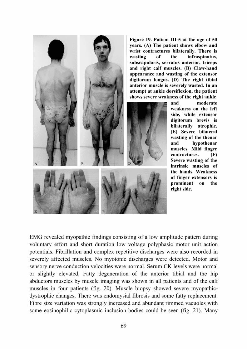
69
Figure 19. Patient III-5 at the age of 50 years. (A) The patient shows elbow and wrist contractures bilaterally. There is wasting of the infraspinatus, subscapularis, serratus anterior, triceps and right calf muscles. (B) Claw-hand appearance and wasting of the extensor digitorum longus. (D) The right tibial anterior muscle is severely wasted. In an attempt at ankle dorsiflexion, the patient shows severe weakness of the right ankle
EMG revealed myopathic findings consisting of a low amplitude pattern during voluntary effort and short duration low voltage polyphasic motor unit action potentials. Fibrillation and complex repetitive discharges were also recorded in severely affected muscles. No myotonic discharges were detected. Motor and sensory nerve conduction velocities were normal. Serum CK levels were normal or slightly elevated. Fatty degeneration of the anterior tibial and the hip abductors muscles by muscle imaging was shown in all patients and of the calf muscles in four patients (fig. 20). Muscle biopsy showed severe myopathic-dystrophic changes. There was endomysial fibrosis and some fatty replacement. Fibre size variation was strongly increased and abundant rimmed vacuoles with some eosinophilic cytoplasmic inclusion bodies could be seen (fig. 21). Many
and moderate weakness on the left side, while extensor digitorum brevis is bilaterally atrophic.(E) Severe bilateral wasting of the thenar and hypothenar muscles. Mild finger contractures. (F) Severe wasting of the intrinsic muscles of the hands. Weakness of finger extensors is prominent on the right side.
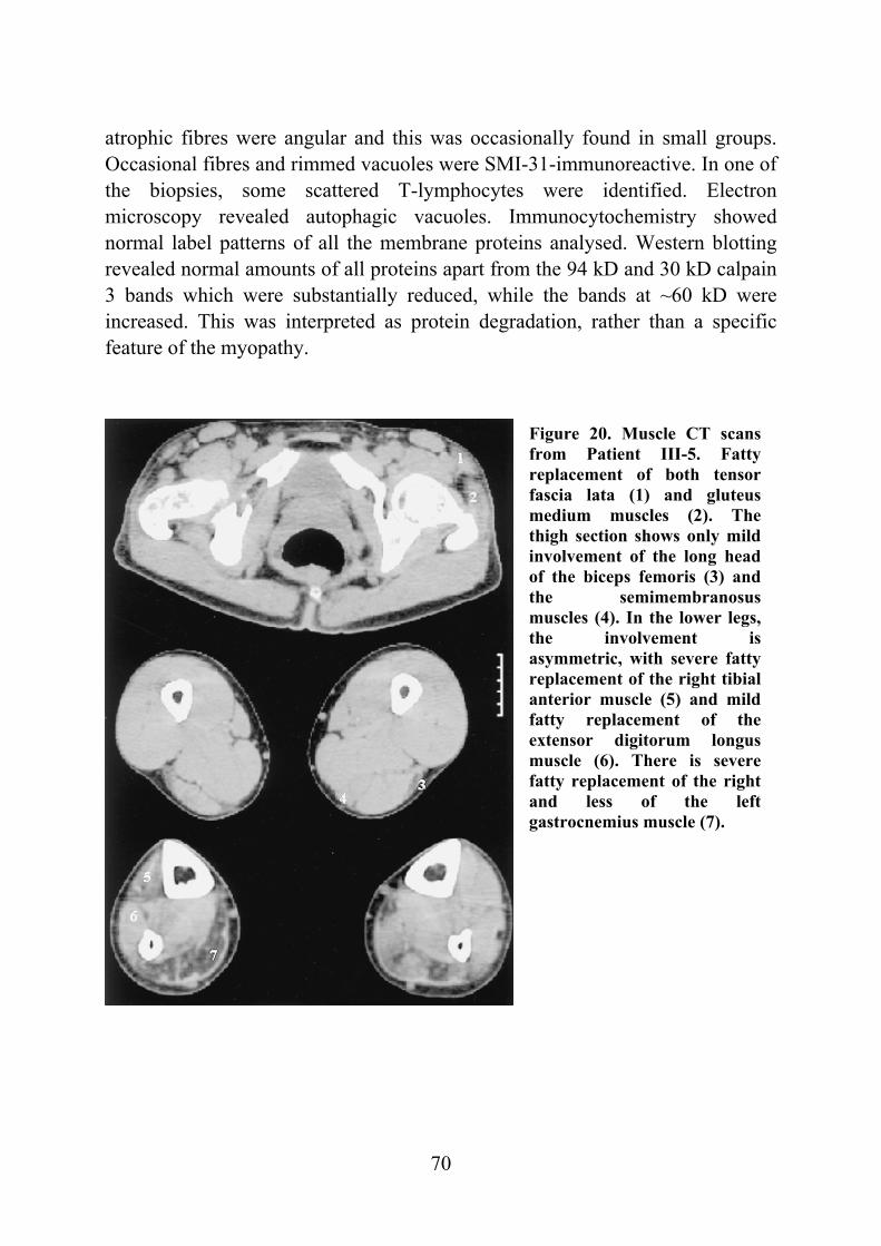
70
Figure 20. Muscle CT scans from Patient III-5. Fatty replacement of both tensor fascia lata (1) and gluteus medium muscles (2). The thigh section shows only mild involvement of the long head of the biceps femoris (3) and the semimembranosus muscles (4). In the lower legs, the involvement is asymmetric, with severe fatty replacement of the right tibial anterior muscle (5) and mild fatty replacement of the extensor digitorum longus muscle (6). There is severe fatty replacement of the right and less of the left gastrocnemius muscle (7).
atrophic fibres were angular and this was occasionally found in small groups. Occasional fibres and rimmed vacuoles were SMI-31-immunoreactive. In one of the biopsies, some scattered T-lymphocytes were identified. Electron microscopy revealed autophagic vacuoles. Immunocytochemistry showed normal label patterns of all the membrane proteins analysed. Western blotting revealed normal amounts of all proteins apart from the 94 kD and 30 kD calpain 3 bands which were substantially reduced, while the bands at ~60 kD were increased. This was interpreted as protein degradation, rather than a specific feature of the myopathy.
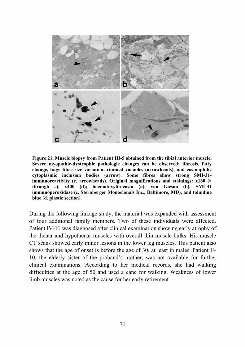
71
Figure 21. Muscle biopsy from Patient III-5 obtained from the tibial anterior muscle. Severe myopathic-dystrophic pathologic changes can be observed: fibrosis, fatty change, huge fibre size variation, rimmed vacuoles (arrowheads), and eosinophilic cytoplasmic inclusion bodies (arrow). Some fibres show strong SMI-31-immunoreactivity (c, arrowheads). Original magnifications and stainings: x160 (a through c), x400 (d); haematoxylin-eosin (a), van Gieson (b), SMI-31 immunoperoxidase (c, Sternberger Monoclonals Inc., Baltimore, MD), and toluidineblue (d, plastic section).
During the following linkage study, the material was expanded with assessment of four additional family members. Two of these individuals were affected. Patient IV-11 was diagnosed after clinical examination showing early atrophy of the thenar and hypothenar muscles with overall thin muscle bulks. His muscle CT scans showed early minor lesions in the lower leg muscles. This patient also shows that the age of onset is before the age of 30, at least in males. Patient II-10, the elderly sister of the proband’s mother, was not available for further clinical examinations. According to her medical records, she had walking difficulties at the age of 50 and used a cane for walking. Weakness of lower limb muscles was noted as the cause for her early retirement.
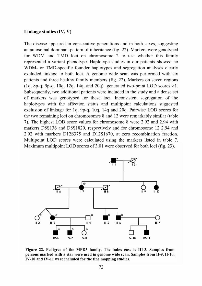
72
Figure 22. Pedigree of the MPD3 family. The index case is III-3. Samples from persons marked with a star were used in genome wide scan. Samples from II-9, II-10, IV-10 and IV-11 were included for the fine mapping studies.
Linkage studies (IV, V) The disease appeared in consecutive generations and in both sexes, suggesting an autosomal dominant pattern of inheritance (fig. 22). Markers were genotyped for WDM and TMD loci on chromosome 2 to test whether this family represented a variant phenotype. Haplotype studies in our patients showed no WDM- or TMD-specific founder haplotypes and segregation analyses clearly excluded linkage to both loci. A genome wide scan was performed with six patients and three healthy family members (fig. 22). Markers on seven regions (1q, 8p-q, 9p-q, 10q, 12q, 14q, and 20q) generated two-point LOD scores >1. Subsequently, two additional patients were included in the study and a dense set of markers was genotyped for these loci. Inconsistent segregation of the haplotypes with the affection status and multipoint calculations suggested exclusion of linkage for 1q, 9p-q, 10q, 14q and 20q. Pairwise LOD scores for the two remaining loci on chromosomes 8 and 12 were remarkably similar (table 7). The highest LOD score values for chromosome 8 were 2.92 and 2.94 with markers D8S136 and D8S1820, respectively and for chromosome 12 2.94 and 2.92 with markers D12S375 and D12S1670, at zero recombination fraction. Multipoint LOD scores were calculated using the markers listed in table 7. Maximum multipoint LOD scores of 3.01 were observed for both loci (fig. 23).
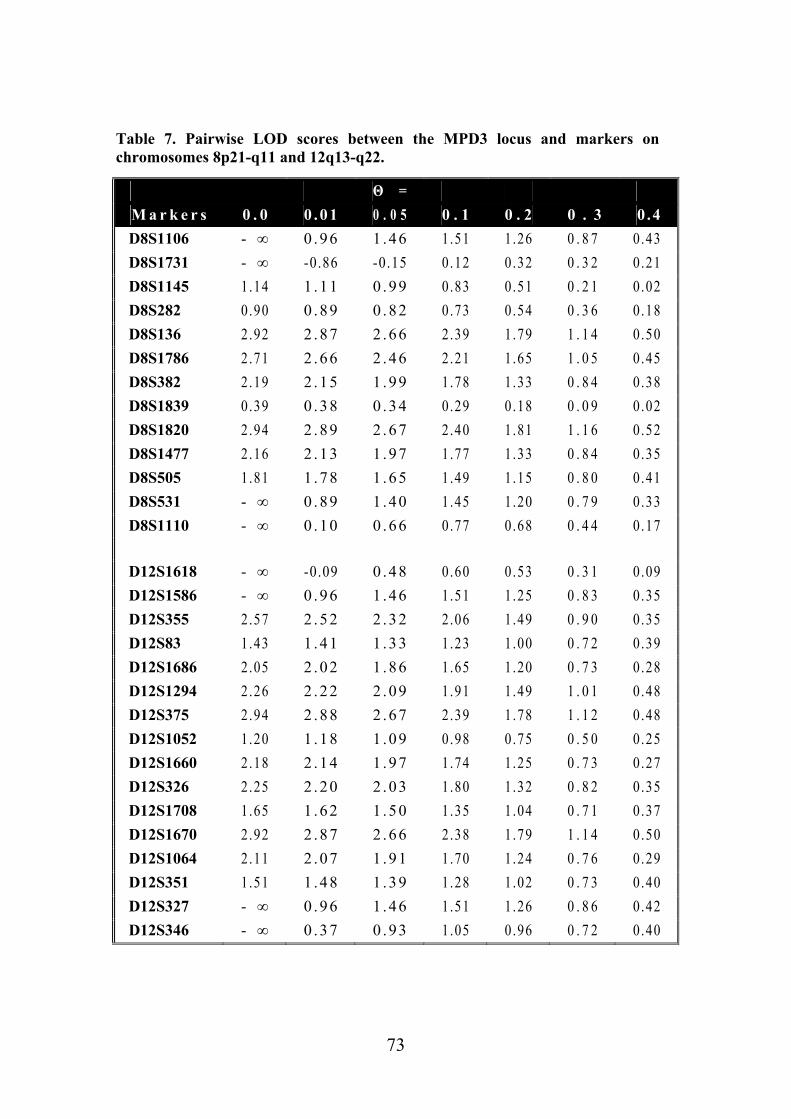
73
Table 7. Pairwise LOD scores between the MPD3 locus and markers on chromosomes 8p21-q11 and 12q13-q22.
Θ = M a r k e r s 0 . 0 0 .01 0 . 0 5 0 . 1 0 . 2 0 . 3 0 .4 D8S1106 - ∞ 0 .96 1 .46 1.51 1.26 0 . 8 7 0.43 D8S1731 - ∞ -0.86 -0.15 0.12 0.32 0 . 3 2 0.21 D8S1145 1.14 1 .11 0 .99 0.83 0.51 0 . 2 1 0.02 D8S282 0.90 0 .89 0 .82 0.73 0.54 0 . 3 6 0.18 D8S136 2.92 2 .87 2 .66 2.39 1.79 1 . 1 4 0.50 D8S1786 2.71 2 .66 2 .46 2.21 1.65 1 . 0 5 0.45 D8S382 2.19 2 .15 1 .99 1.78 1.33 0 . 8 4 0.38 D8S1839 0.39 0 .38 0 .34 0.29 0.18 0 . 0 9 0.02 D8S1820 2.94 2 .89 2 .67 2.40 1.81 1 . 1 6 0.52 D8S1477 2.16 2 .13 1 .97 1.77 1.33 0 . 8 4 0.35 D8S505 1.81 1 .78 1 .65 1.49 1.15 0 . 8 0 0.41 D8S531 - ∞ 0 .89 1 .40 1.45 1.20 0 . 7 9 0.33 D8S1110 - ∞ 0 .10 0 .66 0.77 0.68 0 . 4 4 0.17 D12S1618 - ∞ -0.09 0 .48 0.60 0.53 0 . 3 1 0.09 D12S1586 - ∞ 0 .96 1 .46 1.51 1.25 0 . 8 3 0.35 D12S355 2.57 2 .52 2 .32 2.06 1.49 0 . 9 0 0.35 D12S83 1.43 1 .41 1 .33 1.23 1.00 0 . 7 2 0.39 D12S1686 2.05 2 .02 1 .86 1.65 1.20 0 . 7 3 0.28 D12S1294 2.26 2 .22 2 .09 1.91 1.49 1 . 0 1 0.48 D12S375 2.94 2 .88 2 .67 2.39 1.78 1 . 1 2 0.48 D12S1052 1.20 1 .18 1 .09 0.98 0.75 0 . 5 0 0.25 D12S1660 2.18 2 .14 1 .97 1.74 1.25 0 . 7 3 0.27 D12S326 2.25 2 .20 2 .03 1.80 1.32 0 . 8 2 0.35 D12S1708 1.65 1 .62 1 .50 1.35 1.04 0 . 7 1 0.37 D12S1670 2.92 2 .87 2 .66 2.38 1.79 1 . 1 4 0.50 D12S1064 2.11 2 .07 1 .91 1.70 1.24 0 . 7 6 0.29 D12S351 1.51 1 .48 1 .39 1.28 1.02 0 . 7 3 0.40 D12S327 - ∞ 0 .96 1 .46 1.51 1.26 0 . 8 6 0.42 D12S346 - ∞ 0 .37 0 .93 1.05 0.96 0 . 7 2 0.40
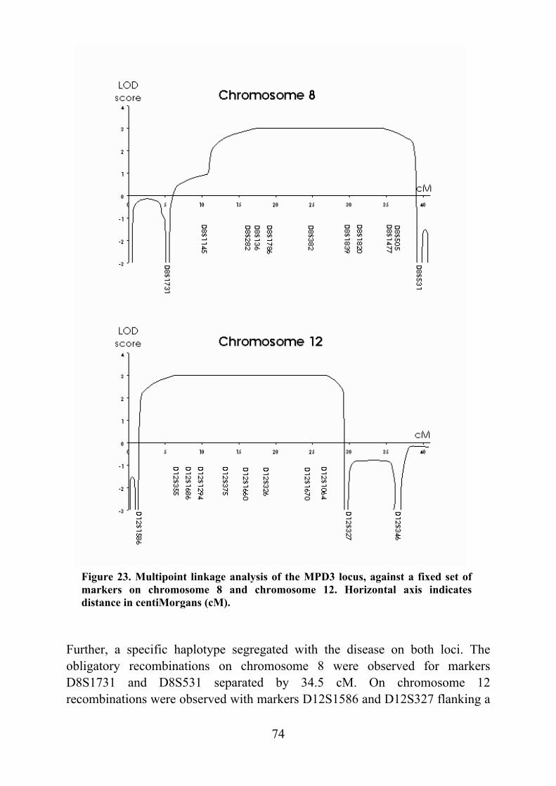
74
Figure 23. Multipoint linkage analysis of the MPD3 locus, against a fixed set of markers on chromosome 8 and chromosome 12. Horizontal axis indicates distance in centiMorgans (cM).
Further, a specific haplotype segregated with the disease on both loci. The obligatory recombinations on chromosome 8 were observed for markers D8S1731 and D8S531 separated by 34.5 cM. On chromosome 12 recombinations were observed with markers D12S1586 and D12S327 flanking a
75
28.6 cM region. Simulation analysis with 1000 replicates predicted the average LOD score for linkage to be 1.92 and the maximum LOD score to be 3.01 for this particular family. Simulation analysis for unlinked markers predicted a LOD score over two in 0.4 percent and over three in 0.1 percent of the simulated markers making the observed linkage evidence at this significance level for the two loci unlikely. Both potentially linked loci are wide. The genomic sequence of the chromosome 8 region is about 34 Mb including 349 known or predicted genes and the chromosome 12 region is about 41 Mb long including 433 known or predicted genes (International Human Genome Sequencing Consortium 2001). The genes expressed specifically in skeletal muscle were explored on these regions. The chromosome 12q MLC1SA gene encodes a myosin light chain 1 slow-twitch muscle isoform A, which is expressed in nonmuscle tissue as well as slow-twitch skeletal muscles (Hailstones and Gunning 1990). The coding region, consisting of eight exons, was sequenced but no differences were identified between DNA samples from affected and healthy individuals or the sequence in the databases. The Ankyrin 1 gene (ANK1) on chromosome 8p, has skeletal muscle specific small splice variants, which are associated with sarcoplasmic reticulum and interact with titin and obscurin (Gallagher and Forget 1998, Zhou et al. 1997, Bagnato et al. 2003, Kontrogianni-Konstantopoulos et al. 2003, Kontrogianni-Konstantopoulos and Bloch 2003). These muscle specific isoforms have their own promoter and an alternative first exon followed by four commonly expressed exons. Again, no sequence variations were observed in the coding region. Three one basepair changes were detected in introns. All of them were concluded to represent polymorphisms, since they were also identified in the DNA samples of the healthy family members. Selfevidently, variants of the non-coding regions of these genes might still be causative for the disease, although for a dominant disease a mutation affecting the protein structure would represent the most plausible molecular defect. The distinction of the new distal myopathy, MPD3, from the other distal myopathies is based on the differences in clinical findings together with genetic exclusion of the known distal myopathy loci. The family material available was thought to be sufficiently informative for successful locus identification. However, an extensive genome wide scan showed linkage to two separate loci, 8p22-q11 and 12q13-q22, with the multipoint LOD score of 3.01 for both loci. Statistical and haplotype analyses could not rule out one or the other of these
76
loci. Further, a LOD score of 3 has been acknowledged as a significant evidence for linkage in monogenic disorder (Conneally et al. 1985). This study shows that even in cases with a seemingly large enough family material, the assignment of linkage may prove to be a difficult task and even well established statistical analysis may show significant results for more than one locus. At present there are no additional family members available for the study, and ascertainment of other families or patients that would represent the same phenotype has not been successful so far. It may be beneficial to genotype the chromosome 8 and 12 markers in patients with unclassified distal myopathy, as they may share the same founder haplotype. In particular, this is appropriate for patients that are excluded to have the TMDFin mutation in titin or the specific founder haplotype of WDM. There are several reports of autosomal dominant distal myopathy families without linkage determination (Sumner et al. 1971, Penisson-Besnier et al. 1998, Felice et al. 1999, Chinnery et al. 2001). Patients from the family reported by Felice et al. developed weakness of the anterior compartment of their lower legs followed by progressive distal upper limb and proximal leg muscle weakness. The muscle affection pattern resembles MPD3 but the age of onset is earlier and the muscle pathology is milder without rimmed vacuoles. Likewise, the disorder in the family reported by Sumner et al. bears some similarities, but the disease showed a more severe course. Despite these clinical differences the 8p22-q11 and 12q13-q22 loci could be tested for linkage in these families. For example, there is a marked clinical variation in the aforementioned chromosome 2q31 linked phenotypes (TMD, LODM, autosomal dominant myopathy with proximal muscle weakness and early respiratory failure, and cardiomyopathy). Identification of the MPD3 causing gene will be difficult without a definitely linked locus and a narrow critical region. The positional candidate gene approach was applied in this study, so far without success. Two genes are known to cause dominant distal myopathies: titin and cardiac beta-myosin (MYH7). In the recessively inherited telethoninopathy (LGMD2G) the initial weakness and atrophy may sometimes be distal and muscle biopsy shows rimmed vacuoles as well. Thus, sarcomeric proteins represent good candidates for causing distal muscle degeneration. Discoveries concerning the pathogenetic pathways involved in the other (distal) myopathies may provide new candidate genes to be screened for the MPD3 causing mutation in the future.
77
CONCLUDING REMARKS The first section of this thesis presents the identification of the defective gene and the mutations causing tibial muscular dystrophy (TMD) by a positional candidate approach. The second section presents the characterization and linkage studies for a novel distal myopathy (MPD3). The TMD locus was assigned to chromosome 2q31 in Finnish families and the genetic homogeneity was shown by linkage to the same locus in a French family. Further, a similar late-onset distal myopathy (LODM) phenotype in an American family was linked to chromosome 2q31. Sequencing of the giant TTN gene identified TMD causing mutations in the Mex6 exon, which encodes Ig-domain M10 in the ultimate C-terminus of titin. M10 is located in the M-line region of the sarcomere. The identified mutations possibly disrupt the Ig-domain structure. Interestingly, three patients with a severe limb-girdle muscular dystrophy (LGMD2J) phenotype were homozygous for the TMDFin mutation. Immunohistochemical studies indicated incorporation of the mutant titin molecules to the sarcomere, but an altered structure of titin C-terminus located in the periphery of the M-line. Further, the amount of titin ligand calpain 3 varied in the muscle samples from heterozygous TMD patients and there was a severe loss of calpain 3 in homozygous patient. Myonuclear apoptosis and perturbation of the IκBα/NF-κB pathway that are similar to the primary calpainopathy (LGMD2A) were observed as well. However, the animal studies of calpain 3 deficient mice and the spontaneous titin mutant MDM-mouse suggest that the TMD phenotype might not result from the calpain 3 deficiency alone. The contribution of titin to the complex M-line structure and its role in signalling and regulatory pathways for sarcomeric protein turnover have begun to emerge. This includes interaction with recently discovered M-line molecules, such as MURFs and obscurin. The goal for the future will be to clarify the details of how the mutations affect titin interaction with calpain 3 and the other ligands. This may shed light on the molecular mechanisms regulated by the M-line components that are involved in the muscle degeneration in TMD and other myopathies. In the second part of this study the MPD3 phenotype was characterized in a Finnish family. Molecular genetic studies excluded linkage to the known distal
78
myopathy loci. Unexpectedly, a genome wide scan provided significant evidence for linkage to two chromosomal regions, 8p22-q11 and 12q13-q22, and it was impossible to make any distinction between them. Unclassified distal myopathy patients and unlinked families reported by others may be tested for these loci in order to confirm the locus assignment. Identification of the MPD3 disease gene and mutation will be intriguing, particularly finding the pathogenetic pathways involved and the possible connections to TMD and the other muscular dystrophies.
79
ACKNOWLEDGMENTS This thesis study was carried out between years 1996 and 2003 in the Department of Molecular Medicine at the National Public Health Institute in Helsinki. I wish to thank the former head of the institute, Professor Jussi Huttunen, and the new director, Pekka Puska, for providing the excellent research facilities. I have been privileged to have two distinguished supervisors. I wish to express my gratitude to Professor Leena Peltonen-Palotie for the inspiring atmosphere in the lab. I truly admire her capability of resolving all the obstacles as well as her ability to focus on the big scientific picture beyond details. I would like to acknowledge Docent Hannu Somer for his pioneering work with the muscle disease research in Finland. It has been educative to have conversations about the schemes how the basic research could benefit the patients. This study would have been impossible without the contribution of my unofficial supervisor Docent Bjarne Udd. He has made a tremendous work in characterizing the TMD phenotype and establishing the diagnoses for the patients. His enthusiastic drive has carried this project all along and his capability to combine clinical work and research is admirable. The final steps of this study have been guided by Docent Marjo Kestilä. I warmly thank her for the caring support and gentle pushing forward at the times most needed. Professor Nigel G. Laing and Docent Katarina Pelin are warmly thanked for reviewing this thesis on a tight schedule and making all the constructive comments. BSc, MA Donald Smart is thanked for revising the language. I have been fortunate to work with Anna Vihola, whose work with immunohistochemistry has truly completed our studies. I wish to thank Peter Hackman for joining in this study at a critical time and for his energetic spirit with titin sequencing project. Jaakko Sarparanta is thanked for continuing the work with TMD. I am grateful to Dr. Ibrahim Mahjeh for his excellent and perceptive clinical skills with the muscle diseases and for distinguishing the new phenotype. I wish to thank Annika Siitonen for skilfully keeping the MPD study going on and for taking over the rapadilino project. I have had a pleasure to collaborate with many distinguished scientists all around the world. I would like to acknowledge Professor Robert C. Griggs and Dr. Denise A. Figlewicz for the collaboration with the LODM family material, Dr. Jerome de Seze for the work with the Lille family, Dr. Volker Straub and Professor Thomas Voit for sharing their results of the MDM mouse study, Dr. Isabelle Richard and Sylvie Marchand for sequencing the giant, Dr. Siegfried Labeit for generously sharing his knowledge about titin, Dr. Louise V.B. Anderson for expert immunohistochemical works, and Professor Jacques S. Beckmann for overall interest towards this study.
80
Professor Hannu Kalimo, Professor Juhani Partanen, Dr. Ari Saarinen and Dr. Anders Paetau are thanked for collaboration and interest in this study. I warmly thank Docent Irma Järvelä for introducing me the field of molecular genetics, Dr. Päivi Mäkelä-Bengs for bench side teaching the lab work, and Dr. Pekka Nokelainen for launching the TMD study in KTL. The seniors in our lab Docent Anu Jalanko and Docent Ismo Ulmanen are thanked for helping out with various practical problems all along. I wish to thank Maikki Parkkonen, Mari Hakkarainen, Ritva Timonen, Elli Kempas, Pekka Ellonen and Merja Soininen for the technical assistance. Sari Kivikko, Sari Mustala, Tuija Svahnbäck and Helena Knuuttila are thanked for their secretarial help and keeping the business running. Aki Suomalainen and Teemu Perheentupa are thanked for their help with the computers crashing. It has been great to work with all the people in our lab. I especially want to thank Mari Auranen for sharing project with muscular dystrophies and Ilona Visapää for sharing the world of monogenic diseases (always not so straightforward). Both are thanked for their friendship also outside the lab. If changing office rooms and meeting new people would be sports… I wish to thank Niklas and Mira for sharing the seventh one and further, the numerous people for keeping up the good humour in the lab. I would like to thank Kirsti, Katja, Mikko, Mervi, Toni, Tuomas, Eeva, Jarno, Aili, Tero, Miia and Anne for our friendship originating from cursus parallelus. Chicas Anu, Milena, Lilli and Maria are thanked for sharing the world of flamenco. I am thankful to my parents Leena and Pekka, brother Jarkko, grandmothers Elvi and Elsi, in-laws Riitta and Seppo, my other family Karen and John, grandmother Sherk, Sara, Nicolas and Oen and all the other relatives for their unfailing love and support. I am very fortunate to have Pasi as my husband. His love, support and encouragement have helped me through this project. You and Ella are the first, the last, my everything. I am indebted to the patients and the families for participating in this study. This study was financially supported by the Daisy and Yrjö Eskola Fond for the Paulo Foundation, the Finnish Cultural Foundation, the Finnish Medical Foundation, the Finnish Neurological Society, the Maud Kuistila Foundation, and the Ulla Hjelt Fund. Järvenpää, November 2003 Henna Haravuori
81
ELECTRONIC DATABASE INFORMATION National Human Genome Research Institute (NHGRI), http://www.genome.gov/ National Center for Biotechnology Information (NCBI), http://www.ncbi.nlm.nih.gov/ Genemap’99 at NCBI, http://www.ncbi.nlm.nih.gov/genemap99/ The Map Viewer at NCBI, http://www.ncbi.nlm.nih.gov/mapview/map_search.cgi? GeneBank at NCBI, http://www.ncbi.nlm.nih.gov/Genbank/ BLAST service at NCBI, http://www.ncbi.nlm.nih.gov/BLAST/ dbSNP Home page, http://www.ncbi.nlm.nih.gov/SNP/ Online Mendelian Inheritance in Man (OMIM), http://www.ncbi.nlm.nih.gov/Omim/ The SNP Consortium Ltd., http://snp.cshl.org/ Whitehead Institute, http://www-genome.wi.mit.edu/ Primer3 software, http://www-genome.wi.mit.edu/genome_software/other/primer3.html Genome Data Base (GDB), http://www.gdb.org/ Human genome organization (HUGO), http://www.gene.ucl.ac.uk/hugo/ Généthon, http://www.genethon.fr/php/index.php The Center for Medical Genetics, http://research.marshfieldclinic.org/genetics/ European Neuromuscular Centre, http://www.enmc.org/default4.html Neuromuscular Disease Center, http://www.neuro.wustl.edu/neuromuscular/ Titin information at EMBL, http://www.embl-heidelberg.de/ExternalInfo/Titin/ Celera, http://www.celera.com
82
REFERENCES Aaltonen J, Komulainen J, Vikman A, Palotie A, Wadelius C, Perheentupa J, Peltonen L.
Autoimmune polyglandular disease type I. Exclusion map using amplifiable multiallelic markers in a microtiter well format. Eur J Hum Genet 1:164-171, 1993
Aittomäki K, Lucena JL, Pakarinen P, Sistonen P, Tapanainen J, Gromoll J, Kaskikari R, Sankila EM, Lehväslaiho H, Engel AR. Mutation in the follicle-stimulating hormone receptor gene causes hereditary hypergonadotropic ovarian failure. Cell 82:959-968, 1995
Alderton JM, Steinhardt RA. Calcium influx through calcium leak channels is responsible for the elevated levels of calcium-dependent proteolysis in dystrophic myotubes. J Biol Chem 275:9452-9460, 2000
Allamand V, Sunada Y, Salih MA, Straub V, Ozo CO, Al-Turaiki MH, Akbar M, Kolo T, Colognato H, Zhang X, Sorokin LM, Yurchenco PD, Tryggvason K, Campbell KP. Mild congenital muscular dystrophy in two patients with an internally deleted laminin α2-chain. Hum Mol Genet 6:747-752, 1997
Altschul SF, Madden TL, Schaffer AA, Zhang J, Zhang Z, Miller W, Lipman DJ. Gapped BLAST and PSI-BLAST: a new generation of protein database search programs. Nucleic Acids Res 25:3389-3402, 1997
Anderson LV, Davison K, Moss JA, Richard I, Fardeau M, Tome FM, Hubner C, Lasa A, Colomer J, Beckmann JS. Characterization of monoclonal antibodies to calpain 3 and protein expression in muscle from patients with limb-girdle muscular dystrophy type 2A. Am J Pathol 153:1169-1179, 1998
Anderson LV, Davison K, Moss JA, Young C, Cullen MJ, Walsh J, Johnson MA, Bashir R, Britton S, Keers S, Argov Z, Mahjneh I, Fougerousse F, Beckmann JS, Bushby KM. Dysferlin is a plasma membrane protein and is expressed early in human development. Hum Mol Genet 8:855-861, 1999
Anderson LV, Harrison RM, Pogue R, Vafiadaki E, Pollitt C, Davison K, Moss JA, Keers S, Pyle A, Shaw PJ, Mahjneh I, Argov Z, Greenberg CR, Wrogemann K, Bertorini T, Goebel HH, Beckmann JS, Bashir R, Bushby KM. Secondary reduction in calpain 3 expression in patients with limb girdle muscular dystrophy type 2B and Miyoshi myopathy (primary dysferlinopathies). Neuromusc Disord 10:553-559, 2000
Argov Z, Yarom R. “Rimmed vacuole myopathy” sparing the quadriceps. A unique disorder in Iranian Jews. J Neurol Sci 64:33-43, 1984
Askanas V, Engel WK. New advances in the understanding of sporadic inclusion-body myositis and hereditary inclusion-body myopathies. Curr Opin Rheumatol 7:486-496, 1995
Auerbach D, Bantle S, Keller S, Hinderling V, Leu M, Ehler E, Perriard JC. Different domains of the M-band protein myomesin are involved in myosin binding and M-band targeting. Mol Biol Cell 10:1297-1308, 1999
Baghdiguian S, Martin M, Richard I, Pons F, Astier C, Bourg N, Hay RT, Chemaly R, Halaby G, Loiselet J, Anderson LV, Lopez de Munain A, Fardeau M, Mangeat P, Beckmann JS, Lefranc G. Calpain 3 deficiency is associated with myonuclear apoptosis and
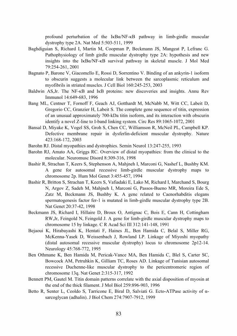
83
profound perturbation of the IκBα/NF-κB pathway in limb-girdle muscular dystrophy type 2A. Nat Med 5:503-511, 1999
Baghdiguian S, Richard I, Martin M, Coopman P, Beckmann JS, Mangeat P, Lefranc G. Pathophysiology of limb girdle muscular dystrophy type 2A: hypothesis and new insights into the IκBα/NF-κB survival pathway in skeletal muscle. J Mol Med 79:254-261, 2001
Bagnato P, Barone V, Giacomello E, Rossi D, Sorrentino V. Binding of an ankyrin-1 isoform to obscurin suggests a molecular link between the sarcoplasmic reticulum and myofibrils in striated muscles. J Cell Biol 160:245-253, 2003
Baldwin AS,Jr. The NF-κB and IκB proteins: new discoveries and insights. Annu Rev Immunol 14:649-683, 1996
Bang ML, Centner T, Fornoff F, Geach AJ, Gotthardt M, McNabb M, Witt CC, Labeit D, Gregorio CC, Granzier H, Labeit S. The complete gene sequence of titin, expression of an unusual approximately 700-kDa titin isoform, and its interaction with obscurin identify a novel Z-line to I-band linking system. Circ Res 89:1065-1072, 2001
Bansal D, Miyake K, Vogel SS, Groh S, Chen CC, Williamson R, McNeil PL, Campbell KP. Defective membrane repair in dysferlin-deficient muscular dystrophy. Nature 423:168-172, 2003
Barohn RJ. Distal myopathies and dystrophies. Semin Neurol 13:247-255, 1993 Barohn RJ, Amato AA, Griggs RC. Overview of distal myopathies: from the clinical to the
molecular. Neuromusc Disord 8:309-316, 1998 Bashir R, Strachan T, Keers S, Stephenson A, Mahjneh I, Marconi G, Nashef L, Bushby KM.
A gene for autosomal recessive limb-girdle muscular dystrophy maps to chromosome 2p. Hum Mol Genet 3:455-457, 1994
Bashir R, Britton S, Strachan T, Keers S, Vafiadaki E, Lako M, Richard I, Marchand S, Bourg N, Argov Z, Sadeh M, Mahjneh I, Marconi G, Passos-Bueno MR, Moreira Ede S, Zatz M, Beckmann JS, Bushby K. A gene related to Caenorhabditis elegans spermatogenesis factor fer-1 is mutated in limb-girdle muscular dystrophy type 2B. Nat Genet 20:37-42, 1998
Beckmann JS, Richard I, Hillaire D, Broux O, Antignac C, Bois E, Cann H, Cottingham RW,Jr, Feingold N, Feingold J. A gene for limb-girdle muscular dystrophy maps to chromosome 15 by linkage. C R Acad Sci III 312:141-148, 1991
Bejaoui K, Hirabayashi K, Hentati F, Haines JL, Ben Hamida C, Belal S, Miller RG, McKenna-Yasek D, Weissenbach J, Rowland LP. Linkage of Miyoshi myopathy (distal autosomal recessive muscular dystrophy) locus to chromosome 2p12-14. Neurology 45:768-772, 1995
Ben Othmane K, Ben Hamida M, Pericak-Vance MA, Ben Hamida C, Blel S, Carter SC, Bowcock AM, Petruhkin K, Gilliam TC, Roses AD. Linkage of Tunisian autosomal recessive Duchenne-like muscular dystrophy to the pericentromeric region of chromosome 13q. Nat Genet 2:315-317, 1992
Bennett PM, Gautel M. Titin domain patterns correlate with the axial disposition of myosin at the end of the thick filament. J Mol Biol 259:896-903, 1996
Betto R, Senter L, Ceoldo S, Tarricone E, Biral D, Salviati G. Ecto-ATPase activity of α-sarcoglycan (adhalin). J Biol Chem 274:7907-7912, 1999
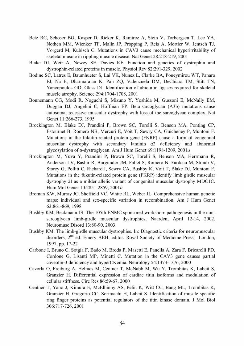
84
Betz RC, Schoser BG, Kasper D, Ricker K, Ramirez A, Stein V, Torbergsen T, Lee YA, Nothen MM, Wienker TF, Malin JP, Propping P, Reis A, Mortier W, Jentsch TJ, Vorgerd M, Kubisch C. Mutations in CAV3 cause mechanical hyperirritability of skeletal muscle in rippling muscle disease. Nat Genet 28:218-219, 2001
Blake DJ, Weir A, Newey SE, Davies KE. Function and genetics of dystrophin and dystrophin-related proteins in muscle. Physiol Rev 82:291-329, 2002
Bodine SC, Latres E, Baumhueter S, Lai VK, Nunez L, Clarke BA, Poueymirou WT, Panaro FJ, Na E, Dharmarajan K, Pan ZQ, Valenzuela DM, DeChiara TM, Stitt TN, Yancopoulos GD, Glass DJ. Identification of ubiquitin ligases required for skeletal muscle atrophy. Science 294:1704-1708, 2001
Bonnemann CG, Modi R, Noguchi S, Mizuno Y, Yoshida M, Gussoni E, McNally EM, Duggan DJ, Angelini C, Hoffman EP. Beta-sarcoglycan (A3b) mutations cause autosomal recessive muscular dystrophy with loss of the sarcoglycan complex. Nat Genet 11:266-273, 1995
Brockington M, Blake DJ, Prandini P, Brown SC, Torelli S, Benson MA, Ponting CP, Estournet B, Romero NB, Mercuri E, Voit T, Sewry CA, Guicheney P, Muntoni F. Mutations in the fukutin-related protein gene (FKRP) cause a form of congenital muscular dystrophy with secondary laminin α2 deficiency and abnormal glycosylation of α-dystroglycan. Am J Hum Genet 69:1198-1209, 2001a
Brockington M, Yuva Y, Prandini P, Brown SC, Torelli S, Benson MA, Herrmann R, Anderson LV, Bashir R, Burgunder JM, Fallet S, Romero N, Fardeau M, Straub V, Storey G, Pollitt C, Richard I, Sewry CA, Bushby K, Voit T, Blake DJ, Muntoni F. Mutations in the fukutin-related protein gene (FKRP) identify limb girdle muscular dystrophy 2I as a milder allelic variant of congenital muscular dystrophy MDC1C. Hum Mol Genet 10:2851-2859, 2001b
Broman KW, Murray JC, Sheffield VC, White RL, Weber JL. Comprehensive human genetic maps: individual and sex-specific variation in recombination. Am J Hum Genet 63:861-869, 1998
Bushby KM, Beckmann JS. The 105th ENMC sponsored workshop: pathogenesis in the non-sarcoglycan limb-girdle muscular dystrophies, Naarden, April 12-14, 2002. Neuromusc Disord 13:80-90, 2003
Bushby KM. The limb-girdle muscular dystrophies. In: Diagnostic criteria for neuromuscular disorders, 2nd ed. Emery AEH, editor. Royal Society of Medicine Press, London, 1997, pp. 17-22
Carbone I, Bruno C, Sotgia F, Bado M, Broda P, Masetti E, Panella A, Zara F, Bricarelli FD, Cordone G, Lisanti MP, Minetti C. Mutation in the CAV3 gene causes partial caveolin-3 deficiency and hyperCKemia. Neurology 54:1373-1376, 2000
Cazorla O, Freiburg A, Helmes M, Centner T, McNabb M, Wu Y, Trombitas K, Labeit S, Granzier H. Differential expression of cardiac titin isoforms and modulation of cellular stiffness. Circ Res 86:59-67, 2000
Centner T, Yano J, Kimura E, McElhinny AS, Pelin K, Witt CC, Bang ML, Trombitas K, Granzier H, Gregorio CC, Sorimachi H, Labeit S. Identification of muscle specific ring finger proteins as potential regulators of the titin kinase domain. J Mol Biol 306:717-726, 2001
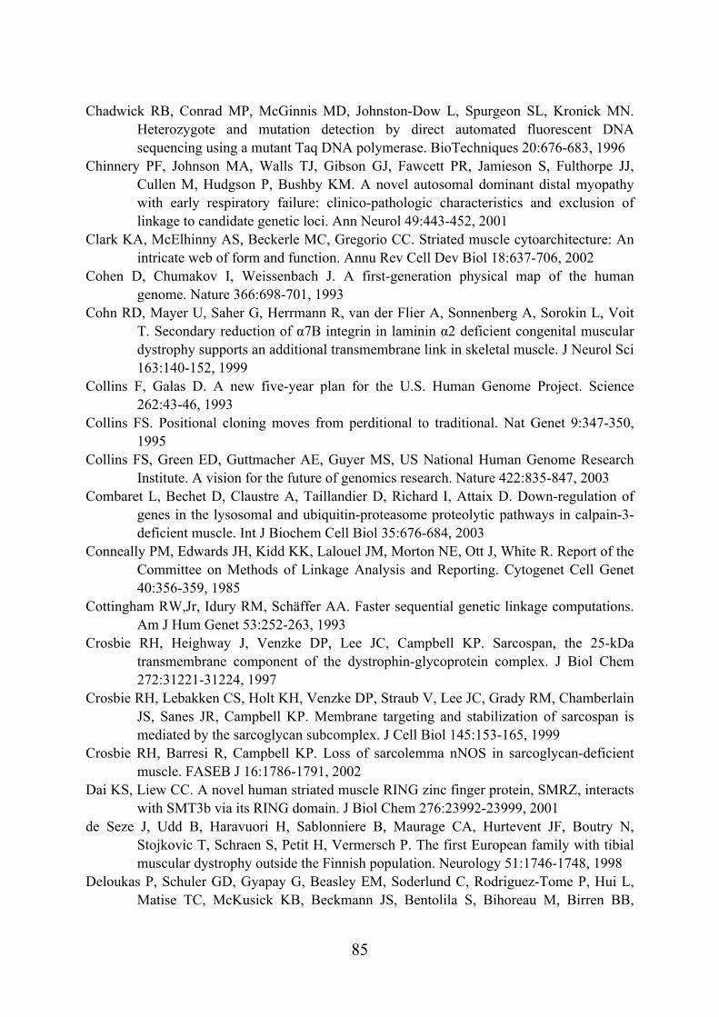
85
Chadwick RB, Conrad MP, McGinnis MD, Johnston-Dow L, Spurgeon SL, Kronick MN. Heterozygote and mutation detection by direct automated fluorescent DNA sequencing using a mutant Taq DNA polymerase. BioTechniques 20:676-683, 1996
Chinnery PF, Johnson MA, Walls TJ, Gibson GJ, Fawcett PR, Jamieson S, Fulthorpe JJ, Cullen M, Hudgson P, Bushby KM. A novel autosomal dominant distal myopathy with early respiratory failure: clinico-pathologic characteristics and exclusion of linkage to candidate genetic loci. Ann Neurol 49:443-452, 2001
Clark KA, McElhinny AS, Beckerle MC, Gregorio CC. Striated muscle cytoarchitecture: An intricate web of form and function. Annu Rev Cell Dev Biol 18:637-706, 2002
Cohen D, Chumakov I, Weissenbach J. A first-generation physical map of the human genome. Nature 366:698-701, 1993
Cohn RD, Mayer U, Saher G, Herrmann R, van der Flier A, Sonnenberg A, Sorokin L, Voit T. Secondary reduction of α7B integrin in laminin α2 deficient congenital muscular dystrophy supports an additional transmembrane link in skeletal muscle. J Neurol Sci 163:140-152, 1999
Collins F, Galas D. A new five-year plan for the U.S. Human Genome Project. Science 262:43-46, 1993
Collins FS. Positional cloning moves from perditional to traditional. Nat Genet 9:347-350, 1995
Collins FS, Green ED, Guttmacher AE, Guyer MS, US National Human Genome Research Institute. A vision for the future of genomics research. Nature 422:835-847, 2003
Combaret L, Bechet D, Claustre A, Taillandier D, Richard I, Attaix D. Down-regulation of genes in the lysosomal and ubiquitin-proteasome proteolytic pathways in calpain-3-deficient muscle. Int J Biochem Cell Biol 35:676-684, 2003
Conneally PM, Edwards JH, Kidd KK, Lalouel JM, Morton NE, Ott J, White R. Report of the Committee on Methods of Linkage Analysis and Reporting. Cytogenet Cell Genet 40:356-359, 1985
Cottingham RW,Jr, Idury RM, Schäffer AA. Faster sequential genetic linkage computations. Am J Hum Genet 53:252-263, 1993
Crosbie RH, Heighway J, Venzke DP, Lee JC, Campbell KP. Sarcospan, the 25-kDa transmembrane component of the dystrophin-glycoprotein complex. J Biol Chem 272:31221-31224, 1997
Crosbie RH, Lebakken CS, Holt KH, Venzke DP, Straub V, Lee JC, Grady RM, Chamberlain JS, Sanes JR, Campbell KP. Membrane targeting and stabilization of sarcospan is mediated by the sarcoglycan subcomplex. J Cell Biol 145:153-165, 1999
Crosbie RH, Barresi R, Campbell KP. Loss of sarcolemma nNOS in sarcoglycan-deficient muscle. FASEB J 16:1786-1791, 2002
Dai KS, Liew CC. A novel human striated muscle RING zinc finger protein, SMRZ, interacts with SMT3b via its RING domain. J Biol Chem 276:23992-23999, 2001
de Seze J, Udd B, Haravuori H, Sablonniere B, Maurage CA, Hurtevent JF, Boutry N, Stojkovic T, Schraen S, Petit H, Vermersch P. The first European family with tibial muscular dystrophy outside the Finnish population. Neurology 51:1746-1748, 1998
Deloukas P, Schuler GD, Gyapay G, Beasley EM, Soderlund C, Rodriguez-Tome P, Hui L, Matise TC, McKusick KB, Beckmann JS, Bentolila S, Bihoreau M, Birren BB,
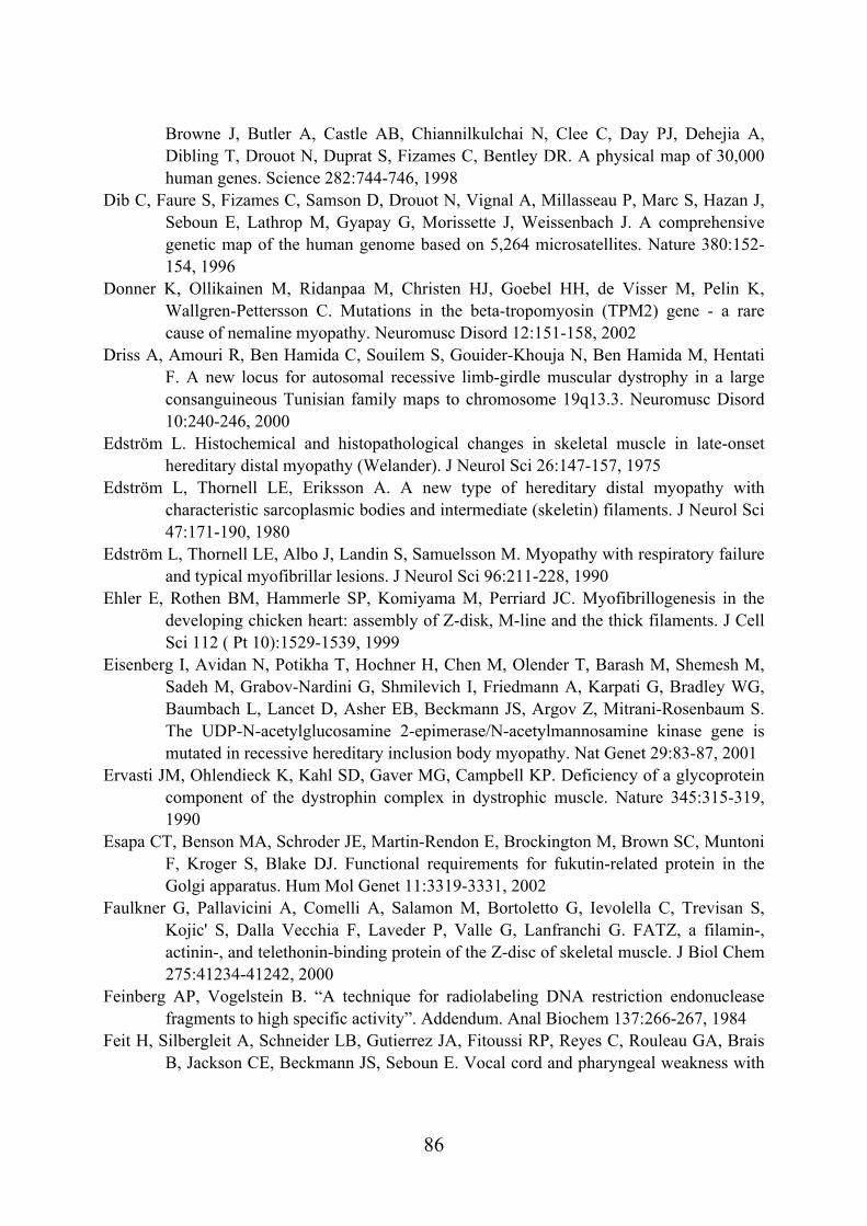
86
Browne J, Butler A, Castle AB, Chiannilkulchai N, Clee C, Day PJ, Dehejia A, Dibling T, Drouot N, Duprat S, Fizames C, Bentley DR. A physical map of 30,000 human genes. Science 282:744-746, 1998
Dib C, Faure S, Fizames C, Samson D, Drouot N, Vignal A, Millasseau P, Marc S, Hazan J, Seboun E, Lathrop M, Gyapay G, Morissette J, Weissenbach J. A comprehensive genetic map of the human genome based on 5,264 microsatellites. Nature 380:152-154, 1996
Donner K, Ollikainen M, Ridanpaa M, Christen HJ, Goebel HH, de Visser M, Pelin K, Wallgren-Pettersson C. Mutations in the beta-tropomyosin (TPM2) gene - a rare cause of nemaline myopathy. Neuromusc Disord 12:151-158, 2002
Driss A, Amouri R, Ben Hamida C, Souilem S, Gouider-Khouja N, Ben Hamida M, Hentati F. A new locus for autosomal recessive limb-girdle muscular dystrophy in a large consanguineous Tunisian family maps to chromosome 19q13.3. Neuromusc Disord 10:240-246, 2000
Edström L. Histochemical and histopathological changes in skeletal muscle in late-onset hereditary distal myopathy (Welander). J Neurol Sci 26:147-157, 1975
Edström L, Thornell LE, Eriksson A. A new type of hereditary distal myopathy with characteristic sarcoplasmic bodies and intermediate (skeletin) filaments. J Neurol Sci 47:171-190, 1980
Edström L, Thornell LE, Albo J, Landin S, Samuelsson M. Myopathy with respiratory failure and typical myofibrillar lesions. J Neurol Sci 96:211-228, 1990
Ehler E, Rothen BM, Hammerle SP, Komiyama M, Perriard JC. Myofibrillogenesis in the developing chicken heart: assembly of Z-disk, M-line and the thick filaments. J Cell Sci 112 ( Pt 10):1529-1539, 1999
Eisenberg I, Avidan N, Potikha T, Hochner H, Chen M, Olender T, Barash M, Shemesh M, Sadeh M, Grabov-Nardini G, Shmilevich I, Friedmann A, Karpati G, Bradley WG, Baumbach L, Lancet D, Asher EB, Beckmann JS, Argov Z, Mitrani-Rosenbaum S. The UDP-N-acetylglucosamine 2-epimerase/N-acetylmannosamine kinase gene is mutated in recessive hereditary inclusion body myopathy. Nat Genet 29:83-87, 2001
Ervasti JM, Ohlendieck K, Kahl SD, Gaver MG, Campbell KP. Deficiency of a glycoprotein component of the dystrophin complex in dystrophic muscle. Nature 345:315-319, 1990
Esapa CT, Benson MA, Schroder JE, Martin-Rendon E, Brockington M, Brown SC, Muntoni F, Kroger S, Blake DJ. Functional requirements for fukutin-related protein in the Golgi apparatus. Hum Mol Genet 11:3319-3331, 2002
Faulkner G, Pallavicini A, Comelli A, Salamon M, Bortoletto G, Ievolella C, Trevisan S, Kojic' S, Dalla Vecchia F, Laveder P, Valle G, Lanfranchi G. FATZ, a filamin-, actinin-, and telethonin-binding protein of the Z-disc of skeletal muscle. J Biol Chem 275:41234-41242, 2000
Feinberg AP, Vogelstein B. “A technique for radiolabeling DNA restriction endonuclease fragments to high specific activity”. Addendum. Anal Biochem 137:266-267, 1984
Feit H, Silbergleit A, Schneider LB, Gutierrez JA, Fitoussi RP, Reyes C, Rouleau GA, Brais B, Jackson CE, Beckmann JS, Seboun E. Vocal cord and pharyngeal weakness with
87
autosomal dominant distal myopathy: clinical description and gene localization to 5q31. Am J Hum Genet 63:1732-1742, 1998
Felice KJ, Meredith C, Binz N, Butler A, Jacob R, Akkari P, Hallmayer J, Laing N. Autosomal dominant distal myopathy not linked to the known distal myopathy loci. Neuromusc Disord 9:59-65, 1999
Freiburg A, Gautel M. A molecular map of the interactions between titin and myosin-binding protein C. Implications for sarcomeric assembly in familial hypertrophic cardiomyopathy. Eur J Biochem 235:317-323, 1996
Freiburg A, Trombitas K, Hell W, Cazorla O, Fougerousse F, Centner T, Kolmerer B, Witt C, Beckmann JS, Gregorio CC, Granzier H, Labeit S. Series of exon-skipping events in the elastic spring region of titin as the structural basis for myofibrillar elastic diversity. Circ Res 86:1114-1121, 2000
Frey N, Olson EN. Calsarcin-3, a novel skeletal muscle-specific member of the calsarcin family, interacts with multiple Z-disc proteins. J Biol Chem 277:13998-14004, 2002
Frosk P, Weiler T, Nylen E, Sudha T, Greenberg CR, Morgan K, Fujiwara TM, Wrogemann K. Limb-girdle muscular dystrophy type 2H associated with mutation in TRIM32, a putative E3-ubiquitin-ligase gene. Am J Hum Genet 70:663-672, 2002
Furukawa T, Ono Y, Tsuchiya H, Katayama Y, Bang ML, Labeit D, Labeit S, Inagaki N, Gregorio CC. Specific interaction of the potassium channel beta-subunit minK with the sarcomeric protein T-cap suggests a T-tubule-myofibril linking system. J Mol Biol 313:775-784, 2001
Fürst DO, Osborn M, Nave R, Weber K. The organization of titin filaments in the half-sarcomere revealed by monoclonal antibodies in immunoelectron microscopy: a map of ten nonrepetitive epitopes starting at the Z line extends close to the M line. J Cell Biol 106:1563-1572, 1988
Fürst DO, Obermann WM, van der Ven PF. Structure and assembly of the sarcomeric M band. Rev Physiol Biochem Pharmacol 138:163-202, 1999
Galbiati F, Engelman JA, Volonte D, Zhang XL, Minetti C, Li M, Hou H,Jr, Kneitz B, Edelmann W, Lisanti MP. Caveolin-3 null mice show a loss of caveolae, changes in the microdomain distribution of the dystrophin-glycoprotein complex, and t-tubule abnormalities. J Biol Chem 276:21425-21433, 2001
Gallagher PG, Forget BG. An alternate promoter directs expression of a truncated, muscle-specific isoform of the human ankyrin 1 gene. J Biol Chem 273:1339-1348, 1998
Garvey SM, Rajan C, Lerner AP, Frankel WN, Cox GA. The muscular dystrophy with myositis (mdm) mouse mutation disrupts a skeletal muscle-specific domain of titin. Genomics 79:146-149, 2002
Gautel M, Castiglione Morelli MA, Pfuhl M, Motta A, Pastore A. A calmodulin-binding sequence in the C-terminus of human cardiac titin kinase. Eur J Biochem 230:752-759, 1995
Gautel M, Goulding D, Bullard B, Weber K, Fürst DO. The central Z-disk region of titin is assembled from a novel repeat in variable copy numbers. J Cell Sci 109 ( Pt 11):2747-2754, 1996
Gautel M, Mues A, Young P. Control of sarcomeric assembly: the flow of information on titin. Rev Physiol Biochem Pharmacol 138:97-137, 1999
88
Gerull B, Gramlich M, Atherton J, McNabb M, Trombitas K, Sasse-Klaassen S, Seidman JG, Seidman C, Granzier H, Labeit S, Frenneaux M, Thierfelder L. Mutations of TTN, encoding the giant muscle filament titin, cause familial dilated cardiomyopathy. Nat Genet 30:201-204, 2002
Gitschier J, Wood WI, Goralka TM, Wion KL, Chen EY, Eaton DH, Vehar GA, Capon DJ, Lawn RM. Characterization of the human factor VIII gene. Nature 312:326-330, 1984
Golenhofen N, Arbeiter A, Koob R, Drenckhahn D. Ischemia-induced association of the stress protein alpha B-crystallin with I-band portion of cardiac titin. J Mol Cell Cardiol 34:309-319, 2002
Gotthardt M, Hammer RE, Hubner N, Monti J, Witt CC, McNabb M, Richardson JA, Granzier H, Labeit S, Herz J. Conditional expression of mutant M-line titins results in cardiomyopathy with altered sarcomere structure. J Biol Chem 278:6059-6065, 2003
Gowers WR. A lecture on myopathy and distal form. Br Med J 2:89, 1902 Granzier H, Labeit S. Cardiac titin: an adjustable multi-functional spring. J Physiol 541:335-
342, 2002 Gregorio CC, Trombitas K, Centner T, Kolmerer B, Stier G, Kunke K, Suzuki K, Obermayr
F, Herrmann B, Granzier H, Sorimachi H, Labeit S. The NH2 terminus of titin spans the Z-disc: its interaction with a novel 19-kD ligand (T-cap) is required for sarcomeric integrity. J Cell Biol 143:1013-1027, 1998
Gregorio CC, Granzier H, Sorimachi H, Labeit S. Muscle assembly: a titanic achievement? Curr Opin Cell Biol 11:18-25, 1999
Griggs RC, Askanas V, DiMauro S, Engel A, Karpati G, Mendell JR, Rowland LP. Inclusion body myositis and myopathies. Ann Neurol 38:705-713, 1995
Grove BK, Kurer V, Lehner C, Doetschman TC, Perriard JC, Eppenberger HM. A new 185,000-dalton skeletal muscle protein detected by monoclonal antibodies. J Cell Biol 98:518-524, 1984
Gyapay G, Morissette J, Vignal A, Dib C, Fizames C, Millasseau P, Marc S, Bernardi G, Lathrop M, Weissenbach J. The 1993-94 Genethon human genetic linkage map. Nat Genet 7:246-339, 1994
Hack AA, Cordier L, Shoturma DI, Lam MY, Sweeney HL, McNally EM. Muscle degeneration without mechanical injury in sarcoglycan deficiency. Proc Natl Acad Sci USA 96:10723-10728, 1999
Hackman P, Vihola A, Haravuori H, Sarparanta J, Udd B. Orsaken till tibial muskeldystrofi - mutationer i den gigantiska muskelgenen titin. Finska Läkaresällskapets Handlingar 162:20-25, 2002
Hailstones DL, Gunning PW. Characterization of human myosin light chains 1sa and 3nm: implications for isoform evolution and function. Mol Cell Biol 10:1095-1104, 1990
Hanke M, Wink M. Direct DNA sequencing of PCR-amplified vector inserts following enzymatic degradation of primer and dNTPs. BioTechniques 17:858-860, 1994
Hauser MA, Horrigan SK, Salmikangas P, Torian UM, Viles KD, Dancel R, Tim RW, Taivainen A, Bartoloni L, Gilchrist JM, Stajich JM, Gaskell PC, Gilbert JR, Vance
89
JM, Pericak-Vance MA, Carpen O, Westbrook CA, Speer MC. Myotilin is mutated in limb girdle muscular dystrophy 1A. Hum Mol Genet 9:2141-2147, 2000
Hauser MA, Conde CB, Kowaljow V, Zeppa G, Taratuto AL, Torian UM, Vance J, Pericak-Vance MA, Speer MC, Rosa AL. myotilin Mutation found in second pedigree with LGMD1A. Am J Hum Genet 71:1428-1432, 2002
Hayashi YK, Chou FL, Engvall E, Ogawa M, Matsuda C, Hirabayashi S, Yokochi K, Ziober BL, Kramer RH, Kaufman SJ, Ozawa E, Goto Y, Nonaka I, Tsukahara T, Wang JZ, Hoffman EP, Arahata K. Mutations in the integrin α7 gene cause congenital myopathy. Nat Genet 19:94-97, 1998
Heimann P, Menke A, Rothkegel B, Jockusch H. Overshooting production of satellite cells in murine skeletal muscle affected by the mutation “muscular dystrophy with myositis” (mdm, Chr 2). Cell Tissue Res 283:435-441, 1996
Helbling-Leclerc A, Zhang X, Topaloglu H, Cruaud C, Tesson F, Weissenbach J, Tome FM, Schwartz K, Fardeau M, Tryggvason K. Mutations in the laminin alpha 2-chain gene (LAMA2) cause merosin-deficient congenital muscular dystrophy. Nat Genet 11:216-218, 1995
Herasse M, Ono Y, Fougerousse F, Kimura E, Stockholm D, Beley C, Montarras D, Pinset C, Sorimachi H, Suzuki K, Beckmann JS, Richard I. Expression and functional characteristics of calpain 3 isoforms generated through tissue-specific transcriptional and posttranscriptional events. Mol Cell Biol 19:4047-4055, 1999
Hodges BL, Hayashi YK, Nonaka I, Wang W, Arahata K, Kaufman SJ. Altered expression of the α7β1 integrin in human and murine muscular dystrophies. J Cell Sci 110 ( Pt 22):2873-2881, 1997
Hoffman EP, Brown RH,Jr, Kunkel LM. Dystrophin: the protein product of the Duchenne muscular dystrophy locus. Cell 51:919-928, 1987
Holt KH, Campbell KP. Assembly of the sarcoglycan complex. Insights for muscular dystrophy. J Biol Chem 273:34667-34670, 1998
Horowits R, Kempner ES, Bisher ME, Podolsky RJ. A physiological role for titin and nebulin in skeletal muscle. Nature 323:160-164, 1986
Horowits R. The physiological role of titin in striated muscle. Rev Physiol Biochem Pharmacol 138:57-96, 1999
Houmeida A, Holt J, Tskhovrebova L, Trinick J. Studies of the interaction between titin and myosin. J Cell Biol 131:1471-1481, 1995
Hudson TJ, Stein LD, Gerety SS, Ma J, Castle AB, Silva J, Slonim DK, Baptista R, Kruglyak L, Xu SH. An STS-based map of the human genome. Science 270:1945-1954, 1995
Hästbacka J, de la Chapelle A, Mahtani MM, Clines G, Reeve-Daly MP, Daly M, Hamilton BA, Kusumi K, Trivedi B, Weaver A. The diastrophic dysplasia gene encodes a novel sulfate transporter: positional cloning by fine-structure linkage disequilibrium mapping. Cell 78:1073-1087, 1994
Ibraghimov-Beskrovnaya O, Milatovich A, Ozcelik T, Yang B, Koepnick K, Francke U, Campbell KP. Human dystroglycan: skeletal muscle cDNA, genomic structure, origin of tissue specific isoforms and chromosomal localization. Hum Mol Genet 2:1651-1657, 1993
90
Ikonen E, Baumann M, Gron K, Syvänen AC, Enomaa N, Halila R, Aula P, Peltonen L. Aspartylglucosaminuria: cDNA encoding human aspartylglucosaminidase and the missense mutation causing the disease. EMBO J 10:51-58, 1991
Ilkovski B, Cooper ST, Nowak K, Ryan MM, Yang N, Schnell C, Durling HJ, Roddick LG, Wilkinson I, Kornberg AJ, Collins KJ, Wallace G, Gunning P, Hardeman EC, Laing NG, North KN. Nemaline myopathy caused by mutations in the muscle alpha-skeletal-actin gene. Am J Hum Genet 68:1333-1343, 2001
Illa I, Serrano-Munuera C, Gallardo E, Lasa A, Rojas-Garcia R, Palmer J, Gallano P, Baiget M, Matsuda C, Brown RH. Distal anterior compartment myopathy: a dysferlin mutation causing a new muscular dystrophy phenotype. Ann Neurol 49:130-134, 2001
Illarioshkin SN, Ivanova-Smolenskaya IA, Greenberg CR, Nylen E, Sukhorukov VS, Poleshchuk VV, Markova ED, Wrogemann K. Identical dysferlin mutation in limb-girdle muscular dystrophy type 2B and distal myopathy. Neurology 55:1931-1933, 2000
Improta S, Politou AS, Pastore A. Immunoglobulin-like modules from titin I-band: extensible components of muscle elasticity. Structure 4:323-337, 1996
Improta S, Krueger JK, Gautel M, Atkinson RA, Lefevre JF, Moulton S, Trewhella J, Pastore A. The assembly of immunoglobulin-like modules in titin: implications for muscle elasticity. J Mol Biol 284:761-777, 1998
International Human Genome Sequencing Consortium. Initial sequencing and analysis of the human genome. Nature 409:860-921, 2001
Itoh-Satoh M, Hayashi T, Nishi H, Koga Y, Arimura T, Koyanagi T, Takahashi M, Hohda S, Ueda K, Nouchi T, Hiroe M, Marumo F, Imaizumi T, Yasunami M, Kimura A. Titin mutations as the molecular basis for dilated cardiomyopathy. Biochem Biophys Res Commun 291:385-393, 2002
Jia Z, Petrounevitch V, Wong A, Moldoveanu T, Davies PL, Elce JS, Beckmann JS. Mutations in calpain 3 associated with limb girdle muscular dystrophy: analysis by molecular modeling and by mutation in m-calpain. Biophys J 80:2590-2596, 2001
Joazeiro CA, Weissman AM. RING finger proteins: mediators of ubiquitin ligase activity. Cell 102:549-552, 2000
Jobsis GJ, Keizers H, Vreijling JP, de Visser M, Speer MC, Wolterman RA, Baas F, Bolhuis PA. Type VI collagen mutations in Bethlem myopathy, an autosomal dominant myopathy with contractures. Nat Genet 14:113-115, 1996
Johnston JJ, Kelley RI, Crawford TO, Morton DH, Agarwala R, Koch T, Schaffer AA, Francomano CA, Biesecker LG. A novel nemaline myopathy in the Amish caused by a mutation in troponin T1. Am J Hum Genet 67:814-821, 2000
Jorde LB. Linkage disequilibrium as a gene-mapping tool. Am J Hum Genet 56:11-14, 1995 Kayashima T, Matsuo H, Satoh A, Ohta T, Yoshiura K, Matsumoto N, Nakane Y, Niikawa N,
Kishino T. Nonaka myopathy is caused by mutations in the UDP-N-acetylglucosamine-2-epimerase/N-acetylmannosamine kinase gene (GNE). J Hum Genet 47:77-79, 2002
91
Keppler OT, Hinderlich S, Langner J, Schwartz-Albiez R, Reutter W, Pawlita M. UDP-GlcNAc 2-epimerase: a regulator of cell surface sialylation. Science 284:1372-1376, 1999
Kinbara K, Sorimachi H, Ishiura S, Suzuki K. Muscle-specific calpain, p94, interacts with the extreme C-terminal region of connectin, a unique region flanked by two immunoglobulin C2 motifs. Arch Biochem Biophys 342:99-107, 1997
Knupp C, Luther PK, Squire JM. Titin organisation and the 3D architecture of the vertebrate-striated muscle I-band. J Mol Biol 322:731-739, 2002
Knöll R, Hoshijima M, Hoffman HM, Person V, Lorenzen-Schmidt I, Bang ML, Hayashi T, Shiga N, Yasukawa H, Schaper W, McKenna W, Yokoyama M, Schork NJ, Omens JH, McCulloch AD, Kimura A, Gregorio CC, Poller W, Schaper J, Schultheiss HP, Chien KR. The cardiac mechanical stretch sensor machinery involves a Z disc complex that is defective in a subset of human dilated cardiomyopathy. Cell 111:943-955, 2002
Koenig M, Hoffman EP, Bertelson CJ, Monaco AP, Feener C, Kunkel LM. Complete cloning of the Duchenne muscular dystrophy (DMD) cDNA and preliminary genomic organization of the DMD gene in normal and affected individuals. Cell 50:509-517, 1987
Kolmerer B, Olivieri N, Witt CC, Herrmann BG, Labeit S. Genomic organization of M line titin and its tissue-specific expression in two distinct isoforms. J Mol Biol 256:556-563, 1996
Kontrogianni-Konstantopoulos A, Bloch RJ. The hydrophilic domain of small ankyrin-1 interacts with the two N-terminal immunoglobulin domains of titin. J Biol Chem 278:3985-3991, 2003
Kontrogianni-Konstantopoulos A, Jones EM, Van Rossum DB, Bloch RJ. Obscurin is a ligand for small ankyrin 1 in skeletal muscle. Mol Biol Cell 14:1138-1148, 2003
Kruglyak L, Daly MJ, Reeve-Daly MP, Lander ES. Parametric and nonparametric linkage analysis: a unified multipoint approach. Am J Hum Genet 58:1347-1363, 1996
Kulke M, Fujita-Becker S, Rostkova E, Neagoe C, Labeit D, Manstein DJ, Gautel M, Linke WA. Interaction between PEVK-titin and actin filaments: origin of a viscous force component in cardiac myofibrils. Circ Res 89:874-881, 2001
Labeit S, Gautel M, Lakey A, Trinick J. Towards a molecular understanding of titin. EMBO J 11:1711-1716, 1992
Labeit S, Kolmerer B. Titins: giant proteins in charge of muscle ultrastructure and elasticity. Science 270:293-296, 1995
Laemmli UK. Cleavage of structural proteins during the assembly of the head of bacteriophage T4. Nature 227:680-685, 1970
Laing NG, Laing BA, Meredith C, Wilton SD, Robbins P, Honeyman K, Dorosz S, Kozman H, Mastaglia FL, Kakulas BA. Autosomal dominant distal myopathy: linkage to chromosome 14. Am J Hum Genet 56:422-427, 1995a
Laing NG, Wilton SD, Akkari PA, Dorosz S, Boundy K, Kneebone C, Blumbergs P, White S, Watkins H, Love DR. A mutation in the alpha tropomyosin gene TPM3 associated with autosomal dominant nemaline myopathy. Nat Genet 9:75-79, 1995b

92
Lamande SR, Bateman JF, Hutchison W, McKinlay Gardner RJ, Bower SP, Byrne E, Dahl HH. Reduced collagen VI causes Bethlem myopathy: a heterozygous COL6A1 nonsense mutation results in mRNA decay and functional haploinsufficiency. Hum Mol Genet 7:981-989, 1998
Lane PW. Muscular dystrophy with myositis (mdm). Mouse News Lett 73:18, 1985 Lange S, Auerbach D, McLoughlin P, Perriard E, Schafer BW, Perriard JC, Ehler E.
Subcellular targeting of metabolic enzymes to titin in heart muscle may be mediated by DRAL/FHL-2. J Cell Sci 115:4925-4936, 2002
Lathrop GM, Lalouel JM. Easy calculations of lod scores and genetic risks on small computers. Am J Hum Genet 36:460-465, 1984
Laulumaa V, Partanen J, Palvajärvi L. Tibialis anterior - myopatia. In: Lihastautien kehittyvä tutkimus ja hoito. Lang H, editor. Kiasma, Turku, 1989, pp. 63-66
Lennon NJ, Kho A, Bacskai BJ, Perlmutter SL, Hyman BT, Brown RH Jr. Dysferlin interacts with annexins A1 and A2 and mediates sarcolemmal wound-healing. J Biol Chem in press, published online ahead of print, 2003
Li H, Oberhauser AF, Redick SD, Carrion-Vazquez M, Erickson HP, Fernandez JM. Multiple conformations of PEVK proteins detected by single-molecule techniques. Proc Natl Acad Sci USA 98:10682-10686, 2001
Lim LE, Duclos F, Broux O, Bourg N, Sunada Y, Allamand V, Meyer J, Richard I, Moomaw C, Slaughter C. Beta-sarcoglycan: characterization and role in limb-girdle muscular dystrophy linked to 4q12. Nat Genet 11:257-265, 1995
Lin Z, Lu MH, Schultheiss T, Choi J, Holtzer S, DiLullo C, Fischman DA, Holtzer H. Sequential appearance of muscle-specific proteins in myoblasts as a function of time after cell division: evidence for a conserved myoblast differentiation program in skeletal muscle. Cell Motil Cytoskeleton 29:1-19, 1994
Linke WA, Rudy DE, Centner T, Gautel M, Witt C, Labeit S, Gregorio CC. I-band titin in cardiac muscle is a three-element molecular spring and is critical for maintaining thin filament structure. J Cell Biol 146:631-644, 1999
Linke WA, Kulke M, Li H, Fujita-Becker S, Neagoe C, Manstein DJ, Gautel M, Fernandez JM. PEVK domain of titin: an entropic spring with actin-binding properties. J Struct Biol 137:194-205, 2002
Linssen WH, de Visser M, Notermans NC, Vreyling JP, Van Doorn PA, Wokke JH, Baas F, Bolhuis PA. Genetic heterogeneity in Miyoshi-type distal muscular dystrophy. Neuromusc Disord 8:317-320, 1998
Lisanti MP, Tang Z, Scherer PE, Kubler E, Koleske AJ, Sargiacomo M. Caveolae, transmembrane signalling and cellular transformation. Mol Membr Biol 12:121-124, 1995
Liu J, Aoki M, Illa I, Wu C, Fardeau M, Angelini C, Serrano C, Urtizberea JA, Hentati F, Hamida MB, Bohlega S, Culper EJ, Amato AA, Bossie K, Oeltjen J, Bejaoui K, McKenna-Yasek D, Hosler BA, Schurr E, Arahata K, de Jong PJ, Brown RH,Jr. Dysferlin, a novel skeletal muscle gene, is mutated in Miyoshi myopathy and limb girdle muscular dystrophy. Nat Genet 20:31-36, 1998
Liversage AD, Holmes D, Knight PJ, Tskhovrebova L, Trinick J. Titin and the sarcomere symmetry paradox. J Mol Biol 305:401-409, 2001
93
Luther PK, Squire JM. Muscle Z-band ultrastructure: titin Z-repeats and Z-band periodicities do not match. J Mol Biol 319:1157-1164, 2002
Ma K, Wang K. Interaction of nebulin SH3 domain with titin PEVK and myopalladin: implications for the signaling and assembly role of titin and nebulin. FEBS Letters 532: 273-278, 2002
Machado C, Andrew DJ. Titin as a chromosomal protein. Adv Exp Med Biol 481:221-32; discussion 232-236, 2000
Mahjneh I. Limb-girdle muscular dystrophy 2B: a new clinical entity and clinical comparison with other limb-girdle muscular dystrophies (dissertation). Helsinki, 1999
Markesbery WR, Griggs RC, Leach RP, Lapham LW. Late onset hereditary distal myopathy. Neurology 24:127-134, 1974
Markesbery WR, Griggs RC, Herr B. Distal myopathy: electron microscopic and histochemical studies. Neurology 27:727-735, 1977
Martinsson T, Oldfors A, Darin N, Berg K, Tajsharghi H, Kyllerman M, Wahlstrom J. Autosomal dominant myopathy: missense mutation (Glu-706 → Lys) in the myosin heavy chain IIa gene. Proc Natl Acad Sci USA 97:14614-14619, 2000
Maruyama K, Matsubara S, Natori R, Nonomura Y, Kimura S. Connectin, an elastic protein of muscle. Characterization and Function. J Biochem (Tokyo) 82:317-337, 1977
Maruyama K, Kimura S, Ohashi K, Kuwano Y. Connectin, an elastic protein of muscle. Identification of “titin” with connectin. J Biochem (Tokyo) 89:701-709, 1981
Mastaglia FL, Phillips BA, Cala LA, Meredith C, Egli S, Akkari PA, Laing NG. Early onset chromosome 14-linked distal myopathy (Laing). Neuromusc Disord 12:350-357, 2002
Matsuda C, Hayashi YK, Ogawa M, Aoki M, Murayama K, Nishino I, Nonaka I, Arahata K, Brown RH Jr. The sarcolemmal proteins dysferlin and caveolin-3 interact in skeletal muscle. Hum Mol Genet 10:1761-1766, 2001
Mayans O, van der Ven PF, Wilm M, Mues A, Young P, Fürst DO, Wilmanns M, Gautel M. Structural basis for activation of the titin kinase domain during myofibrillogenesis. Nature 395:863-869, 1998
McElhinny AS, Kakinuma K, Sorimachi H, Labeit S, Gregorio CC. Muscle-specific RING finger-1 interacts with titin to regulate sarcomeric M-line and thick filament structure and may have nuclear functions via its interaction with glucocorticoid modulatory element binding protein-1. J Cell Biol 157:125-136, 2002
McKusick VA. HUGO news. The Human Genome Organisation: history, purposes, and membership. Genomics 5:385-387, 1989
McNally EM, Duggan D, Gorospe JR, Bonnemann CG, Fanin M, Pegoraro E, Lidov HG, Noguchi S, Ozawa E, Finkel RS, Cruse RP, Angelini C, Kunkel LM, Hoffman EP. Mutations that disrupt the carboxyl-terminus of gamma-sarcoglycan cause muscular dystrophy. Hum Mol Genet 5:1841-1847, 1996
McNally EM, de Sa Moreira E, Duggan DJ, Bonnemann CG, Lisanti MP, Lidov HG, Vainzof M, Passos-Bueno MR, Hoffman EP, Zatz M, Kunkel LM. Caveolin-3 in muscular dystrophy. Hum Mol Genet 7:871-877, 1998
Meredith C. Molecular genetic investigation of autosomal dominant muscular dystrophy (dissertation). Edith Cowan University, Australia, 2001
94
Messina DN, Speer MC, Pericak-Vance MA, McNally EM. Linkage of familial dilated cardiomyopathy with conduction defect and muscular dystrophy to chromosome 6q23. Am J Hum Genet 61:909-917, 1997
Michele DE, Barresi R, Kanagawa M, Saito F, Cohn RD, Satz JS, Dollar J, Nishino I, Kelley RI, Somer H, Straub V, Mathews KD, Moore SA, Campbell KP. Post-translational disruption of dystroglycan-ligand interactions in congenital muscular dystrophies. Nature 418:417-422, 2002
Millevoi S, Trombitas K, Kolmerer B, Kostin S, Schaper J, Pelin K, Granzier H, Labeit S. Characterization of nebulette and nebulin and emerging concepts of their roles for vertebrate Z-discs. J Mol Biol 282:111-123, 1998
Minetti C, Sotgia F, Bruno C, Scartezzini P, Broda P, Bado M, Masetti E, Mazzocco M, Egeo A, Donati MA, Volonte D, Galbiati F, Cordone G, Bricarelli FD, Lisanti MP, Zara F. Mutations in the caveolin-3 gene cause autosomal dominant limb-girdle muscular dystrophy. Nat Genet 18:365-368, 1998
Minetti C, Bado M, Broda P, Sotgia F, Bruno C, Galbiati F, Volonte D, Lucania G, Pavan A, Bonilla E, Lisanti MP, Cordone G. Impairment of caveolae formation and T-system disorganization in human muscular dystrophy with caveolin-3 deficiency. Am J Pathol 160:265-270, 2002
Miyoshi K, Kawai H, Iwasa M, Kusaka K, Nishino H. Autosomal recessive distal muscular dystrophy as a new type of progressive muscular dystrophy. Seventeen cases in eight families including an autopsied case. Brain 109 ( Pt 1):31-54, 1986
Monici MC, Aguennouz M, Mazzeo A, Messina C, Vita G. Activation of nuclear factor-kappaB in inflammatory myopathies and Duchenne muscular dystrophy. Neurology 60:993-997, 2003
Moreira ES, Vainzof M, Marie SK, Sertie AL, Zatz M, Passos-Bueno MR. The seventh form of autosomal recessive limb-girdle muscular dystrophy is mapped to 17q11-12. Am J Hum Genet 61:151-159, 1997
Moreira ES, Wiltshire TJ, Faulkner G, Nilforoushan A, Vainzof M, Suzuki OT, Valle G, Reeves R, Zatz M, Passos-Bueno MR, Jenne DE. Limb-girdle muscular dystrophy type 2G is caused by mutations in the gene encoding the sarcomeric protein telethonin. Nat Genet 24:163-166, 2000
Morgan MJ, Madgwick AJ. The LIM proteins FHL1 and FHL3 are expressed differently in skeletal muscle. Biochem Biophys Res Commun 255:245-250, 1999
Muchir A, Bonne G, van der Kooi AJ, van Meegen M, Baas F, Bolhuis PA, de Visser M, Schwartz K. Identification of mutations in the gene encoding lamins A/C in autosomal dominant limb girdle muscular dystrophy with atrioventricular conduction disturbances (LGMD1B). Hum Mol Genet 9:1453-1459, 2000
Mues A, van der Ven PF, Young P, Furst DO, Gautel M. Two immunoglobulin-like domains of the Z-disc portion of titin interact in a conformation-dependent way with telethonin. FEBS Lett 428:111-114, 1998
Mühle-Goll CM, Pastore A, Nilges M. The three-dimensional structure of a type I module from titin: a prototype of intracellular fibronectin type III domains. Structure 6:1291-1302, 1998
95
Mühle-Goll C, Habeck M, Cazorla O, Nilges M, Labeit S, Granzier H. Structural and functional studies of titin's fn3 modules reveal conserved surface patterns and binding to myosin S1- a possible role in the Frank-Starling mechanism of the heart. J Mol Biol 313:431-447, 2001
Müller-Seitz M, Kaupmann K, Labeit S, Jockusch H. Chromosomal localization of the mouse titin gene and its relation to “muscular dystrophy with myositis” and nebulin genes on chromosome 2. Genomics 18:559-561, 1993
Neagoe C, Kulke M, del Monte F, Gwathmey JK, de Tombe PP, Hajjar RJ, Linke WA. Titin isoform switch in ischemic human heart disease. Circulation 106:1333-1341, 2002
Nicholas G, Thomas M, Langley B, Somers W, Patel K, Kemp CF, Sharma M, Kambadur R. Titin-cap associates with, and regulates secretion of, Myostatin. J Cell Physiol 193:120-131, 2002
Nicolao P, Xiang F, Gunnarsson LG, Giometto B, Edström L, Anvret M, Zhang Z. Autosomal dominant myopathy with proximal weakness and early respiratory muscle involvement maps to chromosome 2q. Am J Hum Genet 64:788-792, 1999
Nigro V, de Sa Moreira E, Piluso G, Vainzof M, Belsito A, Politano L, Puca AA, Passos-Bueno MR, Zatz M. Autosomal recessive limb-girdle muscular dystrophy, LGMD2F, is caused by a mutation in the delta-sarcoglycan gene. Nat Genet 14:195-198, 1996
Noguchi S, McNally EM, Ben Othmane K, Hagiwara Y, Mizuno Y, Yoshida M, Yamamoto H, Bonnemann CG, Gussoni E, Denton PH. Mutations in the dystrophin-associated protein gamma-sarcoglycan in chromosome 13 muscular dystrophy. Science 270:819-822, 1995
Nokelainen P, Udd B, Somer H, Peltonen L. Linkage analyses in tibial muscular dystrophy. Hum Hered 46:98-107, 1996
Nonaka I, Sunohara N, Ishiura S, Satoyoshi E. Familial distal myopathy with rimmed vacuole and lamellar (myeloid) body formation. J Neurol Sci 51:141-155, 1981
Norio R. Finnish disease heritage I: characteristics, causes, background. Hum Genet 112:441-456, 2003a
Norio R. Finnish Disease Heritage II: population prehistory and genetic roots of Finns. Hum Genet 112:457-469, 2003b
Norio R. The Finnish disease heritage III: the individual diseases. Hum Genet 112:470-526, 2003c
Nowak KJ, Wattanasirichaigoon D, Goebel HH, Wilce M, Pelin K, Donner K, Jacob RL, Hubner C, Oexle K, Anderson JR, Verity CM, North KN, Iannaccone ST, Muller CR, Nurnberg P, Muntoni F, Sewry C, Hughes I, Sutphen R, Lacson AG, Swoboda KJ, Vigneron J, Wallgren-Pettersson C, Beggs AH, Laing NG. Mutations in the skeletal muscle alpha-actin gene in patients with actin myopathy and nemaline myopathy. Nat Genet 23:208-212, 1999
Obermann WM, Gautel M, Weber K, Fürst DO. Molecular structure of the sarcomeric M band: mapping of titin and myosin binding domains in myomesin and the identification of a potential regulatory phosphorylation site in myomesin. EMBO J 16:211-220, 1997
96
O'Connell Jr., Weeks DE. The VITESSE algorithm for rapid exact multilocus linkage analysis via genotype set-recoding and fuzzy inheritance. Nat Genet 11:402-408, 1995
Ohlendieck K, Matsumura K, Ionasescu VV, Towbin JA, Bosch EP, Weinstein SL, Sernett SW, Campbell KP. Duchenne muscular dystrophy: deficiency of dystrophin-associated proteins in the sarcolemma. Neurology 43:795-800, 1993
Ohlendieck K. Towards an understanding of the dystrophin-glycoprotein complex: linkage between the extracellular matrix and the membrane cytoskeleton in muscle fibers. Eur J Cell Biol 69:1-10, 1996
Ono Y, Shimada H, Sorimachi H, Richard I, Saido TC, Beckmann JS, Ishiura S, Suzuki K. Functional defects of a muscle-specific calpain, p94, caused by mutations associated with limb-girdle muscular dystrophy type 2A. J Biol Chem 273:17073-17078, 1998
Ott J. Computer-simulation methods in human linkage analysis. Proc Natl Acad Sci USA 86:4175-4178, 1989
Ott J. Analysis of Human Genetic Linkage. The Johns Hopkins University press, Baltimore and London, 1991
Ozawa E, Noguchi S, Mizuno Y, Hagiwara Y, Yoshida M. From dystrophinopathy to sarcoglycanopathy: evolution of a concept of muscular dystrophy. Muscle Nerve 21:421-438, 1998
Palenzuela L, Andreu AL, Gamez J, Vila MR, Kunimatsu T, Meseguer A, Cervera C, Fernandez Cadenas I, Van Der Ven PF, Nygaard TG, Bonilla E, Hirano M. A novel autosomal dominant limb-girdle muscular dystrophy (LGMD 1F) maps to 7q32.1-32.2. Neurology 61:404-406, 2003
Pan TC, Zhang RZ, Pericak-Vance MA, Tandan R, Fries T, Stajich JM, Viles K, Vance JM, Chu ML, Speer MC. Missense mutation in a von Willebrand factor type A domain of the alpha 3(VI) collagen gene (COL6A3) in a family with Bethlem myopathy. Hum Mol Genet 7:807-812, 1998
Partanen J, Laulumaa V, Partanen K, Palvajärvi L. Autosomal dominant tardive foot-drop dystrophy. J Neurol Sci 98S:460, 1990
Partanen J, Laulumaa V, Paljarvi L, Partanen K, Naukkarinen A. Late onset foot-drop muscular dystrophy with rimmed vacuoles. J Neurol Sci 125:158-167, 1994
Parton RG. Caveolae and caveolins. Curr Opin Cell Biol 8:542-548, 1996 Parton RG, Way M, Zorzi N, Stang E. Caveolin-3 associates with developing T-tubules
during muscle differentiation. J Cell Biol 136:137-154, 1997 Passos-Bueno MR, Moreira ES, Vainzof M, Marie SK, Zatz M. Linkage analysis in
autosomal recessive limb-girdle muscular dystrophy (AR LGMD) maps a sixth form to 5q33-34 (LGMD2F) and indicates that there is at least one more subtype of AR LGMD. Hum Mol Genet 5:815-820, 1996
Pelin K, Ridanpää M, Donner K, Wilton S, Krishnarajah J, Laing N, Kolmerer B, Millevoi S, Labeit S, de la Chapelle A, Wallgren-Petterson C. Refined localisation of the genes for nebulin and titin on chromosome 2q allows the assignment of nebulin as a candidate gene for autosomal recessive nemaline myopathy. Eur J Hum Genet 5:229-234, 1997
Pelin K, Hilpelä P, Donner K, Sewry C, Akkari PA, Wilton SD, Wattanasirichaigoon D, Bang ML, Centner T, Hanefeld F, Odent S, Fardeau M, Urtizberea JA, Muntoni F,
97
Dubowitz V, Beggs AH, Laing NG, Labeit S, de la Chapelle A, Wallgren-Pettersson C. Mutations in the nebulin gene associated with autosomal recessive nemaline myopathy. Proc Natl Acad Sci USA 96:2305-2310, 1999
Peltonen L, Jalanko A, Varilo T. Molecular genetics of the Finnish disease heritage. Hum Mol Genet 8:1913-1923, 1999
Penisson-Besnier I, Dumez C, Chateau D, Dubas F, Fardeau M. Autosomal dominant late adult onset distal leg myopathy. Neuromusc Disord 8:459-466, 1998
Piccolo F, Roberds SL, Jeanpierre M, Leturcq F, Azibi K, Beldjord C, Carrie A, Recan D, Chaouch M, Reghis A. Primary adhalinopathy: a common cause of autosomal recessive muscular dystrophy of variable severity. Nat Genet 10:243-245, 1995
Pizon V, Iakovenko A, Van Der Ven PF, Kelly R, Fatu C, Furst DO, Karsenti E, Gautel M. Transient association of titin and myosin with microtubules in nascent myofibrils directed by the MURF2 RING-finger protein. J Cell Sci 115:4469-4482, 2002
Rando TA. The dystrophin-glycoprotein complex, cellular signaling, and the regulation of cell survival in the muscular dystrophies. Muscle Nerve 24:1575-1594, 2001
Richard I, Broux O, Allamand V, Fougerousse F, Chiannilkulchai N, Bourg N, Brenguier L, Devaud C, Pasturaud P, Roudaut C. Mutations in the proteolytic enzyme calpain 3 cause limb-girdle muscular dystrophy type 2A. Cell 81:27-40, 1995
Richard I, Roudaut C, Marchand S, Baghdiguian S, Herasse M, Stockholm D, Ono Y, Suel L, Bourg N, Sorimachi H, Lefranc G, Fardeau M, Sebille A, Beckmann JS. Loss of calpain 3 proteolytic activity leads to muscular dystrophy and to apoptosis-associated IkappaBalpha/nuclear factor kappaB pathway perturbation in mice. J Cell Biol 151:1583-1590, 2000
Roberds SL, Leturcq F, Allamand V, Piccolo F, Jeanpierre M, Anderson RD, Lim LE, Lee JC, Tome FM, Romero NB. Missense mutations in the adhalin gene linked to autosomal recessive muscular dystrophy. Cell 78:625-633, 1994
Rommens JM, Iannuzzi MC, Kerem B, Drumm ML, Melmer G, Dean M, Rozmahel R, Cole JL, Kennedy D, Hidaka N. Identification of the cystic fibrosis gene: chromosome walking and jumping. Science 245:1059-1065, 1989
Rosa G, Ceccarini M, Cavaldesi M, Zini M, Petrucci TC. Localization of the dystrophin binding site at the carboxyl terminus of beta-dystroglycan. Biochem Biophys Res Commun 223:272-277, 1996
Rozen S, Skaletsky H. Primer3 on the WWW for general users and for biologist programmers. Methods Mol Biol 132:365-386, 2000
Russell MW, Raeker MO, Korytkowski KA, Sonneman KJ. Identification, tissue expression and chromosomal localization of human Obscurin-MLCK, a member of the titin and Dbl families of myosin light chain kinases. Gene 282:237-246, 2002
Rybakova IN, Patel JR, Ervasti JM. The dystrophin complex forms a mechanically strong link between the sarcolemma and costameric actin. J Cell Biol 150:1209-1214, 2000
Salmikangas P, Mykkanen OM, Gronholm M, Heiska L, Kere J, Carpen O. Myotilin, a novel sarcomeric protein with two Ig-like domains, is encoded by a candidate gene for limb-girdle muscular dystrophy. Hum Mol Genet 8:1329-1336, 1999
Salmikangas P, van der Ven PF, Lalowski M, Taivainen A, Zhao F, Suila H, Schroder R, Lappalainen P, Fürst DO, Carpen O. Myotilin, the limb-girdle muscular dystrophy
98
1A (LGMD1A) protein, cross-links actin filaments and controls sarcomere assembly. Hum Mol Genet 12:189-203, 2003
Sanger JW, Chowrashi P, Shaner NC, Spalthoff S, Wang J, Freeman NL, Sanger JM. Myofibrillogenesis in skeletal muscle cells. Clin Orthop (403 Suppl):S153-62, 2002
Sanoudou D, Beggs AH. Clinical and genetic heterogeneity in nemaline myopathy - a disease of skeletal muscle thin filaments. Trends Mol Med 7:362-368, 2001
Satoh M, Takahashi M, Sakamoto T, Hiroe M, Marumo F, Kimura A. Structural analysis of the titin gene in hypertrophic cardiomyopathy: identification of a novel disease gene. Biochem Biophys Res Commun 262:411-417, 1999
Satoyoshi E, Kinoshita M. Oculopharyngodistal myopathy. Arch Neurol 34:89-92, 1977 Scacheri PC, Gillanders EM, Subramony SH, Vedanarayanan V, Crowe CA, Thakore N,
Bingler M, Hoffman EP. Novel mutations in collagen VI genes: expansion of the Bethlem myopathy phenotype. Neurology 58:593-602, 2002
Schäffer AA, Gupta SK, Shriram K, Cottingham RW Jr. Avoiding recomputation in linkage analysis. Hum Hered 44:225-237, 1994
Seidman JG, Seidman C. The genetic basis for cardiomyopathy: from mutation identification to mechanistic paradigms. Cell 104:557-567, 2001
Servidei S, Capon F, Spinazzola A, Mirabella M, Semprini S, de Rosa G, Gennarelli M, Sangiuolo F, Ricci E, Mohrenweiser HW, Dallapiccola B, Tonali P, Novelli G. A distinctive autosomal dominant vacuolar neuromyopathy linked to 19p13. Neurology 53:830-837, 1999
Siu BL, Niimura H, Osborne JA, Fatkin D, MacRae C, Solomon S, Benson DW, Seidman JG, Seidman CE. Familial dilated cardiomyopathy locus maps to chromosome 2q31. Circulation 99:1022-1026, 1999
Sjöberg G, Saavedra-Matiz CA, Rosen DR, Wijsman EM, Borg K, Horowitz SH, Sejersen T. A missense mutation in the desmin rod domain is associated with autosomal dominant distal myopathy, and exerts a dominant negative effect on filament formation. Hum Mol Genet 8:2191-2198, 1999
Somer H. Distal myopathies. 25th ENMC International Workshop, 18-20 November 1994, Naarden, The Netherlands. Neuromusc Disord 5:249-252, 1995
Somer H. Distal myopathies. In: Diagnostic criteria for neuromuscular disorders, 2nd ed., Emery AEH, editor. Royal Society of Medicine Press, London, 1997, pp. 61-63
Sorimachi H, Imajoh-Ohmi S, Emori Y, Kawasaki H, Ohno S, Minami Y, Suzuki K. Molecular cloning of a novel mammalian calcium-dependent protease distinct from both m- and mu-types. Specific expression of the mRNA in skeletal muscle. J Biol Chem 264:20106-20111, 1989
Sorimachi H, Kinbara K, Kimura S, Takahashi M, Ishiura S, Sasagawa N, Sorimachi N, Shimada H, Tagawa K, Maruyama K. Muscle-specific calpain, p94, responsible for limb girdle muscular dystrophy type 2A, associates with connectin through IS2, a p94-specific sequence. J Biol Chem 270:31158-31162, 1995
Sorimachi H, Freiburg A, Kolmerer B, Ishiura S, Stier G, Gregorio CC, Labeit D, Linke WA, Suzuki K, Labeit S. Tissue-specific expression and alpha-actinin binding properties of the Z-disc titin: implications for the nature of vertebrate Z-discs. J Mol Biol 270:688-695, 1997
99
Sotgia F, Lee JK, Das K, Bedford M, Petrucci TC, Macioce P, Sargiacomo M, Bricarelli FD, Minetti C, Sudol M, Lisanti MP. Caveolin-3 directly interacts with the C-terminal tail of beta -dystroglycan. Identification of a central WW-like domain within caveolin family members. J Biol Chem 275:38048-38058, 2000
Southern EM. Detection of specific sequences among DNA fragments separated by gel electrophoresis. J Mol Biol 98:503-517, 1975
Speer MC, Yamaoka LH, Gilchrist JH, Gaskell CP, Stajich JM, Vance JM, Kazantsev A, Lastra AA, Haynes CS, Beckmann JS. Confirmation of genetic heterogeneity in limb-girdle muscular dystrophy: linkage of an autosomal dominant form to chromosome 5q. Am J Hum Genet 50:1211-1217, 1992
Speer MC, Vance JM, Grubber JM, Lennon Graham F, Stajich JM, Viles KD, Rogala A, McMichael R, Chutkow J, Goldsmith C, Tim RW, Pericak-Vance MA. Identification of a new autosomal dominant limb-girdle muscular dystrophy locus on chromosome 7. Am J Hum Genet 64:556-562, 1999
Spencer JA, Eliazer S, Ilaria RL,Jr, Richardson JA, Olson EN. Regulation of microtubule dynamics and myogenic differentiation by MURF, a striated muscle RING-finger protein. J Cell Biol 150:771-784, 2000
Spencer MJ, Guyon JR, Sorimachi H, Potts A, Richard I, Herasse M, Chamberlain J, Dalkilic I, Kunkel LM, Beckmann JS. Stable expression of calpain 3 from a muscle transgene in vivo: immature muscle in transgenic mice suggests a role for calpain 3 in muscle maturation. Proc Natl Acad Sci USA 99:8874-8879, 2002
Squire JM. Architecture and function in the muscle sarcomere. Curr Opin Struct Biol 7:247-257, 1997
Stewart EA, McKusick KB, Aggarwal A, Bajorek E, Brady S, Chu A, Fang N, Hadley D, Harris M, Hussain S, Lee R, Maratukulam A, O'Connor K, Perkins S, Piercy M, Qin F, Reif T, Sanders C, She X, Sun WL, Tabar P, Voyticky S, Cowles S, Fan JB, Cox DR. An STS-based radiation hybrid map of the human genome. Genome Res 7:422-433, 1997
Stromer MH. The cytoskeleton in skeletal, cardiac and smooth muscle cells. Histol Histopathol 13:283-291, 1998
Stryer L. Molecular motors. In: Biochemistry, 4th ed. W.H. Freeman and Company, New York, 1995, pp. 391-404
Sumner D, Crawfurd MD, Harriman DG. Distal muscular dystrophy in an English family. Brain 94:51-60, 1971
Sunada Y, Ohi H, Hase A, Ohi H, Hosono T, Arata S, Higuchi S, Matsumura K, Shimizu T. Transgenic mice expressing mutant caveolin-3 show severe myopathy associated with increased nNOS activity. Hum Mol Genet 10:173-178, 2001
Tajsharghi H, Thornell LE, Darin N, Martinsson T, Kyllerman M, Wahlstrom J, Oldfors A. Myosin heavy chain IIa gene mutation E706K is pathogenic and its expression increases with age. Neurology 58:780-786, 2002
Tan P, Briner J, Boltshauser E, Davis MR, Wilton SD, North K, Wallgren-Pettersson C, Laing NG. Homozygosity for a nonsense mutation in the alpha-tropomyosin slow gene TPM3 in a patient with severe infantile nemaline myopathy. Neuromusc Disord 9:573-579, 1999
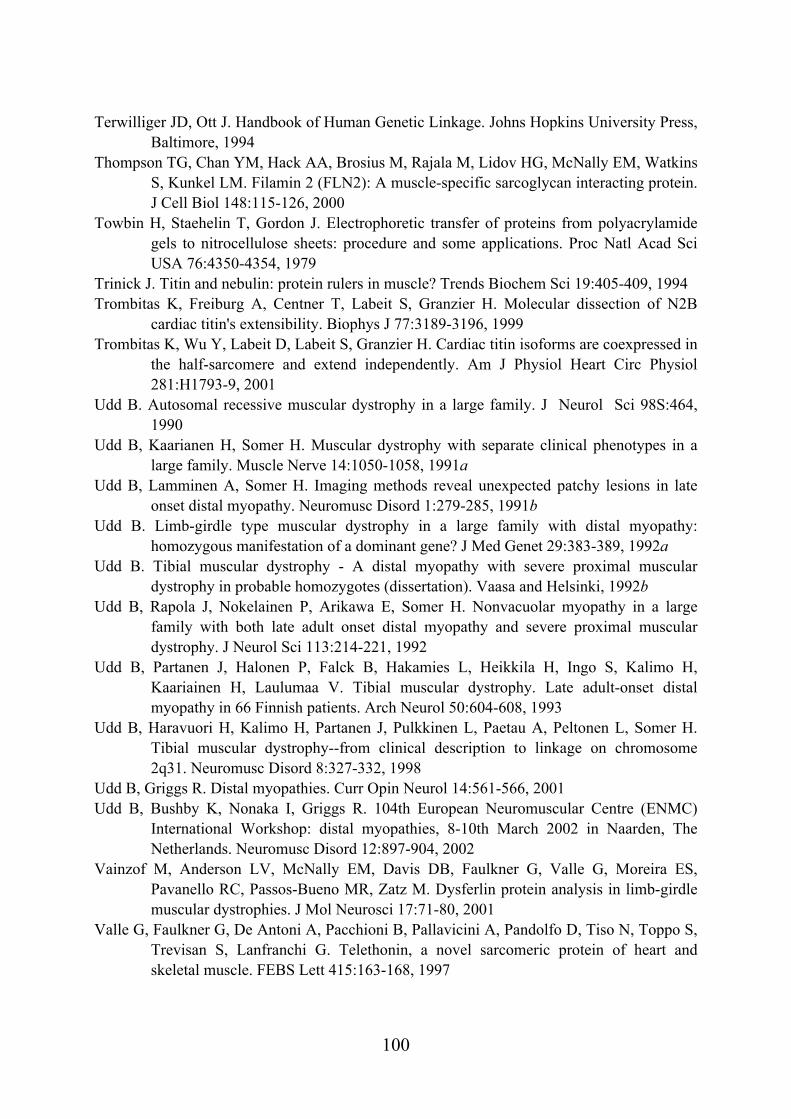
100
Terwilliger JD, Ott J. Handbook of Human Genetic Linkage. Johns Hopkins University Press, Baltimore, 1994
Thompson TG, Chan YM, Hack AA, Brosius M, Rajala M, Lidov HG, McNally EM, Watkins S, Kunkel LM. Filamin 2 (FLN2): A muscle-specific sarcoglycan interacting protein. J Cell Biol 148:115-126, 2000
Towbin H, Staehelin T, Gordon J. Electrophoretic transfer of proteins from polyacrylamide gels to nitrocellulose sheets: procedure and some applications. Proc Natl Acad Sci USA 76:4350-4354, 1979
Trinick J. Titin and nebulin: protein rulers in muscle? Trends Biochem Sci 19:405-409, 1994 Trombitas K, Freiburg A, Centner T, Labeit S, Granzier H. Molecular dissection of N2B
cardiac titin's extensibility. Biophys J 77:3189-3196, 1999 Trombitas K, Wu Y, Labeit D, Labeit S, Granzier H. Cardiac titin isoforms are coexpressed in
the half-sarcomere and extend independently. Am J Physiol Heart Circ Physiol 281:H1793-9, 2001
Udd B. Autosomal recessive muscular dystrophy in a large family. J Neurol Sci 98S:464, 1990
Udd B, Kaarianen H, Somer H. Muscular dystrophy with separate clinical phenotypes in a large family. Muscle Nerve 14:1050-1058, 1991a
Udd B, Lamminen A, Somer H. Imaging methods reveal unexpected patchy lesions in late onset distal myopathy. Neuromusc Disord 1:279-285, 1991b
Udd B. Limb-girdle type muscular dystrophy in a large family with distal myopathy: homozygous manifestation of a dominant gene? J Med Genet 29:383-389, 1992a
Udd B. Tibial muscular dystrophy - A distal myopathy with severe proximal muscular dystrophy in probable homozygotes (dissertation). Vaasa and Helsinki, 1992b
Udd B, Rapola J, Nokelainen P, Arikawa E, Somer H. Nonvacuolar myopathy in a large family with both late adult onset distal myopathy and severe proximal muscular dystrophy. J Neurol Sci 113:214-221, 1992
Udd B, Partanen J, Halonen P, Falck B, Hakamies L, Heikkila H, Ingo S, Kalimo H, Kaariainen H, Laulumaa V. Tibial muscular dystrophy. Late adult-onset distal myopathy in 66 Finnish patients. Arch Neurol 50:604-608, 1993
Udd B, Haravuori H, Kalimo H, Partanen J, Pulkkinen L, Paetau A, Peltonen L, Somer H. Tibial muscular dystrophy--from clinical description to linkage on chromosome 2q31. Neuromusc Disord 8:327-332, 1998
Udd B, Griggs R. Distal myopathies. Curr Opin Neurol 14:561-566, 2001 Udd B, Bushby K, Nonaka I, Griggs R. 104th European Neuromuscular Centre (ENMC)
International Workshop: distal myopathies, 8-10th March 2002 in Naarden, The Netherlands. Neuromusc Disord 12:897-904, 2002
Vainzof M, Anderson LV, McNally EM, Davis DB, Faulkner G, Valle G, Moreira ES, Pavanello RC, Passos-Bueno MR, Zatz M. Dysferlin protein analysis in limb-girdle muscular dystrophies. J Mol Neurosci 17:71-80, 2001
Valle G, Faulkner G, De Antoni A, Pacchioni B, Pallavicini A, Pandolfo D, Tiso N, Toppo S, Trevisan S, Lanfranchi G. Telethonin, a novel sarcomeric protein of heart and skeletal muscle. FEBS Lett 415:163-168, 1997
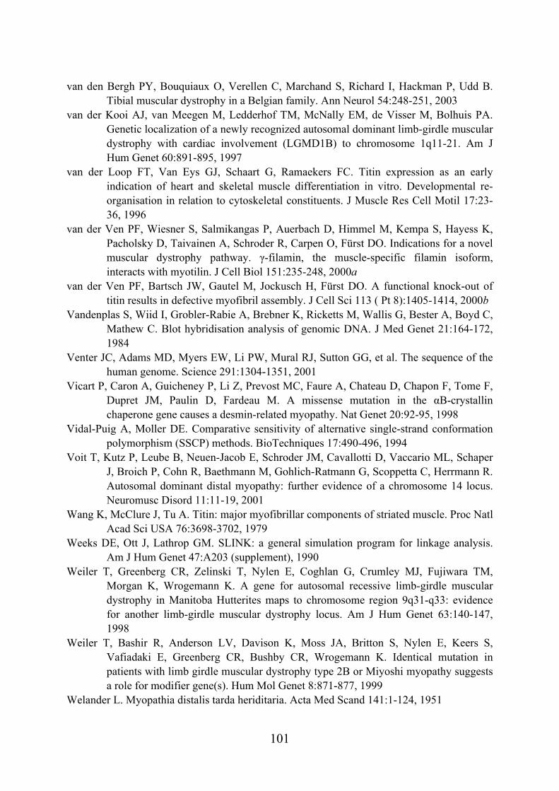
101
van den Bergh PY, Bouquiaux O, Verellen C, Marchand S, Richard I, Hackman P, Udd B. Tibial muscular dystrophy in a Belgian family. Ann Neurol 54:248-251, 2003
van der Kooi AJ, van Meegen M, Ledderhof TM, McNally EM, de Visser M, Bolhuis PA. Genetic localization of a newly recognized autosomal dominant limb-girdle muscular dystrophy with cardiac involvement (LGMD1B) to chromosome 1q11-21. Am J Hum Genet 60:891-895, 1997
van der Loop FT, Van Eys GJ, Schaart G, Ramaekers FC. Titin expression as an early indication of heart and skeletal muscle differentiation in vitro. Developmental re-organisation in relation to cytoskeletal constituents. J Muscle Res Cell Motil 17:23-36, 1996
van der Ven PF, Wiesner S, Salmikangas P, Auerbach D, Himmel M, Kempa S, Hayess K, Pacholsky D, Taivainen A, Schroder R, Carpen O, Fürst DO. Indications for a novel muscular dystrophy pathway. γ-filamin, the muscle-specific filamin isoform, interacts with myotilin. J Cell Biol 151:235-248, 2000a
van der Ven PF, Bartsch JW, Gautel M, Jockusch H, Fürst DO. A functional knock-out of titin results in defective myofibril assembly. J Cell Sci 113 ( Pt 8):1405-1414, 2000b
Vandenplas S, Wiid I, Grobler-Rabie A, Brebner K, Ricketts M, Wallis G, Bester A, Boyd C, Mathew C. Blot hybridisation analysis of genomic DNA. J Med Genet 21:164-172, 1984
Venter JC, Adams MD, Myers EW, Li PW, Mural RJ, Sutton GG, et al. The sequence of the human genome. Science 291:1304-1351, 2001
Vicart P, Caron A, Guicheney P, Li Z, Prevost MC, Faure A, Chateau D, Chapon F, Tome F, Dupret JM, Paulin D, Fardeau M. A missense mutation in the αB-crystallin chaperone gene causes a desmin-related myopathy. Nat Genet 20:92-95, 1998
Vidal-Puig A, Moller DE. Comparative sensitivity of alternative single-strand conformation polymorphism (SSCP) methods. BioTechniques 17:490-496, 1994
Voit T, Kutz P, Leube B, Neuen-Jacob E, Schroder JM, Cavallotti D, Vaccario ML, Schaper J, Broich P, Cohn R, Baethmann M, Gohlich-Ratmann G, Scoppetta C, Herrmann R. Autosomal dominant distal myopathy: further evidence of a chromosome 14 locus. Neuromusc Disord 11:11-19, 2001
Wang K, McClure J, Tu A. Titin: major myofibrillar components of striated muscle. Proc Natl Acad Sci USA 76:3698-3702, 1979
Weeks DE, Ott J, Lathrop GM. SLINK: a general simulation program for linkage analysis. Am J Hum Genet 47:A203 (supplement), 1990
Weiler T, Greenberg CR, Zelinski T, Nylen E, Coghlan G, Crumley MJ, Fujiwara TM, Morgan K, Wrogemann K. A gene for autosomal recessive limb-girdle muscular dystrophy in Manitoba Hutterites maps to chromosome region 9q31-q33: evidence for another limb-girdle muscular dystrophy locus. Am J Hum Genet 63:140-147, 1998
Weiler T, Bashir R, Anderson LV, Davison K, Moss JA, Britton S, Nylen E, Keers S, Vafiadaki E, Greenberg CR, Bushby CR, Wrogemann K. Identical mutation in patients with limb girdle muscular dystrophy type 2B or Miyoshi myopathy suggests a role for modifier gene(s). Hum Mol Genet 8:871-877, 1999
Welander L. Myopathia distalis tarda heriditaria. Acta Med Scand 141:1-124, 1951
102
Welander L. Homozygous appearance of distal myopathy. Acta Genet 7:321, 1957 Werle E, Schneider C, Renner M, Volker M, Fiehn W. Convenient single-step, one tube
purification of PCR products for direct sequencing. Nucleic Acids Res 22:4354-4355, 1994
Winegrad S. Myosin binding protein C, a potential regulator of cardiac contractility. Circ Res 86:6-7, 2000
Xu X, Meiler SE, Zhong TP, Mohideen M, Crossley DA, Burggren WW, Fishman MC. Cardiomyopathy in zebrafish due to mutation in an alternatively spliced exon of titin. Nat Genet 30:205-209, 2002
Yamasaki R, Berri M, Wu Y, Trombitas K, McNabb M, Kellermayer MS, Witt C, Labeit D, Labeit S, Greaser M, Granzier H. Titin-actin interaction in mouse myocardium: passive tension modulation and its regulation by calcium/S100A1. Biophys J 81:2297-2313, 2001
Yamasaki R, Wu Y, McNabb M, Greaser M, Labeit S, Granzier H. Protein kinase A phosphorylates titin's cardiac-specific N2B domain and reduces passive tension in rat cardiac myocytes. Circ Res 90:1181-1188, 2002
Yoshida T, Pan Y, Hanada H, Iwata Y, Shigekawa M. Bidirectional signaling between sarcoglycans and the integrin adhesion system in cultured L6 myocytes. J Biol Chem 273:1583-1590, 1998
Young P, Ferguson C, Banuelos S, Gautel M. Molecular structure of the sarcomeric Z-disk: two types of titin interactions lead to an asymmetrical sorting of alpha-actinin. EMBO J 17:1614-1624, 1998
Young P, Gautel M. The interaction of titin and alpha-actinin is controlled by a phospholipid-regulated intramolecular pseudoligand mechanism. EMBO J 19:6331-6340, 2000
Young P, Ehler E, Gautel M. Obscurin, a giant sarcomeric Rho guanine nucleotide exchange factor protein involved in sarcomere assembly. J Cell Biol 154:123-136, 2001
Zhou D, Birkenmeier CS, Williams MW, Sharp JJ, Barker JE, Bloch RJ. Small, membrane-bound, alternatively spliced forms of ankyrin 1 associated with the sarcoplasmic reticulum of mammalian skeletal muscle. J Cell Biol 136:621-631, 1997
Zimprich F, Djamshidian A, Hainfellner JA, Budka H, Zeitlhofer J. An autosomal dominant early adult-onset distal muscular dystrophy. Muscle Nerve 23:1876-1879, 2000
Zou P, Gautel M, Geerlof A, Wilmanns M, Koch MH, Svergun DI. Solution scattering suggests cross-linking function of telethonin in the complex with titin. J Biol Chem 278:2636-2644, 2003
Åhlberg G, von Tell D, Borg K, Edström L, Anvret M. Genetic linkage of Welander distal myopathy to chromosome 2p13. Ann Neurol 46:399-404, 1999