Embed Size (px)
Citation preview
Molecular Pathways
Molecular Pathways: BCR-ABL
Daniela Cilloni and Giuseppe Saglio
AbstractAberrant tyrosine kinase activity plays a critical role in many hematologic disorders, including chronic
myeloid leukemia characterized by the constitutive activity of BCR-ABL. ABL therefore represents a crucial
target for new therapeutic strategies. Here, we summarize the molecular pathways that are abnormally
activated by the oncoprotein. Such pathways may provide additional opportunities to develop new drugs
to overcome the resistance to tyrosine kinase inhibitors. In particular, the phosphoinositide 3-kinase
(PI3K)/AKTpathway canbe effectively blockedbymTOR inhibitors, and several compounds canhit theRAS
pathway and the resulting mitogen-activated protein (MAP) extracellular signal-regulated kinase (ERK)1/2
(MEK) andMAPkinase activation. Furthermore,mitotic kinases canbe blocked byAurora kinase inhibitors,
and Pim kinases can be blocked by selective serine-threonine kinase inhibitors. Finally, the abnormal
pathways that sustain the self-renewal of leukemic stem cells are described as possible targets to completely
eradicate leukemic clones. Such pathways include the Hedgehog pathway, which can be blocked by
Smoothened inhibitors, and the CXCR4/SDF1 axis, which can be targeted by specific antagonists. Clin
Cancer Res; 18(4); 1–8. �2011 AACR.
Background
Chronic myeloid leukemia (CML) is a clonal myelopro-liferative disorder characterized by the Philadelphia (Ph)chromosome, which results from t(9;22)(q34;q11) bal-anced reciprocal translocation (1). The molecular conse-quence of the Ph chromosome is the generation of the BCR-ABL oncogene that encodes for the chimeric BCR-ABLoncoprotein,with constitutive kinase activity that promotesthe growth advantage of leukemic cells (2).The deregulated tyrosine kinase activity of BCR-ABL has
been shown to be necessary and sufficient to maintain theleukemia phenotype of CML (3–5). Activation of the ABLtyrosine kinase is a primary event in the genesis of CML, asshown by the retrovirally mediated insertion of a humanBCR-ABL gene into murine hematopoietic stem cells andthe creationofBCR-ABL transgenicmice (3). This representsa critical issue in the effort to design molecular therapies.
BCR-ABL oncogenetic pathwayThe ABL protein physiologically shuttles between the
nucleus and the cytoplasm; however, when fused to BCR,the oncoprotein loses this property and is mainly retainedwithin the cytoplasm, where it interacts with themajority ofproteins involved in the oncogenic pathway. ABL tyrosine
kinase activity is constitutively activated by the juxtaposi-tion of BCR, thus favoring dimerization or tetramerizationand subsequent autophosphorylation. This increases thenumber of the phosphotyrosine residues on BCR-ABL and,as a consequence, the binding sites for the SH2 domains ofother proteins (6, 7).
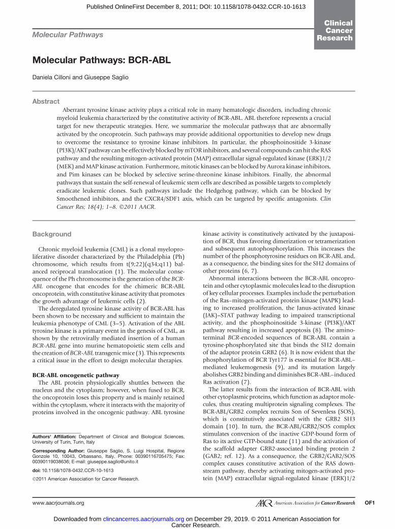
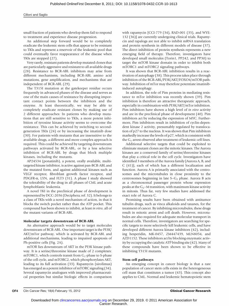
Abnormal interactions between the BCR-ABL oncopro-tein and other cytoplasmicmolecules lead to the disruptionof key cellular processes. Examples include the perturbationof the Ras–mitogen-activated protein kinase (MAPK) lead-ing to increased proliferation, the Janus-activated kinase(JAK)–STAT pathway leading to impaired transcriptionalactivity, and the phosphoinositide 3-kinase (PI3K)/AKTpathway resulting in increased apoptosis (8). The amino-terminal BCR-encoded sequences of BCR-ABL contain atyrosine-phosphorylated site that binds the SH2 domainof the adaptor protein GRB2 (6). It is now evident that thephosphorylation of BCR Tyr177 is essential for BCR-ABL–mediated leukemogenesis (9), and its mutation largelyabolishesGRB2binding and diminishes BCR-ABL–inducedRas activation (7).
The latter results from the interaction of BCR-ABL withother cytoplasmic proteins,which function as adaptormole-cules, thus creating multiprotein signaling complexes. TheBCR-ABL/GRB2 complex recruits Son of Sevenless (SOS),which is constitutively associated with the GRB2 SH3domain (10). In turn, the BCR-ABL/GRB2/SOS complexstimulates conversion of the inactive GDP-bound form ofRas to its active GTP-bound state (11) and the activation ofthe scaffold adapter GRB2-associated binding protein 2(GAB2; ref. 12). As a consequence, the GRB2/GAB2/SOScomplex causes constitutive activation of the RAS down-stream pathway, thereby activating mitogen-activated pro-tein (MAP) extracellular signal-regulated kinase (ERK)1/2
Authors' Affiliation: Department of Clinical and Biological Sciences,University of Turin, Turin, Italy
Corresponding Author: Giuseppe Saglio, S. Luigi Hospital, RegioneGonzole 10, 10043, Orbassano, Italy. Phone: 00390116705475; Fax:00390119038636; E-mail: [email protected]
doi: 10.1158/1078-0432.CCR-10-1613
�2011 American Association for Cancer Research.
ClinicalCancer
Research
www.aacrjournals.org OF1
Cancer Research. on December 29, 2019. © 2011 American Association forclincancerres.aacrjournals.org Downloaded from
Published OnlineFirst December 8, 2011; DOI: 10.1158/1078-0432.CCR-10-1613
(MEK) and MAP kinase proteins and resulting in abnormalcell proliferation. In addition, this complex activates thePI3K/AKT pathway (ref. 13, Fig. 1), which promotes survivalby suppressing the activity of the forkhead O (FOXO) tran-scription factor, and enhances cell proliferation by inducingp27 proteosomal degradation and by mTOR activation.
In addition, BCR-ABL, through PI3K/AKT/FOXO4 andfinally through upregulation of mTOR, potently blocksimportant cellular processes, such as autophagy. BCR-ABL may activate PI3K by more than one pathway,because Crk and Crkl have also been shown to connectBCR-ABL with PI3K (14, 15). Once activated, PI3K acti-vates AKT kinase, which serves as a key downstreameffector by exerting many cellular effects through the
phosphorylation of downstream substrates that regulatethe apoptotic machinery [e.g., Bad, caspase 9, Mdm2, andAsk1 (16)], resulting in prolonged survival and expansionof the abnormal clone.
Key transcription factors are involved in BCR-ABL signal-ing. Among these a key role is played by STAT1 and STAT5(signal transducer and activation of transcription), whichare constantly active in BCR-ABL–positive cell lines and inprimary cells from CML patients, contributing to the induc-tion of cytokine independence (17).
In normal cells, nuclear translocation of STATs occursexclusively after cytokine binding to receptors and ismediated by activation of the receptor-associated JAKkinases. By contrast, in CML, STATs seem to be activated
© 2011 American Association for Cancer Research© 2011 American Association for Cancer Research
MYC
PIM inhibitors
Bcr-Abl BCR
Y Y
Y177 Y1294
CRKL
ATP
P
SH3 SH2 SH1 Proline rich
NLS
DB AB
Bcr-Abl BCR
Y YATP
SH3 SH2 SH1 Proline rich
NLS
DB AB
RASGDP
JUN
Nucleus
Bcr-Abl inhibitors
MAPK
MEK1/2
ERK
RAF1
SOS
RASGTP
GAB2
SHCGRB2
MYC
STAT-1
STAT-5
STAT-1
STAT-5
Figure 1. Schematic representation of the molecular pathway activated by BCR-ABL. Bcr-Abl phosphorylation of BCR Tyr177 is essential for BCR-ABL–mediated leukemogenesis. The BCR-ABL/GRB2 complex recruits SOS,which is constitutively associatedwith theGRB2SH3 domain. TheBCR-ABL/GRB2/SOS complex stimulates conversion of the inactive GDP-bound form of Ras to its active GTP-bound state, and activation of the scaffold adapterGAB2. As a consequence, the GRB2/GAB2/SOS complex causes constitutive activation of the RAS downstream pathway, thereby activating MEK1/2and MAPK proteins and resulting in abnormal cell proliferation. In addition, this complex activates the PI3K/AKT pathway (see Fig. 2B).
Cilloni and Saglio
Clin Cancer Res; 18(4) February 15, 2012 Clinical Cancer ResearchOF2
Cancer Research. on December 29, 2019. © 2011 American Association forclincancerres.aacrjournals.org Downloaded from
Published OnlineFirst December 8, 2011; DOI: 10.1158/1078-0432.CCR-10-1613
in a JAK-independent manner through a direct associa-tion of STAT SH2 domains with phosphorylated tyrosineson BCR-ABL (18).Activation of STAT5 is at least partially responsible for
protection from programmed cell death through the upre-gulation of the antiapoptotic molecule BCL-xL togetherwith the inactivation of the proapoptotic molecule BADby AKT (8).Another postulated nuclear target of the transforming
activity of the BCR-ABL protein is the protooncogeneMYC,which is expressed at a high level in CML cells. MYCactivation seems to be independent of the RAS pathwaybut directly upregulated by theABL SH2 region (19). Severallines of evidence indicate that Myc is often overexpressed inblast crisis compared with the chronic phase, thus linkingMYC to progression (19). In vitro inhibition of c-Myc withantisense oligonucleotides or dominant-negative con-structs can inhibit BCR-ABL transformation or leukemo-genesis (19).All reported activated signaling pathways converge into a
unique terminal point: loss of control of proliferation andexpansion of the leukemic clone. Defining the relativecontribution of each signal transduction pathway to theleukemic process is an important area of research becausethe combination of a tyrosine kinase inhibitor (TKI) with adownstream inhibitor may prove to be a clinically success-ful strategy.Despite the seemingly endless expansion of the list of
pathways that are activated by BCR-ABL, and the increasingcomplexity that is being revealed in these pathways, it seemsthat all of the transforming functions of BCR-ABL dependon its tyrosine kinase activity (20). This precondition has anincredible intrinsic clinical potential with regard to thedevelopment of more-sophisticated targeted therapies.
Clinical–Translational Advances
Kinase inhibitorsImatinib, a small-molecule TKI, was the first drug to be
developed that was able to directly target BCR-ABL tyrosinekinase activity and to be tested in CML (21). In a short time,it has become the standard first-line therapy for all CMLpatients in early chronic phase based on the response ratesand the good tolerability shown (22).Despite the exciting results obtained with imatinib, after
8 years of follow-up, the cumulative complete cytogeneticresponse (CCyR) rate for first-line imatinib-treated patientswas 83%, the event-free survival rate was 81%, and theestimated overall survival rate was 85%.However, it should be considered that if a CCyR is not
achieved after 12 months of imatinib therapy, the proba-bility of progression or loss of response increases to 38%.Indeed, after 8 years of follow-up, it was concluded thatearly cytogenetic response is predictive of long-term out-come, and the cytogenetic response is therefore considered aprognostic indicator of lack of events, whereas nonoptimalresponders had a poorer prognosis (23). In addition, thedegree of response is critical. The IRIS study (22) also
confirmed that the achievement of a major molecularresponse at 12 months predicts a low risk of events orprogression, thus emphasizing the value of achieving amolecular response early during treatment.
On the basis of these data, it appears clear that for at leastone third of patients, the potential exists to improve onwhat can be achieved with standard imatinib treatment(400mg daily). Therefore, researchers have investigated thepossibility of inhibiting BCR-ABL activity in a more potentmanner to improve the results of first-line therapy. Thetherapeutic strategies assessed include modified imatinib-based regimens and first-line administration of next-gener-ation TKIs.
Several highly potent next-generation BCR-ABL inhibi-tors have been developed to overcome imatinib resistanceand improve the prognosis of patients with CML. Theseinclude novel and more potent multi-TKIs such asdasatinib, an orally bioavailable dual BCR-ABL and Srcinhibitor, and potent selective BCR-ABL inhibitors suchas nilotinib. Both nilotinib and dasatinib induce signi-ficant clinical responses. Dasatinib blocks BCR-ABL atlow concentrations but is less selective than imatinib.Similarly to imatinib, it inhibits BCR-ABL, Kit, and plate-let-derived growth factor receptor (PDGFR), but in con-trast, it also blocks Src, Tec, and Eph kinases, as well asmany other kinases. Nilotinib blocks BCR-ABL at lowerconcentrations than does imatinib, but, like imatinib, itappears to be more selective than dasatinib in targetingtyrosine kinases.
In phase II trials of patients with chronic-phase CML,dasatinib and nilotinib were associated with 2-year CCyRrates of 45% and 41%, respectively, in a group of ima-tinib-resistant patients (24). Following the impressiveresults obtained in patients with imatinib failure, dasati-nib and nilotinib were assessed in phase II studies ofpatients with newly diagnosed CML. The rate of CCyR at12 months was 80%, and the rate of major molecularresponse was 44%, thus showing superiority over imati-nib standard treatment and a favorable toxic profile (25).The results of these trials seem to indicate a superiorefficacy of the more-potent second-generation TKIs withrespect to imatinib, particularly in terms of higher rates ofCCyR and major molecular response, which, of moreimportance, also seem to lead to a decreased number ofdisease progressions. Although these results still need tobe fully evaluated after a longer period of follow-up, theuse of next-generation agents in the first-line therapy ofCML is likely to become a key area of clinical researchduring the next few years.
Considering the impressive results obtainedwith second-generation TKIs in first-line treatment, there are only a fewfields of interventionwe can still envisage thatmayoptimizetreatment of CML.
The first is to target what is commonly defined as theAchilles’ heel, which is represented by the emergence ofBCR-ABL mutant clones, including T315I, that arecompletely insensitive to second-generation TKIs (26).Although these insensitive clones occur very rarely, the
BCR-ABL
www.aacrjournals.org Clin Cancer Res; 18(4) February 15, 2012 OF3
Cancer Research. on December 29, 2019. © 2011 American Association forclincancerres.aacrjournals.org Downloaded from
Published OnlineFirst December 8, 2011; DOI: 10.1158/1078-0432.CCR-10-1613
small fraction of patients who develop them fail to respondto treatment and experience disease progression.
An additional step forward would be to completelyeradicate the leukemic stem cells that appear to be resistantto TKIs and represent a reservoir of the leukemic pool thatcould eventually favor reappearance of the disease whenTKIs are stopped (27).
Very rarely, resistant patients developmutated clones thatare particularly aggressive and resistant to all available drugs(28). Resistance to BCR-ABL inhibitors may arise fromdifferent mechanisms, including BCR-ABL amino acidmutations, gene amplification, and mechanisms that areindependent of BCR-ABL (29).
The T315I mutation at the gatekeeper residue occursfrequently in advanced phases of the disease and serves asone of the main causes of resistance by disrupting impor-tant contact points between the inhibitors and theenzyme. At least theoretically, we may be able tocompletely eradicate resistant clones by making use of2 different approaches: In patients who develop muta-tions that are still sensitive to TKIs, a more potent inhi-bition of tyrosine kinase activity seems to overcome theresistance. This can be achieved by switching to second-generation TKIs (24) or by increasing the imatinib dose(30). For patients with mutants that are insensitive to theavailable drugs, a different and more complex approach isrequired. This could be achieved by targeting downstreampathways activated by BCR-ABL, or by a less selectiveinhibition of BCR-ABL by drugs that block multiplekinases, including the mutants.
AP24534 (ponatinib), a potent, orally available, multi-targeted kinase inhibitor, is active against pan-BCR-ABL andthe mutated form, and against additional kinases such asVEGF receptor, fibroblast growth factor receptor, andPDGFR-a, LYN, and FLT3 (31). A phase I study showedthe tolerability of the drug in all phases of CML and acutelymphoblastic leukemia.
A novel TKI in the preclinical phase of development isrepresented byDCC-2036 (Deciphera; ref. 32). It belongs toa class of TKIs with a novel mechanism of action, in that itblocks the switch pocket rather than the ATP pocket. Thiscompound has been shown to inhibit, at least in vitro, all ofthe mutant variants of BCR-ABL.
Molecular targets downstream of BCR-ABLAn alternative approach would be to target molecules
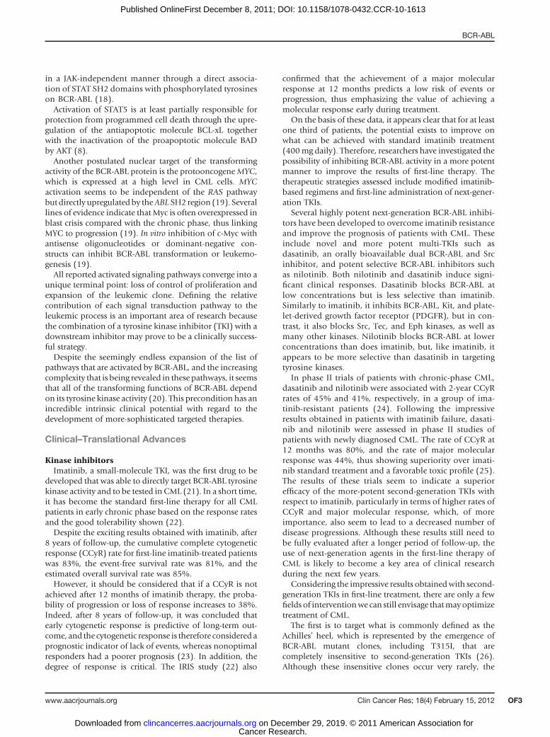
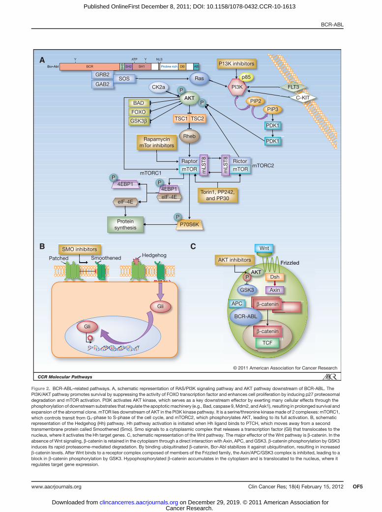
downstream of BCR-ABL. One important target is the PI3K/AKT/mTor pathway, which is activated by BCR-ABL andadditional mechanisms, leading to impaired apoptosis ofPh-positive cells (Fig. 2A).
mTOR lies downstream of AKT in the PI3K kinase path-way. It is a serine/threonine kinase made of 2 complexes:mTORC1, which controls transit from G1-phase to S-phaseof the cell cycle, and mTORC2, which phosphorylates AKT,leading to its full activation (33). Rapamycin (sirolimus)has emerged as a potent inhibitor ofmTORC signaling (34).Several rapamycin analogues with improved pharmaceuti-cal properties but similar biologic effects in comparison
with rapamycin [CCI-779 (34), RAD-001 (35), and WYE-132 (36)] are currently undergoing clinical trials. Rapamy-cin and rapalogs are not able to inhibit mRNA translationand protein synthesis in different models of disease (37).The direct inhibition of protein synthesis represents a newemerging field of therapy. Therefore, investigators havedeveloped small molecules (Torin1, PP242, and PP30) totarget the mTOR kinase domain in order to inhibit bothmTORC1 and mTORC2 signaling pathways.
It was shown that BCR-ABL inhibition results in a reac-tivation of autophagy (38). This process takes place throughinhibition of the BCR-ABL/PI3K/AKT/FOXO4/mTOR path-way. Inhibition of mTormay therefore potentiate imatinib-induced autophagy.
In addition, the role of Pim proteins in mediating resis-tance to mTor inhibition was recently shown (39). Piminhibition is therefore an attractive therapeutic approach,especially in combination with PI3K/AKT/mTor inhibition.Pim inhibitors have shown a high level of in vitro activityand are in the preclinical phase of development (40). Piminhibitors act by reducing the expression of MYC. Further-more, Pim inhibitors result in inhibition of cyclin-depen-dent kinase 2 activity, presumably regulated by transloca-tion of p27 to the nucleus. It was shown that Pim inhibitorsmarkedly increase the levels of p27,which is consistent withthe G1 arrest observed after treatment of leukemic cell lines.
Additional selective targets that could be exploited toeliminatemutant clones are themitotic kinases. The Aurorakinases are a conserved family of serine/threonine kinasesthat play a critical role in the cell cycle. Investigators haveidentified 3members of the Aurora family [Aurora A, B, andC (41)], each of which has a different localization andfunction. Aurora A is primarily associated with the centro-somes and the microtubules in close proximity to thecentrosomes beginning in late S–G2 phase. Aurora B actsas a chromosomal passenger protein whose expressionpeaks at theG2–M transition, withmaximumkinase activityin mitosis. Thus far, very few studies have addressed theexact role of Aurora C.
Promising results have been obtained with antitumortubulin drugs, such as vinca alkaloids and taxanes, for thetreatment of cancer. By inhibitingmicrotubules, these drugsresult in mitotic arrest and cell death. However, microtu-bules are also required for adequate molecular transport innormal cells. Therefore, investigators are searching for spe-cific targets to more selectively kill leukemic cells, and havedeveloped different Aurora kinase inhibitors (42), includ-ing hesperidin, MK-0457, ZM447439, MLN8054, andAZD1152. These inhibitors act by blocking enzymatic activ-ity by occupying the catalytic ATPbinding site (42).Manyofthese compounds have been shown to be effective ininhibiting T315I mutants.
Stem cell pathwaysAn emerging concept in cancer biology is that a rare
population of cancer stem cells exists in the heterogeneouscell mass that constitutes a tumor (43). This concept alsoapplies to CML. Normal and leukemic hematopoietic stem
Cilloni and Saglio
Clin Cancer Res; 18(4) February 15, 2012 Clinical Cancer ResearchOF4
Cancer Research. on December 29, 2019. © 2011 American Association forclincancerres.aacrjournals.org Downloaded from
Published OnlineFirst December 8, 2011; DOI: 10.1158/1078-0432.CCR-10-1613
© 2011 American Association for Cancer Research
P
P
P
P
P
P
Bcr-Abl
A
B C
BCR
Y YATP
SH
3
SH2 SH1 Proline rich
NLS
DB AB
CK2a
Ras
PI3K
PIP2
PIP3
PDK1
PDK1
C-KIT
FLT3
p85
AKT
TSC1 TSC2
Rheb
GSK3β
FOXO
Rapamycin
mTor inhibitors
mTORC2
4EBP14EBP1
Torin1, PP242,
and PP30
Protein
synthesis
Hedgehog
Gli
Gli
SmoothenedPatched
SMO inhibitors
P13K inhibitors
AKT inhibitors
Wnt
Dsh
APC
BCR-ABL
TCF
β-catenin
β-catenin
AxinGSK3
Frizzled
AKT
P70S6K
eIF-4EeIF-4E
mLS
T8
mLS
T8
mTORC1mTORmTOR
RictorRaptor
BAD
SOSGAB2
GRB2
Figure 2. BCR-ABL–related pathways. A, schematic representation of RAS/PI3K signaling pathway and AKT pathway downstream of BCR-ABL. ThePI3K/AKT pathway promotes survival by suppressing the activity of FOXO transcription factor and enhances cell proliferation by inducing p27 proteosomaldegradation and mTOR activation. PI3K activates AKT kinase, which serves as a key downstream effector by exerting many cellular effects through thephosphorylation of downstream substrates that regulate the apoptoticmachinery (e.g., Bad, caspase 9,Mdm2, and Ask1), resulting in prolonged survival andexpansion of the abnormal clone. mTOR lies downstream of AKT in the PI3K kinase pathway. It is a serine/threonine kinase made of 2 complexes: mTORC1,which controls transit from G1-phase to S-phase of the cell cycle, and mTORC2, which phosphorylates AKT, leading to its full activation. B, schematicrepresentation of the Hedgehog (Hh) pathway. Hh pathway activation is initiated when Hh ligand binds to PTCH, which moves away from a secondtransmembrane protein called Smoothened (Smo). Smo signals to a cytoplasmic complex that releases a transcription factor (Gli) that translocates to thenucleus, where it activates the Hh target genes. C, schematic representation of the Wnt pathway. The major effector of the Wnt pathway is b-catenin. In theabsence of Wnt signaling, b-catenin is retained in the cytoplasm through a direct interaction with Axin, APC, and GSK3. b-catenin phosphorylation by GSK3induces its rapid proteasome-mediated degradation. By binding ubiquitinated b-catenin, Bcr-Abl stabilizes it against ubiquitination, resulting in increasedb-catenin levels. After Wnt binds to a receptor complex composed of members of the Frizzled family, the Axin/APC/GSK3 complex is inhibited, leading to ablock in b-catenin phosphorylation by GSK3. Hypophosphorylated b-catenin accumulates in the cytoplasm and is translocated to the nucleus, where itregulates target gene expression.
BCR-ABL
www.aacrjournals.org Clin Cancer Res; 18(4) February 15, 2012 OF5
Cancer Research. on December 29, 2019. © 2011 American Association forclincancerres.aacrjournals.org Downloaded from
Published OnlineFirst December 8, 2011; DOI: 10.1158/1078-0432.CCR-10-1613
cell functions are defined by a common set of criticalstemness genes that regulate self-renewal (44). Hematopoi-etic stem cells (HSC) and leukemic stem cells (LSs) sharecommon features, including self-renewal, the capacity todifferentiate, resistance to apoptosis, and limitless prolifer-ative potential (44). Despite these similarities, however,several stemness factors, such as Notch, BMI-1, and Wntshow differential activation in HSC versus LSC. Such differ-ences could be exploited therapeutically (45).
It is important to consider that stemness in leukemia islinked to self-renewal. It is extremely likely that LSCs under-go self-renewal and are capable of recapitulating leukemia,and thus maintenance of the LSC pool would play a criticalrole in the success of any therapeutic intervention. Targetingstemness factors could be the key factor for a successfultherapy.
HSCs reside mainly in specialized bone marrow micro-environments calledHSCniches. The niche provides appro-priate signals thatmaintain the balance between self-renew-al and differentiation of stem/progenitor cells. Hedgehog(Hh) is one of the major regulators of the cell-fate decision.The Hh signal is critical for HSC and progenitor differen-tiation (Fig. 2B). Hedgehog pathway activation is initiatedwhen Hh ligand binds to Patched (PTCH), which movesaway froma second transmembraneprotein called Smooth-ened (Smo). Smo signals to a cytoplasmic complex thatreleases a transcription factor (Gli) that translocates to thenucleus, where it activates the Hh target genes (46). Leu-kemic cells are believed to rely on autocrine signaling, andthe Hh ligand produced by leukemic cells acts on neigh-boring leukemic cells to stimulate their growth or survival.
This model is supported by in vitro evidence that prolif-eration of tumor cell lines is accelerated by the addition ofHh ligand (47) and inhibited by the addition of Hh neu-tralizing antibody or by cyclopamine, an Hh pathwayantagonist. Finally, Hh also acts through a paracrine mech-anism. Hh ligand secreted by neoplastic cells signals to themicroenvironment, and the stromal cell compartment sig-nals back to the leukemic cells (48).
The clear link between the Hh pathway and humanleukemias led to an effort to identify small molecules toblock the pathway. Investigators have identified differentclasses of small-molecule Hh antagonists through cell-based screens using an Hh reporter assay (46).
Oneof thesemolecules, CUR61414, an aminoprolineHhantagonist, canblockelevatedHhsignalingactivity resultingfrom oncogenic mutations within the Patched-1 sequence(49). In addition, a novel series of Hh pathway inhibitors,1-amino-4-benzylphthalazines (Novartis, Institutes for Bio-medical Research, Cambridge,MA), has been identified andconfirmed to act via antagonism of the Smo receptor.
Wnt-mediated signaling has been shown to regulate cell-fate determination, proliferation, adhesion, migration, andpolarity during development (Fig. 2C). In addition to theircrucial role in embryogenesis, Wnt proteins and theirdownstream signaling molecules have been implicated inleukemogenesis. Wnt signaling has been implicated inmaintaining and amplifying stem cells, as well as in defin-
ing the stem-cell fate. The major effector of the Wntpathway is b-catenin. In the absence of Wnt signaling,b-catenin is retained in the cytoplasm through a directinteraction with Axin, APC, and GSK3. The phosphoryla-tion of b-catenin by GSK3 was shown to induce its rapidproteasome-mediated degradation (50). After Wnt bindsto a receptor complex composed of members of theFrizzled family, Axin/APC/GSK3 complex is inhibited, lead-ing to a block in b-catenin phosphorylation by GSK3.Hypophosphorylated b-catenin accumulates in the cyto-plasm and is translocated to the nucleus, where it regulatestarget gene expression (50). Deregulation of the Wnt signal-ing pathway is a hallmark of several types of leukemias (51).Different molecular mechanisms have been implicatedin abnormal activation of the Wnt pathway, including thefunctional loss of Wnt antagonists resulting in leukemogen-esis through deregulation of cell proliferation and differen-tiation. Although the Wnt signaling pathway is now recog-nized as one of the major players in the genesis of leukemia(52), it remains to be determined whether this pathway canbe targeted by drugs.
The niche is anatomically and functionally defined, andhas an endosteal and perivascular compartment within thebone marrow. Within the niche, critical bidirectional sig-nals ensure the regulation of normal HSC numbers andmaintenance of the quiescent long-term HSC pool. Adven-titial reticular cells, which are of mesenchymal origin, havebeen shown to alter stem-cell function. These cells expresshigh levels of the CX chemokine ligand12 [CXCL12; alsocalled stromal-derived factor 1 (SDF1)] and are locatedbetween vessels and bone (53). Targeted deletion of CXCR4(the ligand for CXCL12) led to a severe reduction in HSCnumbers and increased chemosensitivity (53). Interactionsbetween CXCR4 and SDF1 are important for the localiza-tion and retention of HSC and progenitor cells within theniche. In addition, they play a critical role in colonization ofthe bone marrow by HSCs during early development, asrevealed by the observation that SDF1-deficient embryoshave severely reduced HSC numbers and function (54).
The interaction between CXCR4 and CXCL12 appears tobe critical for leukemic cell maintenance. The leukemic cellsseem to have the unique capacity to directly modulate theniche at the expense of normal hematopoietic stem andprogenitor cells (55) by downregulating CXCL12 levels inthe areas of leukemia infiltration. Stem cell factor, which issecreted by the leukemic cells, is a niche regulator that leadsto abnormal engraftment of normal HSCs in the tumor-infiltrated microenvironment. Emerging evidence indicatesthat we may be able to target the complex interactionsbetween LSCs and their niche to selectively deplete therepopulating ability of LSCs and favor the normal HSCcounterparts. To achieve a selective eradication of LSCs, aniche-targeted therapy would require a high degree of selec-tivity toward the aberrant interaction between the leukemicclone and the microenvironment (56). These could includeself-renewal pathways such as Notch and Wnt, homingmechanisms, and cell adhesion molecules. Published datasupport the role of theNotch pathway in themaintenance of
Cilloni and Saglio
Clin Cancer Res; 18(4) February 15, 2012 Clinical Cancer ResearchOF6
Cancer Research. on December 29, 2019. © 2011 American Association forclincancerres.aacrjournals.org Downloaded from
Published OnlineFirst December 8, 2011; DOI: 10.1158/1078-0432.CCR-10-1613
leukemic stem cells and Wnt signaling in blast crisis CMLstem cells (57). Of importance, evidence indicates that bothof these pathways are at least partially regulated by the niche.Adhesion molecules are attractive candidate targets for LSC-specific therapies. Direct targeting of CXCR4 with chemicalcompoundsmay represent anotherdeveloping strategy (58).The CXCR4 antagonist AMD3465 has been shown to over-come the protective effects of stromal cells toward chemo-therapeutic agents. Further studies have also shown thattreatment with AMD3100, another CXCR4 antagonist thatis currently usedmainly tomobilize normal HSCs, results in
mobilization of leukemic cells and increased chemosensi-tivity (59). Furthermore, targeting CXCR4 appears to havebeneficial effects against leukemic clones.
Disclosure of Potential Conflicts of Interest
G. Saglio has received honoraria from the Speakers Bureau and is aconsultant to Novartis and Bristol-Myers Squibb. D. Cilloni disclosed nopotential conflicts of interest.
Received October 19, 2010; revised November 2, 2011; acceptedNovember 3, 2011; published OnlineFirst December 8, 2011.
References1. Nowell PC, Hungerford DA. A minute chromosome in human chronic
granulocitic leukemia. Science 1960;32:1497–501.2. Melo JV, Barnes DJ. Chronic myeloid leukaemia as amodel of disease
evolution in human cancer. Nat Rev Cancer 2007;7:441–53.3. Daley GQ, Van Etten RA, Baltimore D. Induction of chronic myelog-
enous leukemia in mice by 210 bcr-abl gene of the Philadelphiachromosome. Science 1990;87:6649–53.
4. Kelliher MA, McLaughlin J, Witte ON, Rosenberg N. Induction of achronic myelogenous leukemia-like syndrome in mice with v-abl andBCR/ABL. Proc Natl Acad Sci U S A 1990;87:6649–53.
5. Heisterkamp N, Jenster G, ten Hoeve J, Zovich D, Pattengale PK,Groffen J. Acute leukaemia in bcr/abl transgenic mice. Nature 1990;344:251–3.
6. Pendergast AM, Muller AJ, Havlik MH, Maru Y, Witte ON. BCRsequences essential for transformation by the BCR-ABL oncogenebind to the ABL SH2 regulatory domain in a non-phosphotyrosine-dependent manner. Cell 1991;66:161–71.
7. Pendergast AM, Quilliam LA, Cripe LD, Bassing CH, Dai Z, Li N,et al. BCR-ABL-induced oncogenesis is mediated by direct inter-action with the SH2 domain of the GRB-2 adaptor protein. Cell1993;75:175–85.
8. Melo JV, DeiningerMWN. Biology of chronicmyelogenous leukemia—signaling pathways of initiation and transformation. Hematol OncolClin North Am 2004;18:545–68, vii–viii.
9. Zhang X, Subrahmanyam R, Wong R, Gross AW, Ren R. The NH(2)-terminal coiled-coil domain and tyrosine 177 play important roles ininduction of a myeloproliferative disease in mice by Bcr-Abl. Mol CellBiol 2001;21:840–53.
10. Cortez D, Reuther GW, Pendergast AM. The Bcr-Abl tyrosine kinaseactivatesmitogenic signaling pathways and stimulates G1-to-S phasetransition in hematopoietic cells. Oncogene 1997;15:2333–42.
11. Ren R. Mechanisms of BCR-ABL in the pathogenesis of chronicmyelogenous leukaemia. Nat Rev Cancer 2005;5:172–83.
12. Sattler M, Mohi MG, Pride YB, Quinnan LR, Malouf NA, Podar K, et al.Critical role for Gab2 in transformation by BCR/ABL. Cancer Cell2002;1:479–92.
13. Skorski T, Kanakaraj P, Nieborowska-Skorska M, Ratajczak MZ, WenSC, Zon G, et al. Phosphatidylinositol-3 kinase activity is regulated byBCR/ABL and is required for the growth of Philadelphia chromosome-positive cells. Blood 1995;86:726–36.
14. Varticovski L, Daley GQ, Jackson P, Baltimore D, Cantley LC. Activa-tion of phosphatidylinositol 3-kinase in cells expressing abl oncogenevariants. Mol Cell Biol 1991;11:1107–13.
15. Jain SK, Susa M, Keeler ML, Carlesso N, Druker B, Varticovski L. PI 3-kinase activation in BCR/abl-transformed hematopoietic cells doesnot require interaction of p85 SH2 domains with p210 BCR/abl. Blood1996;88:1542–50.
16. Franke TF, Kaplan DR, Cantley LC. PI3K: downstream AKTion blocksapoptosis. Cell 1997;88:435–7.
17. Ilaria RL Jr, Van Etten RA. P210 and P190(BCR/ABL) induce thetyrosine phosphorylation and DNA binding activity of multiple specificSTAT family members. J Biol Chem 1996;271:31704–10.
18. Carlesso N, Frank DA, Griffin JD. Tyrosyl phosphorylation and DNAbinding activity of signal transducers and activators of transcription(STAT) proteins in hematopoietic cell lines transformed by Bcr/Abl. JExp Med 1996;183:811–20.
19. Sawyers CL, Callahan W, Witte ON. Dominant negative MYC blockstransformation by ABL oncogenes. Cell 1992;70:901–10.
20. Lugo TG, Pendergast AM,Muller AJ,WitteON. Tyrosine kinase activityand transformation potency of bcr-abl oncogene products. Science1990;247:1079–82.
21. O'Brien SG, Guilhot F, Larson RA, Gathmann I, Baccarani M, Cer-vantes F, et al. Imatinib compared with interferon and low-dosecytarabine for newly diagnosed chronic-phase chronic myeloid leu-kemia. N Engl J Med 2003;348:994–1004.
22. Druker BJ, Guilhot F, O'Brien SG, Gathmann I, Kantarjian H, Gatter-mann N, et al. IRIS Investigators. Five-year follow-up of patientsreceiving imatinib for chronic myeloid leukemia. N Engl J Med2006;355:2408–17.
23. Iacobucci I, Saglio G, Rosti G, Testoni N, Pane F, Amabile M, et al.GIMEMA Working Party on Chronic Myeloid Leukemia. Achieving amajor molecular response at the time of a complete cytogeneticresponse (CCgR) predicts abetter duration ofCCgR in imatinib-treatedchronicmyeloid leukemia patients. Clin Cancer Res 2006;12:3037–42.
24. Hochhaus A, Baccarani M, Deininger M, Apperley JF, Lipton JH,Goldberg SL, et al. Dasatinib induces durable cytogenetic responsesin patients with chronic myelogenous leukemia in chronic phase withresistance or intolerance to imatinib. Leukemia 2008;22:1200–6.
25. Saglio G, Kim DW, Issaragrisil S, le Coutre P, Etienne G, Lobo C, et al.ENESTnd Investigators. Nilotinib versus imatinib for newly diagnosedchronic myeloid leukemia. N Engl J Med 2010;362:2251–9.
26. O'Hare T, Deininger MWN, Eide CA, Clackson T, Druker BJ. Targetingthe BCR-ABL signaling pathway in therapy-resistant Philadelphiachromosome-positive leukemia. Clin Cancer Res 2011;17:212–21.
27. Carter BZ, Mak DH, Cortes J, Andreeff M. The elusive chronic myeloidleukemia stem cell: does it matter and how do we eliminate it? SeminHematol 2010;47:362–70.
28. KhorashadJS, deLavalladeH,Apperley JF,MilojkovicD,ReidAG,BuaM, et al. Finding of kinase domain mutations in patients with chronicphase chronic myeloid leukemia responding to imatinib may identifythose at high risk of disease progression. J Clin Oncol 2008;26:4806–13.
29. Quint�as-Cardama A, Kantarjian HM, Cortes JE. Mechanisms of pri-mary and secondary resistance to imatinib in chronic myeloid leuke-mia. Cancer Contr 2009;16:122–31.
30. Castagnetti F, Palandri F, Amabile M, Testoni N, Luatti S, Soverini S,et al. GIMEMA CML Working Party. Results of high-dose imatinibmesylate in intermediate Sokal risk chronic myeloid leukemia patientsin early chronic phase: a phase 2 trial of the GIMEMA CML WorkingParty. Blood 2009;113:3428–34.
31. O'Hare T, Shakespeare WC, Zhu X, Eide CA, Rivera VM, Wang F, et al.AP24534, a pan-BCR-ABL inhibitor for chronic myeloid leukemia,potently inhibits the T315I mutant and overcomes mutation-basedresistance. Cancer Cell 2009;16:401–12.
BCR-ABL
www.aacrjournals.org Clin Cancer Res; 18(4) February 15, 2012 OF7
Cancer Research. on December 29, 2019. © 2011 American Association forclincancerres.aacrjournals.org Downloaded from
Published OnlineFirst December 8, 2011; DOI: 10.1158/1078-0432.CCR-10-1613
32. Van Etten RA, Chan WW, Zaleskas VM, Walz C, Evangelista P,Lazarides K, et al. Switch pocket inhibitors of the ABL tyrosine kinase:distinct kinome inhibition profiles and in vivo efficacy in mousemodelsof CML and B-lymphoblastic leukemia induced by BCR-ABL T315I[abstract]. Blood 2008;112:576.
33. Jiang BH, Liu LZ. PI3K/PTEN signaling in angiogenesis and tumori-genesis. Adv Cancer Res 2009;102:19–65.
34. Geoerger B, Kerr K, Tang CB, Fung KM, Powell B, Sutton LN, et al.Antitumor activity of the rapamycin analog CCI-779 in human primitiveneuroectodermal tumor/medulloblastomamodels as single agent andin combination chemotherapy. Cancer Res 2001;61:1527–32.
35. Tamburini J, Chapuis N, Bardet V, Park S, Sujobert P, Willems L, et al.Mammalian target of rapamycin (mTOR) inhibition activates phospha-tidylinositol 3-kinase/Akt by up-regulating insulin-like growth factor-1receptor signaling in acute myeloid leukemia: rationale for therapeuticinhibition of both pathways. Blood 2008;111:379–82.
36. Yu K, Shi C, Toral-Barza L, Lucas J, Shor B, Kim JE, et al. Beyondrapalog therapy: preclinical pharmacology and antitumor activity ofWYE-125132, an ATP-competitive and specific inhibitor of mTORC1and mTORC2. Cancer Res 2010;70:621–31.
37. Tamburini J, Green AS, Bardet V, Chapuis N, Park S, Willems L, et al.Protein synthesis is resistant to rapamycin and constitutes a promisingtherapeutic target in acute myeloid leukemia. Blood 2009;114:1618–27.
38. Bellodi C, Lidonnici MR, Hamilton A, Helgason GV, Soliera AR,Ronchetti M, et al. Targeting autophagy potentiates tyrosine kinaseinhibitor-induced cell death in Philadelphia chromosome-positivecells, including primary CML stem cells. J Clin Invest 2009;119:1109–23.
39. Hammerman PS, Fox CJ, Birnbaum MJ, Thompson CB. Pim and Aktoncogenes are independent regulators of hematopoietic cell growthand survival. Blood 2005;105:4477–83.
40. Lin YW, Beharry ZM, Hill EG, Song JH, Wang W, Xia Z, et al. A smallmolecule inhibitor of Pim protein kinases blocks the growth of pre-cursor T-cell lymphoblastic leukemia/lymphoma. Blood 2010;115:824–33.
41. Carmena M, Earnshaw WC. The cellular geography of aurora kinases.Nat Rev Mol Cell Biol 2003;4:842–54.
42. Carvajal RD, Tse A, Schwartz GK. Aurora kinases: new targets forcancer therapy. Clin Cancer Res 2006;12:6869–75.
43. Witters LM, Witkoski A, Planas-Silva MD, Berger M, Viallet J, LiptonA. Synergistic inhibition of breast cancer cell lines with a dualinhibitor of EGFR-HER-2/neu and a Bcl-2 inhibitor. Oncol Rep2007;17:465–9.
44. Bonnet D. Normal and leukaemic stem cells. Br J Haematol 2005;130:469–79.
45. Huntly BJ, Gilliland DG. Cancer biology: summing up cancer stemcells. Nature 2005;7046:1169–70.
46. Chen JK, Taipale J, Cooper MK, Beachy PA. Inhibition of Hedgehogsignaling by direct binding of cyclopamine to Smoothened. GenesDev2002;16:2743–8.
47. Berman DM, Karhadkar SS, Maitra A, Montes De Oca R, GerstenblithMR, Briggs K, et al. Widespread requirement for Hedgehog ligandstimulation in growth of digestive tract tumours. Nature 2003;425:846–51.
48. Kawahara T, Kawaguchi-IharaN,Okuhashi Y, ItohM,NaraN, TohdaS.Cyclopamine and quercetin suppress the growth of leukemia andlymphoma cells. Anticancer Res 2009;29:4629–32.
49. Williams JA, Guicherit OM, Zaharian BI, Xu Y, Chai L, Wichterle H, et al.Identification of a small molecule inhibitor of the hedgehog signalingpathway: effects on basal cell carcinoma-like lesions. Proc Natl AcadSci U S A 2003;100:4616–21.
50. Wodarz A, Nusse R. Mechanisms of Wnt signaling in development.Annu Rev Cell Dev Biol 1998;14:59–88.
51. Kikuchi A. Tumor formation by geneticmutations in the components ofthe Wnt signaling pathway. Cancer Sci 2003;94:225–9.
52. Wang Y, Krivtsov AV, Sinha AU, North TE, Goessling W, Feng Z, et al.The Wnt/beta-catenin pathway is required for the development ofleukemia stem cells in AML. Science 2010;327:1650–3.
53. Sugiyama T, Kohara H, Noda M, Nagasawa T. Maintenance of thehematopoietic stemcell pool byCXCL12-CXCR4chemokine signalingin bone marrow stromal cell niches. Immunity 2006;25:977–88.
54. Ara T, Tokoyoda K, Sugiyama T, Egawa T, Kawabata K, Nagasawa T.Long-term hematopoietic stem cells require stromal cell-derived fac-tor-1 for colonizing bone marrow during ontogeny. Immunity 2003;19:257–67.
55. Colmone A, Amorim M, Pontier AL, Wang S, Jablonski E, Sipkins DA.Leukemic cells create bonemarrow niches that disrupt the behavior ofnormal hematopoietic progenitor cells. Science 2008;322:1861–5.
56. AdamsGB,Martin RP, Alley IR, Chabner KT, CohenKS, Calvi LM, et al.Therapeutic targeting of a stem cell niche. Nat Biotechnol 2007;25:238–43.
57. Jamieson CH, Ailles LE, Dylla SJ, Muijtjens M, Jones C, Zehnder JL,et al. Granulocyte-macrophage progenitors as candidate leukemicstem cells in blast-crisis CML. N Engl J Med 2004;351:657–67.
58. Zeng Z, Shi YX, Samudio IJ, Wang RY, Ling X, Frolova O, et al.Targeting the leukemia microenvironment by CXCR4 inhibition over-comes resistance to kinase inhibitors and chemotherapy in AML.Blood 2009;113:6215–24.
59. Nervi B, Ramirez P, Rettig MP, DiPersio JF. Chemosensitization ofacute myeloid leukemia (AML) following mobilization by the CXCR4antagonist AMD3100. Blood 2009;113:6206–14.
Cilloni and Saglio
Clin Cancer Res; 18(4) February 15, 2012 Clinical Cancer ResearchOF8
Cancer Research. on December 29, 2019. © 2011 American Association forclincancerres.aacrjournals.org Downloaded from
Published OnlineFirst December 8, 2011; DOI: 10.1158/1078-0432.CCR-10-1613
Published OnlineFirst December 8, 2011.Clin Cancer Res Daniela Cilloni and Giuseppe Saglio Molecular Pathways: BCR-ABL
Updated version
10.1158/1078-0432.CCR-10-1613doi:
Access the most recent version of this article at:
E-mail alerts related to this article or journal.Sign up to receive free email-alerts
Subscriptions
Reprints and
To order reprints of this article or to subscribe to the journal, contact the AACR Publications
Permissions
Rightslink site. (CCC)Click on "Request Permissions" which will take you to the Copyright Clearance Center's
.http://clincancerres.aacrjournals.org/content/early/2012/02/01/1078-0432.CCR-10-1613To request permission to re-use all or part of this article, use this link
Cancer Research. on December 29, 2019. © 2011 American Association forclincancerres.aacrjournals.org Downloaded from
Published OnlineFirst December 8, 2011; DOI: 10.1158/1078-0432.CCR-10-1613