Embed Size (px)
Citation preview
3 Quantum Photochemistry
3 - 1 . Properties of Photons and Electrons 35 3-2. Covalent Bonds
38
3-3. Photon Effects on Orbital Electrons 42 3-4. Dissipation of Photon Energy 43 3-5. Role of Molecular Orientation 49 3-6. Hypochromicity 51 3-7. Energy Transfer 54
General References 56
3-1. P R O P E R T I E S O F P H O T O N S A N D E L E C T R O N S
In this chapter we will consider what happens at the electronic level when a photon is absorbed by a molecule; why it is absorbed in the first place, and why the absorption event can lead to chemical reaction under the proper c i rcumstances . This should then provide some basis for the understanding of the photon induced changes in nucleic acids and proteins to be discussed later.
T h e transition from classical physics to atomic physics is intimately linked with the transition from cont inuous to discrete atomic phenomena. This is true for radiation as well as for matter . Whereas classical physics is concerned with light waves that are cont inuous in space and time (e.g., a spherical wave emitted by a light source) , atomic phenomena such as the interaction be tween radiation and matter can only be explained by assuming that radiation is emitted and absorbed as single quanta. According to Planck 's fundamental relationship (E = hv), the energy of these quanta (which we call photons) is proport ional to the frequency of a radiating dipole. Classically, the origin of all e lectromagnet ic radiat ions is in the oscilla-
35
36 3. QUANTUM PHOTOCHEMISTRY
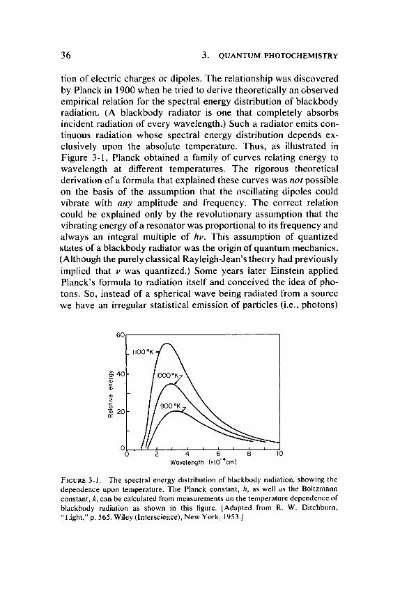
tion of electric charges or dipoles. T h e relationship was discovered by Planck in 1900 when he tried to derive theoretically an observed empirical relation for the spectral energy distribution of blackbody radiation. (A blackbody radiator is one that completely absorbs incident radiation of every wavelength.) Such a radiator emits continuous radiation whose spectral energy distribution depends exclusively upon the absolute temperature . Thus , as illustrated in Figure 3-1 , Planck obtained a family of curves relating energy to wavelength at different temperatures . T h e rigorous theoretical derivation of a formula that explained these curves was not possible on the basis of the assumption that the oscillating dipoles could vibrate with any amplitude and frequency. T h e correct relation could be explained only by the revolutionary assumption that the vibrating energy of a resonator was proportional to its frequency and always an integral multiple of hv. This assumption of quantized states of a blackbody radiator was the origin of quantum mechanics . (Although the purely classical Rayleigh-Jean's theory had previously implied that ν was quantized.) Some years later Einstein applied Planck's formula to radiation itself and conceived the idea of photons. So, instead of a spherical wave being radiated from a source we have an irregular statistical emission of particles (i.e., photons)
601
FIGURE 3 - 1 . The spectral energy distribution of blackbody radiation, showing the dependence upon temperature. The Planck constant, A, as well as the Boltzmann constant, k, can be calculated from measurements on the temperature dependence of blackbody radiation as shown in this figure. [Adapted from R. W. Ditchburn, "Light," p. 5 6 5 . Wiley (Interscience), New York, 1953 . ]
3-1. PROPERTIES OF PHOTONS AND ELECTRONS 37
into all directions of space. Of course photons also have wave aspects and these propert ies can be demons t ra ted in o ther experiments . T h e contradictory character is t ics of part icles and waves require that one cannot s imultaneously apply a particle descr iption and a wave description in the same exper iment . T h e wave model can be used to descr ibe exper iments on interference and diffraction while the particle model is necessary to explain certain other phenomena (e.g., photoelectr ic effect, the Compton effect, pair production, and pair annihilation). T h e theoretical principle of complementarity, formulated by Niels Bohr in 1928, resolves the wave-particle paradox by concluding that it is necessary to attr ibute both wave characterist ics and particle characteris t ics to electromagnetic radiation and also to material particles such as electrons.
Having discussed the particulate aspect of radiation we should also briefly consider the wave nature of electrons. Interference pat terns of electron beams (or even of helium ion beams for that matter) provide quite a convincing demonstra t ion of the wave propert ies of particles. T h e most successful theoretical t reatment of the motion of electrons in a toms has been that of wave mechanics . In fact the basic wave equation for the hydrogen atom has been solved to give exact answers —that is, to express precisely the probability of finding the electron at any given point in space. T h e solution for this problem comes from a considerat ion of the motion of an electron in the coulombic field of the positive proton nucleus. F o r more complex systems exact solutions are not obtainable but approximation methods have been worked out on the assumption that the solutions will resemble in form those for the hydrogen atom.
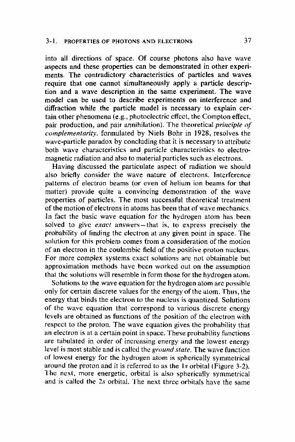
Solutions to the wave equation for the hydrogen atom are possible only for certain discrete values for the energy of the a tom. T h u s , the energy that binds the electron to the nucleus is quantized. Solutions of the wave equation that correspond to various discrete energy levels are obtained as functions of the position of the electron with respect to the proton. T h e wave equation gives the probability that an electron is at a certain point in space. These probability functions are tabulated in order of increasing energy and the lowest energy level is most stable and is called the ground state. T h e wave function of lowest energy for the hydrogen atom is spherically symmetrical around the proton and it is referred to as the \s orbital (Figure 3-2). T h e next, more energetic, orbital is also spherically symmetrical and is called the 2s orbital. T h e next three orbitals have the same
38 3. QUANTUM PHOTOCHEMISTRY
One Is orbital
Three perpendicular 2p orbitals
FIGURE 3-2. The orbitals for the hydrogen atom.
energy values as the 2s orbital, but they are symmetrical , respectively about the three perpendicular axes. They are called the 2px, 2ρμ, and 2pz orbitals, respectively. Each orbital has three quantum numbers (e.g., Cartesian coordinates) that describe the positioning of the electron in space. T o complete the description of the state of the electron we need a fourth quantum number called the spin. In addition to its orbital motion about the nucleus the electron can be considered to rotate about its own axis. Since rotation of a charged sphere about its axis is equivalent to a circular current , it gives rise to a magnetic field whose direction is in that of the axis. This may be positive or negative, depending upon which way the electron is spinning. Finally, in addition to the quantization of physical parameters a further restriction must be imposed that is known as the Pauli exclusion principle. It specifies that no two electrons in an atom can be in the same detailed state (i.e., no two electrons can have the same four quantum numbers) . Therefore, two electrons might occupy the same orbital but their spins would have to be in opposite directions. Also no more than two electrons can occupy the same orbital.
3-2. C O V A L E N T B O N D S
So far we have been considering only a single atom. Chemical
3-2. COVALENT BONDS 39
"^BP ^ ^ ^ ^ ^ ^ ^
Two hydrogen Is orbitals Addition of two hydrogen Is orbitals to form H*
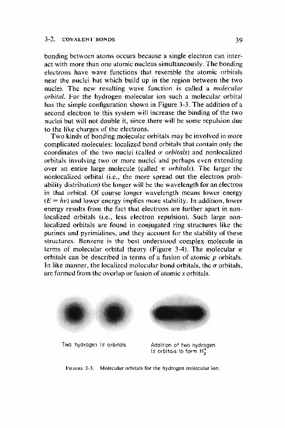
FIGURE 3-3. Molecular orbitals for the hydrogen molecular ion.
bonding between a toms occurs because a single electron can interact with more than one atomic nucleus simultaneously. T h e bonding electrons have wave functions that resemble the atomic orbitals near the nuclei but which build up in the region between the two nuclei. T h e new resulting wave function is called a molecular orbital. Fo r the hydrogen molecular ion such a molecular orbital has the simple configuration shown in Figure 3-3. T h e addition of a second electron to this system will increase the binding of the two nuclei but will not double it, since there will be some repulsion due to the like charges of the electrons.
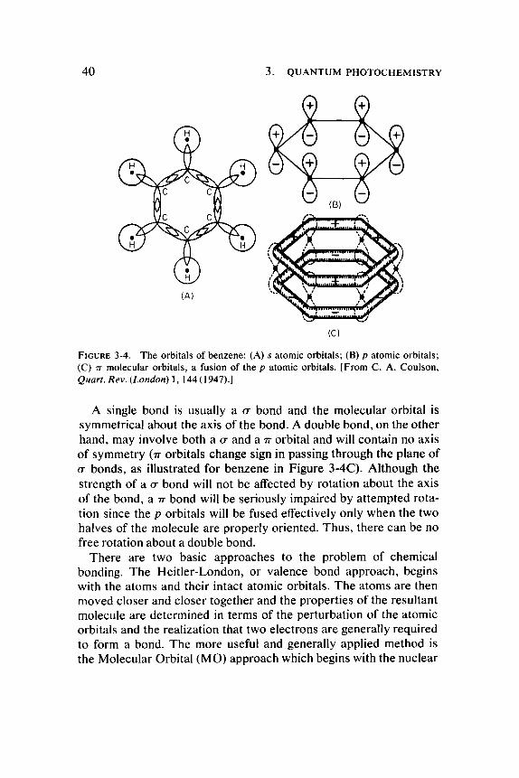
T w o kinds of bonding molecular orbitals may be involved in more complicated molecules: localized bond orbitals that contain only the coordinates of the two nuclei (called σ orbitals) and nonlocalized orbitals involving two or more nuclei and perhaps even extending over an entire large molecule (called π orbitals). T h e larger the nonlocalized orbital (i.e., the more spread out the electron probability distribution) the longer will be the wavelength for an electron in that orbital. Of course longer wavelength means lower energy (E = hv) and lower energy implies more stability. In addition, lower energy results from the fact that electrons are further apart in non-localized orbitals (i.e., less electron repulsion). Such large non-localized orbitals are found in conjugated ring structures like the purines and pyrimidines, and they account for the stability of these structures. Benzene is the best unders tood complex molecule in terms of molecular orbital theory (Figure 3-4). T h e molecular π orbitals can be described in terms of a fusion of atomic ρ orbitals. In like manner , the localized molecular bond orbitals, the σ orbitals, are formed from the overlap or fusion of atomic s orbitals.
40 3. QUANTUM PHOTOCHEMISTRY
FIGURE 3-4. The orbitals of benzene: (A) s atomic orbitals; (Β) ρ atomic orbitale; (C) π molecular orbitals, a fusion of the ρ atomic orbitals. [From C. A. Coulson, Quart. Rev. (London) 1, 144 (1947).]
A single bond is usually a σ bond and the molecular orbital is symmetrical about the axis of the bond. A double bond, on the other hand, may involve both a σ and a π orbital and will contain no axis of symmetry (π orbitals change sign in passing through the plane of σ bonds , as illustrated for benzene in Figure 3-4C). Al though the strength of a σ bond will not be affected by rotation about the axis of the bond, a π bond will be seriously impaired by a t tempted rotation since the ρ orbitals will be fused effectively only when the two halves of the molecule are properly oriented. Thus , there can be no free rotation about a double bond.
There are two basic approaches to the problem of chemical bonding. T h e Hei t ler-London, or valence bond approach, begins with the atoms and their intact atomic orbitals. T h e a toms are then moved closer and closer together and the propert ies of the resultant molecule are determined in terms of the perturbation of the atomic orbitals and the realization that two electrons are generally required to form a bond. T h e more useful and generally applied method is the Molecular Orbital (MO) approach which begins with the nuclear
3-2. COVALENT BONDS 41
framework. T h e nuclei of the a toms are placed at their equilibrium separations and the electrons are then fed into the resulting force field, observing the Pauli exclusion principle. It is then possible by various approximation methods to calculate the molecular energy levels. It should be mentioned that while distinct in approach, both the valence bond and M O method yield the same result.
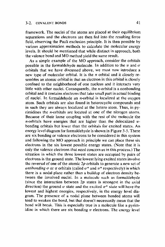
As a simple example of the M O approach, consider the orbitals possible in the formaldehyde molecule. In addition to the π and σ orbitals that we have discussed above , we must now introduce a new type of molecular orbital. It is the η orbital and it closely resembles an atomic orbital in that an electron in this orbital is closely confined to the neighborhood of one nucleus and it interacts very little with other nuclei. Consequent ly , the «-orbital is a nonbonding orbital and it contains electrons that take small part in actual binding of nuclei. In formaldehyde an «-orbital is localized at the oxygen atom. Such orbitals are also found in heterocyclic compounds and in such they are always localized at the hetero atom. T h u s , in py-rimidines the Ai-orbitals are located at one of the nitrogen a toms. Because of their loose coupling with the rest of the molecule the Az-orbitals have energies that are higher than the delocalized π-bonding orbitals but lower than the orbitals for excited states. An energy level diagram for formaldehyde is shown in Figure 3-5. There are six bonding or valence electrons to be considered in this system and following the M O approach in principle we can place these six electrons in the six lowest possible energy states. (Note that it is only the valence electrons that need concern us in this process.) T h e situation in which the three lowest states are occupied by pairs of electrons is the ground state. T h e lowest lying excited states involve the reversal of one of the atomic 2p orbitals to generate a new set of antibonding σ or π orbitals (called σ* and π * respectively) in which there is a nodal plane rather than a buildup of electron density between the involved nuclei. In a molecule such as formaldehyde (since the interaction be tween 2p s tates is strongest in the axial direction) the ground σ state and the excited σ* s tate will have the lowest and highest energies, respectively, in the energy level diagram. T h e presence of a nodal plane be tween bonded a toms will tend to weaken the bond, but that doesn ' t necessarily mean that the bond will break. This is especially true in a molecule like a pyrim-idine in which there are six bonding π e lectrons. T h e energy level
42 3. Q U A N T U M P H O T O C H E M I S T R Y
Ο Ο σ
FIGURE 3-5. Energy level diagram for formaldehyde. The symbols are defined in the text.
diagram as shown in Figure 3-5 can be useful for illustrating the transitions of electrons from one molecular orbital to another. However , it should be pointed out that such a diagram is not strictly correct since the actual energies of particular orbitals will depend upon which other orbitals have been vacated. Thus , the energy of an electron in the π * orbital will depend upon whether there is a vacancy in the η or π orbitals.
3-3. P H O T O N E F F E C T S O N O R B I T A L E L E C T R O N S
T h e general effect of an incoming photon of the appropriate energy will be to promote an electron to an orbital of greater energy (e.g., from a bonding or nonbonding orbital to an antibonding orbital). T h e energies involved in these transitions between low lying molecular states are those of the visible and ultraviolet quanta, hence the importance of these spectral regions to photochemistry.
Α π π * transition involves the excitation of a π e lectron into a π * state. Likewise, an mr* transition involves the excitation of an η electron into a π * orbital, crcr* transit ions can generally be ignored as a first approximation and need not concern us in the present discussion. Sigma electrons can not be neglected in detailed molecular orbital calculations, however , T h e π π * transit ions are responsible for the most intense absorption bands in molecular spectra. This is due to a high degree of overlap between the ground state and excited state wave functions and to a strong dipole oscillation. Al-
3-4. D I S S I P A T I O N O F P H O T O N E N E R G Y 43
though the π and π * orbitals differ in symmetry they cover approximately the same regions of the molecule. In contrast there is very poor spatial overlap be tween the localized nonbonding and the delocalized π * orbitals so that the transition probability for an mr* transition is correspondingly low. Because of this the absorption bands of mr* transitions are much less intense than those of ππ* transit ions by a factor of 10- to 100-fold. Also , the radiative lifetimes of ηπ* s tates should be greater than those of π π * states . H o w ever, the actual lifetime of the ηπ* state may be shortened considerably because of another property of these transit ions: a large electron displacement that leads to a high degree of polarization of charge in the transition. Such a polarization leads to increased chemical reactivity (as an electron donor or acceptor) and makes the molecule especially susceptible to the deexcitation processes that involve chemical reaction.
3-4. D I S S I P A T I O N O F P H O T O N E N E R G Y
Of course most photochemical excitations of molecules do not lead to chemical reaction. The re are a number of different pa thways that the energy of absorbed photons may take after it has appeared as internal energy of molecular excitation. Tha t the energy does not remain long in the absorbing molecule is illustrated by the fact that the color of most substances does not change during illumination. This must mean that the excited molecules return very quickly to the ground state where they can again absorb the same wavelengths as at the first instant of illumination.
What happens between the time a photon is absorbed and the return of the excited molecule to the stable ground state? Absorption occurs in about 1 0 "
15 seconds and the intrinsic lifetimes of
ππ* and ηπ* excited states are about 10~8 and 10~
6 seconds re
spectively. The simplest way in which the energy may be dissipated is in the reemission of light. If this happens within 10"
6 seconds after
absorption it is called fluorescence. However , not all of the energy comes back as light and the fluorescent quantum is thus at a longer wavelength than the one that was absorbed. T h e energy deficiency can be unders tood in terms of the Franck-Condon principle which recognizes that the time required for the absorption of photons is about Vioo that of the period of vibration of a molecule. So nuclei do
44 3. QUANTUM PHOTOCHEMISTRY
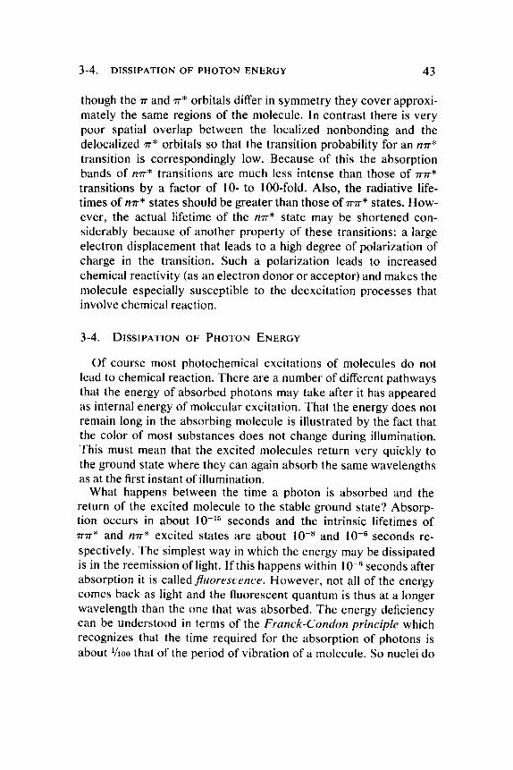
not appreciably alter their relative positions or kinetic energies during the act of absorption. T h e total energy of a molecule involves rotational and vibrational modes as well as electronic ones . T h e energies of the rotational modes are sufficiently smaller than the electronic ones so that they can be neglected, but those of vibration can not. T o take a simple example, consider the ground state of a diatomic molecule. The overall energy can be plotted as a function of some molecular coordinate such as the internuclear separation. Different quantized modes of vibration can be represented as levels within such a potential energy curve as shown in Figure 3-6. T h e first excited electronic state (singlet state) can be represented by another potential energy curve on the same energy scale. It should be noted that, in general, the equilibrium separation of nuclei will be different in the excited state because of the changed electronic distribution which results in an altered charge distribution in the molecule. (In fact, any photon induced transition will result in some change in the charge distribution in the excited molecule.) As a consequence of the Franck-Condon principle the most probable electronic transitions are those for which the average interatomic distances are the same in initial and final states. Thus , the absorption of light does not necessarily bring the molecule to the lowest vibrational state in the new electronic configuration because the interatomic distances in this state may not be the same as in the ground state. Instead, the primary excitation will leave the molecule in a vibrating state in which interatomic distances correspond most closely to those in the ground state. T h e excess vibrational energy in the excited state will be dissipated as heat by impacts with the solvent and other molecules. F luorescence will then generally occur from the lowest vibrational energy level of the excited singlet state as illustrated in Figure 3-6. Incidentally, this explains why the wavelength of fluorescence is independent of the wavelength of the exciting light. It is also evident from the foregoing that vibrational energy modes contribute to the spectral line width in absorption spectra.
Some molecules, particularly those with highly conjugated structures have the ability to radiate from an electronic state that is intermediate between the ground state and the fluorescent state. This type of luminescence is called phosphorescence and for a number of cases this has been shown to be a triplet state. T h e triplet state is
3-4. D I S S I P A T I O N O F P H O T O N E N E R G Y 45
Internal coordinate
FIGURE 3-6. A diagramatic representation of photon absorption and fluorescence. A representative internal coordinate for a diatomic molecule might be internuclear distance. A potential well might then schematically represent the possible inter-nuclear separations for a given electronic state of the molecule as shown in this figure. The minimum energy value or bottom of the well indicates the equilibrium internuclear separation. Numbered bands within these potential wells represent the different vibrational states of the molecule. (Rotational energy states could have been indicated in this figure as a fine structure of bands between the various vibrational levels.) At higher vibrational energy states the variation in internuclear separation increases until sufficient energy is attained to overcome the attraction between the nuclei, and this results in the breaking apart of the molecule. Note that the minimum internuclear distance approaches a limiting value as the vibrational energy increases. Vibrational deexcitation in the excited electronic state is indicated by the wavy line.
one in which the spin of the electron has flipped during the transition so that the system has two electrons with unpaired spins. N o violation of the Pauli principle is involved, however , since the spatial quantum numbers for the two electrons are different. Transi t ions from the triplet state to the ground state are forbidden (i.e., flipping of the electron's spin upon returning to ground state is forbidden). As with many forbidden things this does not really mean that such a transition will never occur but merely that the probability of its occurrence is very small, such that the lifetime of a triplet state may be as long as 10"
3 seconds or even a second. In terms of chemical
reaction a second is a rather long time (in fact 10"3 seconds is a
long time!). An important aspect of the triplet state is that it allows
46 3. QUANTUM PHOTOCHEMISTRY
more time for chemistry to occur before the return to the ground state. Although there does not seem to be good theoretical justification for the temperature dependence of triplet state lifetimes, it turns out that phosphorescence is strongly temperature dependent , such that at very low temperatures lifetimes of the order of several hours can be observed.
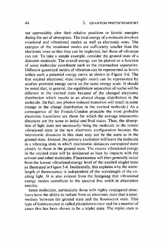
As with fluorescence we can illustrate the phenomenon of phosphorescence in terms of an energy potential diagram as shown in Figure 3-7. Because the excited electron and its (former) par tner have parallel spins, triplet states are of lower energy than their singlet counterpar ts . (Lower energy in this case results from a lower electron-electron repulsion by virtue of the greater spatial separation of the electrons in the triplet state.) Since the spin moment of an excited electron interacts with magnetic forces other than that of its ground state partner the probability of single-triplet transit ions and consequently the lifetimes of the triplet states are markedly influenced by the electrical and magnetic environment of the excited molecules.
The transition from the singlet to the triplet state can be seen in the potential energy diagram (Figure 3-7) in terms of a point at which the potential curve for the singlet state crosses that for the triplet state. Collisional deactivation can drop the energy to this point and
Internal coordinate
FIGURE 3-7 . Potential energy diagramatic representation for triplet state excitation and phosphorescence.
3-4. DISSIPATION OF PHOTON ENERGY 47
if further collisions do not carry the molecule too rapidly past this point, transition to the triplet state may occur. Such nonradiat ive transitions be tween states of different multiplicity are called inter-system crossing. T o restate: the obvious importance of the triplet state to photochemistry is its long lifetime. T h e longer a molecule is in an excited reactive state, the more likely that chemical reaction will occur.
There is another long-lived state that can sometimes occur following photon absorption and which leads to a delayed emission of a fluorescent energy photon. Such a state is not necessarily a triplet state, so it might be more generally referred to simply as a metastable state. Transi t ions from this state to the ground state are not merely forbidden but are apparently impossible. Therefore , the system must remain in this state until a quantum of sufficient energy is absorbed to restore it to the singlet state. Then the molecule can return to ground state by photon emission. Finally there is still another mode of delayed fluorescence observed in some organic crystals in which a population of molecules in triplet s tates can interact in such a way that two triplet molecules are conver ted into one molecule in the ground state and the other in the excited singlet state. T h e excited singlet then emits a photon in returning to the ground state.
We have discussed absorption and emission of photons in terms of oscillators. A useful parameter in the discussion of electronic absorption bands is that of the oscillator strength or absorption probability (/). Classically, the oscillator strength measures the effective number of electrons whose oscillations give rise to a particular absorption or emission band. This dimensionless quanti ty is determined simply by integrating the area under an experimentally obtained absorption band. This area is then proportional to the absorption probability and related to it by a constant . Thus :
The shapes of most absorption bands are such that the area can be approximated by the half-width of the band multiplied by the molar absorptivity.
where / is the oscillator strength e is the molar absorptivity ~v is the average wave number
48 3. QUANTUM PHOTOCHEMISTRY
The intrinsic probabilities of absorption and emission are proportional and thus, the intrinsic lifetime of an excited state varies inversely as the probability of absorption. T h e lifetime (τ) is given by the relationship τ = k/V
2f where the constant k is about 1.5 and ν is
the average wave number for the absorption band. Values of the oscillator strength range be tween 1.0 (very strong absorpt ion bands) to less than 0.1 for very weak and sometimes forbidden transi t ions.
It is important to realize that the intrinsic lifetime is that which would prevail if the only means for leaving the excited state were fluorescence. T h e actual lifetime of the excited state will be less than the intrinsic lifetime because pathways other than fluorescence are avilable for vacating the state. We have mentioned phosphorescence as one such pathway.
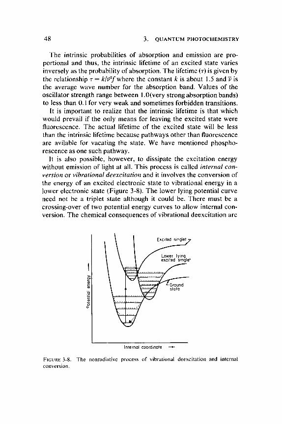
It is also possible, however , to dissipate the excitation energy without emission of light at all. This process is called internal conversion or vibrational deexcitation and it involves the conversion of the energy of an excited electronic state to vibrational energy in a lower electronic state (Figure 3-8). T h e lower lying potential curve need not be a triplet state although it could be. There must be a crossing-over of two potential energy curves to allow internal conversion. The chemical consequences of vibrational deexcitation are
Internal coordinate
FIGURE 3-8. The nonradiative process of vibrational deexcitation and internal conversion.
3-5. R O L E O F M O L E C U L A R O R I E N T A T I O N 49
important because the result is a " h o t " molecule of much the same type that might be produced by a high tempera ture collision activation. Thus , photochemistry may result in the same products obtained in an ordinary chemical reaction, but with the photon replacing thermal agitation to supply the necessary energy of activation. One might wonder , then, why an ultraviolet photon of 4.9 ev (2537 Ä) does not break chemical bonds if it is totally conver ted to vibrational energy, since it certainly carries more than enough energy to break most bonds. T h e evident reason is that the energy is not usually localized in a particular bond, but ra ther that it is distributed over many of the possible vibrational modes of many a toms in the molecule. Cases have been reported, however , where the absorbed electronic energy is transferred without dispersal to the vibrational energy of a particular bond, thus breaking the bond.
3-5. R O L E O F M O L E C U L A R O R I E N T A T I O N
At this point we should move from theory to exper iment . T h e r e is a UV-induced photoproduct of thymine that consists of two thymine residues linked covalently through their respect ive 5 and 6 carbon a toms to form a dimer. This photoproduct was first demonstrated in irradiated frozen solutions of thymine by Beukers and Berends. Thymine in solution at room tempera ture is quite resistant to photochemical alteration. The reactivity of the thymine in frozen solution is explained in large part by the fact that the thymine residues are favorably oriented for dimerization. T h e importance of orientation may relate to the lifetime of the excited state. In solution the reaction would be diffusion controlled and the lifetime of the excited state of thymine under these condit ions appears to be too short to allow for a high probability that two molecules of thymine will become properly oriented for dimer formation to occur. In support of this concept is the fact that thymine dimers formed in ice can be split to monomeric thymine by continued irradiation if the solution is allowed to thaw (see Section 4-5). T h e explanation is that as the dimers are split by U V (the formation and splitting of dimers are in equilibrium) the thymine monomers diffuse apart and are therefore not available for the reformation of dimers .
The yield of thymine dimers in solution can be enhanced if oxygen is excluded from the system. Oxygen and other paramagnet ic substances quench the triplet state. In the absence of the quenching
50 3. QUANTUM PHOTOCHEMISTRY
effect of oxygen, the lifetime of the triplet state is greatly lengthened, thereby giving the excited thymine molecules greater time to collide with other thymine molecules to form dimers. These results support the idea that the formation of the dimer can proceed through the triplet state of thymine. However , there is no oxygen effect on the production of thymine dimers in irradiated polynucleotides. In polynucleotides the thymine residues are already so close to one another that even in the presence of oxygen the dimerization rate is maximal. Unde r situations where the thymine residues are optimally oriented (i.e., thymine dimers that have been split in an ice matrix so that the thymine residues cannot diffuse apart) Lamola has shown that dimerization appears to go via the excited singlet state. Incidentally, oxygen does not quench the singlet state.
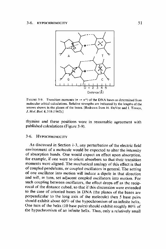
Although the orientation of molecules is important in the chemistry that follows photon absorption, it is also important in the photon absorption event itself. There is a net displacement of charge involved in any probable photon induced transition. This , of course , implies a direction for the excitation and in fact, an induced dipole. T h e direction and magnitude of this induced dipole is expressed in the vector quanti ty, the transition moment. T h e preferred direction for excitation is a direct consequence of the fact that the dipole has a preferred direction for absorption. T h e direction of the transition moment can sometimes be predicted from theory, but it is very difficult to determine it experimentally since determinations must be made on well-oriented systems (i.e., crystals) using polarized light. The re will be preferred directions for absorption depending upon the particular transitions allowed. Figure 3-9 shows the calculated transition moments for the four bases in D N A . N o t e that there are at least two ππ* transitions possible in each base. (The ηπ* transitions are perpendicular to the plane of the base and are much weaker.) The ππ* transition is considerably stronger in heterocyclics (f= 0.20 in thymidine) than in benzene (f= 0.0014).
An elegant determination of the transition moments of purines and pyrimidines has been performed upon single crystals of 1-methyl thymine and upon a mixed crystal of 1-methyl thymine and 9-methyl adenine by Stewart and Davidson. Thin sections (0.1 μ
thickness) were cut from these crystals by ul t ramicrotome and then examined in polarized ultraviolet light. T w o ππ* t ransit ions were determined, at 14° and 19° respectively, from nitrogen a tom one in
3-6. HYPOCHROMICITY 51
0 1 2 3 4
Distance (A)
FIGURE 3-9 . Transition moments (π -* π*) of the DNA bases as determined from molecular orbital calculations. Relative strengths are indicated by the lengths of the arrows shown in the planes of the bases. [Redrawn from H. DeVoe and I. Tinoco, J. Mol £/<?/. 4 , 5 1 8 ( 1 9 6 2 ) . ]
thymine and these positions were in reasonable agreement with published calculations (Figure 3-9).
3-6. HYPOCHROMICITY
As discussed in Section 1-3, any perturbat ion of the electric field environment of a molecule would be expected to alter the intensity of absorption bands . One would expect an effect upon absorpt ion, for example, if one were to orient absorbers so that their transition moments were aligned. T h e mechanical analogy of this effect is that of coupled pendulums, or coupled oscillators in general. T h e setting of one oscillator into motion will induce a dipole in that direction and will, in turn, set adjacent coupled oscillators into motion. Fo r such coupling between oscillators, the effect drops off as the reciprocal of the distance cubed, so that if this discussion were extended to the case of oriented bases in D N A (the planes of the bases are perpendicular to the long axis of the molecule) then 5 base pairs should exhibit about 6 0 % of the hypochromism of an infinite helix. One turn of the helix (10 base pairs) should exhibit roughly 8 0 % of the hypochromism of an infinite helix. T h u s , only a relatively small
5 2 3. QUANTUM PHOTOCHEMISTRY
number of bases in a polynucleotide strand are required to approximate the effect in a large polymer. T h e fact that hyperchromism is also observed upon the hydrolysis of single stranded D N A indicates that some base interaction (i.e., stacking) occurs in the single-strand configuration (Figure 1-8).
Although it is possible to calculate an expected hypochromism on the basis of coulombic interactions of the stacked bases alone, it has also been demonstra ted that solvent interactions with the absorbers can give rise to hypochromism. Fo r example, inosine exhibits less absorbance in the nonpolar solvent, acetonitrile, than in water . When the absorbance of isotactic polystyrene is compared to that of atactic or disordered configurations of this polymer in a nonpolar solvent, some agreement is seen with the theory that coulombic interactions of monomers is responsible for hypochromism. H o w ever, an additional hypochromic effect is seen if the comparison is carried out in a polar solvent.
It is evident that at least two factors are involved in the phenomenon of hypochromicity in D N A : (1) The partial alignment of transition moments by base stacking, and ( 2 ) the additional enhancement of the coupling of such interactions in polar solvents. The solvent itself may also affect the base stacking since this is a hydrophobic interaction. Such stacking is evident even for dinucleotides in polar solvents, but is not as evident if they are in nonpolar solvents. T h e synthetic polynucleotide, polyribouridylic acid (poly rU) , does not stack at all in aqueous solution above about 16°C; consequently it exhibits very little hyperchromicity. On the other hand polyribo-adenylic acid (poly rA) does stack to a significant degree and correspondingly exhibits a rather large hyperchromic effect when the stacking is disrupted thermally. Since poly rA also forms a multi-stranded hydrogen-bonded complex with itself, one might wonder what the contribution of hydrogen bonding might be to the hyperchromic effect. Evidence that this contribution is negligible is seen in the observation of hyperchromicity with polyribohydroxyethyl-adenylic acid which is sterically unable to form hydrogen-bonded base pairs. Thus , in D N A it is likely that the double-helix configuration is important to hypochromicity only by stabilizing the orientation of the stacked bases in the individual polynucleotide s trands.
Although hypochromism cannot be used in a simple way to look at the compositional character of D N A it is possible to demonstra te
3-6. HYPOCHROMICITY 53
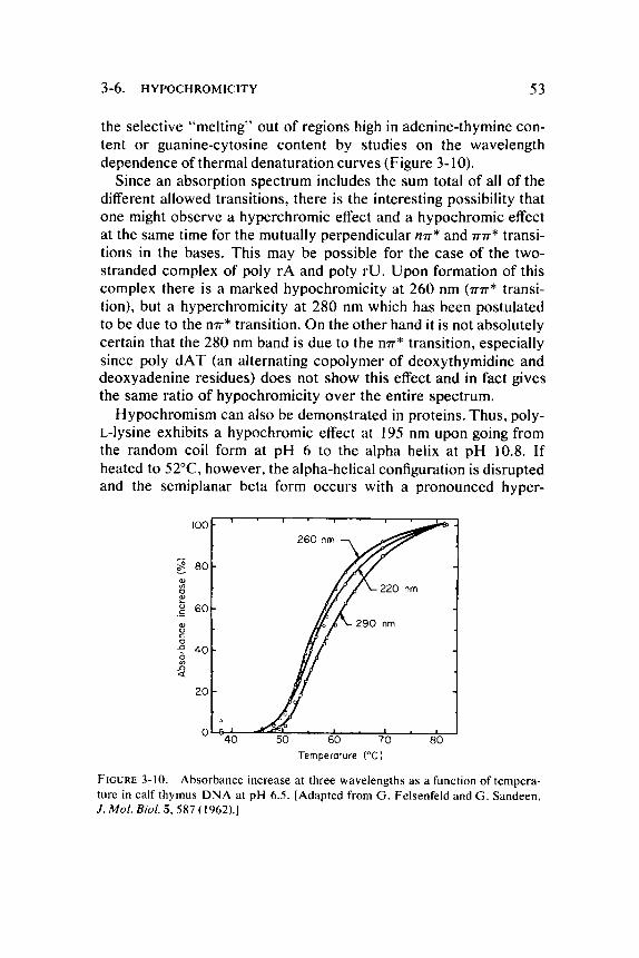
the selective "mel t ing" out of regions high in adenine-thymine content or guanine-cytosine content by studies on the wavelength dependence of thermal denaturat ion curves (Figure 3-10).
Since an absorption spectrum includes the sum total of all of the different allowed transitions, there is the interesting possibility that one might observe a hyperchromic effect and a hypochromic effect at the same time for the mutually perpendicular mr* and π π * transitions in the bases . This may be possible for the case of the two-stranded complex of poly rA and poly r U . U p o n formation of this complex there is a marked hypochromici ty at 260 nm ( π π * transition), but a hyperchromicity at 280 nm which has been postulated to be due to the η π * transition. On the other hand it is not absolutely certain that the 280 nm band is due to the η π * transition, especially since poly d A T (an alternating copolymer of deoxythymidine and deoxyadenine residues) does not show this effect and in fact gives the same ratio of hypochromici ty over the entire spectrum.
Hypochromism can also be demonst ra ted in proteins. T h u s , poly-L-lysine exhibits a hypochromic effect at 195 nm upon going from the random coil form at p H 6 to the alpha helix at p H 10.8. If heated to 52°C, however , the alpha-helical configuration is disrupted and the semiplanar beta form occurs with a pronounced hyper-
Temperature (°c)
FIGURE 3-10. Absorbance increase at three wavelengths as a function of temperature in calf thymus DNA at pH 6.5. [Adapted from G. Felsenfeld and G. Sandeen, J. Mol. Biol. 5 , 587 (1962).]
54 3. Q U A N T U M P H O T O C H E M I S T R Y
chromicity. T h e hyperchromic effect might be used in the determination of the helical content of proteins were it not for the interfering absorption bands of the aromatic amino acids.
3-7. E N E R G Y T R A N S F E R
There is another consequence of the coupling of absorbers that is of importance to photochemistry and that is the possibility of energy transfer. This represents a new mode for dissipation of absorbed photon energy which might be called "passing the buck ." A photon is absorbed in molecule A, the excitation energy is then transferred to molecule B, and molecule Β must then dispose of it. All of the usual avenues for dissipation of this energy are then available, as discussed in Section 3-4. If the energy of molecule Β is dissipated by fluorescence, the process is called sensitized fluorescence. It is best observed in situations in which the fluorescent spectra and absorption spectra of molecular species A and Β are different. Such an energy transfer process is distinct from the trivial possibility that a fluorescent quantum emitted by molecule A might be absorbed and then reemitted by molecule B. T h e transfer process is in competition with the other pathways for deexcitation. The time required for such transfer can be much shorter than fluorescence lifetimes and the efficiency of transfer can be much greater than even the maximum efficiency of fluorescence of the primary absorber . Sensitized fluorescence has been demonstra ted in a number of dye complexes and the distance of energy transfer has been shown to sometimes exceed 100 Ä although it never exceeds the wavelength of the sensitizing photons . Sensitized phosphorescence has also been demonstrated. It is not really necessary to think of the energy transfer process as involving the localization of the incident energy in one unit and then the subsequent transfer of this packet of energy to another unit. If there are strong electric field interactions between the units, then the transfer can be described in terms of a delocalized excitation energy that is distributed over the entire ensemble of units.
Evidence for energy transfer in D N A is the fact that the phosphorescence of native D N A is not the sum of the expected emission from individual nucleotides. Using electron spin resonance and optical emission it has been demonstra ted that the emission is pre-
3-7. ENERGY TRANSFER 55
dominantly from thymine. Samples were dissolved in an ethylene glycol-water glass at 77°K in order to stabilize triplet and other metastable states. It was found that at neutral p H , adenylic acid (AMP) and guanylic acid ( G M P ) , but not thymidylic acid ( T M P ) or cytidylic acid (CMP) , exhibit phosphorescence . T h e decay times were of the order of 1 and 2 seconds , respectively, for G M P and A M P . However , the observed decay time in D N A phosphorescence was only 0.3 seconds. Also, the quantum yield for phosphorescence in D N A was only 0.002 as compared with 0.02 for G M P . Poly d A T gave phosphorescence spectra and decay times quite similar to those for D N A although it was apparent that adenine contr ibuted less than 1 5 % of the observed phosphorescence . This all pointed very strongly to thymine as the phosphorescing moiety. U p o n closer examination of the photochemistry of thymine it was found that a triplet state with a 0.5 second lifetime could be obtained when the irradiation was performed at p H 9.8. U n d e r these c i rcumstances a proton is removed from nitrogen-1 position of thymine. Since this proton is involved in the normal hydrogen bonding of thymine to adenine in D N A , it might be expected that this proton could be shifted to adenine with the expected thymine excitation in D N A . In summary, it is apparent that photons absorbed in either thymine or adenine in D N A can be equally effective in promoting thymine phosphorescence .
T o anticipate our later discussion of biological effects of photons , we might cite the interesting example in which energy transfer apparently provides a protect ive effect in a biological inactivation process . The presence of the fluorescent dye , acridine orange, during U V irradiation enhanced the survival of E. coli strain B/r. It apparently acted as an energy sink for the harmless dissipation by sensitized fluorescence of photons absorbed in D N A . T h e enhanced survival of the bacteria was found to be somewhat greater if the irradiation was carried out in a nitrogen a tmosphere rather than in the presence of oxygen. Perhaps an additional inactivation effect is occurring in the presence of oxygen by photodynamic action (Chapter 9).
It should be emphasized again that environmenta l condit ions which alter the lifetimes of the excited s tates change the distribution of decay modes and the resultant photochemistry . It is conceivable that the quenching of triplet state excitation could reduce
56 3. QUANTUM PHOTOCHEMISTRY
U V mutagenesis in a biological system by reducing the yield of a particular photoproduct . In the next chapter we will give a number of specific examples of the effects of environment upon photochemical reactions.
G E N E R A L R E F E R E N C E S
D. Cram and G. Hammond, 'Organic Chemistry," Chapters 5 and 27. McGraw-Hill, New York, 1959.
G. M. Barrow, "Introduction to Molecular Spectroscopy." McGraw-Hill, New York, 1962.
J. B. Thomas, "Primary Photoprocesses in Biology." Wiley, New York, 1965. R. K. Clayton, "Molecular Physics in Photosynthesis," Chapter 7. Ginn (Blaisdell),
Boston, Massachusetts, 1965. J. C. Speakman, "Molecules." McGraw-Hill, New York, 1966. J. G. Calvert and J. N. Pitts, Jr., "Photochemistry." Wiley, New York, 1966. N.J. Turro, "Molecular Photochemistry." Benjamin, New York, 1967.


![Journal of Photochemistry & Photobiology, B: Biology · tion and migration [32–Thus, the scratch assay presents a simple34]. Journal of Photochemistry & Photobiology, B: Biology](https://img.pdfslide.net/doc/110x75/5f71d8ece961ec0ce1378c73/journal-of-photochemistry-photobiology-b-biology-tion-and-migration-32athus.jpg)


![Journal of Photochemistry & Photobiology, B: Biologybose.res.in/~skpal/papers/priya_JPBB1.pdf · Journal of Photochemistry & Photobiology, B: Biology 157 (2016) 105–112 ... [32]τrot](https://img.pdfslide.net/doc/110x75/5f71d8ece961ec0ce1378c74/journal-of-photochemistry-photobiology-b-skpalpaperspriyajpbb1pdf.jpg)