Embed Size (px)
Citation preview
786 HELVETICA CHIMICA ACTA - Vol. 80 (1997)
60. Molecular Recognition of NADP(H) and ATP by Macrocyclic Polyamines Bearing Acridine Groups
by Hicham Fenniri I), Mir Wais Hosseini I) , and Jean-Marie Lehn * Laboratoire de Chimie Supramoleculaire (CNRS URA 422), Institut Le Bel, Universite Louis Pasteur,
4, rue Blaise Pascal, 67000 F-Strasbourg.
(10.11.97)
The macrocyclic polyamine-based receptor BA bearing two acridine units makes use of combined electrostat- ic and stacking interactions for the binding of nucleotide polyphosphates and for the recognition of ATP and of NADPH (k, > 3 . lo8 M), with a high selectivity for NADPH vs. NADP (ca. lo3) and NAD(H) (> lo6). The binding properties of this receptor towards a variety of substrates led to its in vitro application as a fluorescent probe for ATP. BA also interacts strongly with nucleic acids as shown by spectrophotometric, spectrofluorimetic, and electrophoretic mobility methods.
1. Introduction. - The design of synthetic receptors for the nucleotide ATP and the dinucleotide NADP(H) represents an important target in molecular recognition chem- istry [l-121. A receptor that can bind with high affinity and selectivity to NADPH vs. NADP, or to ATP vs. other nucleotide phosphates (e.g. CTP, GTP, UTP, ADP, AMP) may allow the design of supramolecular catalysts or sensors [2] of interest for biotechnol- ogy and biomedical diagnosis [3]. The cases of ATP and NAD(P)H are particularly interesting, since they represent the ubiquitous energy source to all living organisms. Indeed, receptors and sensors for ATP and NADPH should allow the quantification of this energy in real time, in different tissues as a function of their activity, and depending on their physiological, physicochemical, and pathological states [4].
We describe here the design, synthesis, and physicochemical properties of the macro- cyclic receptor BA and its parent monoacridine compound MA [5], as well as their ability to bind a variety of nucleotide phosphates, dinucleotide phosphates, and polynucleotide phosphates.
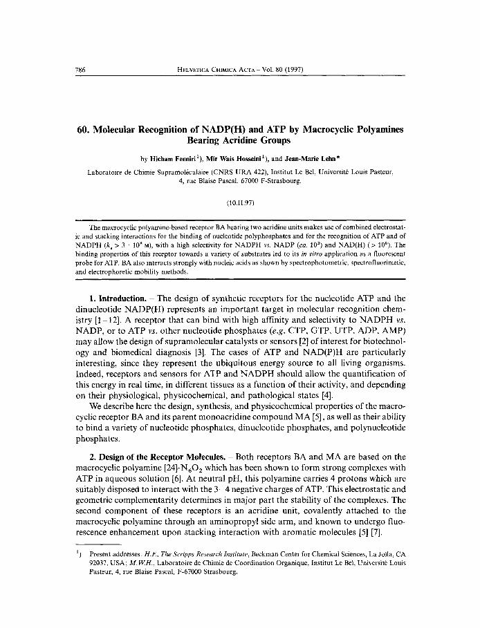
2. Design of the Receptor Molecules. - Both receptors BA and MA are based on the macrocyclic polyamine [24]-N,O, which has been shown to form strong complexes with ATP in aqueous solution [6] . At neutral pH, this polyamine carries 4 protons which are suitably disposed to interact with the 3-4 negative charges of ATP. This electrostatic and geometric complementarity determines in major part the stability of the complexes. The second component of these receptors is an acridine unit, covalently attached to the macrocyclic polyamine through an aminopropyl side arm, and known to undergo fluo- rescence enhancement upon stacking interaction with aromatic molecules [5] [7].
') Present addresses: H I , The Scripps Research Institute, Beckman Center for Chemical Sciences, La Jolla, CA 92037, USA; M . W H . , Laboratoire de Chimie de Coordination Organique, Institut Le Bel, Universite Louis Pasteur, 4, rue Blaise Pascal, F-67000 Strasbourg.
787
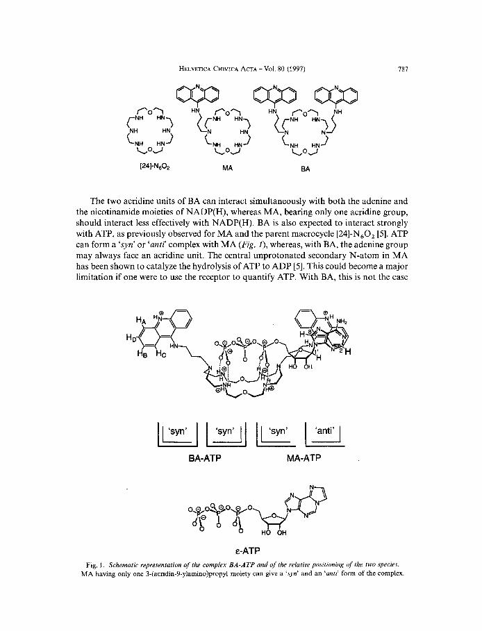
The two acridine units of BA can interact simultaneously with both the adenine and the nicotinamide moieties of NADP(H), whereas MA, bearing only one acridine group, should interact less effectively with NADP(H). BA is also expected to interact strongly with ATP, as previously observed for MA and the parent macrocycle [24]-N,O, [5]. ATP can form a ‘syn’ or ‘anti) complex with MA (Fig. I ) , whereas, with BA, the adenine group may always face an acridine unit. The central unprotonated secondary N-atom in MA has been shown to catalyze the hydrolysis of ATP to ADP [5 ] . This could become a major limitation if one were to use the receptor to quantify ATP. With BA, this is not the case
BA-ATP MA-ATP
E-ATP Fig. 1. Schematic representation of the complex BA-ATP and of the relative positioning of the two species.
MA having only one 3-(acridin-9-ylamino)propyl moiety can give a ‘syn’ and an ‘anti’ form of the complex.
788 HELVETICA CHIMICA ACTA - Vol. 80 (1997)
since the amine site responsible for this hydrolytic activity is now tertiary [8]. Finally, the fluorescence enhancement is greater upon complex formation with BA, in part as a result of partial quenching of its intrinsic fluorescence (see below). All these properties make BA a suitable candidate for its use as a fluorescent probe for ATP.
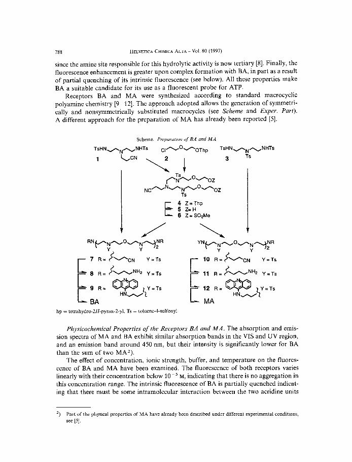
Receptors BA and MA were synthesized according to standard macrocyclic polyamine chemistry 19- 121. The approach adopted allows the generation of symmetri- cally and nonsymmetrically substituted macrocycles (see Scheme and Exper. Part). A different approach for the preparation of MA has already been reported [5].
Scheme. Preparation of’ BA and M A
TsHNdN-NHTS ~ 1 - ~ + 0 ~ h ~ TsHN + N ~ N H T ~
3 Ts
4 Z = T h p E 5 Z=H 6 Z=S02Me
11 R=<-NH2 Y=Ts ‘ 12 R = W
HN-$’ = Ts BA MA
hp = tetrahydro-2H-pyran-2-yl, Ts = toluene-4-sulfonyl
Physicochemical Properties of the Receptors BA and MA. The absorption and emis- sion spectra of MA and BA exhibit similar absorption bands in the VIS and UV region, and an emission band around 450 nm, but their intensity is significantly lower for BA than the sum of two MA2).
The effect of concentration, ionic strength, buffer, and temperature on the fluores- cence of BA and MA have been examined. The fluorescence of both receptors varies linearly with their concentration below lo-* M, indicating that there is no aggregation in this concentration range. The intrinsic fluorescence of BA is partially quenched indicat- ing that there must be some intramolecular interaction between the two acridine units
’) Part of the physical properties of MA have already been described under different experimental conditions, see [5 ] .
HELVETICA CHIMICA ACTA - Vol. 80 (1997) 789
leading to energy transfer. The existence of such a stacking interaction is further support- ed by NMR data (see Table 1) which indicate a substantially higher-field resonance for the aromatic protons of BA ( A 6 from + 0.14 to + 0.37 ppm) as compared to those of MA. Increasing the ionic strength (NaCl) induces a small increase in the fluorescence of BA (18 % at 50 mM NaCl), possibly resulting from decrease of the intramolecular inter- action between the two acridine units. Under the same conditions, MA undergoes a fluorescence quenching (13 % at 50 mM NaCl). Other anions lead to the same result (see Sect. 3). The effect of several buffer systems at various concentrations induces similar behavior, the fluorescence being dependent upon the nature of the buffer, its concentra- tion, or both. Except when indicated, 5 mM Tris acetate buffer (pH 7.6) was selected because it led to the highest fluorescence enhancements in the present study.
While the fluorescence intensity of BA is not altered by decreasing the temperature from 37 to O", that of MA increases by 68%.
3. Complexation Studies. - 3.1. Complexation of Nucleotide Phosphates: 'H- and M solution of BA in D,O (20",
pD 7 or 4) induces a higher upfield shift of the aromatic 'H-NMR signals of ATP than observed for MA ( A S from + 0.58 to + 0.62 pprn vs. + 0.30 to + 0.37 ppm), probably due to the possible formation of 'syn' and 'anti' complexes with MA (Fig. I ) . For the acridine protons, the opposite situation is observed ( A 6 from + 0.18 to + 0.30 ppm for MA and -0.04 to + 0.06 ppm for BA). This behavior is also in agreement with a preexisting interaction between the acridine units of BA, which is inhibited upon complex formation with ATP. Binding of ADP leads to similar effects (see Table f).
'P-NMR Studies. Addition of 1 equiv. of ATP to a
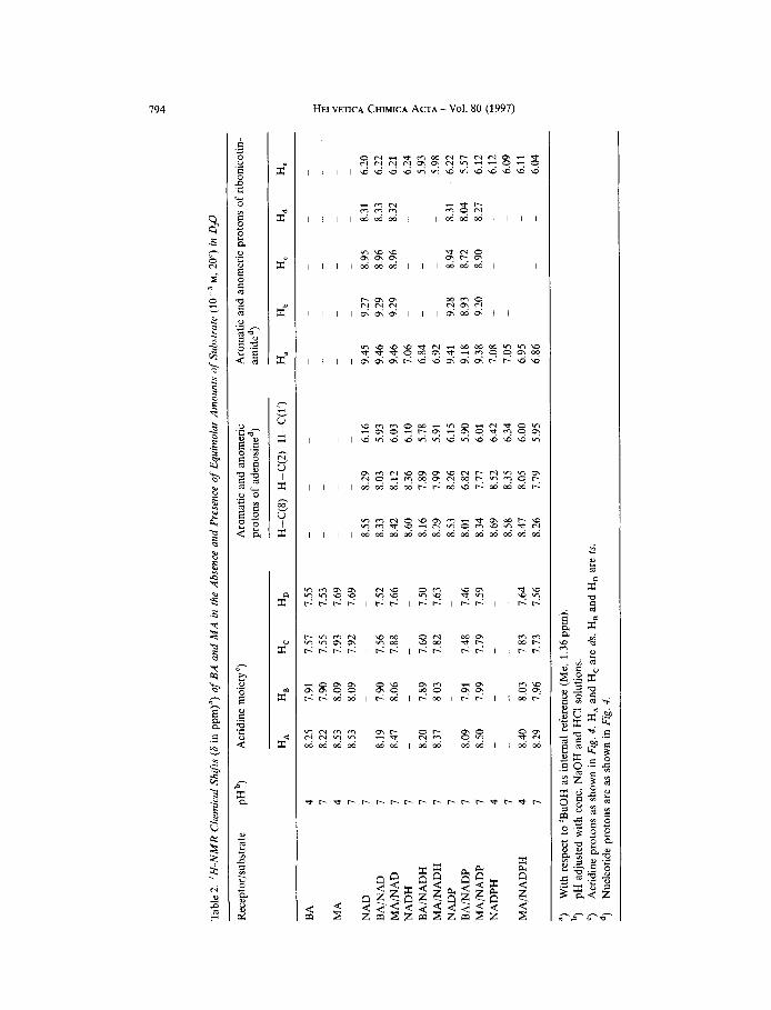
Table 1. 'H-NMR Chemical Shifts (6 in pprn)") of BA and M A in the Absence and Presence of Equimolar Amounts of Substrate M, 20") in D,O
Receptor/ pHb) Acridine moietyc) Aromatic and anomeric protons substrate of nucleotidesd)
H.4 HLI Hc H D H-C(8) H-C(2) H-C(l')
BA
MA
ATP BAIATP MAjATP ADP BAjADP BA/PPPe) MA/PPPe)
4 8.25 7.91 7.57 7.55 7 8.22 7.90 7.55 7.53 4 8.53 8.09 7.93 7.69 7 8.53 8.09 7.92 7.69 I 7 8.27 7.87 7.61 7.49 7 8.23 7.90 7.63 7.51
- - - -
I - - - - 1 8.16 7.78 7.52 7.42 4 8.16 7.17 1.41 7.39 4 8.49 8.09 7.88 7.68
- - -
- - -
- - -
8.66 8.38 6.26 8.08 7.76 5.67 8.36 8.01 5.91 8.65 8.38 6.26 8.03 7.15 5.71 - -
- -
~ ~~
") b,
") d, ") PPP = triphosphate.
With respect to 'BuOH as internal reference (Me, 1.36 ppm). pH adjusted with conc. NaOH and HCI solutions. Acridine protons as shown in Fig. 1, HA and H, are ds, H, and H, are 2s. Nucleotide protons are as shown in Fig. f ,
790 HELVETICA CHIMICA ACTA - Vol. 80 (1997)
The complexation of triphosphate (PPP) could not be studied at neutral pH because precipitation occurred at the concentration used M). The 'H-NMR upfield shifts of the acridine moiety upon complexation of PPP to MA at a pH where precipitation did not occur (pH 4) are insignificant ( A 6 from 0 to + 0.05 ppm) but appreciable with BA ( A 6 from + 0.09 to + 0.16 ppm).
Under the same experimental conditions, the 31P-NMR spectra show an upfield shift in the resonance of P(a), P(p), and P(y) of ATP upon complexation to MA [ 5 ] ( A 6 -0.07 + 1.32, and + 2.20 ppm, resp.) and BA ( A 6 + 0.05, + 1.27, and + 2.92 ppm resp.)
The NMR studies indicate that BA may exist in a 'folded' structure, in which the acridines interact in an intramolecular fashion, and that, upon addition of ATP, a complex is formed where the adenine moiety of ATP interacts with the acridine units of the receptors through stacking interactions, and the triphosphate moiety with the polyammonium macrocycle through electrostatic interactions.
3.2. Complexation qf Nucleotide Phosphates: UVj VIS Absorption Properties and Flu- orescence Studies. The UVjVIS spectra of both receptors remain unchanged upon addi- tion of 1 equiv. of ATP. In the presence of a large excess of ATP (20 equiv.), the bands at 221 and 265 nm show an important hypochromic effect (30%) and a weak bathochromic shift (2 nm) with respect to a solution of ATP in the buffer ([recep- tor] = lo-' M in 5 mM Tris acetate buffer, pH 7.6, 20").
At high ionic strength (NaCl), competitive binding of chloride may alter the binding ability of both receptors. Indeed, it has been shown that protonated polyamines complex C1- [12]. Thus, in the presence of 50 mM NaCl, the fluorescence of the ATP complexes of BA and MA are quenched by 50 and 26 %, respectively (5 . lo- ' M, 5 mM Tris acetate, pH 7.6, 20"). Similar results were obtained from the study of different buffers and their concentration effect on the fluorescence of the ATP complexes (data not shown).
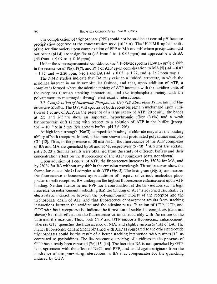
Upon addition of 1 equiv. of ATP, the fluorescence increases by 150 YO for MA, and by 250% for BA without any shift in the emission wavelength. Titration curves show the formation of a stable 1 : 1 complex with ATP (Fig. 2). The histogram (Fig. 3) summarizes the fluorescence enhancement upon addition of 1 equiv. of various nucleotide phos- phates to both receptors. BA undergoes the highest fluorescence enhancement upon ATP binding. Neither adenosine nor PPP nor a combination of the two induces such a high fluorescence enhancement, indicating that the binding of ATP is governed essentially by electrostatic interaction between the polyammonium moiety of the receptor and the triphosphate chain of ATP and that fluorescence enhancement results from stacking interactions between the acridine and the adenine parts. Titration of CTP, UTP, and GTC with both receptors also indicate the formation of stable 1 : 1 complexes (data not shown) but their effects on the fluorescence varies considerably with the nature of the base and the receptor. Thus, both CTP and UTP induce a fluorescence enhancement, whereas GTP quenches the fluorescence of MA, and slightly increases that of BA. The higher fluorescence enhancement obtained with ATP as compared to the other nucleotide triphosphates could be the result of a better stacking interaction with purines [13] as compared to pyrimidines. The fluorescence quenching of acridines in the presence of GTP has already been reported [7c] [13] [14]. The fact that BA is not quenched by GTP is in agreement with the effect of NaC1, and PPP, and could again originate from the hindrance of the preexisting interactions in BA that compensates for the quenching induced by GTP.
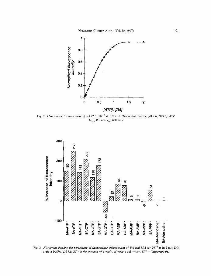
HELVETICA CHIMICA ACTA - Vol. 80 (1997) 791
Fig. 2. Fhorimetric
0 0.5 1 1.5 2
[ATP]/[BA] titration curve of BA (2.5 . M in 2.5 mM Tris acetate buffer, pH 7.6, 20") by ATP
(Aexc 412 nm, i,, 450 nrn)
Fig. 3 . Histogram showing the percentage ofjluorescence enhancement of BA and M A (5 M in 5 mM Tris acetate buffer, pH 7.6, 20") in the presence of I equiv. of various substrates. PPP = Triphosphate.
792 HELVETICA CHIMICA ACTA - Vol. 80 (1997)
In order to further characterize the interaction with these receptors, we also carried out a study of a fluorescent analogue of ATP, E-ATP [l5] (Fig. I ) . The excitation wave- length (300 nm) of this substrate is distant from those of the receptors, but it emits very close to them (410 nm). Fluorescence titration experiments demonstrate the formation of 1 : 1 complexes with BA and MA (see also [5]). In the presence of 1 equiv. of E-ATP, the fluorescence of BA is increased by 96 % and quenched by 36 % in the case of MA. This behavior resembles the one observed with GTP, i.e., a fluorescence quenching with MA, and a fluorescence enhancement with BA. As discussed above, the inhibition of an intramolecular interaction in BA which induces a fluorescence quenching in the free receptor, could compensate for the fluorescence quenching induced by this substrate on one of the acridines. When the substrate was excited (300 nm), the titration curves in the presence of MA and BA were superimposable, indicating that the substrate interacts similarly with both receptors. In both cases the fluorescence of E-ATP is quenched (78 %). The emission spectra upon excitation at 300nm show a tailing down to 550 nm (maximum at 410nm) which may be due to an energy transfer from E-ATP to the acridine.
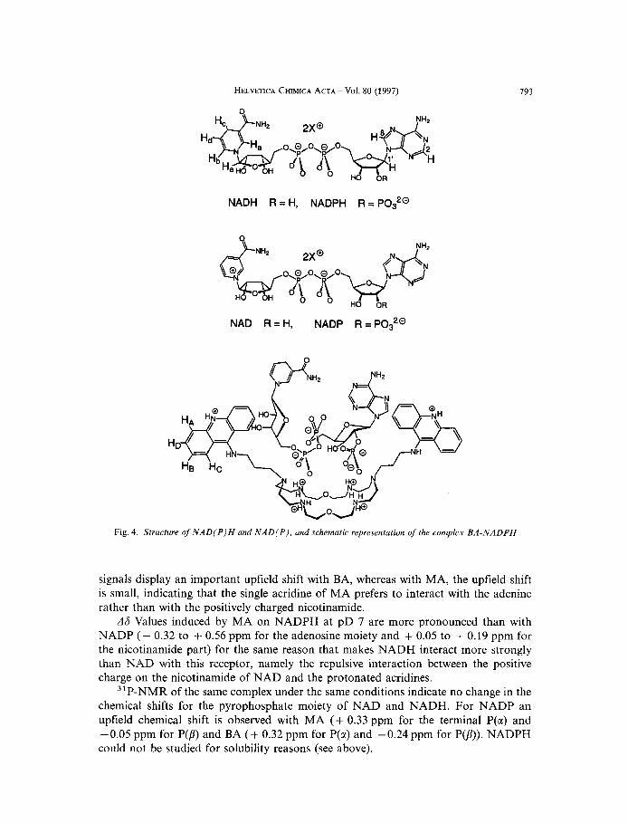
3.3. Complexation of Dinucleotide Phosphates: 'H-NMR and 31P-NMR Studies. NMR studies were carried out at neutral pD when the complex was soluble; alternatively pD 4 was used for less soluble complexes. The NADPH complexe with BA (see Fig. 4 ) could not be studied by NMR (200 MHz) since at the concentration used M) the complex was insoluble, even at pD 4; below pD 4, NADPH is highly unstable [16]. It was, however, possible to study this complex at pH 7.6 by spectrofluorimetry because of the low concentration required for this technique (see Sect. 3.4).
As summarized in Tuble2, the complexation induces small upfield shifts on the adenosine protons of NAD (A6 from + 0.22 to + 0.26 pprn with BA and + 0.13 to + 0.17 ppm with MA) and has essentially no effect on the nicotinamide moiety, indicat- ing that this part of the substrate does not interact with the acridine groups of the receptors. This could result from the repulsive interaction between the positive charge carried by this group and the protonated acridine. As a result of the presence of two acridine units in BA, the chemical shifts are substantially higher. The upfield shifts of the acridine proton signals of both receptors upon complex formation are quite small (A6 from -0.01 to + 0.03 ppm for BA and + 0.03 to + 0.06 ppm for MA), in agreement with a weak interaction between the receptors and NAD.
NADH which is lacking the positive charge of the nicotinamide interacts with the receptors via both its adenosine and nicotinamide moieties. The upfield 'H-NMR chemical shifts ( A 6 ) induced by BA vary from + 0.32 to + 0.47 ppm for the adenosine portion and + 0.22 to + 0.31 ppm for the nicotinamide group. With MA, A 6 ranges from + 0.19 to + 0.37 ppm for the adenosine part and from + 0.14 to + 0.26 ppm for the nicotinamide moiety. Here again, the chemical shifts induced by BA are more pronounced.
NADP which differs from NAD by the phosphoryl group on the 2'-hydroxy group of the adenosine (Fig. 4) displays much larger effects with both receptors (pD 7). The upfield shifts ( A h ) induced by BA vary from + 0.25 to + 1.44 ppm for the adenosine moiety and from + 0.22 to + 0.65 ppm for the nicotinamide, while with MA, they range from + 0.14 to + 0.49 ppm for the adenosine moiety and from + 0.03 to + 0.10 ppm for the nicotinamide. Although the nicotinamide is positively charged, its 'H-NMR
HELVETICA CHIMICA ACTA - Vol. 80 (1997) 793
NAD R = H , NADP R=P0s2@
Fig. 4. Structure of NADIPJH and N A D ( P ) . and schematic representation of the complex BA-NADPH
signals display an important upfield shift with BA, whereas with MA, the upfield shift is small, indicating that the single acridine of MA prefers to interact with the adenine rather than with the positively charged nicotinamide.
A 6 Values induced by MA on NADPH at pD 7 are more pronounced than with NADP (+ 0.32 to + 0.56 ppm for the adenosine moiety and + 0.05 to + 0.19 ppm for the nicotinamide part) for the same reason that makes NADH interact more strongly than NAD with this receptor, namely the repulsive interaction between the positive charge on the nicotinamide of NAD and the protonated acridines.
31P-NMR of the same complex under the same conditions indicate no change in the chemical shifts for the pyrophosphate moiety of NAD and NADH. For NADP an upfield chemical shift is observed with MA (+ 0.33 ppm for the terminal P(a) and -0.05 ppm for P(p) and BA (+ 0.32 ppm for P(cc) and -0.24 ppm for P(p)). NADPH could not be studied for solubility reasons (see above).
4
W
P
Tabl
e 2. '
H-N
MR
Che
mic
al S
hifts
(6
in p
pm)"
) of
BA
and
MA
in t
he A
bsen
ce a
nd P
rese
nce
of E
quim
olar
Am
ount
s of
Subs
trat
e M,
20")
in D@
~ ~~
Aro
mat
ic a
nd a
nom
eric
pr
oton
s of
ade
nosi
ned)
am
ided
) A
rom
atic
and
ano
mer
ic p
roto
ns o
f rib
onic
otin
- R
ecep
tor/s
ubst
rate
pH
b,
Acn
dine
moi
ety'
)
HA
H
B H
C
HD
H
-C(8
) H
-C(2
) H
-C(1
') H
a H
, H
, H
, He
BA
MA
NA
D
BA,"A
D M
A/N
AD
N
AD
H
BA
/NA
DH
M
A/N
AD
H
NA
DP
BA
/NA
DP
MA
/NA
DP
NA
DPH
MA
INA
DPH
4 7 4 7 7 7 7 7 7 7 7 7 I 4 7 4 7
8.25
8.
22
8.53
8.
53
8.19
8.
47
8.20
8.
37
8.09
8.
50
~ -
~ -
- 8.40
8.
29
7.91
7.
90
8.09
8.
09
7.90
8.
06
7.89
8.
03
7.91
7.
99
- - - - - 8.03
7.
96
7.57
7.
55
7.93
7.
92
7.56
7.
88
7.60
7.
82
7.48
7.
79
-
-
~ -
- 7.83
7.
73
7.55
-
7.53
-
7.69
-
7.69
-
-
8.55
7.
52
8.33
7.
66
8.42
-
8.60
7.
50
8.16
7.
63
8.29
-
8.53
7.
46
8.01
7.
59
8.34
~
8.69
-
8.58
7.
64
8.47
7.
56
8.26
~ -
-
- 8.29
8.
03
8.12
8.
36
7.89
7.
99
8.26
6.
82
7.77
8.
52
8.35
8.
05
1.79
-
-
~ -
-
-
-
-
6.16
9.
45
5.93
9.
46
6.03
9.
46
6.10
7.
06
5.78
6.
84
5.91
6.
92
6.15
9.
41
5.90
9.
18
6.01
9.
38
6.42
7.
08
6.34
7.
05
6.00
6.
95
5.95
6.
86
-
~ -
~ 9.27
9.
29
9.29
~ -
~ 9.28
8.
93
9.20
-
-
~ -
~ -
-
- 8.95
8.
96
8.96
-
-
- 8.94
8.
72
8.90
~ ~ ~ -
-
-
-
- 8.31
8.
33
8.32
-
-
- 8.31
8.
04
8.27
-
~ -
-
- g
-
$ -
6
6.20
6.21
6.
24
5.93
6.22
E B ij > 9
5.98
? I 2
6.12
W
6.22
5.
57
7
6.12
0
6.09
6.
1 1
6.04
m
h
L
W 4
v
")
b, ') d,
With
res
pect
to
'BuO
H a
s in
tern
al r
efer
ence
(Me,
1.3
6 ppm
). pH
adj
uste
d w
ith c
onc.
NaO
H a
nd H
CI
solu
tions
. A
crid
ine
prot
ons
as s
how
n in
Fig
. 4, H
A an
d H
, ar
e ds
, H,
and
HD
are
ts.
Nuc
leot
ide
prot
ons
are
as s
how
n in
Fig
. 4.
HELVETICA CHIMICA ACTA - Vol. 80 (1997) 795
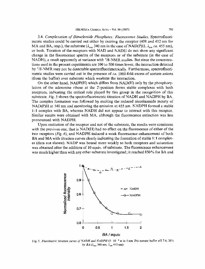
3.4. Complexation of Dinucleotide Phosphates: Fluorescence Studies. Spectrofluori- metric studies could be carried out either by exciting the receptor (408 and 412 nm for MA and BA, resp.), the substrate (A,,, 340 nm in the case of NAD(P)H, ,Iern ca. 455 nm), or both. Titration of the receptors with NAD and NADH do not show any significant change in the fluorescence spectra of the receptors or of the substrate (in the case of NADH), a result apparently at variance with 'H-NMR studies. But since the concentra- tions used in the present experiments are 100 to 500 times lower, the interaction detected by 'H-NMR may not be detectable spectrofluorimetrically. Furthermore, spectrofluori- metric studies were carried out in the presence of ca. 1000-fold excess of acetate anions (from the buffer) over substrate which weakens the interaction.
On the other hand, NADP(H) which differs from NAD(H) only by the phosphory- lation of the adenosine ribose at the 2'-position forms stable complexes with both receptors, indicating the critical role played by this group in the recognition of this substrate. Fig. 5 shows the spectrofluorimetric titration of NADH and NADPH by BA. The complex formation was followed by exciting the reduced nicotinamide moiety of NAD(P)H at 340 nm and monitoring the emission at 455 nm. NADPH formed a stable 1 : 1 complex with BA, whereas NADH did not appear to interact with this receptor. Similar results were obtained with MA, although the fluorescence extinction was less pronounced with NADPH.
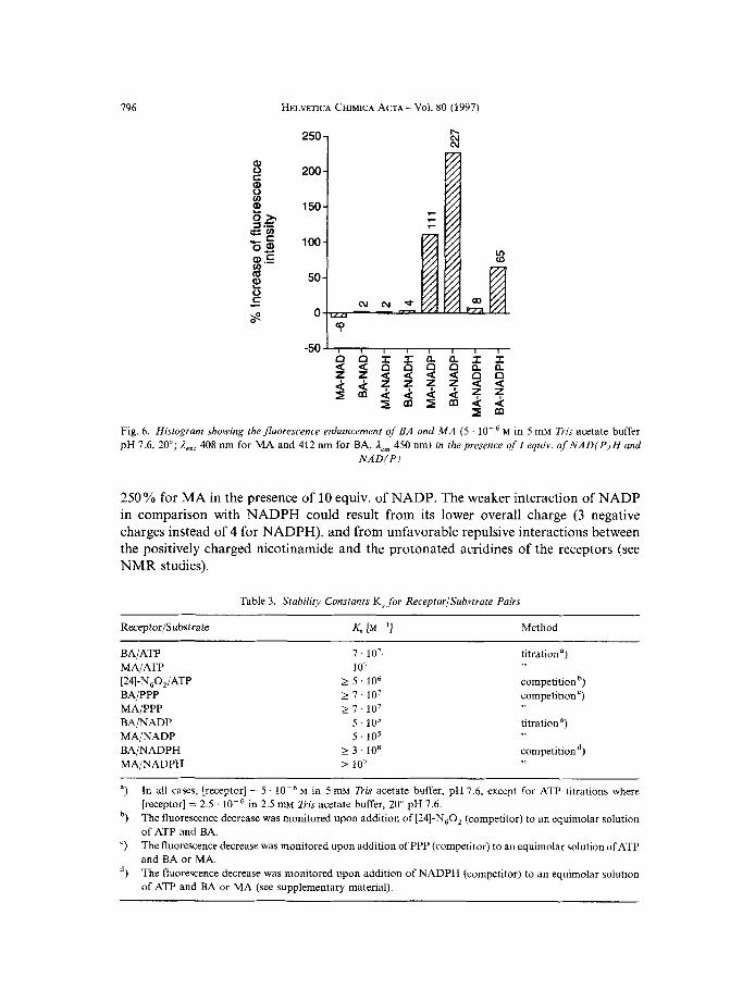
Upon excitation of the receptor and not of the substrate, the results were consistent with the previous one, that is NAD(H) had no effect on the fluorescence of either of the two receptors (Fig. 6), and NADPH induced a weak fluorescence enhancement of both BA and MA with titration curves clearly indicating the formation of stable 1 : 1 complex- es (data not shown). NADP was bound more weakly to both receptors and saturation was obtained after the addition of 10 equiv. of substrate. The fluorescence enhancement was much higher than with any other substrate investigated; it reached 850 % for BA and
1
0 0.5 1 1.5 2
BA / equiv. Fig. 5. Fluorimerric titration curves of NADH and NADPH (5 M in 5 mM Tris acetate buffer pH 1.6, 20")
by BA (A,,, 340 nm, A,, 455 nm)
196 HELVETICA CHIMICA ACTA - Vol. 80 (1997)
250 1 b N N
200
150
100
50
0
Fig. 6. Histogram showing the fluorescence enhancement of BA and M A (5 M in 5 mM Tris acetate buffer pH 1.6, 20"; A,,, 408 nm for MA and 412 nm for BA, Aem 450 nm) in the presence of 1 equiv. of NAD(P)H and
N A D ( P )
250% for MA in the presence of 10 equiv. of NADP. The weaker interaction of NADP in comparison with NADPH could result from its lower overall charge (3 negative charges instead of 4 for NADPH), and from unfavorable repulsive interactions between the positively charged nicotinamide and the protonated acridines of the receptors (see NMR studies).
Table 3 . Stability Constants K, for ReceptorlSubstrate Pairs
Receptor/S u bs tra te K, [M ~ '1 Method
BAlATP 7.107 t i t r a t h a )
[24]-N60,/ATP 2 5 ' 106 competition b,
BAjPPP 2 7.107 competition ')
BA/NADP 5 . 1 0 5 titration*)
BAlNADPH 2 3 ' 108 competitiond)
MAiATP 107
MAjPPP 2 I . 107
MA/NADP 5 . 1 0 5
MAINADPH 2 107
")
b,
")
d,
In all cases, [receptor] = 5 . M in 5 mM Tris acetate buffer, pH 7.6, except for ATP titrations where [receptor] = 2.5 . The fluorescence decrease was monitored upon addition of [24]-N,O, (competitor) to an equimolar solution of ATP and BA. The fluorescence decrease was monitored upon addition of PPP (competitor) to an equimolar solution of ATP and BA or MA. The fluorescence decrease was monitored upon addition of NADPH (competitor) to an equimolar solution of ATP and BA or MA (see supplementary material).
in 2.5 mM Tris acetate buffer, 20" pH 7.6.
HELVETICA CHIMICA ACTA - Vol. 80 (1997)
3.5. Stability of the Complexes. The stability of some of the complex studied was evaluated either directly by spectrofluorimetric titration or indirectly via competition experiments. The results obtained are summarized in Table 3. It is seen that very strong complexes are formed.
A comparison of the stability constants of [24]-N,O,-ATP and BA-ATP indicates that the acridine groups increase the stability b y more than an order of magnitude. A similar effect was observed in a study of adenine complexation by receptors bearing anthracene units [17]. BA binds PPP as strongly as ATP, because PPP possesses an extra negative charge that compensates for the stacking interaction observed with ATP.
PPP, ATP, and NADPH form the strongest complexes with MA and BA. The selectivity of binding of NADPH over NADP by BA amounts to a factor of 600. This selectivity as well as the stability achieved with NADPH compare well with those ob- tained with NAD enzymes3).
Finally, notwithstanding the structural dissimilarities between ATP and NADPH, BA binds both substrates with almost the same strength suggesting a structural adapta- tion of the receptor to these substrates as a result of its flexibility, and also showing that the stability of the complexes is determined mainly by electrostatic interactions.
797
4. Interaction with Double-Stranded DNA. - 4.1. General. Apart from its ability to strongly bind ATP and NADPH, BA also presents analogies with the natural bis-inter- calator triostin A [19] [20]. The macrocyclic moiety of BA and MA forms stable ternary complexes with two Cu" atoms and one molecule of phosphate (& = 3 . lo4 M-') [21]. It has also been shown that simple oligo(ethy1enediamines) catalyze RNA hydrolysis under physiological conditions [22]. BA possesses a binding site for two metals, as well as two acridine moieties which upon intercalation into double-stranded nucleic acids could bring the metal ions in close proximity to the phosphate backbone and thus catalyze its hydrolysis [23].
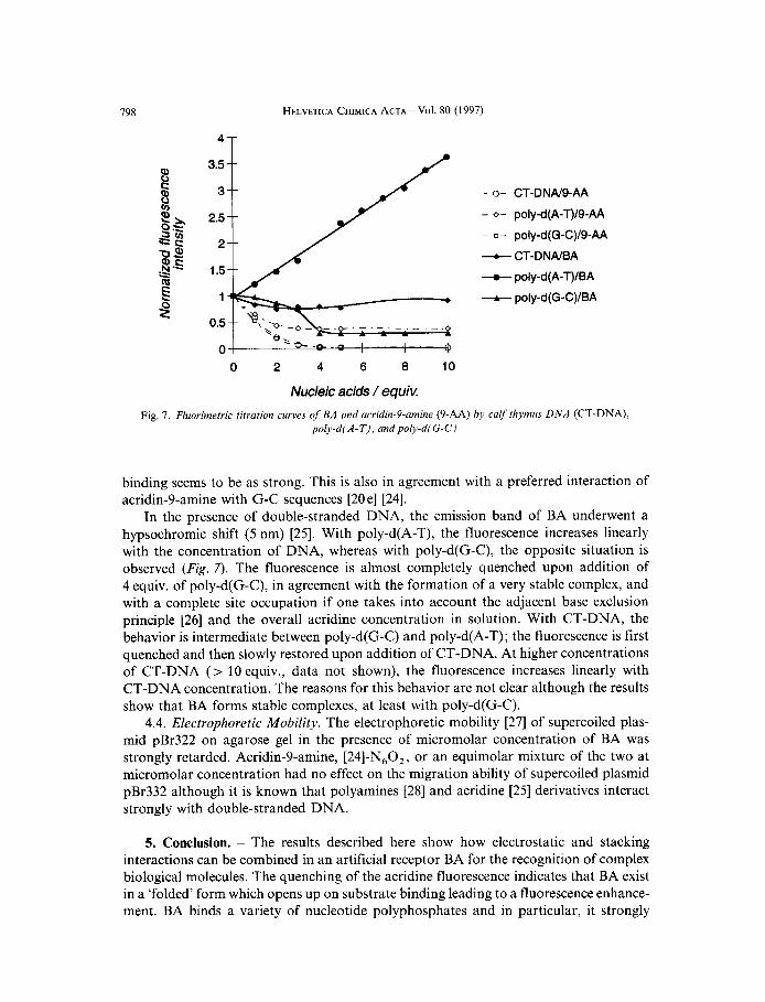
4.2. UVI VIS Spectroscopy. Three isosbestic points (229, 236, and 282 nm) were obtained upon titration of calf thymus DNA (CT-DNA) with BA indicating the existence of two major species in solution and thus that the complex with DNA is very stable. A strong hypochromic effect was recorded at 359 nm (73 "/o) in the presence of 0.3 equiv.
M) with CT-DNA and monitoring the changes around 400 nm. The maximum bathochromic shift (4nm) was obtained after adding 2.8 equiv. of CT-DNA. An important hypochromic effect was also recorded (39 YO).
4.3. Spectrofluorimetry. The interaction of BA was also studied spectrofluorimetrical- ly with CT-DNA, poly-d(G-C), and poly-d(A-T) as shown in Fig. 7. The results with acridin-Pamine (9-AA) were added for comparison as this compound is known to interact with double-stranded DNA by intercalation [20 el [24]; they indicate a strong interaction of acridin-9-amine with double-stranded DNA, in agreement with [20 el [24]. The titration curves of acridin-9-amine with poly-d(G-C) and CT-DNA are superimpos- able, whereas with poly-d(A-T), the fluorescence quenching is not complete although the
of BA to CT-DNA. The same experiment was performed by titrating the receptor
3, Eg., lactate dehydrogenase has a selectivity of 15 in favor of NADH, and a K,,, of lo-' M for this cofactor D81.
798 HELVETICA CHIMICA ACTA - Vol. 80 (1997)
3.5
3
2.5
2
1.5
1
0.5
0
- 0- CT-DNN9-AA
- 0- poly-d(A-T)IS-AA
- Q- poly-d(G-C)/S-AA
--t CT-DNNBA
+ poly-d(A-T)IBA
+ pOly-d(G-C)/BA
0 2 4 6 8 10
Nucleic acids / equiv. Fig. 7. Fluorimetric titration curves of BA and acridin-Pamine (9-AA) by calf thymus D N A (CT-DNA),
poly-dfA-T), and poly-d(G-C)
binding seems to be as strong. This is also in agreement with a preferred interaction of acridin-9-amine with G-C sequences [20 el [24].
In the presence of double-stranded DNA, the emission band of BA underwent a hypsochromic shift ( 5 nm) [25]. With poly-d(A-T), the fluorescence increases linearly with the concentration of DNA, whereas with poly-d(G-C), the opposite situation is observed (Fig. 7 ) . The fluorescence is almost completely quenched upon addition of 4 equiv. of poly-d(G-C), in agreement with the formation of a very stable complex, and with a complete site occupation if one takes into account the adjacent base exclusion principle [26] and the overall acridine concentration in solution. With CT-DNA, the behavior is intermediate between poly-d(G-C) and poly-d(A-T) ; the fluorescence is first quenched and then slowly restored upon addition of CT-DNA. At higher concentrations of CT-DNA (> 10 equiv., data not shown), the fluorescence increases linearly with CT-DNA concentration. The reasons for this behavior are not clear although the results show that BA forms stable complexes, at least with poly-d(G-C).
4.4. Electrophoretic Mobility. The electrophoretic mobility [27] of supercoiled plas- mid pBr322 on agarose gel in the presence of micromolar concentration of BA was strongly retarded. Acridin-9-amine, [24]-N,O,, or an equimolar mixture of the two at micromolar concentration had no effect on the migration ability of supercoiled plasmid pBr332 although it is known that polyamines [28] and acridine [25] derivatives interact strongly with double-stranded DNA.
5. Conclusion. - The results described here show how electrostatic and stacking interactions can be combined in an artificial receptor BA for the recognition of complex biological molecules. The quenching of the acridine fluorescence indicates that BA exist in a ‘folded’ form which opens up on substrate binding leading to a fluorescence enhance- ment. BA binds a variety of nucleotide polyphosphates and in particular, it strongly
HELVETICA CHIMICA ACTA - Vol. 80 (1997)
complexes ATP. Furthermore, NADPH which differs from NADP only by the nicotin- amide moiety is bound 2 600 times more tightly. Whilst NAD(H) interacts weakly with BA, the stability of the complex BA-NADPH (K, 2 3 . lo8) and the selectivity obtained for NADPH vs. NADP is comparable to, and even higher in some cases than that measured for NAD enzymes [18].
It is clear that the combination of different types of intermolecules interactions in suitably designed receptor molecules allows strong and selective binding, i.e., molecular recognition of a variety of substrates of chemical and biological interest.
799
Experimental Part
1. General. M.p. : Digital Thomas-Hoover apparatus (Electrotherma). Chromatography: silica gel Merck 60 (0.063-0.200 mm), silica gel 'flash Merck 60 (0.040-0.063 mm), or alumina Merckact. 11-111 (0.063-0.200 mm); FC = flash chromatography. NMR Spectra: Bruker AC2OO (200.1 MHz for 'H and 50.3 MHz for I3C); b in ppm and Zin Hz, with the solvent as internal reference; spectra in D,O with 2-methyl-propan-2-01 as internal reference (Me, 1.36 ppm; HOC(CH,),, 68.7 and 31.6 ppm, resp. The fast atom bomdardment (FAB) mass spectra were performed by the Service de Spectrometrie de Masse de I'Universite Louis Pasteur de Strasbourg and elemental analyses by the Service de Microanalyse de 1'Universite Louis Pasteur de Strasbourg.
2. Synthesis ofthe Receptors. Acridin-9 (1 OH)-one, acrylonitrile, toluene-4-sulfonyl chloride, methanesulfonyl chloride, and 3,4-dihydro-2H-pyran were purchased from Aldrich. The synthesis of 2 [29], 3 [30], 10-12, MA [S], and 9-chloroacridine [31] has already been reported. MA was obtained via 10 which was prepared in this study by a different synthetic route.
N,N-{[(2-Cyanoethyl) imino]di(ethane-2,l-diyl)}bis[4-methylbenzenesu~onamide] (1). Acrylonitrile (24 ml, 365 rnrnol) was added to a soln. of N,K-[iminodi(ethane-2,1 -diyl)]bis[4-methylbenzenesulfonamide] [32] (15 g, 36.5 mmol) in benzene (230 ml). The mixture was refluxed under N, for 5 days. After cooling to r.t., evaporation yielded a colorless oil which was dried under vacuum for 24 h. Compound 1, was obtained as white crystals from CHCl,/hexane (15.3 g, 90%). R, 0.4 (SiO,, 3% MeOH/CH,CI,). M.p. 106-107". 'H-NMR (200 MHz, CDCI,, 25 ', SiMe,): 2.33 ( f , 3J = 6.6, CH,CN); 2.41 (s, 2 MeC6H,);2.54 ( t , ,J = 4.8,2TsNHCH,CH2N); 2.61 ( t , ' J = 9, CH,CH,CN); 2.88 ( 1 , 3J = 4.8 Hz, 2 CH,NHTs); 5.76 ( t . 2 NKTs); 7.3 (d, 3J = 8.1,4 arom. H); 7.76 (d, ' J = 8.2, 4 arom. H). ',C-NMR (50 MHz, CDCI,, 25", SiMe,): 16.24(CHZN); 21.5(MeC6H,); 40.85(CH2NTs); 49.34(CH,CH2CN); 53.42 (TsNHCH,CH,N); 119.34 (CN); 129.93, 127.22 (arom. CH); 136.69, 143.64 (arom. CSO, arom. CMe). FAB-MS (pos.): 465.0 ( [ M + 11'). Anal. calc. for C,,H,,N,O,S, (464.6): C 54.29, H 6.07, N 12.06; found: C 54.35, H 6.01, N 11.91.
N,N'- { [(2- Cyanoethyl) imino ]di(ethene-2,1 -diyl) }bis[4-methyl-N- { 2- { Z-[(tetrahydro-2H-pyran-2-y1) oxy]- ethoxy}ethyl}benzenesulfonamide] (4). Under N,, 1 (10 g, 21.5 mmol), 2-[2-(2-chloroethoxy)ethoxy]tetrahydro- 2H-pyran (2) [29] (17.97 g, 86 mmol), K,CO, (29.72 g, 215 mmol), Cs,CO, (41.5 g, 129 mmol), and NaI (6.5 g, 43 mmol) were refluxed in MeCN (500 ml) for 19 h. After cooling, the mixture was filtered over Celite and the solid washed with CH,CI,. The combined org. layers were concentrated to 200 ml and extracted with H,O (2 x 200 ml). The org. layer was dried (MgSO,) and evaporated. FC (SiO,, 0-1 % MeOH/CH,Cl,) of the residual brownish oil gave 4 (11.5 g, 66%). Colorless oil. R f 0.4 (SO,, 3% MeOH/CH,CI,). 'H-NMR (200 MHz, CDCl,, 25", SiMe,): 1.4-1.9 (m, 2 CH,CH,CH,); 2.41 (s, 2 MeC,H,); 2.46 (t. 3J = 6.0 CH,CN); 2.8 (m, 3 CH,N); 3.21 ( t , ,J = 7.5, 2 NCH,CH,NTs); 3.33 ( f , ' J = 5, 2 OCH,CH,NTs); 3.4-3.65 (m, 2 NTsCH,CH,OCH,CH,OCHO); 3.7-3.9 (m. 2 CH,CH,CHOCHO); 4.57 (t. 2 OCHO); 7.29 (d, ,J = 9, 4 arom. H); 7.7 (d, ,J = 9, 4 arom. H). ',C-NMR (50 MHz, CDCI,, 25", SiMe,): 16.9 (CH,CN); 21.46(MeC6H,); 19.55, 25.32, 30.57(CH,CH,CHZ); 47.99, 49.2(CH2NTs); 50.41 (CH,CH,CN); 53.66(CH2N); 62.36, 66.44, 70.38(CH20); 98.98(OCHO); 118.88 (CN); 127.08, 129.69 (arom. CH); 136.48, 143.28 (arom. CSO,, arom. CMe). FAB-MS: 809.2 ( [ M + I]+). Anal. calc. for ~,H6,N,0, ,S, + 1 H,O (827.07): C 56.64, H 7.56, N 6.77; found: C 56.91, H 7.28, N6.89.
N,N-{[(2- Cyanoethyl) imino]di(ethane-2,l-diyl) )bis[N-[2- (2-hydroxyethoxy)ethyl]4-methylbenaenesulfon- umide] (5). At r.t. 4 (9.37 g, 11.6 mmol) and toluene-4-sulfonic acid monohydrate (2.87 g, 15.08 mmol) were stirred in MeOH (400 ml) under N, for 12 h. After evaporation, the residual yellow oil was taken up in CH,CI, (300 ml) and extracted with 10% (w/v) aq. K,CO, soh. (400ml). The org. layer was dried (MgSO,), filtered, and evaporated. Compound 5 (7.2 g, 97%) was obtained as a colorless viscous oil after FC (SO,, 2.5% MeOH/ CH,CI,) and drying under high vacuum (24 h). R, 0.24 (SO,, 5 % MeOH/CH,CI,). 'H-NMR (200 MHz, CDCI,, 25", SiMe,): 2.39 (s, MeC,H,); 2.44 ( t . 35 = 6.0, CH,CN); 2.78 (m, 3 CH,N); 3.22 (t. ' J = 7.5,
800 HELVETICA CHIMICA ACTA - VOl. 80 (1997)
2 NCH,CH,NTs); 3.33 (t, ' J = 5, 2 OCH,CH,NTs); 3.45 (t. ' J = 5, 2 NTsCH,CH,O); 3.55 (1, ' J = 5, 2 CHCH,OH); 3.63 ( t , = 8 , 4 arom. H). 13C-NMR (50 MHz, CDCl,, 25", SiMe,): 16.55(CH2CN); 21.36(MeC,H4); 47.76, 48.99(CH2NTs); 50.29(CH2CH,CN); 53.5(NTsCH,CH2N); 61.34,70.09, 72.38(CHZO); 119.05 (CN); 126.98,129.89(arom. CH); 136.22,143.45 (arom. CSO,, arom. CMe). FAB-MS (pos.): 641.2 ( [ M + I]'). Anal. calc. for C,,H,,N,O,S, + 112 H,O (649.89): C53.6, H6.98, N8.62; found: C53.79, H7.16,N8.67.
N,N'-{[(2-Cynnoethyl) imino]di(ethnne-2,l-diyl)}bis[4-merhyl-N-{2-{2-[(methylsulfonyl)oxy]ethoxy}ethyl}- benzenesulfonnmide] (6) . A soh. of 5 (14 g, 21.9 mmol) in anh. CH,Cl, (distilled over CaH,: 440 ml) under N, was cooled to 0" in an ice bath. Et,N (18.3 ml, 131.1 mmol) was added dropwise followed by methanesulfonyl chloride (5.1 ml, 65.6 mmol) in anh. CH,Cl, (distilled over CaH,; 120 ml) within 45 min. After the addition, the temp. was maintained at 0" for 2 h and at r.t. for another 2 h. The mixture was then washed with cold (ca. 4") distilled H,O (150 ml), cold (cn. 4") 10% HCl soh. (100 ml), sat. NaHCO, soh. (150 ml), and then with sat. NaCl soh. (150 ml). The org. layer was dried (MgSO,) and evaporated at r.t. and the residue dried under high vacuum (24 h). This crude 6 (17 g, quant.) was used in the next step without further purification. Colorless oil. R, 0.32 (SO,, 3% MeOH/CH,Cl,). 'H-NMR (200 MHz, CDCl,, 25", SiMe,): 2.4 (s, 2 MeC,H,); 2.43 ( t , CH,CN); 2.65-2.9 (m. 3 CH,N); 3.02 (s, 2 MeSO,); 3.21 (i, ' J = 6 , 2 NCH,CH,NTs); 3.32 (t. ' J = 5, 2 OCH,CH,NTs); 3.5-3.7(m,2CH,OCH,);4.2-4.35(m,2CH2OSO,Me);7.29(d,3J= 7.5,4arom.H);7.66(d,3J= 7.5,4arom. H). ',C-NMR (50 MHz, CDCl,, 25", SiMe,): 17.1 (CH,CN); 21.95(MeC,H4); 37.71 (MeSO,); 49.35, 48.1 (CH,NTs); 50.6(CH,CH2CN); 53.98(TsNCH,CH2N); 69.16, 68.92(CH20CH,); 70.24(CH,OSO2Me); 119.35(CN); 127.08, 129.95 (arom. CH); 135.99, 143.56 (arom. CSO,, arom. CMe).
4,10.16,22- Tetrakis[(4-methylpheny1)su[fonyl]-1,13-di~xn-4,7,10,16,19,22-hexnnzneyclotetrucosnne-7,19-di- propunenitrile (7). A s o h of 6 (11 g, 13.8 mmol) in anh. DMF (distilled over NaH; 500 ml) was added dropwise under N, pressure within 3 h to a mechanically stirred mixture of 1 (6.4 g, 13.8 mmol) and Cs,CO, (36 g, 110.4 mmol) in anh. DMF (distilled over NaH; 1000 ml) at 100". After the addition, the temp. was maintained for an additional 22 h at 100". DMF was evaporated and the residual solid resuspended in CH,C1, (400 ml), filtered on Celite, and washed with CH,Cl,. The org. layers were washed with dist. H,O, dried (Na,SO,), and evaporated. The brownish oil thus obtained was filtered on silica gel (2% MeOH/CH,Cl,) and purified by FC (SO,, 1 % MeOH/CH,Cl,): 7 (4.4g, 30%). Colorless oil. R, 0.5 (SiO,, 1% MeOH/CH,Cl,). 'H-NMR (200MHz, CDCl,, 25", SiMe,): 2.4(m, 16H, CH,CN, MeC,H,); 2.83(m, 12H, CH,N); 3.21 ( r , 8H, NCH,CH,NTs); 3.29 (t, = 8.2, 8H. arom. H); 7.66 (d, = 8.2,8H, arom. H). I3C-NMR (50 MHz, CDCl,, 25", SiMe,): 16.97(CH2CN); 21.42(MeC6H,); 49.4, 48.14(CH2NTs); 50.09(CH,CH2CN); 53.73(TsNCH,CH2N); 70.06(CH20); 119.17(CN); 127.05, 129.79 (arom. CH); 136.17, 143.43 (arom. CSO,, arom. CMe). FAB-MS (pos.): 1069.4 ( [M + I]+). Anal. calc. for C,,H,,N,O,,S, (1069.39): C 56.16, H 6.4, N 10.4; found: C 56.23, H6.57, N 9.87.
4,10,16,22- Tetrakis[(4-methylphenyl)sulfonyl]- 1.13-dioxa-4,7,10.16,19,22-hexnnzacyclotetrncosune-7,19-di- propanumine (8). A soh. of 7 (1.5 g, 1.4 mmol) in anh. THF (distilled over Na; 30 ml) was stirred under N, for 15 min. To this soln., IM B,H, in THF (70 ml) was added and the mixture stirred under reflux for 22 h. The soh. was cooled to 0" and the excess B,H, carefully destroyed by adding slowly a soh. of H,O/THF I : 1. The resulting mixture was evaporated and the white solid obtained refluxed in 6M HCl(l50 ml) for 7 h. The white solid obtained after evaporation was dried under high vacuum (24 h), dissolved in CH,Cl, (200 ml), and washed with 2~ NaOH (200 ml). The aq. layer was further extracted with CH,Cl, (2 x 150 ml). The org. layers were dried (MgSO,) and evaporated and the residue dried under high vacuum (24 h). 8 as a yellow oil (1.3 g, 86%) which was used in the next step without further purification. 'H-NMR (200 MHz, CDCl,, 25", SiMe,): 1.79 (m, 4H. CH,CH,CH,); 2.1-2.4 (m, 12H, CH,N); 2.41 (s, 12H, MeC,H,); 3.04 (1, 3J = 4,4H, CH,NH,); 3.35 (t, = 5,16H, CH,NTs); 3.58(t,3J=4.5,8H,CH20);7.3(d,3J=9,8arom.H);7.68(d,3J=9,8arom.H).
N,N-Di(acridin-9-yl) -4,10,16,22-tetrakis[(4-methylphenyl)sulfonyl]-1,13-dioxa-4,7,10,16,19,22-hexnazucy- clotetracosane-7,19-dipropaneamine (9). A mixture of 8 (1.3 g, 1.2 mmol), phenol (5.65 g, 60 mmol), and 9-chloroacridine [31] (0.52 g, 2.4 mmol) was heated at 80" under Ar for 8 h. After cooling, the residue was taken up in CH,Cl, (100 ml) and washed with 2M aq. NaOH (40 ml). The aq. layer was further extracted with CH,C1, (2 x 25 ml), the combined org. phase dried (MgSO,) and evaporated, and the residue submitted to chromatogra- phy (alumina, 0 ~ 2 % MeOH/CH,Cl,) followed by crystallization from hot EtOH: 9 (0.43 g, 25%). R, 0.33 (A1,0,, 5 % MeOH/CH,CI,). M.p. 95-96". 'H-NMR (200 MHz, CDCl,, 25", SiMe,): 1.96 (m, 4H, CH,CH,CH,); 2.4 (s, 12H, MeC,H,); 2.76 (t. ' J = 5, 4H, CH,CH,CH,N); 2.92 (t ' J = 7.8, 8H, NCH,CH,NTs); 3.23 (m, 16H, CH,NTs); 3.5 (t, = 4.3, 8H, CH,O); 4.03 (t, 3J = 6, 4H, CH,NHAcr); 7.12 (d, , J = 8 , 8arom. H); 7.21(t, ' J=7, 4H, HB); 7.5(d, 'J=8, 8 arom. H); 7.62(t, ,J=6.5, 4H, HD); 8.13 (t, = 9, 8H, HA, Hc). 13C-NMR (50 MHz, CDCl,, 25", SiMe,): 21.34(MeC6H,); 27.92(CH,CH,CH2); 47.84,
= 5, 2 CH,OH); 7.28 (d, = 8 , 4 arom. H); 7.66 (d,
= 5 , 8H, OCH,CH,NTs); 3.55 (t, ' J = 5, 8H, CH,O); 7.29 (d,
HELVETICA CHIMICA ACTA - Vol. 80 (1997) 801
49.72 (CH,NHAcr, CH,NTs); 54.27, 52.55(CH2N); 70.25(CH20); 115.69, 122.3, 123.26, 129.17, 129.65, 149.28, 151.34 (acridine); 129.76, 126.93 (arom. C of Ts); 135.71, 143.29 (arom. CSO,, arom. CMe). FAB-MS ('0s.): 1431.2 ( [ M + 11'). Anal. calc. for C,,H,,N,,O,,S, + 1 H,O (1449.88): C 62.96, H 6.39, N 9.66; found: C 62.70, H 6.27, N 9.85.
N,N'-Di(acridin-9-yl/-1,13-dioxa-4,7,10,16,19,22-hexaazacyclotetracosane-7,19-dipropaneamine (BA). A mixture of 9 (0.26 g, 0.18 mmol), phenol (0.4 g, 4.3 mmol), and 33% HBr in AcOH (31 ml) was stirred at 80" for 38 h. After cooling, Et,O (60 ml) was added, and the precipitate obtained was filtered, washed with Et,O (100 ml), and dried under high vacuum. The yellow solid was dissolved in distilled H,O (20 ml) and extracted with CH,CI, (2 x 20 ml). The aq. layer was basified to pH ca. 14 with 4M NaOH and extracted with CH,CI, (5 x 20 ml). Evaporation of the org. layer yielded a yellow oil which was taken up in abs. EtOH (10 ml) and acidified with conc. HCI soh. to pH ca. 1. The precipitate formed was isolated by centrifugation, resuspended in EtOH (10 ml), and recentrifuged. This operation was repeated a third time. The yellow solid was dried under vacuum to yield BA 8 HC1 (0.2 g, 66%). M.p. 267" (dec.) NMR: the 6s depend strongly upon the concentration and the pH of the medium. 'H-NMR (200 MHz, lo-' M in D,O at pD 7, 25", SiMe,): 2.31 (m, 4H, CH,CH,CH,); 2.94 ( r , 3.f = 7 4H, CH,CH,CH,N); 3.06 (1, 3J = 8, 8H, CH,CH,CH,NCH,); 3.31 (t, 8H, CH,CH,CH,NCH,CH,N); 3.4 (t,8H,NCH,CH,O);4.0(t,8H,CHzO);4.15(t,'J= 7,4H,CH2NHAcr);7.61 (m,8H,HC,H,);7.98(t, ' J = 8, 4H, HB); 8.28 (d, 3 J = 9, 4H, HA). "C-NMR (50MHz, M in D,O at pD 7, 25", SiMe,): 26.45(CH,CH2CH,); 46.92(CH2NHAcr); 49.06, 49.49, 51.25 (CH,CH,CH,NCH,CH,N); 51.56(NCH2- CH,O); 67.29(CHZO); 120.25, 136.0, 137.26, 159.16, (acridine). FAB-MS (pos.): 815.3 ( M + of unprotonated BA (calc. 815.13)), [M + 1]+ and [ M + 2]'+ are hardly detectable (ca. 5%). Anal. calc. for C,,H,,N,,O, + 8 HCI + 4 H,O (1178.88): C 48.91, H 7.01, N 11.88; found: C49.03, H 7.01, N 11.45.
BA is stable in the solid octahydrochloride form. It can be stored at low temp. (+ 4") for a few months in aq. acidic solns. Under neutral or slightly basic conditions, the product starts decomposing in less than 4 weeks, most likely via the displacement of the acridine unit to yield the corresponding acridin-9(10H)-one.
Preparation of MA. The preparation of this compound has been reported earlier [5]. We describe here a different approach where the key step is the macrocyclization step, leading directly to the functionalized polyamine 10. The following steps leading to 11, 12 and MA (not described here) are similar to those previously reported [5].
4,10,16,19,22-Pentakis[(4-methylphenyl)su~onyl]-1,13-dioxu-4.7,10.16.19,22-hexaazacyclotetrucosane-7-pro- paneizitd (10). A soh. of 6 (9.36 g, 11.74 mmol) in anh. DMF (distilled over NaH; 500 ml) was added dropwise under N, pressure within 3 h to a mechanically stirred mixture of N,N'-{ {[(4-methylphenyl)suIfonyl]- imino}di(ethan-2,1-diyl)}bis[4-methylbenzenesu~fonamide] (3) [30] (6.64 g, 11.74 mmol) and Cs,C03 (30.6 g, 94 mmol) in anh. DMF (distilled over NaH, 1000 ml), at 100". After the addition, the temp. was maintained at 100" for an additional 51 h. DMF was evaporated and the residual solid resuspended in CH,CI, (400 ml), filtered over Celite, and washed with CH,C1,. The org. layers were washed with dist. H,O, dried (Na,SO,), and evaporated. The brownish oil thus obtained was filtered over silica gel (1 % MeOH/CH,CI,) and purified by FC (SO,, 0-0.5% MeOH/CH,CI,): 10 (7 g, 50%). Yellowish oil. R, 0.5 (SO,, 2 % MeOH/CH,CI,). 'H-NMR (200MHz, CDCl,, 25", SiMe,): 2.4(m, 17H, CH,CN, MeC,H,); 2.65-2.9 (m, 6H, CH,N); 3.1-3.45(m, 20H, CH,NTs); 3.56 (m. 8H, CH,O); 7.25-7.4 (m, 10 arom. H); 7.6-7.75 (m, 10, arom. H). I3C-NMR (50 MHz, CDCI,, 25", SiMe,): 16.93(CH2CN); 21.55(MeC6H,); 48.06, 48.21, 49.04, 49.43, 49.74(CH2NTs); 50.05(CH2CH,CN); 53.63(TsNCH2CH,N); 69.63, 69.95(CHZO); 119.1 (CN); 127.32, 127.46, 129.87, 129.93, 135.45, 136.21, 136.51, 143.46, 143.65, 143.89 (arom.).
3. Physicochemicul Measurements. Spectrophotometric and Spectrojluorimetric Data of the Receptors and Their Complexes. UVjVIS Spectra : Cary-III instrument. Fluorescence spectra: Shimadzu RF-540 equipped with a photomultiplicator Humamatsu HTV R928 and a recorder Shimadzu DR-3. The excitation wavelength was produced by a Xe lamp. Except when indicated, the fluorescence measurements were performed in a 1 x 1 cm quartz cell at a concentration of 5 . M for the receptors, in 5 mM Tris acetate buffer, pH 7.6, at 20". The acquisition parameters were the following: A,,, 412 nm for BA, and 408 nm for MA, excitation slit 10 nm, emission slit 10 nm, sensitivity high.
For BA in 1 0 0 m ~ Tris acetate buffer, pH 7.6, at 20": eZz1 40900 mol-'l-'cm-', E , , ~ 78900 mol-' I- ' cm-' , E~~~ 13 200 mol-'l- ' cm- ' , E ~ , , 18 900 mol- ' I-'cm-', E , ~ , , 15 700 mol- ' 1 - cm- '. For MA in 100 mM Tris acetate buffer, pH 7.6, at 20": E , , ~ = 31 900 mol-'l-' cm-' , E , , ~ 68000 m o l ~ ' l - ' c r n ~ ' , E~~~ 6200 mol- ' I I cm- ', e4', 11 500 mol- ' 1- cm- , E , ~ , , 9800mol-'l-1cm-'. Upon excitation at 412 nm in 5 mM Tris acetate buffer, pH 7.6, at 20", BA and MA (5 . lo-, M) exhibited an emission maximum at 450 nm.
NMR Data. 'H- and 31P-NMR Spectra: Bruker AC 200 (200.1 MHz for 'H and 81.028 MHz for "P), 2-methylpropan-2-01 as internal reference for 'H (CH,, 1.36 ppm) and conc. H3P04 soh. as external reference
802 HELVETICA CHIMICA ACTA - Vol. 80 (1997)
(SR = - 10795.12) for ”P. The spectra were recorded under the following conditions; [receptor] = [substrate] = M, at 20” in D,O. The pD was adjusted by addition of few pl of conc. HC1 or NaOH soh.
Interactions with DNA. Titration of BA by CT-DNA. CT-DNA (Sigma) was first extracted with phenol/ CHCI, (3 times) and precipitated by EtOH before use. Poly-d(G-C) and poly-d(A-T) were used as obtained from Sigma. BA was added in increments of 0.06 equiv. to an UV cell containing a soh. of CT-DNA (87.6 ’ M, in 50 mM Tris acetate buffer, pH 7.6, 20”) and to another one containing the buffer only (reference). The spectra were recorded as the difference between the cell containing the CT-DNA and the reference. After the seventh increment (0.42 equiv. negligible volume), DNA started precipitating as a yellow complex with BA.
Titration of CT-DNA by BA. CT-DNA was added in increments of 0.1 equiv. (negligible volume) to a UV cell containing BA M, in 50 mM Tris acetate buffer pH 7.6,20”) and to another one containing the buffer only (reference). The spectra recorded as the difference between the cell containing BA and the reference.
REFERENCES
(11 H. Furuta, D. Magda, J. L. Sessler, J. Am. Chem. SOC. 1991, 113, 978; H.-J. Schneider, T. Blatter, B. Palm, U. Pfingstag, V. Riidiger, I. Theis, ibid. 1992, 114, 7704; S. Claude, J. M. Lehn, F. Schmidt, J.-P. Vigneron, J. Chem. Soc., Chem. Commun. 1991, 1182; M. Kodama, Y. Kuramato, E. Kimura, in ‘Current Topics in Macrocyclic Chemistry in Japan’, Ed. E. Kimura, Hiroshima University School of Medicine, 1987, pp. 136; T, P. Prakash, P. Rajamohanan, W. Ganish, J. Chem. SOC., Perkin Trans. 1 1991, 1273; E. Kimura, Topics Curr. Chem. 1985,128,113; S . M. Lacy, D. M. Rudkevich, V. Verboom, D. N. Reinhoudt, Tetrahedron Lett. 1994, 35, 5953; C. Andreu, A. Galan, K. Kobiro, J. de Mendoza, T. K. Park, J. Rebek Jr., A. Salmeron, N. Usman, J. Am. Chem. SOC. 1994,116,5501; A. V. Eliseev, H.-J. Schneider, ibid. 1994, 1 16,6081 ; M. Dhaenens, J.-M. Lehn, J.-P. Vigneron, .I Chem. Soc., Perkin Trans. 2 1993,1379; M. Shionaya, T. Ikeda, E. Kimura, M. Shiro, J. Am. Chem. SOC. 1994, 116, 3838; C. Coudret, A. Harriman, J. Chem. SOC., Chem. Commun. 1992, 1755; Y Kato, M. M. Conn, J. Rebek Jr., J. Am. Chem. SOC. 1994, 116, 3279.
[2] Y. Umezawa, M. Kataoka, W. Takama, E. Kimura, T. Koike, H. Nada, Anal. Chem. 1988, 60, 2392; D. Cordier, M. W Hosseini, New J. Chem. 1990, 14, 61 1 ; D. Y. Sasaki, K. Kurihara, T. Kunitake, J. Am. Chem. SOC. 1991, 113, 9685; A. W. Czarnik, Acc. Chem. Res. 1994, 27, 302.
[3] P. C. Zamecnik, B. Kim, M. J. Gao, G. Taylor, G. M. Blackburn, Proc. Natl. Acad. Sci. U.S.A. 1992, 89, 2370; F. Grummt, ibid. 1978, 75, 371; J. Walker, P. Dossman, B. R. Lackey, J. K. Zimmerman, M. A. Dimmick, R. H. Hilderman, Biochemistry 1993,32, 14009.
[4J E. Rapoport, J. Fontaine, Proc. Nafl. Acad. Sci. U.S.A. 1989, 86, 1662. [5] M. W. Hosseini, A. J. Blacker, J.-M. Lehn, J. Am. Chem. Soc. 1990, 12, 3896. [6] M. W. Hosseini, J.-M. Lehn, Helv. Chim. Acta 1987, 70, 1312. [7] a) ‘The Acridines’, 2nd edn., Ed. R. M. Acheson, Wiley Interscience, New York, 1973; b) N. J. Pritchard, A.
Blake, A. R. Peacocke, Nature (London) 1966, 212, 1360; c) S. Georghiou, Photochem. Photobiol. 1976, 24, 417; d) N. C. Seeman, R. 0. Day, A. Rich, Nature (London) 1975, 253, 324; e) N. Mataga, T. Kubota, ‘Molecular Interactions and Electronic Spectra’, Marcel Dekker Inc., New York, 1970.
[El M. W Hosseini, K. C. Jones, K. E. Plute, K. Bowman Mertes, M. P. Mertes, J. Am. Chem. Soc. 1989, Iff, 6330.
[9] B. Dietrich, M. W Hosseini, J.-M. Lehn, R. B. Sessions, Helv. Chim. Acta 1983, 66, 1262. [lo] B. Dietrich, Pure Appl. Chem. 1993,65,1457; A. Bianchi, M. Micheloni, P. Paoletti, Coord. Chem. Rev. 1991,
[ l l ] M. W. Hosseini, Bioorg. Chem. Frontiers 1993, 3, 67. [12] M. W. Hosseini, J.-P. Kintzinger, J.-M. Lehn, A. Zahidi, Helv. Chim. Acta 1989, 72, 1078. [13] L. Kittler, G. LGber, F. A. Gollmick, H. Berg, Bioefectrochem. Bioenergetics 1980, 7, 503. 1141 S. Giorghiou, Photochem. Photobiol. 1975. 22, 103. [15] P. D. Sattoangi, J. R. Barris, N. J. Leonard, J. Am. Chem. SOC. 1980, f02, 770. [16] J. R. Micsic, P. R. Brown, Biochemistry 1978, 17, 2235; N. J. Oppenheimer, Biochem. Biophys. Res. Commun.
[17] V. Rotello, E. A. Viani, G. Deslongchamps, B. A. Murray, J. Rebeck Jr., L Am. Chem. SOC. 1993, 115, 797. [18] A. Fersht, ‘Enzyme Structure and Mechanism’, 2nd edn., W. H. Freeman and Co., 1984, p. 328. [19] A. H.-J. Wang, G. Ughetto, G. Qnigley, A. Rich, J. Biomol. Struct. Dynamics 1986, 4, 319. [20] For other mono- and bis-intercalations, see a) W Saenger, ‘Principles of Nucleic Acid Structure’, Springer-
Verlag Inc., New York, 1984, pp. 350; b) M. G. Blackburn, M. J. Gait, ‘Nucleic Acids in Chemistry and Biology’, Oxford University Press, New York, 1990, pp. 297; c) W. D. Dilson, R. J. Jones, in ‘Intercalation
110, 17.
1973, 50, 683; N. J. Oppenheimer, N. 0. Kaplan, Biochemistry 1974, 13, 4675, 4685.
HELVETICA CHIMICA ACTA - Val. 80 (1997) 803
Chemistry’ Eds. M. S. Wittingham and A. J. Jacobson, Academic Press, New York, 1982; d) H. Dugas, ‘Bioorganic Chemistry’, Springer-Verlag, New York, 1989, pp. 140; e) S. C. Zimmermann, C. R. Lamberson, M. Cory, T. A. Fairly, J. Am. Chem. Soc. 1989, 111, 6805.
[21] R. J. Motekaitis, A. E. Martell, Inorg. Chem. 1992, 31, 5534. [22] K. Yoshinari, K. Yamazaki, M. Komiyama, J. Am. Chem. Soc. 1991, 113, 5899. [23] S. Takenaka, T. Ihara, M. Tagaki, 1 Mol. Recogn. 1990, 3, 156. [24] J. Markovits, B. Gaugain, J. Barbet, B. P. Roques, J. B. Le Pecq, Biochemistry 1981, 20, 3042. [25] H. M. Berman, P. R. Young, Ann. Rev. Biophys. Bioeng. 1981, 10, 87; A. R. Peacocke, J. N. H. Skerrett,
[26] P. J. Bond, R. Langridge, K. W. Jennette, S. J. Lippard, Proc. Nutl. Acad. Sci. U.S.A. 1975, 72, 4825. [27] P. E. Nielsen, W. Zhen, V. Henriksen, 0. Buchardt, Biochemistry 1988, 27, 67. [28] H. Damashun, G. Damashun, M. Becker, E. Buder, R. Misselwitz, D. Zirwer, Nucleic Acids Res. 1978, 5,
3801; B. Bunce, E. S. W. Kong, Biophys. Chem. 1978, 8, 357; H.-J. Schneider, T. Blatter, Angew. Chem. Inr. Ed. 1992, 31, 1207.
Trans. Faraduy SOC. 1956, 52, 261 ; G . Lober, G. Achert, Biopolymers 1969, 8, 595.
[29] B. Dietrich, M. W. Hosseini, J.-M. Lehn, R. B. Sessions, Helv. Chim. Actu 1985, 68, 289. [30] J. Comarmond, P. Plumerb, J.-M. Lehn, Y Agnus, R. Louis, R. Weiss, 0. Kahn, I. Morgenstern-Badarau,
[31] A. Albert, B. Richie, ‘Organic Synthesis’, Wiley, New York, 1955 Collect. Vol. No. 3, p. 53. [32] M. W Hosseini, J.-M. Lehn, S. R. Duff, K. Gu, M. P. Mertes, J. Org. Chem. 1987, 52, 1662.
J. Am. Chem. Soc. 1982, 104, 6330.


![Acridine – a Promising Fluorescence Probe of Non-Covalent ... · [acridine-H]+BArF−, λ em =485 nm. Fig.3. Absorption spectra in CH 2 Cl 2 of: (1) acridine (2×10⁻⁵ mol/l) and](https://img.pdfslide.net/doc/110x75/5f4a49f4cafd5240686feade/acridine-a-a-promising-fluorescence-probe-of-non-covalent-acridine-hbarfa.jpg)