Embed Size (px)
Citation preview
Molecular Basis for Failure of “Atypical” C1 Domain of Vav1 toBind Diacylglycerol/Phorbol Ester*□S
Received for publication, November 2, 2011, and in revised form, February 16, 2012 Published, JBC Papers in Press, February 18, 2012, DOI 10.1074/jbc.M111.320010
Tamas Geczy‡, Megan L. Peach§, Saïd El Kazzouli¶, Dina M. Sigano¶, Ji-Hye Kang¶, Christopher J. Valle‡,Julia Selezneva‡, Wonhee Woo‡, Noemi Kedei‡, Nancy E. Lewin‡, Susan H. Garfield�, Langston Lim�,Poonam Mannan�, Victor E. Marquez¶, and Peter M. Blumberg‡1
From the ‡Laboratory of Cancer Biology and Genetics and �Laboratory of Experimental Carcinogenesis, Center for CancerResearch, NCI, National Institutes of Health, Bethesda, Maryland 20892 and the ¶Chemical Biology Laboratory, MolecularDiscovery Program, Center for Cancer Research, and §Basic Research Program, SAIC-Frederick, NCI-Frederick, National Institutes ofHealth, Frederick, Maryland 21702
Background: The C1 domain of Vav1 retains a three-dimensional structure consistent with phorbol ester binding butnevertheless does not bind.Results: Five residues render the C1 domain less lipophilic and interfere with its binding.Conclusion: The C1 domain of Vav1 illustrates a novel mechanism rendering the C1 domain “atypical.”Significance: Ligands exploiting the specific amino acid differences may selectively target Vav1.
C1 domains, the recognition motif of the second messengerdiacylglycerol and of the phorbol esters, are classified as typical(ligand-responsive) or atypical (not ligand-responsive). The C1domain of Vav1, a guanine nucleotide exchange factor, plays acritical role in regulation of Vav activity through stabilization ofthe Dbl homology domain, which is responsible for exchangeactivity of Vav. Although the C1 domain of Vav1 is classified asatypical, it retains a binding pocket geometry homologous tothat of the typical C1 domains of PKCs. This study clarifies thebasis for its failure to bind ligands. Substituting Vav1-specificresidues into the C1b domain of PKC�, we identified five crucialresidues (Glu9, Glu10, Thr11, Thr24, and Tyr26) along the rim ofthe binding cleft that weaken binding potency in a cumulativefashion. Reciprocally, replacing these incompatible residues inthe Vav1 C1 domain with the corresponding residues fromPKC� C1b (�C1b) conferred high potency for phorbol esterbinding. Computer modeling predicts that these unique resi-dues in Vav1 increase the hydrophilicity of the rim of the bind-ing pocket, impairing membrane association and thereby pre-venting formation of the ternary C1-ligand-membrane bindingcomplex. The initial design of diacylglycerol-lactones to exploitthese Vav1 unique residues showed enhanced selectivity for C1domains incorporating these residues, suggesting a strategy forthe development of ligands targeting Vav1.
The lipophilic second messenger sn-1,2-diacylglycerol(DAG)2 plays a central role in cellular signaling. Following the
activation of many receptors, DAG is generated either throughthe hydrolysis of phosphatidylinositol 4,5-bisphosphate viaphospholipase C or indirectly from phosphatidylcholine viaphospholipase D (1). Its downstream effects are mediatedthrough interaction with protein kinase C (PKC), RasGRP, andfive other families of effectors that possess a C1 domain recog-nition motif (2–4). The profound involvement of PKC andthese other families of signaling proteins in proliferation, dif-ferentiation, apoptosis, angiogenesis, and drug resistance hasemphasized the importance of these C1 domain-containingproteins as therapeutic targets for cancer and other diseases,andmultiple agents, e.g. bryostatin 1 or PEP005, targeted to theC1 domains of PKC are currently in clinical trials (4, 5).Structural studies by NMR, x-ray crystallography, and
molecular modeling have afforded substantial insights intoligand recognition by theC1domains (6–9). These domains arecysteine-rich zinc finger structures. The DAG-binding site is ahydrophilic cleft formed from two pulled apart �-sheets,whereas the C1 domain surface surrounding the binding cleft ishydrophobic. Insertion of DAG into the binding cleft serves tocomplete the hydrophobic surface, and additional hydropho-bicity is contributed by hydrophobic substituents on the DAG.This increase in hydrophobicity upon binding promotes inser-tion of the C1 domain into the lipid bilayer, which in turn candrive conformational change in the overall protein structure, asis the case with PKC, as well as promote translocation to themembrane, changing access of the protein to interacting part-ners. Because the lipids of the bilayer interact both with theligand andwith the surface of the C1 domain, great selectivity ispossible. Although DAG represents the endogenous ligand forthe C1 domains, nature has provided a diversity of high affinityanalogs such as the phorbol esters or bryostatins, and DAG-lactones have afforded a powerful synthetic platform for prob-ing structure-function relationships (4, 10).
* This work was supported, in whole or in part, by National Institutes of HealthProject Z1A BC 005270 from the Intramural Research Program, NCI.
□S This article contains supplemental Figs. 1–3.1 To whom correspondence should be addressed: National Institutes of
Health, NCI, Bldg. 37, Rm. 4048, 37 Convent Dr., MSC 4255, Bethesda MD20892-4255. Tel.: 301-496-3189; Fax: 301-496-8709; E-mail: [email protected].
2 The abbreviations used are: DAG, diacylglycerol; GEF, guanyl exchange fac-tor; DH domain, Dbl homology domain; PH domain, pleckstrin homologydomain; PMA, phorbol 12-myristate 13-acetate; PDBu, phorbol 12,13-dibu-
tyrate; MLP, molecular lipophilicity; DOG, 1,2-dioctanoyl glycerol; PS, phos-phatidylserine; PC, phosphatidylcholine; SH, Src homology.
THE JOURNAL OF BIOLOGICAL CHEMISTRY VOL. 287, NO. 16, pp. 13137–13158, April 13, 2012Published in the U.S.A.
APRIL 13, 2012 • VOLUME 287 • NUMBER 16 JOURNAL OF BIOLOGICAL CHEMISTRY 13137
by guest on September 7, 2018
http://ww
w.jbc.org/
Dow
nloaded from
Initially, C1 domains were categorized into two families (11)as follows: (i) those that boundDAG/phorbol esterwere termed“typical,” and (ii) the more divergent members that failed tobind were termed “atypical.” Examples of atypical C1 domainsinclude those of the atypical PKC isozymes (aPKC� and aPKC�),c-Raf, kinase suppressor of Ras (KSR), and Vav (3, 4, 12). Morerecently, it has become evident that atypical C1 domains can befurther categorized. The first subclass, represented by proteinslike c-Raf or KSR, consists of C1 domains that are grossly dis-torted in the binding cleft geometry (e.g. deletions of several keyresidues in the loops making up the binding pocket). Membersof the second subclass, in contrast, retain the binding cleftgeometry but incorporate other factors impeding ligand bind-ing.We showed that, in the case of the atypical PKCs, a series ofthree arginine residues lining the rim of the binding pocketwere able to rotate into the cleft, making it inaccessible toligands (13). Replacement of these arginine residues with thecorresponding residues found in the C1b domain of PKC� gen-erated high affinity ligand binding and ligand-driven mem-brane translocation.Recent structural studies using x-ray crystallography have
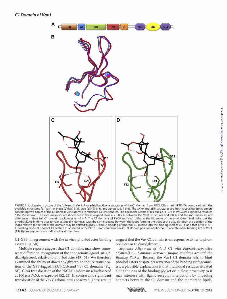
revealed a striking resemblance between the binding pocketgeometry of the C1b domain of the potent phorbol ester recep-tor PKC� and that of the protooncogeneVav1, a guanine nucle-otide exchange factor (14–16). Although early studies on theGEF activity of Vav1 toward small GTPases suggested a DAG/phorbol ester-sensitive function of Vav1 (17), subsequentligand-binding experiments using purified recombinant Vav1and the ultrapotent phorbol ester analog [3H]bryostatin did notsupport those results (18), leading to the classification of thisC1domain as atypical. In this study, we have sought to identify keystructural determinants in the Vav1 C1 sequence that (analo-gous to our findings with atypical PKCs) might account for itsapparent lack of affinity for DAG and phorbol esters.Vav1 is a versatile cellular signal transducer molecule that
plays a pivotal role in various signaling pathways. One of themost important and best characterized functions of Vav1 is theGEF activity toward the Rho/Rac family of small GTPases,which are important molecular transducers in signaling cas-cades that regulate cytoskeleton organization, cell cycle pro-gression, gene transcription, adhesion, migration, cell growth,and survival (19–21). In addition to their activity as exchangefactors for Rho/Rac proteins, Vav proteins can also regulatevarious other cellular processes in a GEF-independent fashion,functioning as adapter molecules to facilitate protein-proteininteractions (22). The expression of Vav1 is restricted almostexclusively to normal cells of hematopoietic origin (23–25).There, T-cell receptor-coupled activation of the guanineexchange function of Vav1 leads to Rac1-induced cytoskeletonorganization, an essential step for the formation of the immu-nological synapse and subsequent proper T-cell activation (20,26). Vav1 further plays a crucial role inT-cell development (27).Although less extensively investigated, recent studies have alsocharacterized a role of Vav1 in human cancer, especially in solidtumors of nonhematopoietic origin (28), where the ectopicexpression of wild-type Vav1 can contribute to the develop-ment and progression of these malignancies (29–32). It there-
fore represents an attractive therapeutic target, both for canceras well as for autoimmunity.Vav1 possesses multiple structural motifs that mediate its
versatile functions in cellular signaling (Fig. 1A) (19). It containsa Dbl homology (DH) domain, which is responsible for catalyz-ing nucleotide exchange (33). The DH domain is flanked by anN-terminal acidic (Ac) motif and a calponin homology (CH)domain together with a C-terminal pleckstrin homology (PH)domain and a C1 domain. These domains surrounding the cat-alytic DH domain in the CH-C1 segment of the molecule reg-ulate the exchange activity of Vav1 (28). In addition, the struc-ture of Vav contains an SH3-SH2-SH3 cassette at the Cterminus, which links it with tyrosine phosphorylation path-ways and mediates its activity as a scaffold protein (22).Initial studies of Vav using site-directed mutagenesis identi-
fied the C1 domain as critical for maintaining efficient guaninenucleotide exchange activity toward Rho/RacGTPases (34, 35).Although this regulatory effect initially was hypothesized toarise from direct contacts between the C1 domain and theGTPases (36), crystallographic studies now provide a differentexplanation (15, 16). An intramolecular network of contactsbetween the PH-C1 unit and the DH domain helps stabilize theconformation of a critical�-helix within theDHdomain, whichis essential for the displacement of guanine nucleotide from theGTPase. The C1 domain (together with the PH domain) thuscontributes to optimal GEF activity by restricting the confor-mational flexibility of the DH domain, keeping it in a stableconformation primed for efficient interaction with the Rac1GTPase. TheC1 domain of Vav1 possesses a three-dimensionalstructure very similar to that of the typical C1b domain ofPKC�. Because its solvent-accessible cavity is located in thevicinity of the �-helix of DH that makes contacts with Rac1,ligand binding to the cleft has the intriguing potential to mod-ulate Vav1 function. Could DAG/phorbol esters or their deriv-atives interact with this binding pocket and thereby disrupt theenzymatic function of Vav1?In this study we wished to characterize the ligand binding
properties of the C1 domain of Vav1.We confirmed the lack ofphorbol ester binding ofVav1.Using site-directedmutagenesis,we identified the central structural determinants in the loopsmaking up the binding cleft responsible for the lack of phorbolester sensitivity. The C1 domain of Vav1 (together with that ofatypical PKCs) thus belongs to that subclass of atypical (nonre-sponsive) C1 domains that retain the proper structure forligand binding. However, the presence of four unique hydro-philic residues around the rim of the binding pocket disruptsthe lipophilic surface of the tip of the binding pocket and,together with an inappropriately hydrophobic residue distal tothe tip of the C1 domain, interferes with the insertion of ligandreceptor complex into the lipid membrane, which is an essen-tial step for stabilizing the ternary binding complex of ligandreceptor and membrane. Mutating these residues in the Vav1C1 domain to correspond to the ones in the potent phorbolester receptor �C1b, we demonstrated almost complete recov-ery of binding affinity in vitro and in vivo, which confirms theprevious structural findings on the conserved binding pocketgeometry of Vav1. Our results raise the possibility that appro-priately modified DAG/phorbol ester analogs that can specifi-
C1 Domain of Vav1
13138 JOURNAL OF BIOLOGICAL CHEMISTRY VOLUME 287 • NUMBER 16 • APRIL 13, 2012
by guest on September 7, 2018
http://ww
w.jbc.org/
Dow
nloaded from
cally target these residues might have the potential to selec-tively bind to Vav1 C1 and to manipulate Vav1 functionthrough this interaction. Finally, we describe some DAG-lac-tones that display modest selectivity for features of the Vav1-C1-like structure.
EXPERIMENTAL PROCEDURES
Materials—[20-3H]Phorbol 12,13-dibutyrate ([3H]PDBu)(17.2 Ci/mmol) was obtained from PerkinElmer Life Sciences.PDBu and phorbol 12-myristate 13-acetate (PMA) were pur-chased from LC Laboratories (Woburn, MA). Phosphatidyl-L-serine (PS), phosphatidylcholine (PC), and 1,2-dioctanoylglyc-erol (DOG) were from Avanti Polar Lipids (Alabaster, AL).LNCaP human prostate cancer cells, fetal bovine serum (FBS),RPMI 1640 medium, and L-glutamine were from the AmericanType Culture Collection (Manassas, VA). Reagents used forculturing bacteria (LBBroth, LB agar plateswith different selec-tion antibiotics, etc.) were from K-D Medical, Inc. (Columbia,MD). The oligonucleotide primers used for PCR cloning,sequence analysis, and site-directedmutagenesiswere obtainedfrom Invitrogen.Construction of GST-fused C1 Domains of PKC� and
Vav1—The wild-type �C1b domain in a pGEX-5�-1 plasmid(GE Healthcare) had previously been constructed in our labo-ratory (48). To generate a recombinant Vav1 C1 domain fusedto glutathione S-transferase (GST) at the N terminus, PCRamplification of the appropriate sequence was performed usingPlatinum�PCRSuperMixHigh Fidelity (Invitrogen), accordingto themanufacturer’s instructions. The full-length cDNAcloneof Vav1 served as template. The following oligonucleotideprimers were used in the PCR as follows: (i) forward primer5�-CGGAATTCAATGCTACAGCCAATGGGC-3� and (ii)reverse primer 5�-CGGAATTCGAAATCTTGCCCATG-GCG-3�. The DNA fragments of the PCR were purified withQIAquick PCR purification kit (Qiagen, Inc., Valencia, CA) andsubsequently digested with EcoRI (New England Biolabs, Bev-erly, MA) to create adhesive ends of the C1 fragments. After anadditional step of purification with theQIAquick PCR purifica-tion kit, the fragments were finally ligated into the GST-con-taining pGEX-5�-1 plasmid (GE Healthcare) using the EcoRIrestriction sites. Analysis of the DNA sequence of the constructwas conducted by the DNA Minicore (Center for CancerResearch, NCI, National Institutes of Health). Verification ofthe sequencing data was performed using the following soft-ware: BioEdit Sequence Alignment Editor Version 7.0.5 andDNA Baser Sequence Assembler Version 2.91.Site-directedMutagenesis of theC1bDomain of PKC�and the
C1 Domain of Vav1—C1 domains consisted of a conserved50–51-amino acid sequence possessing the characteristic cys-teine-rich motif HX12CX2CXnCX2CX4HX2CX7C, where H ishistidine, C is cysteine, X is any other amino acid, and n is 13 or14 (Fig. 2, gray-shaded letters) (11). To facilitate comparisonsbetweenC1 domains, we will refer to residues using numberinginternal to the C1 domain itself, with the N-terminal histidineresidue being labeled His1. This residue corresponds to His516in full-length Vav1 and His231 in the C1b domain of full-lengthPKC�. Point mutations of the amino acid residues at positions9, 10, 11, 22, 24, and 26 of both PKC� C1b and Vav1 C1 were
introduced using the GeneTailorTM site-directed mutagenesissystem (Invitrogen) according to the manufacturer’s instruc-tions. To generate the C1 domain mutants of PKC� and Vav1,the abovementionedwild-typeC1 constructs (PKC� andVav1)in pGEX-5�-1 were used. Single, double, and triple mutationswere introduced in one step, and quadruple and quintuplemutants were generated in a stepwise fashion using triplemutants as templates. The presence of mutations was verifiedby DNA sequencing (DNA Minicore) and analysis (BioEdit,DNA Baser).Construction of GFP-labeled C1 Domains of PKC� and
Vav1—To generate GFP-tagged fluorescent fusion proteins forin vivo translocation studies, pGEX-5�-1 plasmids containingrecombinant C1 domain sequences from either PKC� or Vav1were digested with EcoRI (New England Biolabs). The DNAfragments from this reaction were purified with the QIAquickPCR purification kit (Qiagen). Finally, the C1 fragments wereligated into the pEGFP-C2 vector (Clontech) using the EcoRIrestriction sites, and theDNA sequences of the constructs wereconfirmed by sequence analysis (DNAMinicore).Construction of Full-length Vav1 (Wild-type, Triple, and
Quintuple Mutant) Fused to GFP—The GFP-tagged wild-typeVav1 was generated using the pENTRTM Directional TOPO�cloning kit (Invitrogen), according to the instructions describedin the manual. Briefly, PCR amplification of the appropriatefull-lengthVav1 sequencewas first carried out using Platinum�PCR SuperMix High Fidelity (Invitrogen). The cDNA clone ofVav1 served as a template, and the following oligonucleotideprimers were applied in the PCR: (i) forward primer 5�-CAC-CGAGCTCTGGCGACAGTGC-3� and (ii) reverse primer5�-TCAGCAATATTCGGAATAGTCTTCC-3�. The PCRproduct was then TOPO-cloned into a pENTRTM/D-TOPO�vector, which served as an Entry clone. Next, using the LRrecombination reaction of the Gateway method (Invitrogen),we transferred the appropriate full-length Vav1 sequence fromthe Entry vector into a pcDNA-DEST53 GatewayTM destina-tion vector (Invitrogen), which encodes anN-terminal GFP tag.The triple (E9M/E10S/T11P) and the quintuple (E9M/E10S/T11P/T24L/Y26K) mutants of the full-length Vav1 were gen-erated using the GeneTailorTM site-directed mutagenesis sys-tem (Invitrogen) described above for the C1 domain mutants.The TOPO-Entry clone of the (full length) wild-type Vav1served as a template for constructing the triple and quintuplemutants in a stepwise fashion. The mutated Entry clones werethen subcloned into pcDNA-DEST53 using the above men-tioned LR recombination reaction. The DNA sequences of theGFP-tagged full-length constructs were verified by sequenceanalysis (DNAMinicore).Expression and Purification of GST-tagged C1 Domains from
Escherichia coli—The C1 domains of both PKC� and Vav1 inthe pGEX-5�-1 plasmid were transformed into BL21-AITMOne Shot� chemically competent E. coli (Invitrogen). Trans-formants were grown in LB broth medium (K-D Medical) at37 °C until the optical density of the bacterial suspensionreached 0.6–0.8. Expression of the GST fusion proteins wasinduced with 0.3 mM isopropylO-D-thiogalactopyranoside and0.125% L-arabinose (both from Sigma) for 4 h at 37 °C. Bacterialcells were subjected to sonication in B-PER� bacterial protein
C1 Domain of Vav1
APRIL 13, 2012 • VOLUME 287 • NUMBER 16 JOURNAL OF BIOLOGICAL CHEMISTRY 13139
by guest on September 7, 2018
http://ww
w.jbc.org/
Dow
nloaded from
extraction reagent, supplemented with 50mg/ml lysozyme and2500 units/ml DNase I (all from Pierce). The expressed GST-tagged C1 proteins were purified using a B-PER GST spin puri-fication kit (Pierce) according to the manufacturer’s instruc-tions. Purification efficiency was evaluated by SDS-PAGEanalysis. Protein concentration was assessed with the proteinassay kit from Bio-Rad. Purified fusion proteins were stored in30% glycerol at �80 °C.In Vitro ([3H]PDBu Assays—To assess the affinity of the dif-
ferent C1 domains (wild-type and mutant PKC� and Vav1) tophorbol esters, purified proteins were subjected to an in vitro[3H]PDBu binding assay, and the dissociation constants (Kdvalues) of the individual C1 domains were determined. Mea-surement of [3H]PDBu binding, using the polyethylene glycolprecipitation assay developed in our laboratory, was describedin detail previously (37). Competitive binding assays were car-ried out to assess the affinities (Ki values) of DOG and DAG-lactones as described in detail elsewhere (38). Triton X-100,included in some of the assays, did not exceed 0.003%.Translocation of GFP-labeled Proteins—LNCaP cells
(between passage numbers 5 and 20) were plated at a density of60,000 cells/plate on Ibidi dishes (Ibidi, LLC, Verona, WI) andsubcultured at 37 °C in RPMI 1640medium supplementedwith10% FBS and 2mM L-glutamine. After 48 h in culture, cells weretransfected with GFP-tagged recombinant constructs, usingLipofectamine reagent and Plus reagent (both from Invitrogen)according to the manufacturer’s protocol. Cellular expressionof fluorescent fusion proteins was examined 24 h after transfec-tion on a Zeiss LSM 510 NLO confocal microscopy system(Carl Zeiss, Inc., Thornwood, NY) with excitation from a30-milliwatt argon laser tuned to 488 nm and emission col-lected with a BP 500–530 filter. Intracellular translocation ofthe GFP-labeled C1 domains upon PMA treatment wasdetected sequentially after the administration of the drug.Images were acquired every 30 s for 30 min at varying zoomsettings (1 to 3.5) using Zeiss AIM software and a 63 � 1.4 NAZeiss Plan-Apochromat oil immersion objective.Quantitation of Confocal Images—Three regions of 4 �m2
each were selected in each cell as follows: one in the cytoplasm,one in the cell membrane, and one in the nucleus, avoiding tothe degree possible the selection of subcellular organelles suchas Golgi or the nucleoli. Mean intensities of the GFP-taggedconstructs in the selected regions were calculated using theZeiss AIM software for the images at the different time points;the ratio of the intensities for membrane/cytoplasm and nucle-us/(membrane � cytoplasm) was then calculated and normal-ized to the time 0 values. The increase in the membrane/cyto-plasm ratio and/or the decrease in the nucleus/(membrane �cytoplasm) ratio indicates translocation. For each series ofimages presented, the mean � S.E. of the maximal transloca-tion for all of the replicate experiments for that assay conditionis presented.Chemistry—The DAG-lactone derivatives were synthesized
according to previously published procedures (39). Meltingpoints were determined on an MPA 100 OptiMelt automatedmelting point system (Stanford Research Systems) or a Mel-Temp II apparatus (Laboratory Devices, USA) and are uncor-rected. Column chromatography was performed on a Teledyne
Isco CombiFlash Companion instrument under gradient elu-tion conditions with RediSep disposable flash columns. Analyt-ical TLCwas performed onAnaltechUniplates silica gel GF. 1HNMR spectra were recorded on a Varian Unity Inova instru-ment at 400 MHz. Spectra are referenced to the solvent inwhich they were run (7.24 ppm for CDCl3). Positive ion fastatom bombardment mass spectra (FAB-MS) were obtained ona VG 7070E-HF double-focusing mass spectrometer operatedat an accelerating voltage of 6 kV under the control of aMASPEC-II data system forWindows (Mass Spectrometry Ser-vices, Ltd.). Either glycerol or 3-nitrobenzyl alcohol was used asthe sample matrix, and ionization was effected by a beam ofxenon atoms generated in a saddle-field ion gun at 8.0� 0.5 kV.Nominal mass spectra were obtained at a resolution of 1200,and matrix-derived ions were background-subtracted duringdata system processing. Full experimental details and charac-terization have been reported previously for 6a, 6c, and 8b-e(40). Full characterization of novel DAG-lactones designated ascompounds 6b, 8a, and 8f is described below. NMR spectra forthese compounds can be found in the supplemental material.(E)-(2-(Hydroxymethyl)-4-((1-methyl-1H-indol-3-yl)methyl-
ene)-5-oxotetrahydrofuran-2-yl)-methyl 3-isobutyl-5-methyl-hexanoate (6b)—1H NMR (400 MHz, CDCl3) � 7.94 (t, J � 2.6Hz, 1H, Ar), 7.82 (dt, J � 7.8, 0.9 Hz, 1H, Ar), 7.38 to 7.24 (m,4H, Ar and C � CH), 4.31 (Ab q, J � 11.9 Hz, 2H, CH2), 3.85 (s,3H, CH3), 3.77 (AB q, J � 12.1 Hz, 2H, CH2), 3.06 (dd, J � 17.2,2.7 Hz, 1H, CH4a), 2.84 (dd, J� 17.1, 2.6 Hz, 1H, CH4b), 2.22 (d,J � 6.5 Hz, 3H, CH2 and OH), 1.90 (app sept, 1H, CH), 1.63 to1.48 (m, 2H, 2 � CH), 1.17 to 0.96 (m, 4H, 2 � CH2), 0.83 (dd,J � 6.6 Hz, 3H, CH3), 0.82 (dd, J � 6.6 Hz, 6H, 2 � CH3), and0.78 (d, J � 6.6 Hz, 3H, CH3); FAB-MS (m/z, relative intensity)456 (MH�, 97), 455 (M .�, 100).(E)-4-((5-(Hydroxymethyl)-2-oxo-5-((2-propyl-pentanoyloxy)
methyl)dihydrofuran-3(2H)-ylidene)methyl)-1-propylpyri-dinium Bromide (8a)—1H NMR (400 MHz, CDCl3) � 8.88 (d,J� 6.7Hz, 2H,Ar), 8.16 (d, J� 6.6Hz, 2H,Ar), 7.66 (s, 1H,C�CH), 4.74 to 4.56 (m, 2 H, CH2), 4.33 (s, 2 H, CH2), 3.94 (d, J �12.0 Hz, 1 H, H4a), 3.92 (AB q, J � 15.8 Hz, 2 H, CH2), 3.73 (d,J� 12.0 Hz, 1 H,H4b), 2.56 (v br s, 1 H, OH), 2.34 (tt, J� 8.8, 5.4Hz, 1H,CH), 2.02 (heptet, J� 7.4Hz, 2H,CH2), 1.60 to 1.46 (m,2 H, CH2), 1.43 to 1.32 (m, 2 H, CH2), 1.23 (m, 4 H, 2 � CH2),0.98 (t, J � 7.4 Hz, 3 H, CH3), 0.86 (td, J � 7.3, 1.6 Hz, 6 H, 2 �CH3); FAB-MS (m/z, relative intensity) 404 (M�, 100).(E)-3-((5-(Hydroxymethyl)-5-((3-isobutyl-5-methylhexanoy-
loxy)methyl)-2-oxodihydro-furan-3(2H)-ylidene)methyl)-1-pentyl-pyridinium Bromide (8f)—MP was 142–143 °C. 1HNMR (400 MHz, CDCl3) � 9.76 (s, 1 H, Ar), 8.77 (d, J � 5.7 Hz,1H, Ar), 8.72 (d, J� 8.1Hz, 1H, Ar), 8.08 (irr t, 1 H, Ar), 7.39 (s,1 H, C�CH), 4.89 (br s, 2 H, CH2), 4.19 (AB q, J� 12.3Hz, 2H,CH2), 3.73 (AB q, J � 12.3 Hz, 2 H, CH2), 2.88 (dd, J � 18.6, 2.4Hz, 2 H, H4ab), 2.19 (d, J � 6.5 Hz, 2 H, CH2), 2.04 (br s, 2 H,CH2), 1.88 (irr sept, 1 H, CH), 1.55 (sept, J � 6.7 Hz, 2 H, 2 �CH), 1.38 to 1.35 (m, 4 H, 2 � CH2), 1.15 - 0.94 (m, 4 H, 2 �CH2), 0.92 to 0.75 (m, 15 H, 5 � CH3); FAB-MS (m/z, relativeintensity) 474 (M�, 100).MolecularModeling—Structures for the single- andmultiple
site mutants of the Vav1 and PKC� C1 domains were built byreplacing residues while keeping their side chain � angles as
C1 Domain of Vav1
13140 JOURNAL OF BIOLOGICAL CHEMISTRY VOLUME 287 • NUMBER 16 • APRIL 13, 2012
by guest on September 7, 2018
http://ww
w.jbc.org/
Dow
nloaded from

close as possible to the conformation in the other structure. Forexample, in the Vav1 crystal structure (15), residueGlu9 has theconformation �1 � �74.7, �2 � 166.2, and �3 � 103.1. WhentheM9Emutant of PKC�was built, the Glu residue was rotatedinto the same conformation. In almost all cases, this conforma-tion fit well into the structure, and the only exception was theP11T mutation in PKC�, which required an adjustment ofLeu20 and Phe13 to accommodate the threonine methyl group.This constructionmethod allowed the structure of themutatedVav1 C1 domain to be as similar as possible to the PKC� C1bdomain and vice versa.Docking of phorbol and the DAG-lactones was performed
using the software programGOLDVersion 5.0 (41). The struc-ture of phorbol 13-acetate was extracted from its co-crystalwith the PKC�C1b domain (7), and the C1 domain of Vav1 (15)was clipped from the larger structure.Hydrogenswere added toboth protein and ligands. The binding sitewas defined by atomswithin a 10.0-Å sphere around the N� atom of residue Gln27(Gln542(B) in the full-length structure). Ligand flexibility flagsincluded internal hydrogen bond detection and ring cornerflipping, and the default torsion angle distributions were used.The GoldScore scoring function was used with the defaultparameter file. The genetic algorithm settings were automati-cally optimized according to ligand flexibility with the searchefficiency set to 100%.After docking, the structures of the DAG-lactone-triple
mutant PKC� complexes were subjected to conformationalsearching using MacroModel (42) to identify low energy con-formers for the charged ligand and protein side chains. Thesearches used 50 steps of systematic torsional sampling for eachrotatable bond in the DAG-lactone sn-1 and sn-2 side chains,and in the side chains of residues Glu9, Glu10, and Thr11 in themutated C1 domain. Minimization of each conformer foundwas done using the OPLS 2005 forcefield with octanol implicitsolvent. All atoms in the DAG-lactone and residues 9–13,20–24, and 27 in the binding site were free to move duringminimization and the rest of the protein was held fixed.Lipophilicity analysis was performed using the software pro-
gram VASCo (43). The molecular lipophilicity potential overthe surface of the protein was calculated in the following way.First, an atomic logP value was assigned to each atom. Theatomic logP (AlogP) values used were those from Ghose et al.(44), although the dictionary provided with the VASCo pro-gram was modified to use the ionized forms of Asp, Glu, Arg,and Lys. Next, the solvent-excluded surface was constructedusing MSMS (45), which yielded a set of surface vertices andtriangles. Finally, the lipophilicity of each surface point was cal-culated using a Fermi-type distance function to map andsmooth the logP value of nearby underlying atoms onto thesurface, according to the formulation of Heiden et al. (46) forlarge molecules. The overall molecular lipophilicity potential(MLP) of the binding site area was calculated as the sum of thesurface point lipophilicities for those points whose closestunderlying atom lies on the solvent-accessible surface of the C1domain in the region that inserts into the membrane and inter-acts with phorbol (7). This included the following atoms: resi-due 8, atoms O and C�; residue 9, all atoms; residue 10, allatoms; residue 11, all atoms; residue 12, atoms N and C�; resi-
due 20, all atoms; residue 21, atom O; residue 22, atoms O, C�,C�, C, and C�; residue 23, all atoms; residue 24, all atoms; andresidue 27, atoms C, C�, and N�.
RESULTS
Isolated C1 Domain of Vav1 Does Not Bind Phorbol Esters,Despite Conservation of the Appropriate Geometry of the Bind-ing Pocket—The striking structural resemblance between thebinding cleft of Vav1 C1 and that of PKC� C1b, as revealed byrecent x-ray crystallographic data (15), suggests the potentialfor preserved DAG/phorbol ester responsiveness of Vav1 (Fig.1B). To confirm the ability of the Vav1 C1 domain binding siteto accommodate phorbol ester, we extracted the phorbol13-acetate ligand from the crystal structure of PKC� C1b (7)and docked it into the isolated C1 domain from the highestresolutionVav1 crystal structure (15). As expected based on thestructural overlay of the binding sites, phorbol ester is predictedto be able to bind to the Vav1 C1 domain with essentially thesame binding mode as in the PKC� C1b domain (Fig. 1, C andD). The binding site residues that form direct hydrogen bond-ing interactions with phorbol are identical between the two C1domains, and the pattern of intra- and intermolecular hydrogenbonds formed by bound phorbol is preserved.In contrast to this structural analysis andmodeling, however,
Vav1 has been shown experimentally not to interact with DAGor phorbol esters (18). One explanation for this apparent dis-crepancy could be that the C1 domain is located in the core ofthe Vav1 structure (rather than being exposed on the surface),and other structural domains prevent the access of ligands tothe binding cleft. Examples of such masking of C1 domains,although in both instances not sufficient to fully block binding,are �2-chimerin (47) and PKC� (48).
To test the possibility that the C1 domain is simply masked,we first cloned the isolated Vav1 C1 domain into a pGEX bac-terial plasmid (Clontech) encoding a C-terminal GST tag andassessed the binding properties of the recombinant protein inan in vitro [3H]PDBu binding assay. Compared with the GST-tagged PKC� C1b (�C1b), a high affinity phorbol ester receptorthat served as a positive control in these experiments, the Vav1C1 showed no evidence for [3H]PDBu binding even at very high(�1–2 �M) receptor concentration (data not shown) underthese conditions.Because misfolding of the purified protein from a bacterial
expression system could be a reason for the absence of phorbolester binding in vitro, we examined the phorbol ester respon-siveness of the Vav1 C1 in the intracellular environment aftereukaryotic expression. Phorbol ester causes the translocation oftypical C1 domains to cellular membranes. We expressed aGFP-tagged Vav1 C1 in LNCaP cells, treated with phorbolester, and looked for subcellular redistribution using real timeconfocal microcopy. The GFP-tagged C1b domain of PKC�(�C1b-GFP) served as control, whose translocation dynamicshadpreviously been extensively characterized by our laboratory(13, 49). After the administration of 1 �M PMA, �C1b-GFPshowed a rapid but transient translocation into the cellularmembrane, which was followed by translocation into thenuclear membrane (Fig. 5A). In contrast, PMA treatment failedto induce appreciable intracellular redistribution of Vav1
C1 Domain of Vav1
APRIL 13, 2012 • VOLUME 287 • NUMBER 16 JOURNAL OF BIOLOGICAL CHEMISTRY 13141
by guest on September 7, 2018
http://ww
w.jbc.org/
Dow
nloaded from
C1-GFP, in agreement with the in vitro phorbol ester bindingassays (Fig. 5B).Multiple reports suggest that C1 domains may show some-
what differential recognition of the endogenous ligand, sn-1,2-diacylglycerol, relative to phorbol ester (49–51). We thereforeexamined the ability of dioctanoylglycerol to induce transloca-tion of the GFP-tagged PKC� C1b and Vav C1 domains (Fig.5C). Clear translocation of the PKC�C1b domain was observedat 100 �MDOG, as expected (52, 53). In contrast, no significanttranslocation of theVavC1domainwas observed. These results
suggest that the Vav C1 domain is unresponsive either to phor-bol ester or to diacylglycerol.Sequence Alignment of Vav1 C1 with Phorbol-responsive
(Typical) C1 Domains Reveals Unique Residues around theBinding Pocket—Because the Vav1 C1 domain fails to bindphorbol esters despite preservation of the binding cleft geome-try, a plausible explanation is that individual residues situatedalong the rim of the binding pocket or in close proximity to itmay interfere with ligand-receptor interactions by impedingcontacts between the C1 domain and the membrane lipids,
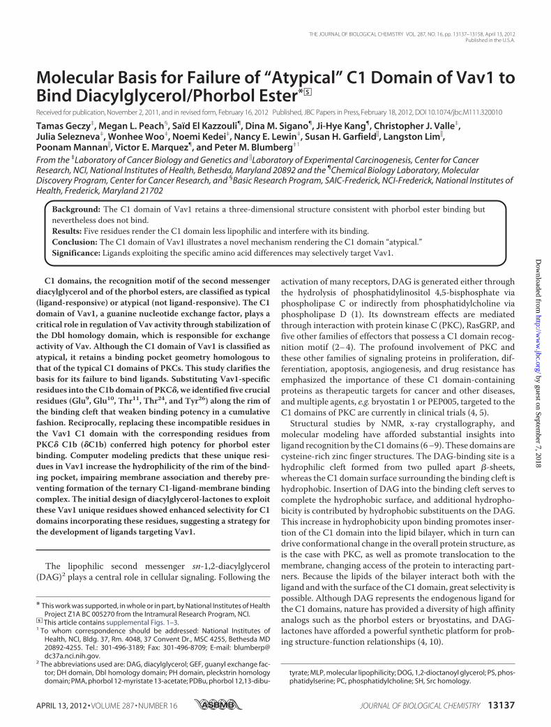
FIGURE 1. A, domain structure of the full-length Vav1. B, overlaid backbone structures of the C1 domain from PKC� C1b in red (1PTR (7)), compared with theavailable structures for Vav1 in green (2VRW) (15), blue (3KY9) (14), and purple (3BJI) (16). The 3KY9 and 3BJI structures are both crystallographic dimerscontaining two copies of the C1 domain. Zinc atoms are rendered as CPK spheres. The backbone atoms of residues 231–274 in PKC� are aligned to residues516 –559 in Vav1. The root mean square difference in these aligned atoms is �0.5 Å between the Vav1 structures and PKC�, and the root mean squaredifference in their full C1 domain backbones is �1.4 Å. The C1 domains of PKC� and Vav1 differ in the tilt angle of the small C-terminal helix, but thephorbol/DAG-binding sites remain essentially identical, with the same spacing between the loops forming the sides of the site, although the position of theloops relative to the rest of the domain may be shifted slightly. C and D, docking of phorbol 13-acetate into the binding cleft of �C1b and that of Vav1 C1.C, binding mode of phorbol 13-acetate as observed in the PKC� C1b crystal structure (7). D, docked position of phorbol 13-acetate in the binding site of Vav1(15). Hydrogen bonds are indicated by dashed lines.
C1 Domain of Vav1
13142 JOURNAL OF BIOLOGICAL CHEMISTRY VOLUME 287 • NUMBER 16 • APRIL 13, 2012
by guest on September 7, 2018
http://ww
w.jbc.org/
Dow
nloaded from
thereby disrupting the ternary binding complex, or by occlud-ing the binding site. This situation would thus be analogous tothat of the atypical C1 domains of the aPKCs (13).C1 domains consist of a conserved 50–51-amino acid
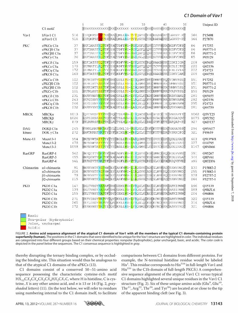
sequence possessing the characteristic cysteine-rich motifHX12CX2CXnCX2CX4HX2CX7C, where H is histidine, C is cys-teine, X is any other amino acid, and n is 13 or 14 (Fig. 2, gray-shaded letters) (11). (In the text below, we will refer to residuesusing numbering internal to the C1 domain itself, to facilitate
comparisons between C1 domains from different proteins. Forexample, the N-terminal histidine residue would be labeledHis1. This residue corresponds toHis516 in full-lengthVav1 andHis231 in the C1b domain of full-length PKC�.) A comprehen-sive sequence alignment of the atypical Vav1 C1 versus typicalC1 domains highlighted several unique residues in the Vav1 C1structure (Fig. 2). Six of these unique amino acids (Glu9, Glu10,Thr11, Arg22, Thr24, and Tyr26) are located at or close to the tipof the apparent binding cleft (see Fig. 3).
FIGURE 2. Amino acid sequence alignment of the atypical C1 domain of Vav1 with all the members of the typical C1 domain-containing proteinsuperfamily (human). The positions in the C1 domains that were identified to be unique for the Vav1 structure are highlighted in color. The individual residuesare categorized into four different groups based on their chemical properties: nonpolar (hydrophobic), polar uncharged, basic, and acidic. The color code isdepicted in the panel below the sequences. The C1 consensus sequence is highlighted in gray.
C1 Domain of Vav1
APRIL 13, 2012 • VOLUME 287 • NUMBER 16 JOURNAL OF BIOLOGICAL CHEMISTRY 13143
by guest on September 7, 2018
http://ww
w.jbc.org/
Dow
nloaded from
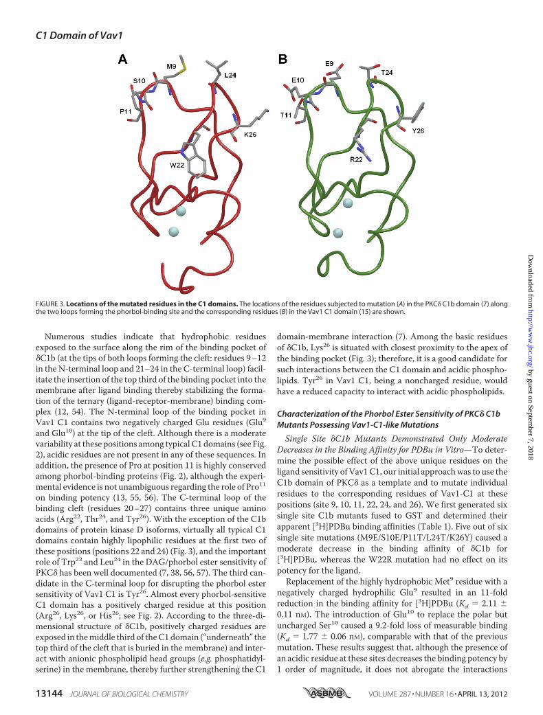
Numerous studies indicate that hydrophobic residuesexposed to the surface along the rim of the binding pocket of�C1b (at the tips of both loops forming the cleft: residues 9–12in the N-terminal loop and 21–24 in the C-terminal loop) facil-itate the insertion of the top third of the binding pocket into themembrane after ligand binding thereby stabilizing the forma-tion of the ternary (ligand-receptor-membrane) binding com-plex (12, 54). The N-terminal loop of the binding pocket inVav1 C1 contains two negatively charged Glu residues (Glu9and Glu10) at the tip of the cleft. Although there is a moderatevariability at these positions among typical C1 domains (see Fig.2), acidic residues are not present in any of these sequences. Inaddition, the presence of Pro at position 11 is highly conservedamong phorbol-binding proteins (Fig. 2), although the experi-mental evidence is not unambiguous regarding the role of Pro11on binding potency (13, 55, 56). The C-terminal loop of thebinding cleft (residues 20–27) contains three unique aminoacids (Arg22, Thr24, and Tyr26). With the exception of the C1bdomains of protein kinase D isoforms, virtually all typical C1domains contain highly lipophilic residues at the first two ofthese positions (positions 22 and 24) (Fig. 3), and the importantrole of Trp22 and Leu24 in the DAG/phorbol ester sensitivity ofPKC� has been well documented (7, 38, 56, 57). The third can-didate in the C-terminal loop for disrupting the phorbol estersensitivity of Vav1 C1 is Tyr26. Almost every phorbol-sensitiveC1 domain has a positively charged residue at this position(Arg26, Lys26, or His26; see Fig. 2). According to the three-di-mensional structure of �C1b, positively charged residues areexposed in themiddle third of theC1domain (“underneath” thetop third of the cleft that is buried in the membrane) and inter-act with anionic phospholipid head groups (e.g. phosphatidyl-serine) in the membrane, thereby further strengthening the C1
domain-membrane interaction (7). Among the basic residuesof �C1b, Lys26 is situated with closest proximity to the apex ofthe binding pocket (Fig. 3); therefore, it is a good candidate forsuch interactions between the C1 domain and acidic phospho-lipids. Tyr26 in Vav1 C1, being a noncharged residue, wouldhave a reduced capacity to interact with acidic phospholipids.
Characterization of the Phorbol Ester Sensitivity of PKC� C1bMutants Possessing Vav1-C1-like Mutations
Single Site �C1b Mutants Demonstrated Only ModerateDecreases in the Binding Affinity for PDBu in Vitro—To deter-mine the possible effect of the above unique residues on theligand sensitivity of Vav1C1, our initial approachwas to use theC1b domain of PKC� as a template and to mutate individualresidues to the corresponding residues of Vav1-C1 at thesepositions (site 9, 10, 11, 22, 24, and 26). We first generated sixsingle site C1b mutants fused to GST and determined theirapparent [3H]PDBu binding affinities (Table 1). Five out of sixsingle site mutations (M9E/S10E/P11T/L24T/K26Y) caused amoderate decrease in the binding affinity of �C1b for[3H]PDBu, whereas the W22R mutation had no effect on itspotency for the ligand.Replacement of the highly hydrophobic Met9 residue with a
negatively charged hydrophilic Glu9 resulted in an 11-foldreduction in the binding affinity for [3H]PDBu (Kd � 2.11 �0.11 nM). The introduction of Glu10 to replace the polar butuncharged Ser10 caused a 9.2-fold loss of measurable binding(Kd � 1.77 � 0.06 nM), comparable with that of the previousmutation. These results suggest that, although the presence ofan acidic residue at these sites decreases the binding potency by1 order of magnitude, it does not abrogate the interactions
FIGURE 3. Locations of the mutated residues in the C1 domains. The locations of the residues subjected to mutation (A) in the PKC� C1b domain (7) alongthe two loops forming the phorbol-binding site and the corresponding residues (B) in the Vav1 C1 domain (15) are shown.
C1 Domain of Vav1
13144 JOURNAL OF BIOLOGICAL CHEMISTRY VOLUME 287 • NUMBER 16 • APRIL 13, 2012
by guest on September 7, 2018
http://ww
w.jbc.org/
Dow
nloaded from
within the ternary binding complex, because both theM9E andS10Emutants still retain a considerable potency for [3H]PDBu.The P11T mutant showed a 9.1-fold weaker affinity for
[3H]PDBu (Kd � 1.76 � 0.21 nM) compared with the wild-type�C1b. Although the replacement of the nonpolar Pro residuewith a polar Thr decreased the binding affinity (to the samedegree as the glutamate mutations M9E and S10E), the rela-tively high residual potency of the P11T mutant argues that athreonine residue at this site is compatible with a significantlyhigh level of phorbol ester affinity. This result is in good agree-ment with previous studies, which demonstrated that thereplacement of Pro11 with arginine caused only a weak (4.4-fold) reduction in the binding potency of �C1b for [3H]PDBu(13), comparable with the results obtained with the P11Tmutant in this study.The mutation at Trp22 had no effect on the phorbol ester
binding of �C1b. TheKd value of theW22Rmutant (0.16� 0.02nM) remained essentially on the same level as that of the wild-type �C1b (0.193 � 0.005 nM). This result agrees with our pre-vious findings, which showed only a very weak reduction in thephorbol ester binding potency of �C1b after the introduction ofLys into this site (38). Therefore, we can conclude that,although almost every typical C1 domain (with the exception ofsomePKD isoforms) conserves a hydrophobic residue at site 22,the presence of positively charged residues at this site generallyseems to be fully compatiblewith a high level of binding affinity,at least for phorbol esters. In contrast with the results for PDBubinding, binding of DAG has been described as being highlydependent on the presence of Trp22 (38, 57, 58).
Mutations at Leu24 (L24T) and Lys26 (K26Y) caused a similardecrease in binding affinity as did the other consequentialmutations. Although our sequence alignment found that apolar residue (like Thr24) essentially never occurs at position 24for typical C1 domains (suggesting a crucial structural role fornonpolar residues), the influence of a polar residue at position24 is thus apparently limited. Likewise, whereas the presence ofa basic residue at site 26 is an almost uniform characteristic oftypical C1 domains (with chimaerins being the sole exceptions),
mutating the Lys26 of �C1b to a noncharged Tyr residue (tocorrespond to Vav1 C1) had only a modest effect.We conclude that none of the unique residues (identified by
our sequence alignment) in the Vav1 C1 structure by itselfaccount for the lack of phorbol ester affinity. Rather, our resultssuggest that the loss of binding reflects the cumulative effect ofmultiple changes, each with more modest effect.Combination of the Five Vav1-like Mutations in �C1b
Resulted in a Dramatic Reduction in the Binding Affinity forPDBu in Vitro—To explore the combined effect of the abovemutations on ligand sensitivity, we generated double, triple,and quintuple �C1b mutants (see Table 1) that contained mul-tiple Vav1 C1-like residues in the same construct. Because thesingle site mutation at Trp22 had no effect on the binding affin-ity of �C1b for phorbol esters, we omitted this mutation fromfurther study. As shown in Table 1, the double mutations at theapex of both loops of the binding cleft caused a considerableloss of binding affinity, with the L24T/K26Y mutant (C-termi-nal loop) showing a somewhat more pronounced reduction inits binding potency compared with that of the M9E/S10Emutant at the N-terminal loop. Except in the case of mutantsincluding P11T, the reductions in binding affinities of the com-bined mutants were greater than the product of the reductionsof the individual mutants. These results argue that the com-bined effect of multiple mutations on the binding affinity can-not be readily predicted based merely on the affinity results ofsingle site mutants. Finally, the combined introduction of allfive mutations (M9E/S10E/P11T/L24T/K26Y) into �C1b abol-ished detectable binding affinity. Under our in vitro assay con-ditions, this would imply a decrease of more than 5 orders ofmagnitude in binding potency (i.e. themutated �C1bwould notbe able to bind [3H]PDBu in the 10 �M range).
We conclude that the five unique residuesGlu9, Glu10, Thr11,Thr24, and Tyr26 in the binding pocket of Vav1 are togethersufficient to account for the lack of detectable phorbol esterbinding activity by the Vav1 C1 domain. Studies describedbelowwill address the question of whether other residues in theVav1 C1 domain further contribute to the lack of measurablephorbol ester binding.The translocation experiments had provided no support for
the suggestion that the Vav1 C1 domain could respond to dia-cylglycerol, despite its lack of response to phorbol ester.Althoughwe could notmeasure binding of diacylglycerol to theVav1 C1 domain directly, we could approach this question byexamining the binding of DOG to several of our multiple site�C1bmutants that still retained some affinity for phorbol ester.The multiple site �C1b mutants in fact showed substantiallydecreased affinities for DOG, although the decreases in affinitywere somewhat less than seen for [3H]PDBu (Table 2).Five Vav1-like Mutations in �C1b Modestly Influence the
Phospholipid Selectivity of the C1 Domain—As describedabove, the five unique residues would be predicted to influencethe interaction of the C1 domain with the phospholipid bilayer.For example, Sossin and co-workers (53) described a role forpositively charged residues at positions 9, 10, and 26 of PKCAplII from Aplysia in the recognition of phosphatidylserine andphosphatidic acid. We therefore examined the binding of[3H]PDBu to wild-type PKC� C1b and the double mutants
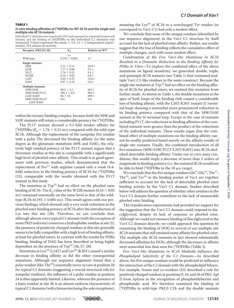
TABLE 1In vitro binding affinities of [3H]PDBu for WT �C1b and the single andmultiple site �C1b mutantsIndividual C1 domains were fusedwithGST and expressed in a bacterial system andisolated, and the binding of [3H]PDBu to the individual C1 domains wasmeasured. Values represent the mean � S.E. (n � 3 independent experi-ments). NA means no activity.
Receptor (PKC� C1b) Kd Relative to WT
nMWild type 0.193 � 0.005 1�
Single mutantsM9E 2.11 � 0.11 10.9�S10E 1.77 � 0.06 9.1�P11T 1.76 � 0.21 9.1�W22R 0.16 � 0.02 0.8�L24T 2.63 � 0.15 13.6�K26Y 0.97 � 0.10 5.0�
Multiple mutantsM9E/S10E 50.5 � 2.1 261�M9E/S10E/P11T 186.1 � 6.0 963�L24T/K26Y 96 � 18 499�M9E/S10E/P11T/
L24T/K26YNA NA
C1 Domain of Vav1
APRIL 13, 2012 • VOLUME 287 • NUMBER 16 JOURNAL OF BIOLOGICAL CHEMISTRY 13145
by guest on September 7, 2018
http://ww
w.jbc.org/
Dow
nloaded from
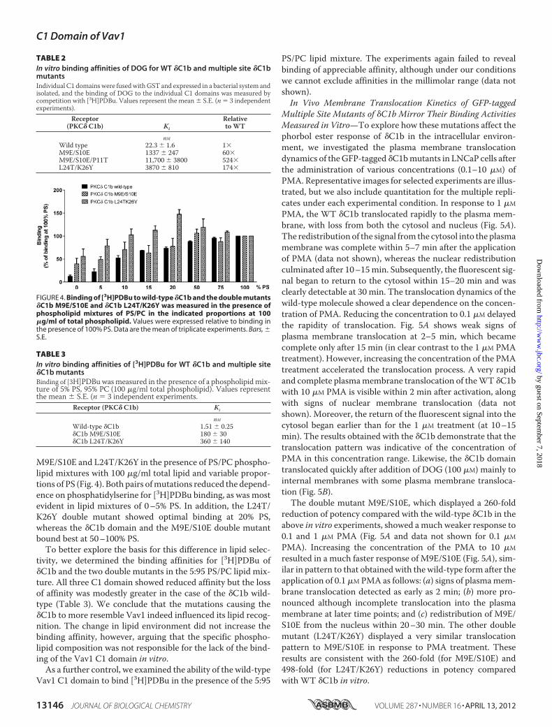
M9E/S10E and L24T/K26Y in the presence of PS/PC phospho-lipid mixtures with 100 �g/ml total lipid and variable propor-tions of PS (Fig. 4). Both pairs ofmutations reduced the depend-ence on phosphatidylserine for [3H]PDBu binding, as was mostevident in lipid mixtures of 0–5% PS. In addition, the L24T/K26Y double mutant showed optimal binding at 20% PS,whereas the �C1b domain and the M9E/S10E double mutantbound best at 50–100% PS.To better explore the basis for this difference in lipid selec-
tivity, we determined the binding affinities for [3H]PDBu of�C1b and the two double mutants in the 5:95 PS/PC lipid mix-ture. All three C1 domain showed reduced affinity but the lossof affinity was modestly greater in the case of the �C1b wild-type (Table 3). We conclude that the mutations causing the�C1b to more resemble Vav1 indeed influenced its lipid recog-nition. The change in lipid environment did not increase thebinding affinity, however, arguing that the specific phospho-lipid composition was not responsible for the lack of the bind-ing of the Vav1 C1 domain in vitro.
As a further control, we examined the ability of the wild-typeVav1 C1 domain to bind [3H]PDBu in the presence of the 5:95
PS/PC lipid mixture. The experiments again failed to revealbinding of appreciable affinity, although under our conditionswe cannot exclude affinities in the millimolar range (data notshown).In Vivo Membrane Translocation Kinetics of GFP-tagged
Multiple Site Mutants of �C1b Mirror Their Binding ActivitiesMeasured in Vitro—To explore how these mutations affect thephorbol ester response of �C1b in the intracellular environ-ment, we investigated the plasma membrane translocationdynamics of theGFP-tagged �C1bmutants in LNCaP cells afterthe administration of various concentrations (0.1–10 �M) ofPMA. Representative images for selected experiments are illus-trated, but we also include quantitation for the multiple repli-cates under each experimental condition. In response to 1 �M
PMA, the WT �C1b translocated rapidly to the plasma mem-brane, with loss from both the cytosol and nucleus (Fig. 5A).The redistribution of the signal from the cytosol into the plasmamembrane was complete within 5–7 min after the applicationof PMA (data not shown), whereas the nuclear redistributionculminated after 10–15min. Subsequently, the fluorescent sig-nal began to return to the cytosol within 15–20 min and wasclearly detectable at 30 min. The translocation dynamics of thewild-type molecule showed a clear dependence on the concen-tration of PMA. Reducing the concentration to 0.1 �M delayedthe rapidity of translocation. Fig. 5A shows weak signs ofplasma membrane translocation at 2–5 min, which becamecomplete only after 15 min (in clear contrast to the 1 �M PMAtreatment). However, increasing the concentration of the PMAtreatment accelerated the translocation process. A very rapidand complete plasmamembrane translocation of theWT �C1bwith 10 �M PMA is visible within 2 min after activation, alongwith signs of nuclear membrane translocation (data notshown). Moreover, the return of the fluorescent signal into thecytosol began earlier than for the 1 �M treatment (at 10–15min). The results obtained with the �C1b demonstrate that thetranslocation pattern was indicative of the concentration ofPMA in this concentration range. Likewise, the �C1b domaintranslocated quickly after addition of DOG (100 �M) mainly tointernal membranes with some plasma membrane transloca-tion (Fig. 5B).The double mutant M9E/S10E, which displayed a 260-fold
reduction of potency compared with the wild-type �C1b in theabove in vitro experiments, showed amuch weaker response to0.1 and 1 �M PMA (Fig. 5A and data not shown for 0.1 �M
PMA). Increasing the concentration of the PMA to 10 �M
resulted in a much faster response of M9E/S10E (Fig. 5A), sim-ilar in pattern to that obtained with the wild-type form after theapplication of 0.1�MPMA as follows: (a) signs of plasmamem-brane translocation detected as early as 2 min; (b) more pro-nounced although incomplete translocation into the plasmamembrane at later time points; and (c) redistribution of M9E/S10E from the nucleus within 20–30 min. The other doublemutant (L24T/K26Y) displayed a very similar translocationpattern to M9E/S10E in response to PMA treatment. Theseresults are consistent with the 260-fold (for M9E/S10E) and498-fold (for L24T/K26Y) reductions in potency comparedwith WT �C1b in vitro.
FIGURE 4. Binding of [3H]PDBu to wild-type �C1b and the double mutants�C1b M9E/S10E and �C1b L24T/K26Y was measured in the presence ofphospholipid mixtures of PS/PC in the indicated proportions at 100�g/ml of total phospholipid. Values were expressed relative to binding inthe presence of 100% PS. Data are the mean of triplicate experiments. Bars, �S.E.
TABLE 2In vitro binding affinities of DOG for WT �C1b and multiple site �C1bmutantsIndividual C1 domains were fusedwithGST and expressed in a bacterial system andisolated, and the binding of DOG to the individual C1 domains was measured bycompetition with [3H]PDBu. Values represent the mean � S.E. (n � 3 independentexperiments).
Receptor(PKC� C1b) Ki
Relativeto WT
nMWild type 22.3 � 1.6 1�M9E/S10E 1337 � 247 60�M9E/S10E/P11T 11,700 � 3800 524�L24T/K26Y 3870 � 810 174�
TABLE 3In vitro binding affinities of [3H]PDBu for WT �C1b and multiple site�C1b mutantsBinding of [3H]PDBuwasmeasured in the presence of a phospholipidmix-ture of 5% PS, 95% PC (100 �g/ml total phospholipid). Values representthe mean � S.E. (n � 3 independent experiments.
Receptor (PKC� C1b) Ki
nMWild-type �C1b 1.51 � 0.25�C1b M9E/S10E 180 � 30�C1b L24T/K26Y 360 � 140
C1 Domain of Vav1
13146 JOURNAL OF BIOLOGICAL CHEMISTRY VOLUME 287 • NUMBER 16 • APRIL 13, 2012
by guest on September 7, 2018
http://ww
w.jbc.org/
Dow
nloaded from
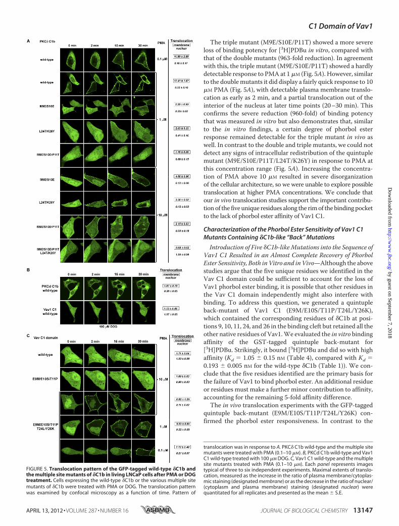
The triple mutant (M9E/S10E/P11T) showed a more severeloss of binding potency for [3H]PDBu in vitro, compared withthat of the double mutants (963-fold reduction). In agreementwith this, the triple mutant (M9E/S10E/P11T) showed a hardlydetectable response to PMA at 1�M (Fig. 5A). However, similarto the doublemutants it did display a fairly quick response to 10�M PMA (Fig. 5A), with detectable plasma membrane translo-cation as early as 2 min, and a partial translocation out of theinterior of the nucleus at later time points (20–30 min). Thisconfirms the severe reduction (960-fold) of binding potencythat was measured in vitro but also demonstrates that, similarto the in vitro findings, a certain degree of phorbol esterresponse remained detectable for the triple mutant in vivo aswell. In contrast to the double and triple mutants, we could notdetect any signs of intracellular redistribution of the quintuplemutant (M9E/S10E/P11T/L24T/K26Y) in response to PMA atthis concentration range (Fig. 5A). Increasing the concentra-tion of PMA above 10 �M resulted in severe disorganizationof the cellular architecture, so we were unable to explore possibletranslocation at higher PMA concentrations. We conclude thatour in vivo translocation studies support the important contribu-tionof the five unique residues along the rimof the bindingpocketto the lack of phorbol ester affinity of Vav1 C1.
Characterization of the Phorbol Ester Sensitivity of Vav1 C1Mutants Containing �C1b-like “Back” Mutations
Introduction of Five �C1b-likeMutations into the Sequence ofVav1 C1 Resulted in an Almost Complete Recovery of PhorbolEster Sensitivity, Both inVitro and inVivo—Although the abovestudies argue that the five unique residues we identified in theVav C1 domain could be sufficient to account for the loss ofVav1 phorbol ester binding, it is possible that other residues inthe Vav C1 domain independently might also interfere withbinding. To address this question, we generated a quintupleback-mutant of Vav1 C1 (E9M/E10S/T11P/T24L/Y26K),which contained the corresponding residues of �C1b at posi-tions 9, 10, 11, 24, and 26 in the binding cleft but retained all theother native residues of Vav1.We evaluated the in vitro bindingaffinity of the GST-tagged quintuple back-mutant for[3H]PDBu. Strikingly, it bound [3H]PDBu and did so with highaffinity (Kd � 1.05 � 0.15 nM (Table 4), compared with Kd �0.193 � 0.005 nM for the wild-type �C1b (Table 1)). We con-clude that the five residues identified are the primary basis forthe failure of Vav1 to bind phorbol ester. An additional residueor residues must make a further minor contribution to affinity,accounting for the remaining 5-fold affinity difference.The in vivo translocation experiments with the GFP-tagged
quintuple back-mutant (E9M/E10S/T11P/T24L/Y26K) con-firmed the phorbol ester responsiveness. In contrast to the
translocation was in response to A. PKC� C1b wild-type and the multiple sitemutants were treated with PMA (0.1–10 �M). B, PKCd C1b wild-type and Vav1C1 wild-type treated with 100 �M DOG. C, Vav1 C1 wild-type and the multiplesite mutants treated with PMA (0.1–10 �M). Each panel represents imagestypical of three to six independent experiments. Maximal extents of translo-cation, measured as the increase in the ratio of plasma membrane/cytoplas-mic staining (designated membrane) or as the decrease in the ratio of nuclear/(cytoplasm and plasma membrane) staining (designated nuclear) werequantitated for all replicates and presented as the mean � S.E.
FIGURE 5. Translocation pattern of the GFP-tagged wild-type �C1b andthe multiple site mutants of �C1b in living LNCaP cells after PMA or DOGtreatment. Cells expressing the wild-type �C1b or the various multiple sitemutants of �C1b were treated with PMA or DOG. The translocation patternwas examined by confocal microscopy as a function of time. Pattern of
C1 Domain of Vav1
APRIL 13, 2012 • VOLUME 287 • NUMBER 16 JOURNAL OF BIOLOGICAL CHEMISTRY 13147
by guest on September 7, 2018
http://ww
w.jbc.org/
Dow
nloaded from
wild-type Vav1 C1, which showed no response to phorbol esterorDOG treatment, the quintuple back-mutant displayed trans-location in response to as little as 0.1�MPMA (Fig. 5C), reflect-ing high phorbol ester sensitivity comparable with thatobtained with WT �C1b. In addition, the translocation kinet-ics/pattern of the quintuple Vav1 back-mutant, like that of thewild-type �C1b, showed a dose-dependent change on PMAconcentration, i.e. increasing the concentration of PMA accel-erated the process of plasma membrane translocation and thatof nuclear translocation.Collectively, the almost complete recovery of the binding
affinity of Vav1 C1 E9M/E10S/T11P/T24L/Y26K (consistentwith the results obtained with �C1b mutants) further supportprevious crystallographic results (15) and our docking results,which demonstrate a retained binding cleft geometry for Vav1C1 and a conserved binding pocket for ligand interaction. Thereason for the lack of ligand affinity is the presence of these fiveresidues at the rim of the binding pocket which presumablyhinder the association of the C1 domain with the cellularmem-brane, thereby destabilizing the ternary binding complex andreducing the potency for phorbol esters.Introduction of Double-, Triple-, and Quadruple �C1b-like
Mutations into the Sequence of Vav1 C1 Further Dissects theRole of the Individual Residues in the LigandAffinity of Vav1C1—To further explore whether all five residues actually contributeto the lack of binding potency in theWTVav1C1, we generateddouble, triple, and quadruple back-mutants of Vav1 C1 (seeTable 4). Introducing two back mutations into the N-terminal(Vav1 C1 E9M/E10S) or the C-terminal (T24L/Y26K) loop ofthe apparent binding cleft of Vav1 C1 was insufficient torecover the phorbol ester response of the molecule. In vitroassays could not detect any measurable [3H]PDBu binding(Table 4), and no translocation could be observed in vivo after10 �M PMA treatment (data not shown). Back-mutating thethree residues in the N-terminal loop (E9M/E10S/T11P) con-ferred only a very weak binding potency (Kd � 7330 � 490 nM)on the triple back-mutant (Table 4), which could only bedetected in vitro but not with translocation assays (data notshown). These results suggest that the presence of compatibleresidues at the tip of both loops is needed for appropriate bind-ing affinity. They stand in partial contrast to the previous find-ings with the double (M9E/S10E; L24T/K26Y) and triple (M9E/S10E/P11T)mutants of �C1b (Table 1 andFig. 5A), which failed
to completely abolish the binding potency of the �C1bmutants.Because the difference lies in the presence of other residues inthe PKC� C1b domain versus Vav1, it emphasizes both thatthese residues make a difference and that the effects of thecombination of residues are not simply additive.Next, using quadruple back-mutants of Vav1 C1 (listed in
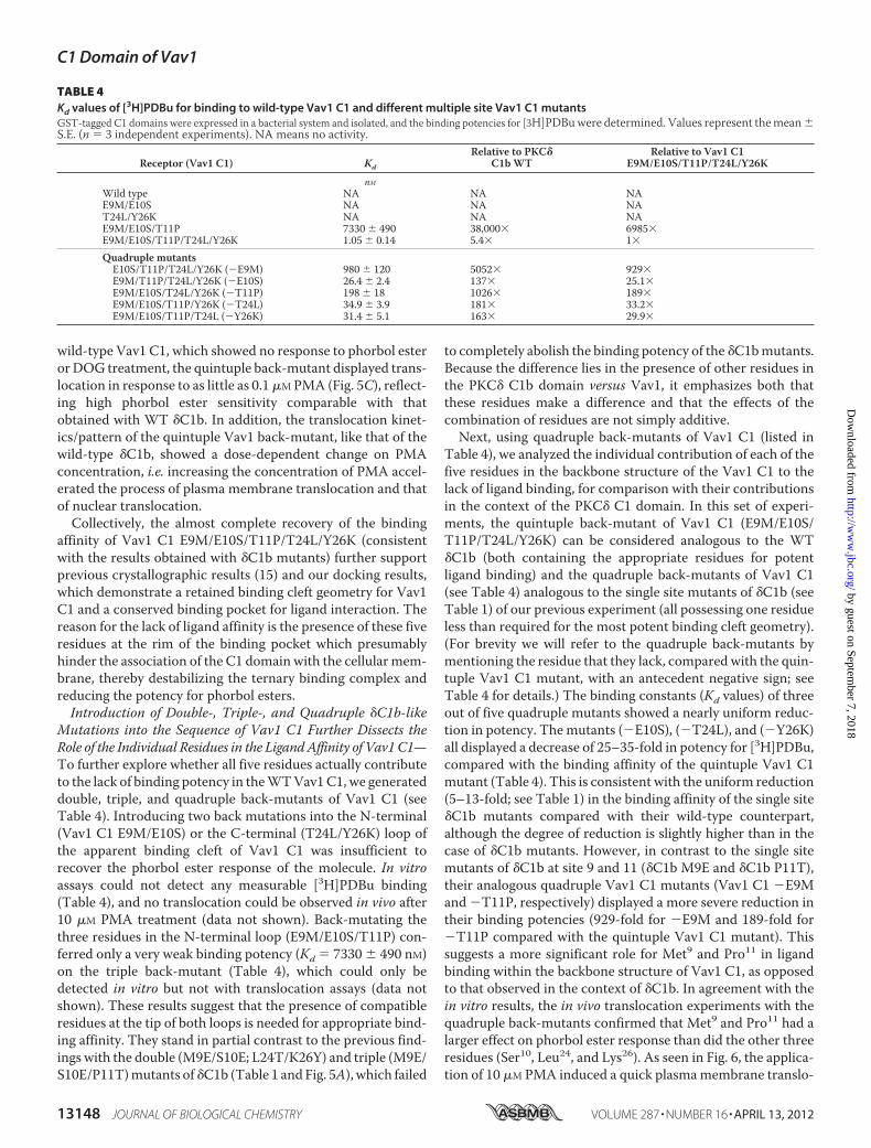
Table 4), we analyzed the individual contribution of each of thefive residues in the backbone structure of the Vav1 C1 to thelack of ligand binding, for comparison with their contributionsin the context of the PKC� C1 domain. In this set of experi-ments, the quintuple back-mutant of Vav1 C1 (E9M/E10S/T11P/T24L/Y26K) can be considered analogous to the WT�C1b (both containing the appropriate residues for potentligand binding) and the quadruple back-mutants of Vav1 C1(see Table 4) analogous to the single site mutants of �C1b (seeTable 1) of our previous experiment (all possessing one residueless than required for the most potent binding cleft geometry).(For brevity we will refer to the quadruple back-mutants bymentioning the residue that they lack, compared with the quin-tuple Vav1 C1 mutant, with an antecedent negative sign; seeTable 4 for details.) The binding constants (Kd values) of threeout of five quadruple mutants showed a nearly uniform reduc-tion in potency. Themutants (�E10S), (�T24L), and (�Y26K)all displayed a decrease of 25–35-fold in potency for [3H]PDBu,compared with the binding affinity of the quintuple Vav1 C1mutant (Table 4). This is consistent with the uniform reduction(5–13-fold; see Table 1) in the binding affinity of the single site�C1b mutants compared with their wild-type counterpart,although the degree of reduction is slightly higher than in thecase of �C1b mutants. However, in contrast to the single sitemutants of �C1b at site 9 and 11 (�C1b M9E and �C1b P11T),their analogous quadruple Vav1 C1 mutants (Vav1 C1 �E9Mand �T11P, respectively) displayed a more severe reduction intheir binding potencies (929-fold for �E9M and 189-fold for�T11P compared with the quintuple Vav1 C1 mutant). Thissuggests a more significant role for Met9 and Pro11 in ligandbinding within the backbone structure of Vav1 C1, as opposedto that observed in the context of �C1b. In agreement with thein vitro results, the in vivo translocation experiments with thequadruple back-mutants confirmed that Met9 and Pro11 had alarger effect on phorbol ester response than did the other threeresidues (Ser10, Leu24, and Lys26). As seen in Fig. 6, the applica-tion of 10 �M PMA induced a quick plasmamembrane translo-
TABLE 4Kd values of [3H]PDBu for binding to wild-type Vav1 C1 and different multiple site Vav1 C1 mutantsGST-tagged C1 domains were expressed in a bacterial system and isolated, and the binding potencies for [3H]PDBuwere determined. Values represent themean�S.E. (n � 3 independent experiments). NA means no activity.
Receptor (Vav1 C1) Kd
Relative to PKC�C1bWT
Relative to Vav1 C1E9M/E10S/T11P/T24L/Y26K
nMWild type NA NA NAE9M/E10S NA NA NAT24L/Y26K NA NA NAE9M/E10S/T11P 7330 � 490 38,000� 6985�E9M/E10S/T11P/T24L/Y26K 1.05 � 0.14 5.4� 1�
Quadruple mutantsE10S/T11P/T24L/Y26K (�E9M) 980 � 120 5052� 929�E9M/T11P/T24L/Y26K (�E10S) 26.4 � 2.4 137� 25.1�E9M/E10S/T24L/Y26K (�T11P) 198 � 18 1026� 189�E9M/E10S/T11P/Y26K (�T24L) 34.9 � 3.9 181� 33.2�E9M/E10S/T11P/T24L (�Y26K) 31.4 � 5.1 163� 29.9�
C1 Domain of Vav1
13148 JOURNAL OF BIOLOGICAL CHEMISTRY VOLUME 287 • NUMBER 16 • APRIL 13, 2012
by guest on September 7, 2018
http://ww
w.jbc.org/
Dow
nloaded from
cation in the case of the �E10S, �T24L, and �Y26K mutants.In addition, although the redistribution from the cytosol intothe plasma membrane was not complete, we could observe afairly strong fluorescent signal in the membrane within 15 minafter the administration of the drug, and an almost completetranslocation out of the interior of the nucleus. In contrast, the�E9M mutant displayed only a weak translocation into theplasma membrane that had much slower kinetics than that ofthe previous threemutants andwas limited to certain regions ofthe plasma membrane. Moreover, the nuclear translocationwas also limited for this mutant. The translocation of the�T11P mutant in response to PMA displayed an intermediatepattern compared with that of the three more potent mutants(�E10S, �T24L, and �Y26K) and �E9M, which showed theweakest response. Although a fairly quick initial plasma mem-brane translocation could be detected, the redistribution fromthe cytosol was only partial, and the fluorescent signal wasabsent from certain regions of the plasma membrane. In addi-tion, translocation out of the nucleus was quite limited.
Collectively, the results obtained with the quadruple Vav1 C1mutants emphasize theprominent contributionofMet9 andPro11to the binding affinity of the quintuple Vav1 C1 mutant (E9M/E10S/T11P/T24L/Y26K) for phorbol esters, which in turn indi-cates that Glu9 andThr11make amore significant contribution tothe reduction of the binding potency of the wild-type Vav1 C1,compared with the other three residues (Glu10, Thr24, and Tyr26).
Rationalizing the Phorbol Binding and MembraneTranslocation Behavior of PKC� and Vav1 Mutants in Terms ofMolecular Lipophilicity
A plausible interpretation of the role played by the fiveunique residues was that the lack of phorbol ester responsive-ness in Vav1 was due to the inability of its C1 domain to asso-ciate with and insert into the membrane, forming the ternaryprotein� ligand� lipid complex that is required for successfulligand binding. To better assess this model, we developed amethod of calculating and quantifying the differences in lipo-philicity between our various C1 domain mutants. We were
FIGURE 6. Translocation pattern of the GFP-tagged quintuple and quadruple Vav1 C1 back-mutants in living LNCaP cells after PMA treatment. Cellsexpressing the quintuple back-mutant Vav1 C1 E9M/E10S/T11P/T24L/Y26K and the indicated quadruple back-mutants were treated with 10 �M PMA. Cellswere imaged by confocal microscopy, and images captured at the indicated times are shown. Each panel represents a typical image of three to five indepen-dent experiments. Maximal extents of translocation, measured as the increase in the ratio of plasma membrane/cytoplasmic staining (designated membrane)or as the decrease in the ratio of nuclear/(cytoplasm and plasma membrane) staining (designated nuclear) were quantitated for all replicates and presented asthe mean � S.E.
C1 Domain of Vav1
APRIL 13, 2012 • VOLUME 287 • NUMBER 16 JOURNAL OF BIOLOGICAL CHEMISTRY 13149
by guest on September 7, 2018
http://ww
w.jbc.org/
Dow
nloaded from
interested in seeing how the membrane affinity of a C1 domainmutant correlatedwith itsKd value for PDBu and itsmembranetranslocation behavior.The standard metric for quantifying the hydrophobicity or
lipophilicity of small molecules is logP, a measure of the parti-tion coefficient between octanol and water. However, logP is awhole-molecule number and as such is not particularly usefulfor characterizingmacromolecules, where the lipophilicitymayvary significantly between different parts of the structure. Thisis especially true for peripheral membrane proteins such as thetypical (DAG/phorbol-responsive) C1 domains. Computa-tional methods for estimating logP generally involve summingup the fractional lipophilicities of individual atoms or smallfragments. These values are based on regression analysis fromlibraries of small molecules with carefully measured experi-mental values of logP (44). The concept of an MLP is an exten-sion of these types of calculations that can be applied to largermolecules such as proteins. The MLP is analogous to an elec-trostatic potential around a protein based on the partial chargeson individual atoms. Each atom is assigned an atomic logP(AlogP) value, and these values are then projected outwardfrom the underlying atoms onto the solvent-accessible molec-ular surface. In otherwords, the lipophilicity value at each pointon the surface is a smoothed distance-dependent function ofthe fractional logP values of all nearby atoms (59). An algorithmfor this calculation has been implemented in the software pro-gramVASCo (43), which also includes a plugin for PyMOL (60)for surface visualization.We built model structures for each of the single andmultiple
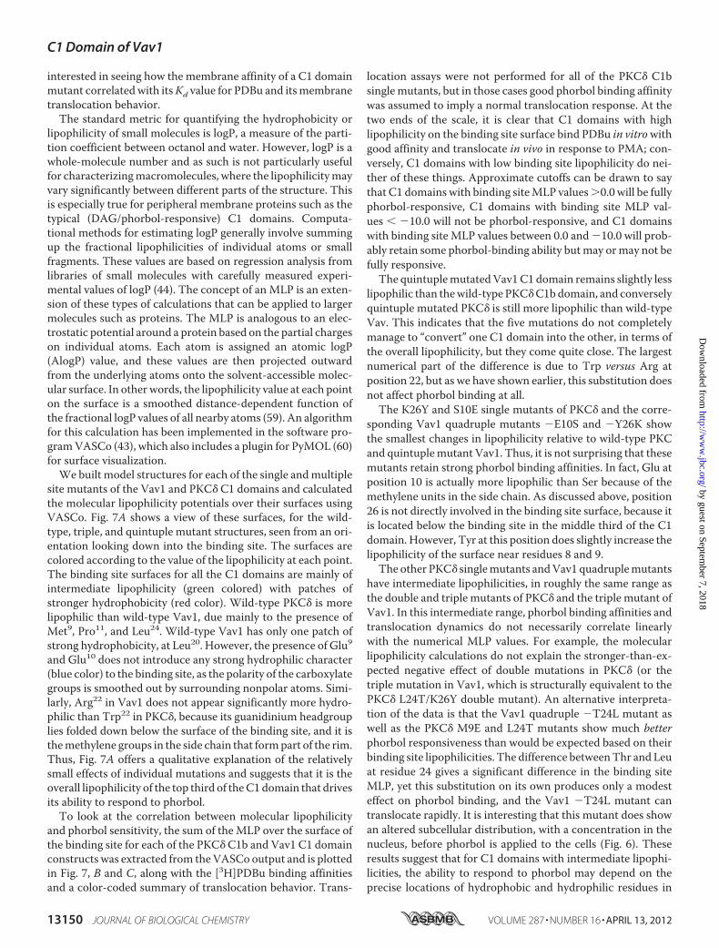
site mutants of the Vav1 and PKC� C1 domains and calculatedthe molecular lipophilicity potentials over their surfaces usingVASCo. Fig. 7A shows a view of these surfaces, for the wild-type, triple, and quintuple mutant structures, seen from an ori-entation looking down into the binding site. The surfaces arecolored according to the value of the lipophilicity at each point.The binding site surfaces for all the C1 domains are mainly ofintermediate lipophilicity (green colored) with patches ofstronger hydrophobicity (red color). Wild-type PKC� is morelipophilic than wild-type Vav1, due mainly to the presence ofMet9, Pro11, and Leu24. Wild-type Vav1 has only one patch ofstrong hydrophobicity, at Leu20. However, the presence of Glu9and Glu10 does not introduce any strong hydrophilic character(blue color) to the binding site, as the polarity of the carboxylategroups is smoothed out by surrounding nonpolar atoms. Simi-larly, Arg22 in Vav1 does not appear significantly more hydro-philic than Trp22 in PKC�, because its guanidinium headgrouplies folded down below the surface of the binding site, and it isthemethylene groups in the side chain that formpart of the rim.Thus, Fig. 7A offers a qualitative explanation of the relativelysmall effects of individual mutations and suggests that it is theoverall lipophilicity of the top third of theC1domain that drivesits ability to respond to phorbol.To look at the correlation between molecular lipophilicity
and phorbol sensitivity, the sum of theMLP over the surface ofthe binding site for each of the PKC� C1b and Vav1 C1 domainconstructs was extracted from theVASCo output and is plottedin Fig. 7, B and C, along with the [3H]PDBu binding affinitiesand a color-coded summary of translocation behavior. Trans-
location assays were not performed for all of the PKC� C1bsinglemutants, but in those cases good phorbol binding affinitywas assumed to imply a normal translocation response. At thetwo ends of the scale, it is clear that C1 domains with highlipophilicity on the binding site surface bind PDBu in vitrowithgood affinity and translocate in vivo in response to PMA; con-versely, C1 domains with low binding site lipophilicity do nei-ther of these things. Approximate cutoffs can be drawn to saythat C1 domains with binding siteMLP values�0.0 will be fullyphorbol-responsive, C1 domains with binding site MLP val-ues �10.0 will not be phorbol-responsive, and C1 domainswith binding site MLP values between 0.0 and �10.0 will prob-ably retain some phorbol-binding ability butmay ormay not befully responsive.The quintuplemutatedVav1C1 domain remains slightly less
lipophilic than thewild-type PKC�C1bdomain, and converselyquintuple mutated PKC� is still more lipophilic than wild-typeVav. This indicates that the five mutations do not completelymanage to “convert” one C1 domain into the other, in terms ofthe overall lipophilicity, but they come quite close. The largestnumerical part of the difference is due to Trp versus Arg atposition 22, but as we have shown earlier, this substitution doesnot affect phorbol binding at all.The K26Y and S10E single mutants of PKC� and the corre-
sponding Vav1 quadruple mutants �E10S and �Y26K showthe smallest changes in lipophilicity relative to wild-type PKCand quintuplemutant Vav1. Thus, it is not surprising that thesemutants retain strong phorbol binding affinities. In fact, Glu atposition 10 is actually more lipophilic than Ser because of themethylene units in the side chain. As discussed above, position26 is not directly involved in the binding site surface, because itis located below the binding site in the middle third of the C1domain. However, Tyr at this position does slightly increase thelipophilicity of the surface near residues 8 and 9.The other PKC� singlemutants andVav1 quadruplemutants
have intermediate lipophilicities, in roughly the same range asthe double and triple mutants of PKC� and the triple mutant ofVav1. In this intermediate range, phorbol binding affinities andtranslocation dynamics do not necessarily correlate linearlywith the numerical MLP values. For example, the molecularlipophilicity calculations do not explain the stronger-than-ex-pected negative effect of double mutations in PKC� (or thetriple mutation in Vav1, which is structurally equivalent to thePKC� L24T/K26Y double mutant). An alternative interpreta-tion of the data is that the Vav1 quadruple �T24L mutant aswell as the PKC� M9E and L24T mutants show much betterphorbol responsiveness than would be expected based on theirbinding site lipophilicities. The difference betweenThr and Leuat residue 24 gives a significant difference in the binding siteMLP, yet this substitution on its own produces only a modesteffect on phorbol binding, and the Vav1 �T24L mutant cantranslocate rapidly. It is interesting that this mutant does showan altered subcellular distribution, with a concentration in thenucleus, before phorbol is applied to the cells (Fig. 6). Theseresults suggest that for C1 domains with intermediate lipophi-licities, the ability to respond to phorbol may depend on theprecise locations of hydrophobic and hydrophilic residues in
C1 Domain of Vav1
13150 JOURNAL OF BIOLOGICAL CHEMISTRY VOLUME 287 • NUMBER 16 • APRIL 13, 2012
by guest on September 7, 2018
http://ww
w.jbc.org/
Dow
nloaded from
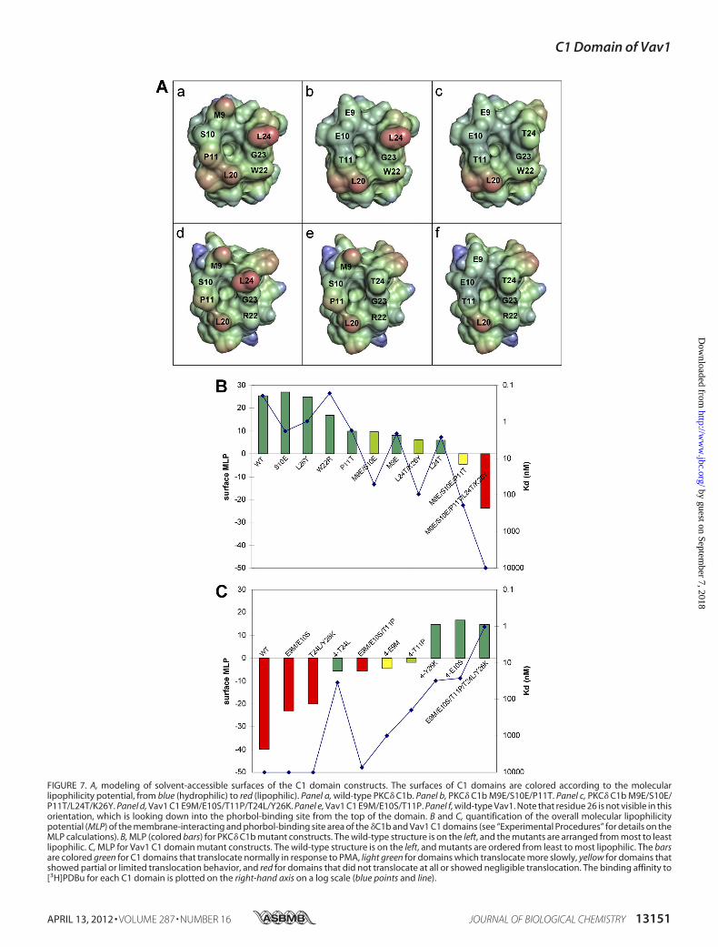
FIGURE 7. A, modeling of solvent-accessible surfaces of the C1 domain constructs. The surfaces of C1 domains are colored according to the molecularlipophilicity potential, from blue (hydrophilic) to red (lipophilic). Panel a, wild-type PKC� C1b. Panel b, PKC� C1b M9E/S10E/P11T. Panel c, PKC� C1b M9E/S10E/P11T/L24T/K26Y. Panel d, Vav1 C1 E9M/E10S/T11P/T24L/Y26K. Panel e, Vav1 C1 E9M/E10S/T11P. Panel f, wild-type Vav1. Note that residue 26 is not visible in thisorientation, which is looking down into the phorbol-binding site from the top of the domain. B and C, quantification of the overall molecular lipophilicitypotential (MLP) of the membrane-interacting and phorbol-binding site area of the �C1b and Vav1 C1 domains (see “Experimental Procedures” for details on theMLP calculations). B, MLP (colored bars) for PKC� C1b mutant constructs. The wild-type structure is on the left, and the mutants are arranged from most to leastlipophilic. C, MLP for Vav1 C1 domain mutant constructs. The wild-type structure is on the left, and mutants are ordered from least to most lipophilic. The barsare colored green for C1 domains that translocate normally in response to PMA, light green for domains which translocate more slowly, yellow for domains thatshowed partial or limited translocation behavior, and red for domains that did not translocate at all or showed negligible translocation. The binding affinity to[3H]PDBu for each C1 domain is plotted on the right-hand axis on a log scale (blue points and line).
C1 Domain of Vav1
APRIL 13, 2012 • VOLUME 287 • NUMBER 16 JOURNAL OF BIOLOGICAL CHEMISTRY 13151
by guest on September 7, 2018
http://ww
w.jbc.org/
Dow
nloaded from
the binding site, and the balance of lipophilicity across the C1domain.
Quintuple Mutated C1 Domain of Vav1 Retains Its PhorbolEster Sensitivity in the Context of the Full-length Molecule andIs Capable of Targeting the Protein to the Cellular Membranein Response to Ligand
To elucidatewhether the binding cleft of theVav1C1 is capa-ble of interacting with phorbol esters in the context of the full-length molecule, we generated a full-length quintuple Vav1back-mutant, which contained the same five back-mutations(E9M/E10S/T11P/T24L/Y26K) in its C1 domain as the quintu-ple mutant of the isolated Vav1 C1. We speculated that if thebinding cleft is not occluded by other domains in the full-lengthmolecule, the introduction of the five crucial residues into theC1 domain would confer phorbol ester sensitivity on the full-length Vav1 protein as well. We cloned the genes of the quin-tuple full-length mutant (Vav1 E9M/E10S/T11P/T24L/Y26K)along with its wild-type counterpart into a vector encoding anN-terminal GFP fusion tag, and we subjected them to in vivotranslocation assays. (Because of technical difficulties with pro-ducing sufficient amounts of purified protein, we were unableto conduct in vitro binding experiments on full-length Vav1.)Fig. 8 shows that, similar to the findings with the isolated Vav1C1, the full-length quintuple back-mutant of Vav1 (Vav1 E9M/E10S/T11P/T24L/Y26K) possessed affinity for phorbol esters,because the application of 10 �M PMA resulted in the translo-cation of the protein into the plasma membrane. In clear con-trast with the quintuple Vav1 mutant, the wild-type and thetriple mutant (Vav1 E9M/E10S/T11P) of the full-length Vav1failed to redistribute from the cytosol into the plasma mem-brane (similar to the results obtained with the isolated Vav1C1). These results demonstrate unambiguously that replacingthe residues of Vav1 at positions 9–11, 24, and 26 (of its C1domain) with �C1b-like amino acids confers phorbol ester sen-
sitivity on the full-length Vav1 protein as well. In the context ofthe interaction of the C1 domain of Vav1 with the DH and PHdomains (Fig. 9A), it can be seen that the three residues on theN-terminal loop point out into solvent. Thus, although mutat-ing these three residues can provide some detectable bindingaffinity for phorbol, their mutation would appear not to disruptthe quaternary packing of the C1 domain. In contrast, both ofthe mutations in the C-terminal loop would affect its interac-tions with the rest of the protein. The T24L mutation clasheswith residue Val373 in helix�6 of the DHdomain, and the Y26Kmutation introduces an unfavorable electrostatic interactionbetween the lysine and either residueArg402 or residue Lys429 inthe PH domain. This suggests that the N-terminal loop muta-tions act to increase the lipophilicity of theC1domain formem-brane binding and the C-terminal loop mutations act to loosenor disrupt the packing of the C1 domain as well as increasinglipophilicity. Both sets of mutations are required for respon-siveness to phorbol.Themodeling has a further implication. Because of the prox-
imity of the C-terminal loop of the C1 domain of Vav1 to the�6helix of the DH domain, a prediction is that an appropriatelybulky ligand binding to the C1 domain should disrupt interac-tion with the DH domain (Fig. 9B). Because the C1 domaininteraction is required to stabilize the GEF activity of the DHdomain, such a ligand might be expected to function as a Vav1inhibitor.
Comparison of the Binding Affinities of Novel DAG-lactonesDesigned to Target the Structure of Vav1 C1
The modeling and site-directed mutagenesis resultsshown above suggested that eliminating the charged Glu res-idues in the N-terminal loop is necessary (but not sufficient)for restoring phorbol sensitivity to Vav1.We theorized that aligand bearing a positive charge might similarly “cancel out”the membrane binding penalty imposed on the C1 domain
FIGURE 8. Translocation pattern of the full-length versions of GFP-tagged wild-type Vav1 and the triple and quintuple Vav1 back-mutants in livingLNCaP cells after PMA treatment. Cells expressing the full-length proteins of the wild-type Vav1, of the triple back-mutant, and of the quintuple back-mutantwere imaged by confocal microscopy after the application of 10 �M PMA. Images captured at the indicated times are shown. Each panel represents a typicalimage of four to six independent experiments. Maximal extents of translocation, measured as the increase in the ratio of plasma membrane/cytoplasmicstaining (designated “membrane”) were quantitated for all replicates and presented as the mean � S.E.
C1 Domain of Vav1
13152 JOURNAL OF BIOLOGICAL CHEMISTRY VOLUME 287 • NUMBER 16 • APRIL 13, 2012
by guest on September 7, 2018
http://ww
w.jbc.org/
Dow
nloaded from
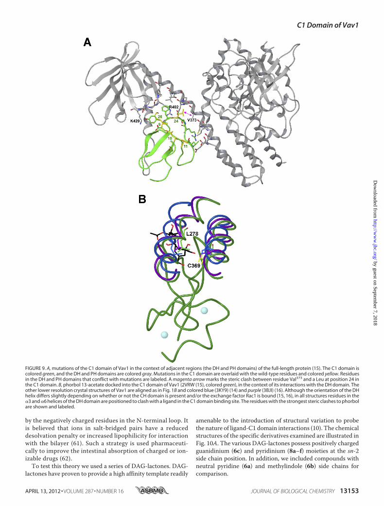
by the negatively charged residues in the N-terminal loop. Itis believed that ions in salt-bridged pairs have a reduceddesolvation penalty or increased lipophilicity for interactionwith the bilayer (61). Such a strategy is used pharmaceuti-cally to improve the intestinal absorption of charged or ion-izable drugs (62).To test this theory we used a series of DAG-lactones. DAG-
lactones have proven to provide a high affinity template readily
amenable to the introduction of structural variation to probethe nature of ligand-C1 domain interactions (10). The chemicalstructures of the specific derivatives examined are illustrated inFig. 10A. The various DAG-lactones possess positively chargedguanidinium (6c) and pyridinium (8a–f) moieties at the sn-2side chain position. In addition, we included compounds withneutral pyridine (6a) and methylindole (6b) side chains forcomparison.
FIGURE 9. A, mutations of the C1 domain of Vav1 in the context of adjacent regions (the DH and PH domains) of the full-length protein (15). The C1 domain iscolored green, and the DH and PH domains are colored gray. Mutations in the C1 domain are overlaid with the wild-type residues and colored yellow. Residuesin the DH and PH domains that conflict with mutations are labeled. A magenta arrow marks the steric clash between residue Val373 and a Leu at position 24 inthe C1 domain. B, phorbol 13-acetate docked into the C1 domain of Vav1 (2VRW (15), colored green), in the context of its interactions with the DH domain. Theother lower resolution crystal structures of Vav1 are aligned as in Fig. 1B and colored blue (3KY9) (14) and purple (3BJI) (16). Although the orientation of the DHhelix differs slightly depending on whether or not the CH domain is present and/or the exchange factor Rac1 is bound (15, 16), in all structures residues in the�3 and �6 helices of the DH domain are positioned to clash with a ligand in the C1 domain binding site. The residues with the strongest steric clashes to phorbolare shown and labeled.
C1 Domain of Vav1
APRIL 13, 2012 • VOLUME 287 • NUMBER 16 JOURNAL OF BIOLOGICAL CHEMISTRY 13153
by guest on September 7, 2018
http://ww
w.jbc.org/
Dow
nloaded from
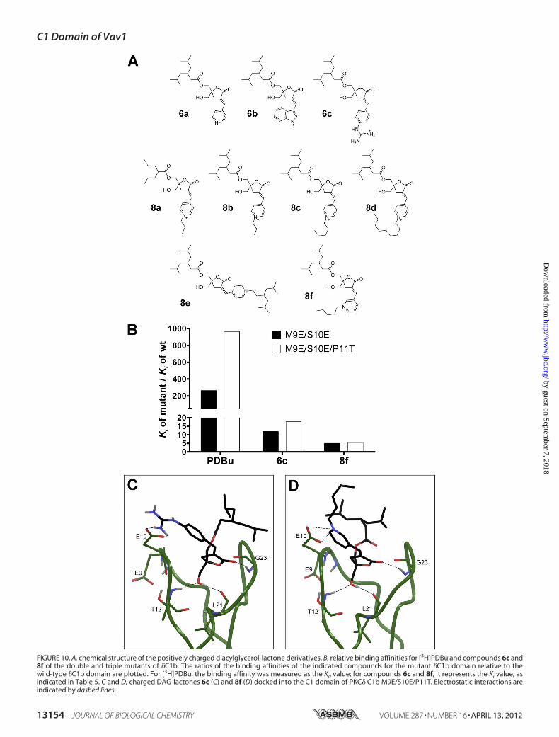
FIGURE 10. A, chemical structure of the positively charged diacylglycerol-lactone derivatives. B, relative binding affinities for [3H]PDBu and compounds 6c and8f of the double and triple mutants of �C1b. The ratios of the binding affinities of the indicated compounds for the mutant �C1b domain relative to thewild-type �C1b domain are plotted. For [3H]PDBu, the binding affinity was measured as the Kd value; for compounds 6c and 8f, it represents the Ki value, asindicated in Table 5. C and D, charged DAG-lactones 6c (C) and 8f (D) docked into the C1 domain of PKC� C1b M9E/S10E/P11T. Electrostatic interactions areindicated by dashed lines.
C1 Domain of Vav1
13154 JOURNAL OF BIOLOGICAL CHEMISTRY VOLUME 287 • NUMBER 16 • APRIL 13, 2012
by guest on September 7, 2018
http://ww
w.jbc.org/
Dow
nloaded from
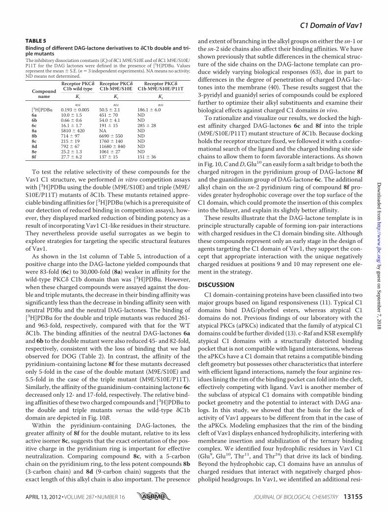
To test the relative selectivity of these compounds for theVav1 C1 structure, we performed in vitro competition assayswith [3H]PDBu using the double (M9E/S10E) and triple (M9E/S10E/P11T) mutants of �C1b. These mutants retained appre-ciable binding affinities for [3H]PDBu (which is a prerequisite ofour detection of reduced binding in competition assays), how-ever, they displayed marked reduction of binding potency as aresult of incorporating Vav1 C1-like residues in their structure.They nevertheless provide useful surrogates as we begin toexplore strategies for targeting the specific structural featuresof Vav1.As shown in the 1st column of Table 5, introduction of a
positive charge into the DAG-lactone yielded compounds thatwere 83-fold (6c) to 30,000-fold (8a) weaker in affinity for thewild-type PKC� C1b domain than was [3H]PDBu. However,when these charged compounds were assayed against the dou-ble and triplemutants, the decrease in their binding affinity wassignificantly less than the decrease in binding affinity seen withneutral PDBu and the neutral DAG-lactones. The binding of[3H]PDBu for the double and triple mutants was reduced 261-and 963-fold, respectively, compared with that for the WT�C1b. The binding affinities of the neutral DAG-lactones 6aand 6b to the doublemutant were also reduced 45- and 82-fold,respectively, consistent with the loss of binding that we hadobserved for DOG (Table 2). In contrast, the affinity of thepyridinium-containing lactone 8f for these mutants decreasedonly 5-fold in the case of the double mutant (M9E/S10E) and5.5-fold in the case of the triple mutant (M9E/S10E/P11T).Similarly, the affinity of the guanidinium-containing lactone 6cdecreased only 12- and 17-fold, respectively. The relative bind-ing affinities of these two charged compounds and [3H]PDBu tothe double and triple mutants versus the wild-type �C1bdomain are depicted in Fig. 10B.Within the pyridinium-containing DAG-lactones, the
greater affinity of 8f for the double mutant, relative to its lessactive isomer 8c, suggests that the exact orientation of the pos-itive charge in the pyridinium ring is important for effectiveneutralization. Comparing compound 8c, with a 5-carbonchain on the pyridinium ring, to the less potent compounds 8b(3-carbon chain) and 8d (9-carbon chain) suggests that theexact length of this alkyl chain is also important. The presence
and extent of branching in the alkyl groups on either the sn-1 orthe sn-2 side chains also affect their binding affinities. We haveshown previously that subtle differences in the chemical struc-ture of the side chains on the DAG-lactone template can pro-duce widely varying biological responses (63), due in part todifferences in the degree of penetration of charged DAG-lac-tones into the membrane (40). These results suggest that the3-pyridyl and guanidyl series of compounds could be exploredfurther to optimize their alkyl substituents and examine theirbiological effects against charged C1 domains in vivo.To rationalize and visualize our results, we docked the high-
est affinity charged DAG-lactones 6c and 8f into the triple(M9E/S10E/P11T) mutant structure of �C1b. Because dockingholds the receptor structure fixed, we followed it with a confor-mational search of the ligand and the charged binding site sidechains to allow them to form favorable interactions. As shownin Fig. 10,C andD, Glu10 can easily forma salt bridge to both thecharged nitrogen in the pyridinium group of DAG-lactone 8fand the guanidinium group of DAG-lactone 6c. The additionalalkyl chain on the sn-2 pyridinium ring of compound 8f pro-vides greater hydrophobic coverage over the top surface of theC1 domain, which could promote the insertion of this complexinto the bilayer, and explain its slightly better affinity.These results illustrate that the DAG-lactone template is in
principle structurally capable of forming ion-pair interactionswith charged residues in the C1 domain binding site. Althoughthese compounds represent only an early stage in the design ofagents targeting the C1 domain of Vav1, they support the con-cept that appropriate interaction with the unique negativelycharged residues at positions 9 and 10 may represent one ele-ment in the strategy.
DISCUSSION
C1 domain-containing proteins have been classified into twomajor groups based on ligand responsiveness (11). Typical C1domains bind DAG/phorbol esters, whereas atypical C1domains do not. Previous findings of our laboratory with theatypical PKCs (aPKCs) indicated that the family of atypical C1domains could be further divided (13). c-Raf andKSR exemplifyatypical C1 domains with a structurally distorted bindingpocket that is not compatible with ligand interactions, whereasthe aPKCs have a C1 domain that retains a compatible bindingcleft geometry but possesses other characteristics that interferewith efficient ligand interactions, namely the four arginine res-idues lining the rim of the binding pocket can fold into the cleft,effectively competing with ligand. Vav1 is another member ofthe subclass of atypical C1 domains with compatible bindingpocket geometry and the potential to interact with DAG ana-logs. In this study, we showed that the basis for the lack ofactivity of Vav1 appears to be different from that in the case ofthe aPKCs. Modeling emphasizes that the rim of the bindingcleft of Vav1 displays enhanced hydrophilicity, interfering withmembrane insertion and stabilization of the ternary bindingcomplex. We identified four hydrophilic residues in Vav1 C1(Glu9, Glu10, Thr11, and Thr24) that drive its lack of binding.Beyond the hydrophobic cap, C1 domains have an annulus ofcharged residues that interact with negatively charged phos-pholipid headgroups. In Vav1, we identified an additional resi-
TABLE 5Binding of different DAG-lactone derivatives to �C1b double and tri-ple mutantsThe inhibitory dissociation constants (Ki) of �C1M9E/S10E and of �C1M9E/S10E/P11T for the DAG lactones were defined in the presence of [3H]PDBu. Valuesrepresent themean� S.E. (n� 3 independent experiments). NAmeans no activity;ND means not determined.
Compoundname
Receptor PKC�C1b wild type
Receptor PKC�C1b M9E/S10E
Receptor PKC�C1b M9E/S10E/P11T
Ki Ki Ki
nM nM nM[3H]PDBu 0.193 � 0.005 50.5 � 2.1 186.1 � 6.06a 10.0 � 1.5 451 � 70 ND6b 0.66 � 0.6 54.0 � 4.1 ND6c 16.1 � 1.7 191 � 15 285 � 288a 5810 � 420 NA ND8b 714 � 97 6690 � 550 ND8c 215 � 19 1760 � 140 ND8d 792 � 67 11680 � 840 ND8e 25.2 � 1.3 1061 � 27 ND8f 27.7 � 6.2 137 � 15 151 � 36
C1 Domain of Vav1
APRIL 13, 2012 • VOLUME 287 • NUMBER 16 JOURNAL OF BIOLOGICAL CHEMISTRY 13155
by guest on September 7, 2018
http://ww
w.jbc.org/
Dow
nloaded from
due (Tyr26) in this annular region that further contributes to theinhibition of binding.A striking finding, for Vav1, as well as for the aPKCs, was that
in neither case did a critical residue cause a major structuralchange in the binding pocket that abrogated ligand affinity.Rather, lack of binding represented the cumulative effect of theotherwise modest impact of multiple individual residues onbinding potency, a mechanism of “death by a thousand cuts.”Our experimental approaches using forward and back
mutagenesis afforded somewhat different conclusions aboutthe importance of different residues. The �C1b single sitemutants indicated an almost uniform contribution for each ofthe five unique residues to the lack of ligand binding. However,the findings with the Vav1C1 quadruplemutants (analogous to�C1bmutants in this setting) suggested amore robust effect foreach residue in the lack of ligand binding, and in addition theyrevealed a more prominent impact for the Glu9 and Pro11 resi-dues on binding potency. These differences, together with thelack of additivity of the effects upon multiple changes in resi-dues, provide a reminder that the context, i.e. the influence ofthe other residues in the C1 domain, on the overall behaviorcannot be ignored. It also is consistent with our calculationsthat the overall lipophilicity of the �C1b is greater than that ofthe Vav1 C1 domain, even after introduction of the mutatedresidues.Because of the structural similarities between the C1
domains of Vav1, Vav2, and Vav3, we speculate that factorssimilar to those that interfere with the phorbol ester binding ofVav1 might also apply in the cases of Vav2 and Vav3. Likewise,Vav orthologs have been described in both Caenorhabditiselegans and in Drosophila melanogaster (64, 65). Evaluation oftheir behavior should further extend our understanding of C1domain structure-function relationships.Anumber of otherC1domains reported either to be weakly sensitive or not sensitiveto phorbol esters or diacylglycerol, such as those of RasGRP2(66) or PKCC1a (67, 68), afford a further opportunity to probefactors influencing C1 domain ligand interactions. As withVav1, such structural differences in turn may afford the oppor-tunity for the design of specific ligands exploiting these struc-tural differences.Vav1 represents a therapeutic target rich in potential appli-
cations. Vav1 is an essential regulator of leukocyte functions,including T-cell activation and development, B-cell function,induction of the killing function of Natural Killer cells, and themigration and phagocytic activity of macrophages (22).Although mutated versions of Vav1 have never been describedin human cancer, the aberrant expression of thewild-type Vav1in several malignancies (e.g.melanoblastoma, pancreatic ductaladenocarcinoma) suggests that it can play a pivotal role intumorigenesis and tumor progression (28, 29). Finally, the actincytoskeleton remodeling mediated by Vav1 through the Rho/Rac1 family of small GTPases may contribute to the increasedinvasive and metastatic potential of neoplastic cells (69).The C1 domain has been shown to play a critical role in the
enzymatic function of Vav1 (34). Our current understanding,from the x-ray crystallographic analysis of Rapley et al. (15), isthat there is a complex network of intramolecular contactsbetween the C1 (and the PH) domain and the DH domain,
which help to restrict the flexibility of a crucial �6 helix in theDH domain, thereby stabilizing it in a conformation that facil-itates maximal exchange activity. The solvent-exposed cavityformed from the apparent binding pocket of the C1 domain liesin the vicinity of these intimate interactions. In fact, the authorsidentified that Arg22, Thr24, and Phe25 (using the C1 domainterminology) form very significant connections with the resi-dues situated at the �6-helix of DH domain within this DH-C1interface. These residues are located at the C-terminal loop ofthe binding pocket of C1. Our findings reveal that the Vav1 C1domain indeed possesses the potential to accommodate DAG/phorbol esters. Although the presence of unique residues alongthe binding cleft renders the molecule noncompatible withmembrane insertion, these residues provide a unique opportu-nity for drug design, engineering specific contacts. Given thehigh selectivity of phorbol esters, DAG-lactones, and relatedcompounds forC1domains (10), the combination of this chem-istry with further features exploiting the specific features of aclass of atypical C1 domain targets such as those of Vav shouldprovide great specificity. Our current understandingmakes thestrong prediction that such ligand binding, by disrupting theC1domain-DH domain interactions, should inhibit the exchangeactivity of Vav.Although these possibilities remain speculative, in this study
we tested a series of diacylglycerol-lactones to begin to explorethis concept. A strategy adopted by theMarquez and Blumberggroup (10) for efficient synthesis of ligands that interact withthe binding pockets of the C1 domain-containing proteins(most notably PKCs) was the cyclization of the structure of thelow affinity endogenous ligandDAG to generate the conforma-tionally constrained DAG-lactones, which with optimizationcan achieve affinities approaching high pM. Using thisapproach, we have previously developed the novel DAG-lac-tone 130C037, which displayed marked selectivity among therecombinant C1a and C1b domains of PKC� and PKC�, as wellas substantial selectivity for the guanyl exchange factor RasGRPrelative to PKC� (49). We have further shown the potential ofcombinatorial chemistry for identification of diacylglycerol-lactones with selectivity for any of a variety of biological endpoints (63). Although the diacylglycerol-lactones describedhere were only of moderate nanomolar affinity, several showedmarkedly enhanced selectivity, compared with the prototypicalligand PDBu, for the �C1b domain containing several of theVav1-specific residues, which we used as a surrogate for thewild-type Vav1 C1 structure. These DAG-lactone derivativescontained a positively charged side chain that had the potentialto interact with the negatively charged glutamic acid residues.Although such compounds provide proof of principle, it shouldbe emphasized that they represent only a first step.
REFERENCES1. Carrasco, S., and Mérida, I. (2007) Diacylglycerol, when simplicity be-
comes complex. Trends Biochem. Sci. 32, 27–362. Newton, A. C. (1995) Protein kinase C. Structure, function, and regula-
tion. J. Biol. Chem. 270, 28495–284983. Kazanietz, M. G. (2002) Novel “nonkinase” phorbol ester receptors. The
C1 domain connection.Mol. Pharmacol. 61, 759–7674. Blumberg, P.M., Kedei, N., Lewin, N. E., Yang, D., Czifra, G., Pu, Y., Peach,
M. L., andMarquez, V. E. (2008)Wealth of opportunity. TheC1domain as
C1 Domain of Vav1
13156 JOURNAL OF BIOLOGICAL CHEMISTRY VOLUME 287 • NUMBER 16 • APRIL 13, 2012
by guest on September 7, 2018
http://ww
w.jbc.org/
Dow
nloaded from
a target for drug development. Curr. Drug Targets 9, 641–6525. Griner, E. M., and Kazanietz, M. G. (2007) Protein kinase C and other
diacylglycerol effectors in cancer. Nat. Rev. Cancer 7, 281–2946. Xu, R. X., Pawelczyk, T., Xia, T.H., andBrown, S. C. (1997)NMR structure
of a protein kinaseC- phorbol-binding domain and study of protein-lipidmicelle interactions. Biochemistry 36, 10709–10717
7. Zhang, G., Kazanietz, M. G., Blumberg, P. M., and Hurley, J. H. (1995)Crystal structure of the Cys-2 activator-binding domain of protein kinaseC� in complex with phorbol ester. Cell 81, 917–924
8. Stewart, M. D., Morgan, B., Massi, F., and Igumenova, T. I. (2011) Probingthe determinants of diacylglycerol binding affinity in the C1B domain ofprotein kinase C�. J. Mol. Biol. 408, 949–970
9. Pak, Y., Enyedy, I. J., Varady, J., Kung, J.W., Lorenzo, P. S., Blumberg, P.M.,and Wang, S. (2001) Structural basis of binding of high affinity ligands toprotein kinase C. Prediction of the binding modes through a new molec-ular dynamics method and evaluation by site-directed mutagenesis.J. Med. Chem. 44, 1690–1701
10. Marquez, V. E., and Blumberg, P. M. (2003) Synthetic diacylglycerols(DAG) and DAG-lactones as activators of protein kinase C (PK-C). Acc.Chem. Res. 36, 434–443
11. Hurley, J. H., Newton, A. C., Parker, P. J., Blumberg, P. M., and Nishizuka,Y. (1997) Taxonomy and function of C1 protein kinase C homology do-mains. Protein Sci. 6, 477–480
12. Colón-González, F., and Kazanietz, M. G. (2006) C1 domains exposed.Fromdiacylglycerol binding to protein-protein interactions.Biochim. Bio-phys. Acta 1761, 827–837
13. Pu, Y., Peach, M. L., Garfield, S. H., Wincovitch, S., Marquez, V. E., andBlumberg, P.M. (2006) Effects on ligand interaction andmembrane trans-location of the positively charged arginine residues situated along the C1domain binding cleft in the atypical protein kinase C isoforms. J. Biol.Chem. 281, 33773–33788
14. Yu, B., Martins, I. R., Li, P., Amarasinghe, G. K., Umetani, J., Fernandez-Zapico, M. E., Billadeau, D. D., Machius, M., Tomchick, D. R., and Rosen,M. K. (2010) Structural and energetic mechanisms of cooperative autoin-hibition and activation of Vav1. Cell 140, 246–256
15. Rapley, J., Tybulewicz, V. L., and Rittinger, K. (2008) Crucial structuralrole for the PH and C1 domains of the Vav1 exchange factor. EMBO Rep.9, 655–661
16. Chrencik, J. E., Brooun, A., Zhang, H., Mathews, I. I., Hura, G. L., Foster,S. A., Perry, J. J., Streiff, M., Ramage, P., Widmer, H., Bokoch, G. M.,Tainer, J. A., Weckbecker, G., and Kuhn, P. (2008) Structural basis ofguanine nucleotide exchangemediated by theT-cell essential Vav1. J.Mol.Biol. 380, 828–843
17. Gulbins, E., Coggeshall, K. M., Baier, G., Telford, D., Langlet, C., Baier-Bitterlich, G., Bonnefoy-Berard, N., Burn, P., Wittinghofer, A., and Alt-man, A. (1994) Direct stimulation of Vav guanine nucleotide exchangeactivity for Ras by phorbol esters and diglycerides. Mol. Cell. Biol. 14,4749–4758
18. Kazanietz, M. G., Bustelo, X. R., Barbacid, M., Kolch, W., Mischak, H.,Wong, G., Pettit, G. R., Bruns, J. D., and Blumberg, P.M. (1994) Zinc fingerdomains and phorbol ester pharmacophore. Analysis of binding to mu-tated form of protein kinase C� and the vav and c-raf proto-oncogeneproducts. J. Biol. Chem. 269, 11590–11594
19. Bustelo, X. R. (2000) Regulatory and signaling properties of theVav family.Mol. Cell. Biol. 20, 1461–1477
20. Hornstein, I., Mortin,M. A., and Katzav, S. (2003) DroVav, theDrosophilamelanogaster homologue of the mammalian Vav proteins, serves as a sig-nal transducer protein in the Rac and DER pathways. Oncogene 22,6774–6784
21. Tybulewicz, V. L. (2005) Vav family proteins in T-cell signaling. Curr.Opin. Immunol. 17, 267–274
22. Bustelo, X. R. (2001) Vav proteins, adaptors and cell signaling. Oncogene20, 6372–6381
23. Katzav, S., Martin-Zanca, D., and Barbacid, M. (1989) vav, a novel humanoncogene derived from a locus ubiquitously expressed in hematopoieticcells. EMBO J. 8, 2283–2290
24. Movilla, N., and Bustelo, X. R. (1999) Biological and regulatory propertiesof Vav-3, a newmember of the Vav family of oncoproteins.Mol. Cell. Biol.
19, 7870–788525. Henske, E. P., Short, M. P., Jozwiak, S., Bovey, C. M., Ramlakhan, S.,
Haines, J. L., andKwiatkowski, D. J. (1995) Identification of VAV2 on 9q34and its exclusion as the tuberous sclerosis gene TSC1. Ann. Hum. Genet.59, 25–37
26. Dustin, M. L., and Shaw, A. S. (1999) Costimulation. Building an immu-nological synapse. Science 283, 649–650
27. Fischer, K. D., Zmuldzinas, A., Gardner, S., Barbacid, M., Bernstein, A.,and Guidos, C. (1995) Defective T-cell receptor signaling and positiveselection of Vav-deficient CD4� CD8� thymocytes. Nature 374,474–477
28. Katzav, S. (2009) Vav1. A hematopoietic signal transduction moleculeinvolved in humanmalignancies. Int. J. Biochem. Cell Biol. 41, 1245–1248
29. Katzav, S. (2007) Flesh and blood. The story ofVav1, a gene that signals inhematopoietic cells but can be transforming in humanmalignancies.Can-cer Lett. 255, 241–254
30. Fernandez-Zapico, M. E., Gonzalez-Paz, N. C., Weiss, E., Savoy, D. N.,Molina, J. R., Fonseca, R., Smyrk, T. C., Chari, S. T., Urrutia, R., andBilladeau, D. D. (2005) Ectopic expression of VAV1 reveals an unexpectedrole in pancreatic cancer tumorigenesis. Cancer Cell 7, 39–49
31. Prieto-Sánchez, R. M., Hernández, J. A., García, J. L., Gutiérrez, N. C., SanMiguel, J., Bustelo, X. R., and Hernández, J. M. (2006) Overexpression ofthe VAV proto-oncogene product is associated with B-cell chronic lym-phocytic leukemia displaying loss on 13q. Br. J. Haematol. 133, 642–645
32. Bartolomé, R. A., Molina-Ortiz, I., Samaniego, R., Sánchez-Mateos, P.,Bustelo, X. R., and Teixidó, J. (2006) Activation of Vav/Rho GTPase sig-naling by CXCL12 controls membrane-type matrix metalloproteinase-dependent melanoma cell invasion. Cancer Res. 66, 248–258
33. Movilla, N., Dosil, M., Zheng, Y., and Bustelo, X. R. (2001) How Vavproteins discriminate theGTPases Rac1 and RhoA fromCdc42.Oncogene20, 8057–8065
34. Booden, M. A., Campbell, S. L., and Der, C. J. (2002) Critical but distinctroles for the pleckstrin homology and cysteine-rich domains as positivemodulators of Vav2 signaling and transformation. Mol. Cell. Biol. 22,2487–2497
35. Zugaza, J. L., López-Lago, M. A., Caloca, M. J., Dosil, M., Movilla, N., andBustelo, X. R. (2002) Structural determinants for the biological activity ofVav proteins. J. Biol. Chem. 277, 45377–45392
36. Heo, J., Thapar, R., and Campbell, S. L. (2005) Recognition and activationof Rho GTPases by Vav1 and Vav2 guanine nucleotide exchange factors.Biochemistry 44, 6573–6585
37. Lewin, N. E., and Blumberg, P. M. (2003) [3H]Phorbol 12,13-dibutyratebinding assay for protein kinase C and related proteins.MethodsMol. Biol.233, 129–156
38. Wang, Q. J., Fang, T. W., Nacro, K., Marquez, V. E., Wang, S., and Blum-berg, P. M. (2001) Role of hydrophobic residues in the C1b domain ofprotein kinase C� on ligand and phospholipid interactions. J. Biol. Chem.276, 19580–19587
39. El Kazzouli, S., Lewin, N. E., Blumberg, P. M., and Marquez, V. E. (2008)Conformationally constrained analogues of diacylglycerol. 30. An investi-gation of diacylglycerol-lactones containing heteroaryl groups revealscompounds with high selectivity for Ras guanyl nucleotide-releasing pro-teins. J. Med. Chem. 51, 5371–5386
40. Raifman, O., Kolusheva, S., El Kazzouli, S., Sigano, D.M., Kedei, N., Lewin,N. E., Lopez-Nicolas, R., Ortiz-Espin, A., Gomez-Fernandez, J. C., Blum-berg, P. M., Marquez, V. E., Corbalan-Garcia, S., and Jelinek, R. (2010)Membrane-surface anchoring of charged diacylglycerol-lactones corre-lates with biological activities. ChemBioChem 11, 2003–2009
41. Jones, G., Willett, P., Glen, R. C., Leach, A. R., and Taylor, R. (1997) De-velopment and validation of a genetic algorithm for flexible docking. J.Mol. Biol. 267, 727–748
42. MacroModel, Version 9.8, Schrödinger, LLC, New York 201043. Steinkellner, G., Rader, R., Thallinger, G. G., Kratky, C., and Gruber, K.
(2009) VASCo. Computation and visualization of annotated protein sur-face contacts. BMC Bioinformatics 10, 32
44. Ghose, A. K., Viswanadhan, V. N., andWendoloski, J. J. (1998) Predictionof hydrophobic (lipophilic) properties of small organic molecules usingfragmentalmethods. An analysis of ALOGP andCLOGPmethods. J. Phys.
C1 Domain of Vav1
APRIL 13, 2012 • VOLUME 287 • NUMBER 16 JOURNAL OF BIOLOGICAL CHEMISTRY 13157
by guest on September 7, 2018
http://ww
w.jbc.org/
Dow
nloaded from
Chem. A 102, 3762–377245. Sanner, M. F., Olson, A. J., and Spehner, J. C. (1996) Reduced surface. An
efficient way to compute molecular surfaces. Biopolymers 38, 305–32046. Heiden, W., Moeckel, G., and Brickmann, J. (1993) A new approach to
analysis and display of local lipophilicity/hydrophilicity mapped on mo-lecular surfaces. J. Comput. Aided Mol. Des. 7, 503–514
47. Canagarajah, B., Leskow, F. C., Ho, J. Y., Mischak, H., Saidi, L. F., Kazani-etz, M. G., and Hurley, J. H. (2004) Structural mechanism for lipid activa-tion of the Rac-specific GAP, �2-chimerin. Cell 119, 407–418
48. Leonard, T. A., Roycki, B., Saidi, L. F., Hummer, G., and Hurley, J. H.(2011) Crystal structure and allosteric activation of protein kinase C �II.Cell 144, 55–66
49. Pu, Y., Perry, N. A., Yang, D., Lewin, N. E., Kedei, N., Braun, D. C., Choi,S. H., Blumberg, P.M., Garfield, S. H., Stone, J. C., Duan, D., andMarquez,V. E. (2005) A novel diacylglycerol-lactone shows marked selectivity invitro among C1 domains of protein kinase C (PKC) isoforms � and � aswell as selectivity for RasGRP compared with PKC�. J. Biol. Chem. 280,27329–27338
50. Stahelin, R. V., Digman, M. A., Medkova, M., Ananthanarayanan, B.,Rafter, J. D., Melowic, H. R., and Cho, W. (2004) Mechanism of diacylg-lycerol-induced membrane targeting and activation of protein kinase C�.J. Biol. Chem. 279, 29501–29512
51. Irie, K., Nakagawa, Y., and Ohigashi, H. (2004) Indolactam and benzolac-tam compounds as new medicinal leads with binding selectivity for C1domains of protein kinase C isozymes. Curr. Pharm. Des. 10, 1371–1385
52. Oancea, E., Teruel, M. N., Quest, A. F., and Meyer, T. (1998) Green fluo-rescent protein (GFP)-tagged cysteine-rich domains from protein kinaseC as fluorescent indicators for diacylglycerol signaling in living cells. J. CellBiol. 140, 485–498
53. Farah, C. A., Nagakura, I., Weatherill, D., Fan, X., and Sossin, W. S. (2008)Physiological role for phosphatidic acid in the translocation of the novelprotein kinase CApl II inAplysia neurons.Mol. Cell. Biol. 28, 4719–4733
54. Cho,W., and Stahelin, R. V. (2005)Membrane-protein interactions in cellsignaling and membrane trafficking. Annu. Rev. Biophys. Biomol. Struct.34, 119–151
55. Ahmed, S., Kozma, R., Lee, J., Monfries, C., Harden, N., and Lim, L. (1991)The cysteine-rich domain of human proteins, neuronal chimaerin, pro-tein kinase C, and diacylglycerol kinase binds zinc. Evidence for the in-volvement of a zinc-dependent structure in phorbol ester binding.Biochem. J. 280, 233–241
56. Kazanietz, M. G., Wang, S., Milne, G. W., Lewin, N. E., Liu, H. L., andBlumberg, P. M. (1995) Residues in the second cysteine-rich region ofprotein kinase C� relevant to phorbol ester binding as revealed by site-directed mutagenesis. J. Biol. Chem. 270, 21852–21859
57. Dries, D. R., Gallegos, L. L., and Newton, A. C. (2007) A single residue in
the C1 domain sensitizes novel protein kinase C isoforms to cellular dia-cylglycerol production. J. Biol. Chem. 282, 826–830
58. Chen, J., Deng, F., Li, J., andWang,Q. J. (2008) Selective binding of phorbolesters and diacylglycerol by individual C1 domains of the PKD family.Biochem. J. 411, 333–342
59. Furet, P., Sele, A., and Cohen, N. C. (1988) 3D molecular lipophilicitypotential profiles. A new tool in molecular modeling. J. Mol. Graphics 6,182–189
60. Delano, W. L. (2002) The PyMOL Molecular Graphics System, Version0.99 Ed., DeLano Scientific, Palo Alto, CA
61. Neubert, R. (1989) Ion pair transport across membranes. Pharm. Res. 6,743–747
62. Miller, J. M., Dahan, A., Gupta, D., Varghese, S., and Amidon, G. L. (2010)Enabling the intestinal absorption of highly polar antiviral agents. Ion-pairfacilitatedmembrane permeation of zanamivir heptyl ester and guanidinooseltamivir.Mol. Pharm. 7, 1223–1234
63. Duan, D., Sigano, D. M., Kelley, J. A., Lai, C. C., Lewin, N. E., Kedei, N.,Peach, M. L., Lee, J., Abeyweera, T. P., Rotenberg, S. A., Kim, H., Kim,Y. H., El Kazzouli, S., Chung, J. U., Young, H. A., Young, M. R., Baker, A.,Colburn, N. H., Haimovitz-Friedman, A., Truman, J. P., Parrish, D. A.,Deschamps, J. R., Perry, N. A., Surawski, R. J., Blumberg, P. M., and Mar-quez, V. E. (2008) Conformationally constrained analogues of diacylglyc-erol. 29. Cells sort diacylglycerol-lactone chemical zip codes to producediverse and selective biological activities. J. Med. Chem. 51, 5198–5220
64. Norman, K. R., Fazzio, R. T., Mellem, J. E., Espelt, M. V., Strange, K.,Beckerle, M. C., and Maricq, A. V. (2005) The Rho/Rac-family guaninenucleotide exchange factor VAV-1 regulates rhythmic behaviors in C.elegans. Cell 123, 119–132
65. Dekel, I., Russek, N., Jones, T., Mortin, M. A., and Katzav, S. (2000) Iden-tification of the Drosophila melanogaster homologue of the mammaliansignal transducer protein, Vav. FEBS Lett. 472, 99–104
66. Johnson, J. E., Goulding, R. E., Ding, Z., Partovi, A., Anthony, K. V., Beau-lieu, N., Tazmini, G., Cornell, R. B., and Kay, R. J. (2007) Differential mem-brane binding and diacylglycerol recognition by C1 domains of RasGRPs.Biochem. J. 406, 223–236
67. Irie, K., Nakagawa, Y., andOhigashi, H. (2005)Toward the development ofnewmedicinal leads with selectivity for protein kinase C isozymes. Chem.Rec. 5, 185–195
68. Melowic, H. R., Stahelin, R. V., Blatner, N. R., Tian, W., Hayashi, K., Alt-man, A., and Cho,W. (2007)Mechanism of diacylglycerol-inducedmem-brane targeting and activation of protein kinase C. J. Biol. Chem. 282,21467–21476
69. Hornstein, I., Alcover, A., and Katzav, S. (2004) Vav proteins, masters ofthe world of cytoskeleton organization. Cell. Signal. 16, 1–11
C1 Domain of Vav1
13158 JOURNAL OF BIOLOGICAL CHEMISTRY VOLUME 287 • NUMBER 16 • APRIL 13, 2012
by guest on September 7, 2018
http://ww
w.jbc.org/
Dow
nloaded from
BlumbergSusan H. Garfield, Langston Lim, Poonam Mannan, Victor E. Marquez and Peter M.Christopher J. Valle, Julia Selezneva, Wonhee Woo, Noemi Kedei, Nancy E. Lewin,
Tamas Geczy, Megan L. Peach, Saïd El Kazzouli, Dina M. Sigano, Ji-Hye Kang,Diacylglycerol/Phorbol Ester
Molecular Basis for Failure of ''Atypical'' C1 Domain of Vav1 to Bind
doi: 10.1074/jbc.M111.320010 originally published online February 18, 20122012, 287:13137-13158.J. Biol. Chem.
10.1074/jbc.M111.320010Access the most updated version of this article at doi:
Alerts:
When a correction for this article is posted•
When this article is cited•
to choose from all of JBC's e-mail alertsClick here
Supplemental material:
http://www.jbc.org/content/suppl/2012/02/18/M111.320010.DC1
http://www.jbc.org/content/287/16/13137.full.html#ref-list-1
This article cites 67 references, 22 of which can be accessed free at
by guest on September 7, 2018
http://ww
w.jbc.org/
Dow
nloaded from