Embed Size (px)
Citation preview
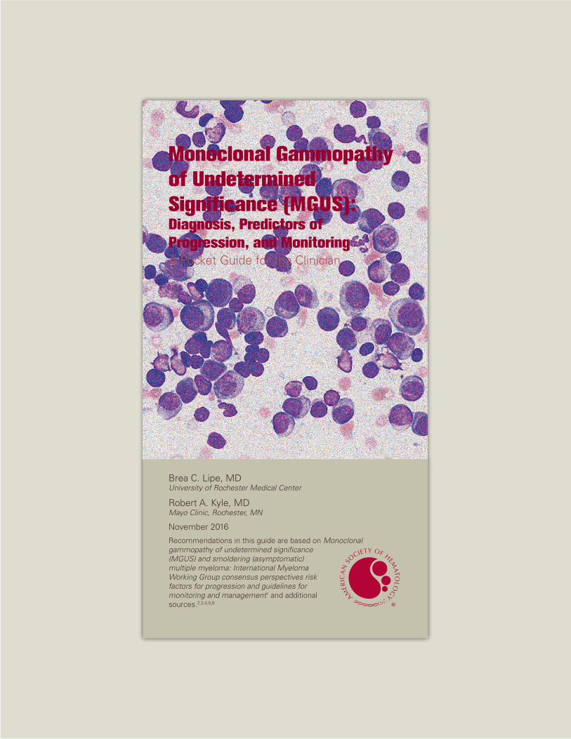
Brea C. Lipe, MDUniversity of Rochester Medical Center
Robert A. Kyle, MDMayo Clinic, Rochester, MN
November 2016
Recommendations in this guide are based on Monoclonal gammopathy of undetermined significance (MGUS) and smoldering (asymptomatic) multiple myeloma: International Myeloma Working Group consensus perspectives risk factors for progression and guidelines for monitoring and management1 and additional sources.2,3,4,5,6
Monoclonal Gammopathy of Undetermined Significance (MGUS):Diagnosis, Predictors of Progression, and MonitoringA Pocket Guide for the Clinician
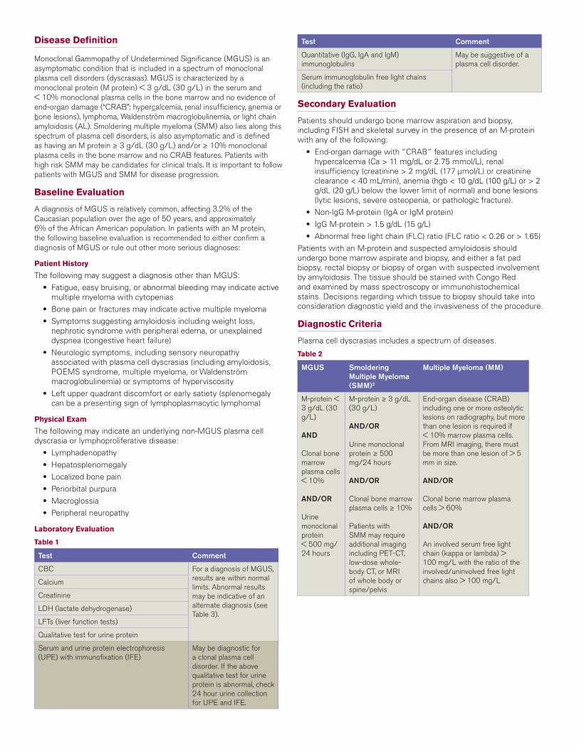
Disease Definition
Monoclonal Gammopathy of Undetermined Significance (MGUS) is an asymptomatic condition that is included in a spectrum of monoclonal plasma cell disorders (dyscrasias). MGUS is characterized by a monoclonal protein (M protein) < 3 g/dL (30 g/L) in the serum and < 10% monoclonal plasma cells in the bone marrow and no evidence of end-organ damage (“CRAB”: hypercalcemia, renal insufficiency, anemia or bone lesions), lymphoma, Waldenström macroglobulinemia, or light chain amyloidosis (AL). Smoldering multiple myeloma (SMM) also lies along this spectrum of plasma cell disorders, is also asymptomatic and is defined as having an M protein ≥ 3 g/dL (30 g/L) and/or ≥ 10% monoclonal plasma cells in the bone marrow and no CRAB features. Patients with high risk SMM may be candidates for clinical trials. It is important to follow patients with MGUS and SMM for disease progression.
Baseline Evaluation
A diagnosis of MGUS is relatively common, affecting 3.2% of the Caucasian population over the age of 50 years, and approximately 6% of the African American population. In patients with an M protein, the following baseline evaluation is recommended to either confirm a diagnosis of MGUS or rule out other more serious diagnoses:
Patient History
The following may suggest a diagnosis other than MGUS: • Fatigue, easy bruising, or abnormal bleeding may indicate active
multiple myeloma with cytopenias • Bone pain or fractures may indicate active multiple myeloma • Symptoms suggesting amyloidosis including weight loss,
nephrotic syndrome with peripheral edema, or unexplained dyspnea (congestive heart failure)
• Neurologic symptoms, including sensory neuropathy associated with plasma cell dyscrasias (including amyloidosis, POEMS syndrome, multiple myeloma, or Waldenström macroglobulinemia) or symptoms of hyperviscosity
• Left upper quadrant discomfort or early satiety (splenomegaly can be a presenting sign of lymphoplasmacytic lymphoma)
Physical Exam
The following may indicate an underlying non-MGUS plasma cell dyscrasia or lymphoproliferative disease: • Lymphadenopathy • Hepatosplenomegaly • Localized bone pain • Periorbital purpura • Macroglossia • Peripheral neuropathy
Laboratory Evaluation
Table 1
Test Comment
CBC For a diagnosis of MGUS, results are within normal limits. Abnormal results may be indicative of an alternate diagnosis (see Table 3).
Calcium
Creatinine
LDH (lactate dehydrogenase)
LFTs (liver function tests)
Qualitative test for urine protein
Serum and urine protein electrophoresis (UPE) with immunofixation (IFE)
May be diagnostic for a clonal plasma cell disorder. If the above qualitative test for urine protein is abnormal, check 24 hour urine collection for UPE and IFE.
Test Comment
Quantitative (IgG, IgA and IgM) immunoglobulins
May be suggestive of a plasma cell disorder.
Serum immunoglobulin free light chains (including the ratio)
Secondary Evaluation
Patients should undergo bone marrow aspiration and biopsy, including FISH and skeletal survey in the presence of an M-protein with any of the following: • End-organ damage with “CRAB” features including
hypercalcemia (Ca > 11 mg/dL or 2.75 mmol/L), renal insufficiency (creatinine > 2 mg/dL (177 µmol/L) or creatinine clearance < 40 mL/min), anemia (hgb < 10 g/dL (100 g/L) or > 2 g/dL (20 g/L) below the lower limit of normal) and bone lesions (lytic lesions, severe osteopenia, or pathologic fracture).
• Non-IgG M-protein (IgA or IgM protein) • IgG M-protein > 1.5 g/dL (15 g/L) • Abnormal free light chain (FLC) ratio (FLC ratio < 0.26 or > 1.65)Patients with an M-protein and suspected amyloidosis should undergo bone marrow aspirate and biopsy, and either a fat pad biopsy, rectal biopsy or biopsy of organ with suspected involvement by amyloidosis. The tissue should be stained with Congo Red and examined by mass spectroscopy or immunohistochemical stains. Decisions regarding which tissue to biopsy should take into consideration diagnostic yield and the invasiveness of the procedure.
Diagnostic Criteria
Plasma cell dyscrasias includes a spectrum of diseases.
Table 2
MGUS Smoldering Multiple Myeloma (SMM)2
Multiple Myeloma (MM)
M-protein < 3 g/dL (30 g/L)
AND
Clonal bone marrow plasma cells < 10%
AND/OR
Urine monoclonal protein < 500 mg/ 24 hours
M-protein ≥ 3 g/dL (30 g/L)
AND/OR
Urine monoclonal protein ≥ 500 mg/24 hours
AND/OR
Clonal bone marrow plasma cells ≥ 10%
Patients with SMM may require additional imaging including PET-CT, low-dose whole-body CT, or MRI of whole body or spine/pelvis
End-organ disease (CRAB) including one or more osteolytic lesions on radiography, but more than one lesion is required if < 10% marrow plasma cells. From MRI imaging, there must be more than one lesion of > 5 mm in size.
AND/OR
Clonal bone marrow plasma cells > 60%
AND/OR
An involved serum free light chain (kappa or lambda) > 100 mg/L with the ratio of the involved/uninvolved free light chains also > 100 mg/L
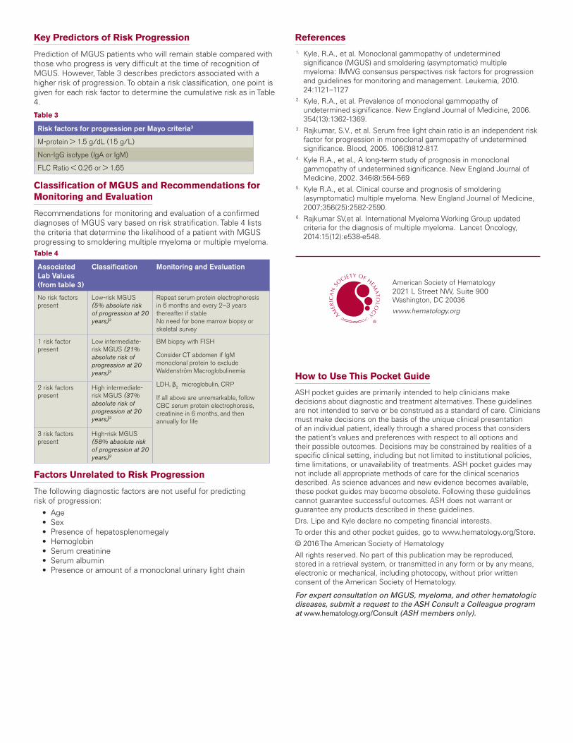
Key Predictors of Risk Progression
Prediction of MGUS patients who will remain stable compared with those who progress is very difficult at the time of recognition of MGUS. However, Table 3 describes predictors associated with a higher risk of progression. To obtain a risk classification, one point is given for each risk factor to determine the cumulative risk as in Table 4.
Table 3
Risk factors for progression per Mayo criteria3
M-protein > 1.5 g/dL (15 g/L)
Non-IgG isotype (IgA or IgM)
FLC Ratio < 0.26 or > 1.65
Classification of MGUS and Recommendations for Monitoring and Evaluation
Recommendations for monitoring and evaluation of a confirmed diagnoses of MGUS vary based on risk stratification. Table 4 lists the criteria that determine the likelihood of a patient with MGUS progressing to smoldering multiple myeloma or multiple myeloma.Table 4
Associated Lab Values (from table 3)
Classification Monitoring and Evaluation
No risk factors present
Low-risk MGUS (5% absolute risk of progression at 20 years)3
Repeat serum protein electrophoresis in 6 months and every 2–3 years thereafter if stableNo need for bone marrow biopsy or skeletal survey
1 risk factor present
Low intermediate-risk MGUS (21% absolute risk of progression at 20 years)3
BM biopsy with FISH
Consider CT abdomen if IgM monoclonal protein to exclude Waldenström Macroglobulinemia
LDH, β2 microglobulin, CRP
If all above are unremarkable, follow CBC serum protein electrophoresis, creatinine in 6 months, and then annually for life
2 risk factors present
High intermediate-risk MGUS (37% absolute risk of progression at 20 years)3
3 risk factors present
High-risk MGUS (58% absolute risk of progression at 20 years)3
Factors Unrelated to Risk Progression
The following diagnostic factors are not useful for predicting risk of progression: • Age • Sex • Presence of hepatosplenomegaly • Hemoglobin • Serum creatinine • Serum albumin • Presence or amount of a monoclonal urinary light chain
References1. Kyle, R.A., et al. Monoclonal gammopathy of undetermined
significance (MGUS) and smoldering (asymptomatic) multiple myeloma: IMWG consensus perspectives risk factors for progression and guidelines for monitoring and management. Leukemia, 2010. 24:1121–1127
2. Kyle, R.A., et al. Prevalence of monoclonal gammopathy of undetermined significance. New England Journal of Medicine, 2006. 354(13):1362-1369.
3. Rajkumar, S.V., et al. Serum free light chain ratio is an independent risk factor for progression in monoclonal gammopathy of undetermined significance. Blood, 2005. 106(3)812-817.
4. Kyle R.A., et al., A long-term study of prognosis in monoclonal gammopathy of undetermined significance. New England Journal of Medicine, 2002. 346(8):564-569
5. Kyle R.A., et al. Clinical course and prognosis of smoldering (asymptomatic) multiple myeloma. New England Journal of Medicine, 2007;356(25):2582-2590.
6. Rajkumar SV,et al. International Myeloma Working Group updated criteria for the diagnosis of multiple myeloma. Lancet Oncology, 2014:15(12):e538-e548.
How to Use This Pocket Guide
ASH pocket guides are primarily intended to help clinicians make decisions about diagnostic and treatment alternatives. These guidelines are not intended to serve or be construed as a standard of care. Clinicians must make decisions on the basis of the unique clinical presentation of an individual patient, ideally through a shared process that considers the patient’s values and preferences with respect to all options and their possible outcomes. Decisions may be constrained by realities of a specific clinical setting, including but not limited to institutional policies, time limitations, or unavailability of treatments. ASH pocket guides may not include all appropriate methods of care for the clinical scenarios described. As science advances and new evidence becomes available, these pocket guides may become obsolete. Following these guidelines cannot guarantee successful outcomes. ASH does not warrant or guarantee any products described in these guidelines.
Drs. Lipe and Kyle declare no competing financial interests.
To order this and other pocket guides, go to www.hematology.org/Store.
© 2016 The American Society of Hematology
All rights reserved. No part of this publication may be reproduced, stored in a retrieval system, or transmitted in any form or by any means, electronic or mechanical, including photocopy, without prior written consent of the American Society of Hematology.
For expert consultation on MGUS, myeloma, and other hematologic diseases, submit a request to the ASH Consult a Colleague program at www.hematology.org/Consult (ASH members only).
American Society of Hematology2021 L Street NW, Suite 900Washington, DC 20036
www.hematology.org