Embed Size (px)
Citation preview
Monoclonal Gammopathy of Undetermined Significance (MGUS) and Smoldering (Asymptomatic)
Multiple Myeloma: IMWG Consensus Perspectives Risk Factors for Progression and Guidelines for
Monitoring and Management
R. A. Kyle, MD1, B.G.M. Durie, MD2, S.V. Rajkumar, MD1, O. Landgren, MD3, J. Blade, MD4, G. Merlini, MD5, N. Kröger, MD6, H. Einsele, MD7, D. H. Vesole, MD8, M. Dimopoulos, MD9, J. San-Miguel, MD10, H. Avet-Loiseau, MD11, R. Hajek, MD12, WM Chen, MD13, K.C. Anderson, MD14 H. Ludwig, MD15, P. Sonneveld, MD16, S. Pavlovsky, MD17, A. Palumbo, MD18, P. G. Richardson, MD14, B. Barlogie, MD19, P. Greipp, MD1, R.Vescio, MD2, I. Turesson, MD20, J. Westin, MD21, M. Boccadoro, MD18
on behalf of the International Myeloma Working Group 1Division of Hematology, Mayo Clinic, Rochester, Minnesota, USA; 2Aptium Oncology, Inc., Cedars-Sinai Outpatient Cancer Center at the Samuel Oschin Comprehensive Cancer Institute, Los Angeles, California, USA;3National Cancer Institute, National Institutes of Health, Bethesda, MD, USA; 4Department of Hematology, Hospital Clinic, IDIBAPS, Barcelona, Spain; 5Department of Biochemistry, University Hospital San Matteo, Italy; 6Department of Stem Cell Transplantation, University Hospital Hamburg, Germany; 7Department of Internal Medicine University of Wurzburg, Wurzburg, Germany; 8Hackensack Cancer Center, Hackensack University Medical Center, Hackensack, NJ, USA; 9Departments of Therapeutics, University of Athens School of Medicine, Athens, Greece; 10Department of Hematology, Servicio de Hematología, Hospital Universitario de Salamanca. CIC, IBMCC (USAL-CSIC). Spain;
11Institut de Biologie, Laboratoire d’Hematologie, Nantes, France; 12Department of Internal Medicine Hematology, University Hospital Brno, Brno, Czech Republic; 13Department of Hematology and Oncology, Beijing Chaoyang Hospital, Beijing, China;14Department of Medical Oncology, Division of Hematologic Malignancies, Dana-Farber Cancer Institute, Boston, Massachusetts, USA;151st Medical Department, Center for Oncology and Hematology, Wilhelminenspital, Wien, Vienna, Austria;
2
16Erasmus MC, Department of Hematology, Rotterdam, The Netherlands;17FUNDALEU, Angelica Ocampo Hospital, Buenos Aires, Argentina;18Divisione di Ematologia, University of Torino, Torino, Italy;
19Departments of Hematology and Pathology, MIRT UAMS, Little Rock, Arkansas, USA; 20Department of Hematology, Malmo University Hospital, Malmo, Sweden; 21Sahlgresnka University Hospital, Gottenburg, Sweden; Abstract Monoclonal gammopathy of undetermined significance (MGUS) was
identified in 3.2% of 21,463 residents of Olmsted County, Minnesota, 50 years of age or
older. The risk of progression to multiple myeloma, Waldenstrom's macroglobulinemia,
AL amyloidosis or a lymphoproliferative disorder is approximately 1% per year. Low-
risk MGUS is characterized by having an M protein < 15 g/L, IgG type and a normal free
light chain (FLC) ratio. Patients should be followed with serum protein electrophoresis at
six months and, if stable, can be followed every two to three years or when symptoms
suggestive of a plasma cell malignancy arise. Patients with intermediate and high-risk
MGUS should be followed in six months and then annually for life.
The risk of smoldering (asymptomatic) multiple myeloma (SMM) progressing to
multiple myeloma or a related disorder is 10% per year for the first 5 years, 3% per year
for the next 5 years and 1%-2% per year for the next 10 years. Testing should be done 2
– 3 months after the initial recognition of SMM. If the results are stable, the patient
should be followed every four to six months for one year and, if stable, every six to
twelve months.
3
MONOCLONAL GAMMOPATHY OF UNDETERMINED SIGNIFICANCE
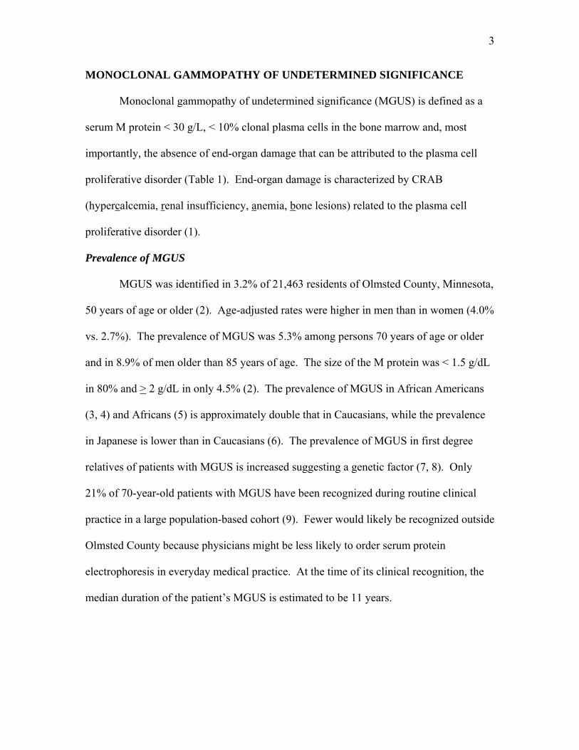
Monoclonal gammopathy of undetermined significance (MGUS) is defined as a
serum M protein < 30 g/L, < 10% clonal plasma cells in the bone marrow and, most
importantly, the absence of end-organ damage that can be attributed to the plasma cell
proliferative disorder (Table 1). End-organ damage is characterized by CRAB
(hypercalcemia, renal insufficiency, anemia, bone lesions) related to the plasma cell
proliferative disorder (1).
Prevalence of MGUS
MGUS was identified in 3.2% of 21,463 residents of Olmsted County, Minnesota,
50 years of age or older (2). Age-adjusted rates were higher in men than in women (4.0%
vs. 2.7%). The prevalence of MGUS was 5.3% among persons 70 years of age or older
and in 8.9% of men older than 85 years of age. The size of the M protein was < 1.5 g/dL
in 80% and > 2 g/dL in only 4.5% (2). The prevalence of MGUS in African Americans
(3, 4) and Africans (5) is approximately double that in Caucasians, while the prevalence
in Japanese is lower than in Caucasians (6). The prevalence of MGUS in first degree
relatives of patients with MGUS is increased suggesting a genetic factor (7, 8). Only
21% of 70-year-old patients with MGUS have been recognized during routine clinical
practice in a large population-based cohort (9). Fewer would likely be recognized outside
Olmsted County because physicians might be less likely to order serum protein
electrophoresis in everyday medical practice. At the time of its clinical recognition, the
median duration of the patient’s MGUS is estimated to be 11 years.
4
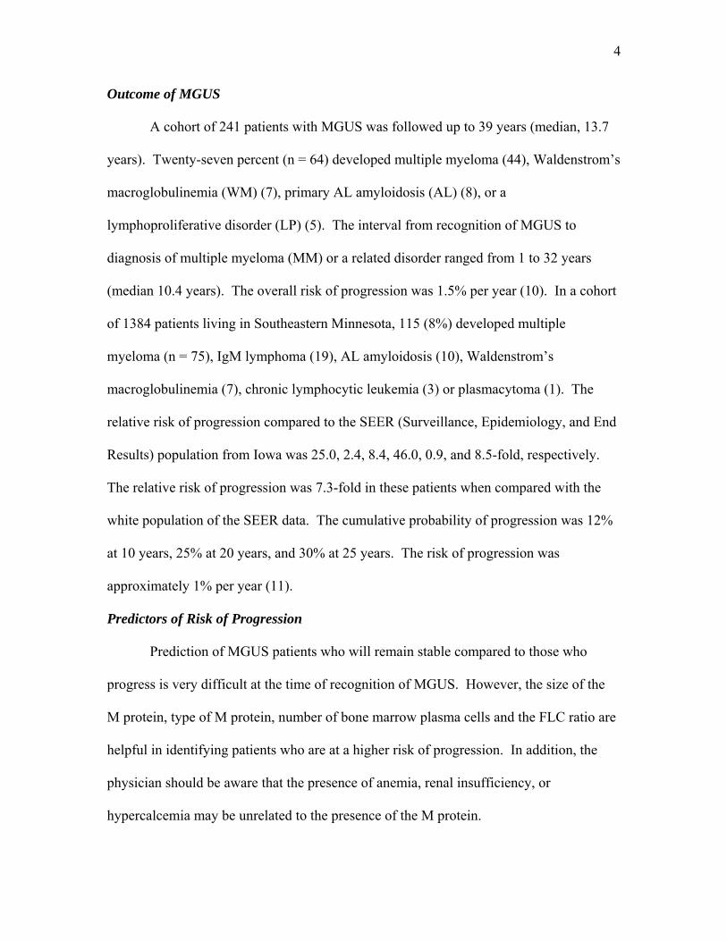
Outcome of MGUS
A cohort of 241 patients with MGUS was followed up to 39 years (median, 13.7
years). Twenty-seven percent (n = 64) developed multiple myeloma (44), Waldenstrom’s
macroglobulinemia (WM) (7), primary AL amyloidosis (AL) (8), or a
lymphoproliferative disorder (LP) (5). The interval from recognition of MGUS to
diagnosis of multiple myeloma (MM) or a related disorder ranged from 1 to 32 years
(median 10.4 years). The overall risk of progression was 1.5% per year (10). In a cohort
of 1384 patients living in Southeastern Minnesota, 115 (8%) developed multiple
myeloma (n = 75), IgM lymphoma (19), AL amyloidosis (10), Waldenstrom’s
macroglobulinemia (7), chronic lymphocytic leukemia (3) or plasmacytoma (1). The
relative risk of progression compared to the SEER (Surveillance, Epidemiology, and End
Results) population from Iowa was 25.0, 2.4, 8.4, 46.0, 0.9, and 8.5-fold, respectively.
The relative risk of progression was 7.3-fold in these patients when compared with the
white population of the SEER data. The cumulative probability of progression was 12%
at 10 years, 25% at 20 years, and 30% at 25 years. The risk of progression was
approximately 1% per year (11).
Predictors of Risk of Progression
Prediction of MGUS patients who will remain stable compared to those who
progress is very difficult at the time of recognition of MGUS. However, the size of the
M protein, type of M protein, number of bone marrow plasma cells and the FLC ratio are
helpful in identifying patients who are at a higher risk of progression. In addition, the
physician should be aware that the presence of anemia, renal insufficiency, or
hypercalcemia may be unrelated to the presence of the M protein.
5
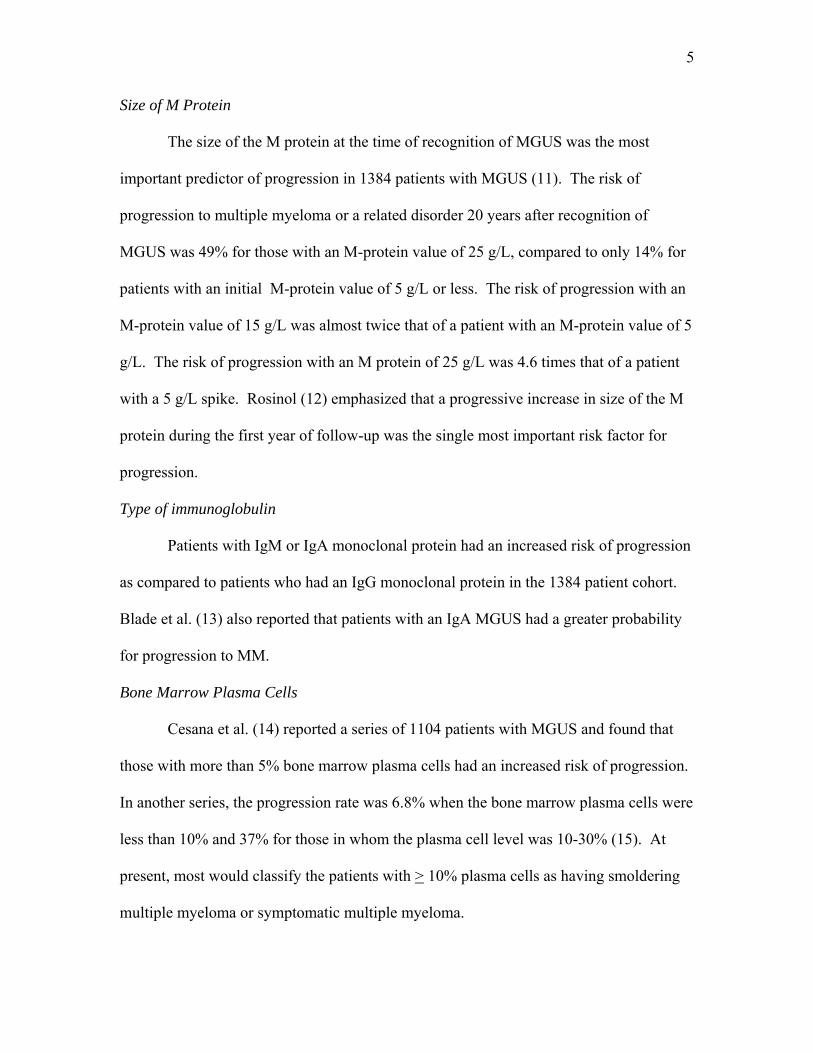
Size of M Protein
The size of the M protein at the time of recognition of MGUS was the most
important predictor of progression in 1384 patients with MGUS (11). The risk of
progression to multiple myeloma or a related disorder 20 years after recognition of
MGUS was 49% for those with an M-protein value of 25 g/L, compared to only 14% for
patients with an initial M-protein value of 5 g/L or less. The risk of progression with an
M-protein value of 15 g/L was almost twice that of a patient with an M-protein value of 5
g/L. The risk of progression with an M protein of 25 g/L was 4.6 times that of a patient
with a 5 g/L spike. Rosinol (12) emphasized that a progressive increase in size of the M
protein during the first year of follow-up was the single most important risk factor for
progression.
Type of immunoglobulin
Patients with IgM or IgA monoclonal protein had an increased risk of progression
as compared to patients who had an IgG monoclonal protein in the 1384 patient cohort.
Blade et al. (13) also reported that patients with an IgA MGUS had a greater probability
for progression to MM.
Bone Marrow Plasma Cells
Cesana et al. (14) reported a series of 1104 patients with MGUS and found that
those with more than 5% bone marrow plasma cells had an increased risk of progression.
In another series, the progression rate was 6.8% when the bone marrow plasma cells were
less than 10% and 37% for those in whom the plasma cell level was 10-30% (15). At
present, most would classify the patients with > 10% plasma cells as having smoldering
multiple myeloma or symptomatic multiple myeloma.
6
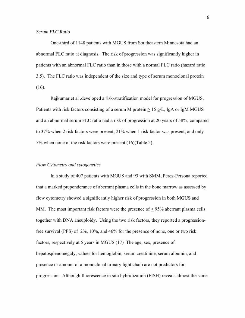
Serum FLC Ratio
One-third of 1148 patients with MGUS from Southeastern Minnesota had an
abnormal FLC ratio at diagnosis. The risk of progression was significantly higher in
patients with an abnormal FLC ratio than in those with a normal FLC ratio (hazard ratio
3.5). The FLC ratio was independent of the size and type of serum monoclonal protein
(16).
Rajkumar et al .developed a risk-stratification model for progression of MGUS.
Patients with risk factors consisting of a serum M protein > 15 g/L, IgA or IgM MGUS
and an abnormal serum FLC ratio had a risk of progression at 20 years of 58%; compared
to 37% when 2 risk factors were present; 21% when 1 risk factor was present; and only
5% when none of the risk factors were present (16)(Table 2).
Flow Cytometry and cytogenetics
In a study of 407 patients with MGUS and 93 with SMM, Perez-Persona reported
that a marked preponderance of aberrant plasma cells in the bone marrow as assessed by
flow cytometry showed a significantly higher risk of progression in both MGUS and
MM. The most important risk factors were the presence of > 95% aberrant plasma cells
together with DNA aneuploidy. Using the two risk factors, they reported a progression-
free survival (PFS) of 2%, 10%, and 46% for the presence of none, one or two risk
factors, respectively at 5 years in MGUS (17) The age, sex, presence of
hepatosplenomegaly, values for hemoglobin, serum creatinine, serum albumin, and
presence or amount of a monoclonal urinary light chain are not predictors for
progression. Although fluorescence in situ hybridization (FISH) reveals almost the same
7
number and type of abnormalities as in multiple myeloma, there is little evidence that this
plays a role in the progression of MGUS to MM. The gene expression profile may be of
benefit in predicting the risk of progression, but no convincing data exists at present.
Patient Management
At first diagnosis, a complete history and physical examination should be done
with emphasis on symptoms and findings that might suggest multiple myeloma or AL
amyloidosis. A complete blood count, serum calcium and creatinine values and a
qualitative test for urine protein should be performed. If proteinuria is found,
electrophoresis and immunofixation is indicated. Serum protein electrophoresis should
be repeated 3 to 6 months after recognition of MGUS in order to exclude multiple
myeloma or Waldenstrom’s macroglobulinemia because the monoclonal protein is
usually recognized by chance.
Low-Risk MGUS
If the serum monoclonal protein is < 15 g/L, IgG type and the FLC ratio is
normal, the risk of eventual progression to myeloma or related malignancy is low. In this
setting, a baseline bone marrow examination or skeletal radiography are not routinely
indicated if the clinical evaluation, complete blood count, serum creatinine and calcium
values suggest MGUS. On the other hand, a bone marrow is required if the patient has
unexplained anemia, renal insufficiency, hypercalcemia, or bone lesions. Patients should
be followed with serum protein electrophoresis in 6 months, and if stable can be followed
every 2 to 3 years or when symptoms suggestive of a plasma cell malignancy arise.
Intermediate and High Risk MGUS
8
If a patient with apparent MGUS has a serum monoclonal protein > 15 g/L, IgA
or IgM protein type, or an abnormal FLC ratio, a bone marrow aspirate and biopsy should
be done at baseline to rule out underlying plasma cell malignancy. As discussed earlier, a
bone marrow is always required if a patient with presumed MGUS has unexplained
anemia, renal insufficiency, hypercalcemia, or bone lesions or a suspicion of AL
amyloidosis. Both conventional cytogenetics and FISH should be performed on the bone
marrow examination. If available, a plasma cell labeling index and a search for
circulating plasma cells in the peripheral blood using flow cytometry are useful (18). A
computed tomographic scan of the abdomen should be done in the presence of an IgM
monoclonal protein because asymptomatic retroperitoneal lymph nodes may be present.
Lactate dehydrogenase, beta-2-microglobulin, and C-reactive proteins should be
determined if there is evidence of multiple myeloma or Waldenstrom's
macroglobulinemia. If the results of these tests are satisfactory, patients should be
followed with serum protein electrophoresis and a complete blood count in 6 months and
then annually for life. Treatment is not indicated unless it is part of a clinical trial (19).
The patient must contact their physician if there is any change in their clinical condition.
SMOLDERING (ASYMPTOMATIC) MULTIPLE MYELOMA
Smoldering (asymptomatic) multiple myeloma (SMM) requires the presence of a
monoclonal protein level of 30 g/L or more or a proportion of clonal plasma cells in the
bone marrow of 10% or more but no end-organ damage (1). It needs to be distinguished
from MGUS because of a higher risk of progression to myeloma or related disorder; 10%
per year for SMM versus 1% per year for MGUS.
Outcome
9
In a cohort of 276 patients fulfilling the criteria for SMM, 163 (59%) developed
symptomatic multiple myeloma or AL amyloidosis during follow-up. The overall risk of
progression was 10% per year for the first 5 years, approximately 3% per year for the
next 5 years, and 1% per year for the last 10 years. The cumulative probability of
progression to active multiple myeloma or AL amyloidosis was 51% at 5 years, 66% at
10 years, and 73% at 15 years. The median time to progression was 4.8 years (20).
The number of patients with progression to symptomatic multiple myeloma was
522 times the number of persons without SMM who would be expected to have active
disease, while the risk of AL amyloidosis was increased by a factor of 50-fold. Ninety-
seven percent of those who progressed developed symptomatic multiple myeloma.
Risk Factors for Progression
The size of the serum monoclonal protein and the number of plasma cells in the
bone marrow are the most important factors for progression. Sex, hemoglobin level, type
of serum heavy chain, serum albumin value, presence and type of urinary light chain,
reduction in levels of uninvolved immunoglobulins and involvement of the interfatty
marrow space were not significant risk factors for progression. The FLC (free light
chain) ratio is an independent additional risk factor for progression (21).
On the basis of the size of the monoclonal protein and number of bone marrow
plasma cells, a risk-stratification model was developed (20). If the patient had both >
10% plasma cells and > 30 g/L of monoclonal protein, the probability of progression at
15 years was 87%; if the patient had > 10% plasma cells and < 30 g/L of monoclonal
protein, the risk of progression was 70% at 15 years, while those patients with < 10%
plasma cells and > 30 g/L monoclonal protein had a risk of progression of 39%. The

10
median time to progression was 2 years, 8 years, and 19 years, respectively in the above 3
groups. The type of serum heavy chain added significantly to the multivariate model
containing the number of bone marrow plasma cells and size of the serum M protein.
Dispenzieri et al found that the serum free light chain assay provides additional
prognostic information. An abnormal FLC ratio (defined as ≤0.125 or ≥8) predicted for
higher rates of progression, hazard ratio, 2.3; 95% CI, 1.6-3.2) (21). A risk model was
constructed, incorporating three risk factors: abnormal FLC ratio, bone marrow plasma
cells ≥ 10%, and serum M protein ≥3 g/dL. Patients with 1, 2, or 3 risk factors had 5-year
progression rates of 25%, 51%, and 76% respectively (Fig. 1). Similarly to what we have
mentioned above for MGUS, the presence of >95% aberrant PC from the total BM PC
compartment detected by flow cytometry together with immunoparesis can discriminate 3
prognostic groups in SMM, with a risk of progression at 5 years of 72%, 46% and 4% if
the patient has two, one or none of these risk factors respectively (17).
Others have found that the presence of occult bone lesions on MRI increases the
risk of progression in patients otherwise defined as having SMM (22) In a recent study,
Wang and colleagues estimated risk of progression in 72 patients with SMM in whom an
MRI of the spine was also performed (23). The median TTP was significantly shorter
with an abnormal MRI compared to normal MRI, 1.5 years versus 5 years, respectively.
Patient Management
Serum protein electrophoresis, complete blood count, measurement of calcium
and creatinine values and 24-hour urine collection for electrophoresis and
immunofixation should be performed at diagnosis and in 2 to 3 months after the initial
11
recognition of SMM. A baseline bone marrow biopsy and skeletal survey are mandatory.
An MRI of the spine and pelvis is recommended because it can detect occult lesions and,
if present, predict for a more rapid progression to symptomatic myeloma. If the results
are stable, the studies should be repeated every 4 to 6 months for 1 year and, if stable,
evaluation can be lengthened to every 6 to 12 months. A skeletal survey should be
performed if there is evidence of progression in the above-mentioned routine studies.
Summary of IMWG recommendations for MGUS and SMM
Based on the discussion above, the summary recommendations that highlight the specific
new recommendations by the IMWG panel are summarized in Table 3. As more data
emerge we will reexamine these guidelines, particularly with regard to follow up and
prophylactic strategies. Understanding the biology and mechanisms of progression of
MGUS/SMM to myeloma will provide major insights towards the goal of finding a cure
for multiple myeloma.
12
Figure Legends
Fig. 1 Risk stratification for smoldering multiple myeloma. The model incorporates
three risk factors: abnormal FLC ratio, bone marrow plasma cells ≥ 10%, and
serum M protein ≥3 g/dL. Patients with 1, 2, or 3 risk factors had 5-year
progression rates of 25%, 51%, and 76% respectively. Corresponding median
times to progression are 10, 5.1, and 1.9 years, respectively.
13
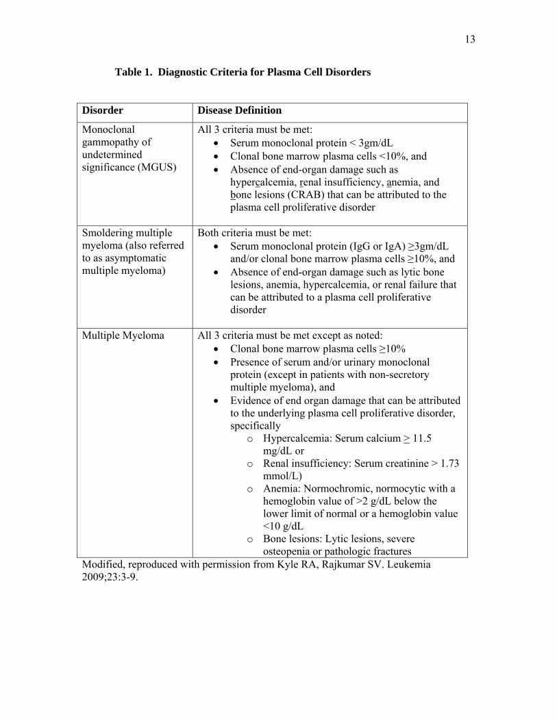
Table 1. Diagnostic Criteria for Plasma Cell Disorders
Disorder Disease Definition
Monoclonal gammopathy of undetermined significance (MGUS)
All 3 criteria must be met: • Serum monoclonal protein < 3gm/dL • Clonal bone marrow plasma cells <10%, and • Absence of end-organ damage such as
hypercalcemia, renal insufficiency, anemia, and bone lesions (CRAB) that can be attributed to the plasma cell proliferative disorder
Smoldering multiple myeloma (also referred to as asymptomatic multiple myeloma)
Both criteria must be met: • Serum monoclonal protein (IgG or IgA) ≥3gm/dL
and/or clonal bone marrow plasma cells ≥10%, and • Absence of end-organ damage such as lytic bone
lesions, anemia, hypercalcemia, or renal failure that can be attributed to a plasma cell proliferative disorder
Multiple Myeloma All 3 criteria must be met except as noted:
• Clonal bone marrow plasma cells ≥10% • Presence of serum and/or urinary monoclonal
protein (except in patients with non-secretory multiple myeloma), and
• Evidence of end organ damage that can be attributed to the underlying plasma cell proliferative disorder, specifically
o Hypercalcemia: Serum calcium > 11.5 mg/dL or
o Renal insufficiency: Serum creatinine > 1.73 mmol/L)
o Anemia: Normochromic, normocytic with a hemoglobin value of >2 g/dL below the lower limit of normal or a hemoglobin value <10 g/dL
o Bone lesions: Lytic lesions, severe osteopenia or pathologic fractures
Modified, reproduced with permission from Kyle RA, Rajkumar SV. Leukemia 2009;23:3-9.
14
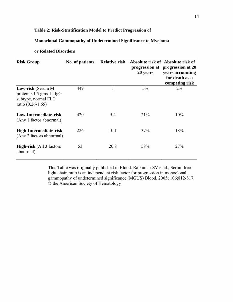
Table 2: Risk-Stratification Model to Predict Progression of
Monoclonal Gammopathy of Undetermined Significance to Myeloma
or Related Disorders
Risk Group No. of patients Relative risk Absolute risk of progression at
20 years
Absolute risk of progression at 20 years accounting
for death as a competing risk
Low-risk (Serum M protein <1.5 gm/dL, IgG subtype, normal FLC ratio (0.26-1.65)
449 1 5% 2%
Low-Intermediate-risk (Any 1 factor abnormal)
420 5.4 21% 10%
High-Intermediate-risk (Any 2 factors abnormal)
226 10.1 37% 18%
High-risk (All 3 factors abnormal)
53 20.8 58% 27%
This Table was originally published in Blood. Rajkumar SV et al., Serum free
light chain ratio is an independent risk factor for progression in monoclonal gammopathy of undetermined significance (MGUS) Blood. 2005; 106;812-817. © the American Society of Hematology
15
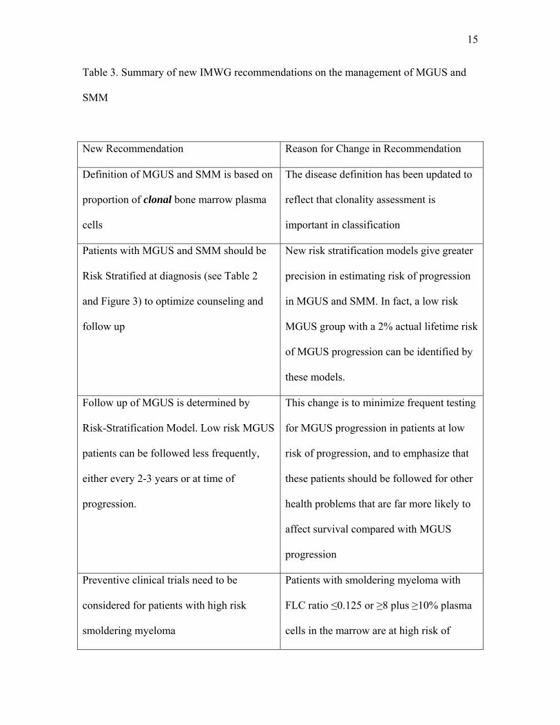
Table 3. Summary of new IMWG recommendations on the management of MGUS and
SMM
New Recommendation Reason for Change in Recommendation
Definition of MGUS and SMM is based on
proportion of clonal bone marrow plasma
cells
The disease definition has been updated to
reflect that clonality assessment is
important in classification
Patients with MGUS and SMM should be
Risk Stratified at diagnosis (see Table 2
and Figure 3) to optimize counseling and
follow up
New risk stratification models give greater
precision in estimating risk of progression
in MGUS and SMM. In fact, a low risk
MGUS group with a 2% actual lifetime risk
of MGUS progression can be identified by
these models.
Follow up of MGUS is determined by
Risk-Stratification Model. Low risk MGUS
patients can be followed less frequently,
either every 2-3 years or at time of
progression.
This change is to minimize frequent testing
for MGUS progression in patients at low
risk of progression, and to emphasize that
these patients should be followed for other
health problems that are far more likely to
affect survival compared with MGUS
progression
Preventive clinical trials need to be
considered for patients with high risk
smoldering myeloma
Patients with smoldering myeloma with
FLC ratio ≤0.125 or ≥8 plus ≥10% plasma
cells in the marrow are at high risk of
16
progression in the first two years following
recognition. These patients should be
considered candidates for chemoprevention
trials. However, off-study, observation is
still the standard even in this group
17
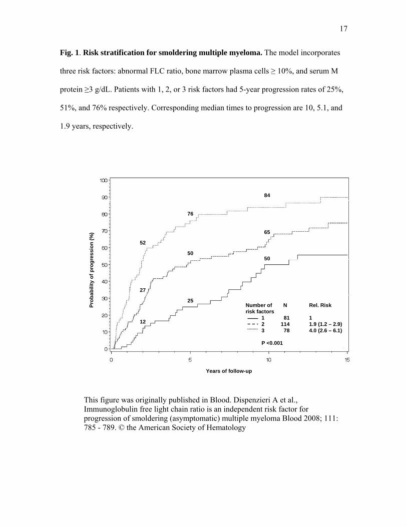
Fig. 1. Risk stratification for smoldering multiple myeloma. The model incorporates
three risk factors: abnormal FLC ratio, bone marrow plasma cells ≥ 10%, and serum M
protein ≥3 g/dL. Patients with 1, 2, or 3 risk factors had 5-year progression rates of 25%,
51%, and 76% respectively. Corresponding median times to progression are 10, 5.1, and
1.9 years, respectively.
50
84
65
25
76
50
12
52
27
Number of N Rel. Riskrisk factors
1 81 12 114 1.9 (1.2 – 2.9)3 78 4.0 (2.6 – 6.1)
P <0.001
Years of follow-up
Prob
abili
ty o
f pro
gres
sion
(%)
This figure was originally published in Blood. Dispenzieri A et al., Immunoglobulin free light chain ratio is an independent risk factor for progression of smoldering (asymptomatic) multiple myeloma Blood 2008; 111: 785 - 789. © the American Society of Hematology
18
References
1. Criteria for the classification of monoclonal gammopathies, multiple myeloma and related disorders: a report of the International Myeloma Working Group. Br J Haematol 2003 June 01, 2003; 121(5): 749-757.
2. Kyle RA, Therneau TM, Rajkumar SV, Larson DR, Plevak MF, Offord JR, et al.
Prevalence of monoclonal gammopathy of undetermined significance. N Engl J Med 2006 Mar 30; 354(13): 1362-1369.
3. Cohen HJ, Crawford J, Rao MK, Pieper CF, Currie MS. Racial differences in the
prevalence of monoclonal gammopathy in a community-based sample of the elderly.[erratum appears in Am J Med 1998 Oct;105(4):362]. American Journal of Medicine 1998; 104(5): 439-444.
4. Singh J, Dudley AW, Jr., Kulig KA. Increased incidence of monoclonal
gammopathy of undetermined significance in blacks and its age-related differences with whites on the basis of a study of 397 men and one woman in a hospital setting. Journal of Laboratory & Clinical Medicine 1990; 116(6): 785-789.
5. Landgren O, Katzmann JA, Hsing AW, Pfeiffer RM, Kyle RA, Yeboah ED, et al.
Prevalence of monoclonal gammopathy of undetermined significance among men in Ghana. Mayo Clin Proc 2007 Dec; 82(12): 1468-1473.
6. Iwanaga M, Tagawa M, Tsukasaki K, Kamihira S, Tomonaga M. Prevalence of
monoclonal gammopathy of undetermined significance: study of 52,802 persons in Nagasaki City, Japan. Mayo Clin Proc 2007 Dec; 82(12): 1474-1479.
7. Landgren O, Kristinsson SY, Goldin LR, Caporaso NE, Blimark C, Mellqvist
UH, et al. Risk of plasma-cell and lymphoproliferative disorders among 14,621 first-degree relatives of 4,458 patients with monoclonal gammopathy of undetermined significance (MGUS) in Sweden. Blood 2009 Jan 30.
8. Vachon CM, Kyle RA, Therneau TM, Foreman BJ, Larson DR, Colby CL, et al.
Increased risk of monoclonal gammopathy in first-degree relatives of patients with multiple myeloma or monoclonal gammopathy of undetermined significance. Blood 2009 Jul 23; 114(4): 785-790.
9. Kyle RA, Therneau TM, Melton iii LJ, Dispenzieri A, Larson D, Benson J, et al.
Monoclonal gammopathy of undetermined significance: Estimated incidence and duration prior to recognition Blood (Abstract) 2007; 110(22): 79a.
10. Kyle RA, Therneau TM, Rajkumar SV, Larson DR, Plevak MF, Melton LJ, 3rd.
Long-term follow-up of 241 patients with monoclonal gammopathy of
19
undetermined significance: the original Mayo Clinic series 25 years later.[see comment]. Mayo Clinic Proceedings 2004; 79(7): 859-866.
11. Kyle RA, Therneau TM, Rajkumar SV, Offord JR, Larson DR, Plevak MF, et al.
A long-term study of prognosis in monoclonal gammopathy of undetermined significance.[see comment]. New England Journal of Medicine 2002; 346(8): 564-569.
12. Rosinol L, Cibeira MT, Montoto S, Rozman M, Esteve J, Filella X, et al.
Monoclonal gammopathy of undetermined significance: predictors of malignant transformation and recognition of an evolving type characterized by a progressive increase in M protein size. Mayo Clin Proc 2007 Apr; 82(4): 428-434.
13. Blade J, Lopez-Guillermo A, Rozman C, Cervantes F, Salgado C, Aguilar JL, et
al. Malignant transformation and life expectancy in monoclonal gammopathy of undetermined significance. British Journal of Haematology 1992; 81(3): 391-394.
14. Cesana C, Klersy C, Barbarano L, Nosari AM, Crugnola M, Pungolino E, et al.
Prognostic factors for malignant transformation in monoclonal gammopathy of undetermined significance and smoldering multiple myeloma. Journal of Clinical Oncology 2002; 20(6): 1625-1634.
15. Baldini L, Guffanti A, Cesana BM, Colombi M, Chiorboli O, Damilano I, et al.
Role of different hematologic variables in defining the risk of malignant transformation in monoclonal gammopathy. Blood 1996; 87(3): 912-918.
16. Rajkumar SV, Kyle RA, Therneau TM, Melton LJ, III, Bradwell AR, Clark RJ, et
al. Serum free light chain ratio is an independent risk factor for progression in monoclonal gammopathy of undetermined significance. Blood 2005 August 1, 2005; 106(3): 812-817.
17. Perez-Persona E, Vidriales MB, Mateo G, Garcia-Sanz R, Mateos MV, de Coca
AG, et al. New criteria to identify risk of progression in monoclonal gammopathy of uncertain significance and smoldering multiple myeloma based on multiparameter flow cytometry analysis of bone marrow plasma cells. Blood 2007 Oct 1; 110(7): 2586-2592.
18. Nowakowski GS, Witzig TE, Dingli D, Tracz MJ, Gertz MA, Lacy MQ, et al.
Circulating plasma cells detected by flow cytometry as a predictor of survival in 302 patients with newly diagnosed multiple myeloma. Blood 2005 Oct 1; 106(7): 2276-2279.
19. Anderson KC, Kyle RA, Rajkumar SV, Stewart AK, Weber D, Richardson P.
Clinically relevant end points and new drug approvals for myeloma. Leukemia 2008 Feb; 22(2): 231-239.
20
20. Kyle RA, Remstein ED, Therneau TM, Dispenzieri A, Kurtin PJ, Hodnefield JM, et al. Clinical course and prognosis of smoldering (asymptomatic) multiple myeloma. N Engl J Med 2007 Jun 21; 356(25): 2582-2590.
21. Dispenzieri A, Kyle RA, Katzmann JA, Therneau TM, Larson D, Benson J, et al.
Immunoglobulin free light chain ratio is an independent risk factor for progression of smoldering (asymptomatic) multiple myeloma. Blood 2008 Jan 15; 111(2): 785-789.
22. Dimopoulos MA, Moulopoulos LA, Maniatis A, Alexanian R. Solitary
plasmacytoma of bone and asymptomatic multiple myeloma. Blood 2000 September 15, 2000; 96(6): 2037-2044.
23. Wang M, Alexanian R, Delasalle K, Weber D. Abnormal MRI of spine is the
dominant risk factor for early progression of asymptomatic multiple myeloma. Blood 2003; 102: 687a (abstract).
**International Myeloma Working Group: Rafat Abonour, Indiana University School of Medicine, Indianapolis, Indiana, USA Ray Alexanian, MD Anderson, Houston, Texas, USA Kenneth C. Anderson, DFCI, Boston, Massachusetts, USA Michael Attal, Purpan Hospital, Toulouse, France Herve Avet-Loiseau, Institute de Biologie, Nantes, France Ashraf Badros, University of Maryland, Baltimore, Maryland, USA Bart Barlogie, M.I.R.T. UAMS Little Rock, Arkanas, USA Regis Batille, Institute de Biologie, Nantes, France Meral Beksac, Ankara University, Ankara, Turkey Andrew Belch, Cross Cancer Institute, Alberta, Canada Bill Bensinger, Fred Hutchinson Cancer Center, Seattle, Washington, USA P. Leif Bergsagel, Mayo Clinic Scottsdale, Scottsdale, Arizona, USA Jenny Bird, Bristol Haematology and Oncology Center, Bristol, UK Joan Bladé, Hospital Clinica, Barcelona, Spain Mario Boccadoro, University of Torino, Torino, Italy Michele Cavo, Universita di Bologna, Bologna, Italy Wen Ming Chen, MM Research Center of Beijing, Beijing, China Tony Child, Leeds General Hospital, Leeds, United Kingdom James Chim, Department of Medicine, Queen Mary Hospital, Hong Kong Wee-Joo Chng, National University Health System, Singapore Ray Comenzo, Tufts Medical School, Boston, Massachusetts, USA John Crowley, Cancer Research and Biostatistics, Seattle, Washington, USA William Dalton, H. Lee Moffitt, Tampa, Florida, USA Faith Davies, Royal Marsden Hospital, London, England Cármino de Souza, Univeridade de Campinas, Caminas, Brazil Michel Delforge, University Hospital Gasthuisberg, Leuven, Belgium Meletios Dimopoulos, University of Athens School of Medicine, Athens, Greece
21
Angela Dispenzieri, Mayo Clinic, Rochester, Minnesota, USA Brian G.M. Durie, Cedars-Sinai Outpatient Cancer Center, Los Angeles, California, USA Hermann Einsele, Universitätsklinik Würzburg, Würzburg, Germany Theirry Facon, Centre Hospitalier Regional Universitaire de Lille, Lille, France Dorotea Fantl, Socieded Argentinade Hematolgia, Buenos Aires, Argentina Jean-Paul Fermand, Hopitaux de Paris, Paris, France Rafael Fonseca, Mayo Clinic Arizona, Scottsdale, Arizona, USA Gosta Gahrton, Karolinska Institute for Medicine, Huddinge, Sweden Christina Gasparetto, Duke University Medical Center, Durham, North Carolina, USA Morie Gertz, Mayo Clinic, Rochester, Minnesota, USA John Gibson, Royal Prince Alfred Hospital, Sydney, Australia Sergio Giralt, MD Anderson Cancer Center, Houston, Texas, USA Hartmut Goldschmidt, University Hospital Heidelberg, Heidelberg, Germany Philip Greipp, Mayo Clinic, Rochester, Minnesota, USA Roman Hajek, Brno University, Brno, Czech Republic Izhar Hardan, Tel Aviv University, Tel Aviv, Israel Jean-Luc Harousseau, Institute de Biologie, Nantes, France Hiroyuki Hata, Kumamoto University Hospital, Kumamoto, Japan Yutaka Hattori, Keio University School of Medicine, Tokyo, Japan Tom Heffner, Emory University, Atlanta, Georgia, USA Joy Ho, Royal Prince Alfred Hospital, Sydney, Australia Vania Hungria, Clinica San Germano, Sao Paolo, Brazil Shinsuke Ida, Nagoya City University Medical School, Nagoya, Japan Peter Jacobs, Constantiaberg Medi-Clinic, Plumstead, South Africa Sundar Jagannath, St. Vincent’s Comprehensive Cancer Center, New York, New York, USA Hou Jian, Shanghai Chang Zheng Hospital, Shanghai, China Douglas Joshua, Royal Prince Alfred Hospital, Sydney, Australia Artur Jurczyszyn, The Myeloma Treatment Foundation, Poland Asher Chanan Kahn, Roswell Park Cancer Institute, Buffalo, New York, USA Michio Kawano, Yamaguchi University, Ube, Japan Nicolaus Kröger, University Hospital Hamburg, Hamburg, Germany Shaji Kumar, Department of Hematology, Mayo Clinic, Minnesota, USA Robert Kyle, Department of Laboratory Med. and Pathology, Mayo Clinic, Minnesota, USA Juan Lahuerta, Grupo Espanol di Mieloma, Hospital Universitario, Madrid, Spain Ola Landgren, National Cancer Institute, Bethesda, Maryland, USA Jacob Laubach, Dana-Farber Cancer Institute, Boston, Massachusetts, USA Jae Hoon Lee, Gachon University Gil Hospital, Incheon, Korea Xavier LeLeu, Hospital Huriez, CHRU Lille, France Suzanne Lentzsch, University of Pittsburgh, Pittsburgh, Pennsylvania, USA Henk Lokhorst, University Medical CenterUtrecht, Utrecht, The Netherlands Sagar Lonial, Emory University Medical School, Atlanta, Georgia, USA Heinz Ludwig, Wilhelminenspital Der Stat Wien, Vienna, Austria
22
Angelo Maiolino, Rua fonte da Saudade, Rio de Janeiro, Brazil Maria Mateos, University of Salamanca, Salamanca, Spain Jayesh Mehta, Northwestern University, Chicago, Illinois, USA GiamPaolo Merlini, University of Pavia, Pavia, Italy Joseph Mikhael, Mayo Clinic Arizona, Scottsdale, Arizona, USA Angelina Rodriquez Morales, Bonco Metro Politano de Sangre, Caracas, Venezuela Philippe Moreau, University Hospital, Nantes, France Gareth Morgan, Royal Marsden Hospital, London, England Nikhil Munshi, Diane Farber Cancer Institute, Boston, Massachusetts, USA Ruben Niesvizky, Weill Medical College of Cornell University, New York, New York, USA Amara Nouel, Hospital Rutz y Paez, Bolivar, Venezuela Yana Novis, Hospital SírioLibanês, Bela Vista, Brazil Robert Orlowski, MD Anderson Cancer Center, Houston, Texas, USA Antonio Palumbo, Cathedra Ematologia, Torino, Italy Santiago Pavlovsky, Fundaleu, Buenos Aires, Argentina Linda Pilarski, University of Alberta, Alberta, Canada Raymond Powles, Leukemia & Myeloma, Wimbledon, England S. Vincent Rajkumar, Mayo Clinic, Rochester, Minnesota, USA Donna Reece, Princess Margaret Hospital, Toronto, Canada Tony Reiman, Cross Cancer Institute, Alberta, Canada Paul G. Richardson, Dana Farber Cancer Institute, Boston, Massachusetts, USA Laura Rosinol, Hospital Clinic, Barcelona, Spain Jesus San Miguel, University of Salamanca, Salamanca, Spain Orhan Sezer, Universitätsklinik Würzburg, Würzburg, Germany Jatin Shah, MD Anderson Cancer Institute, Houston, Texas, USA John Shaughnessy, M.I.R.T. UAMS, Little Rock, Arkansas, USA Kazuyuki Shimizu, Nagoya City Midori General Hospital, Nagoya, Japan Chaim Shustik, McGill University, Montreal, Canada David Siegel, Hackensack, Cancer Center, Hackensack, New Jersey, USA Seema Singhal, Northwestern University, Chicago, Illinois, USA Pieter Sonneveld, Erasmus MC, Rotterdam, The Netherlands Andrew Spencer, The Alfred Hospital, Melbourne, Australia Edward Stadtmauer, University of Pennsylvania, Philadelphia, Pennsylvania, USA Keith Stewart, Mayo Clinic Arizona, Scottsdale, Arizona, USA Evangelos Terpos, University of Athens School of Medicine, Athens, Greece Patrizia Tosi, Italian Cooperative Group, Istituto di Ematologia Seragnoli, Bologna, Italy Guido Tricot, Huntsman Cancer Institute, Salt Lake City, Utah, USA Ingemar Turesson, Department of Hematology, Malmo University, Malmo, Sweden Karin Vanderkerken, Vrije University Brussels VUB, Brussels, Belgium Brian Van Ness, University of Minnesota, Minneapolis, Minnesota, USA Ivan Van Riet, Brussels Vrija University, Brussels, Belgium Robert Vescio, Cedars-Sinai Cancer Center, Los Angeles, California, USA David Vesole, Hackensack Cancer Center, Hackensack, New Jersey, USA Anders Waage, University Hospital, Trondheim, Norway NSMG

23
Michael Wang, M.D. Anderson, Houston, Texas, USA Donna Weber, MD Anderson, Houston, Texas, USA Jan Westin, Sahlgrenska University Hospital, Gothenburg, Sweden Keith Wheatley, University of Birmingham, Birmingham, United Kingdom Dina B. Yehuda, Department of Hematology, Hadassah University Hospital, Hadassah, Israel Jeffrey Zonder, Karmanos Cancer Institute, Detroit, Michigan, USA