Embed Size (px)
Citation preview
2015
MONOCLONAL
GAMMOPATHIES
Monoclonal gammopathies
MONOCLONAL GAMMOPATHIES (MG)
MG – plasma cell or lymphoplasmocytic dyscrasies characterized by the production of the identical whole immunoglobulin chain or chain fragment, which is evidence for monoclonality
• the monoclonal protein product of plasma cell, lymphocyte cell population is called as M-protein, monoclonal immunoglobulin (MIG) and paraprotein respectivelly
• clinical situations characterized by the occurence of an M-protein may be malignant or nonmalignant
Laboratory evaluation of M-proteins and/or plasma cell dyscrasias
• serum/urine protein electrophoresis
− a common screening test for an M-protein depends on the rate of migration of proteins in an electric field
− molecules of each M-protein have identical size and charge and thus migrate as a narrow band
• immunoelectrophoresis and immunofixation electrophoresis
− used to identify the exact heavy chain class and light chain type in M-proteins
• serum viscosity – IgM and/or IgA paraproteins form multimers and elevate the serum viscosity
− the relative viscosity of normal serum in relation to destilled water is 1.8
• serum free lights chains – Freelite test, measurement of serum concentrations of kappa and lambda chains and its ratio (kappa/lambda ratio)
• cryoglobulins – proteins that precipitate in the cold (< 370C) and redissolve when heated
Classification of monoclonal gammopathies (R.A.Kyle, 1996)
MG –characterized clonal plasma cell proliferation with production of monoclonal immunoglobulin (MIG, „paraprotein“) or chain fragments
I. MONOCLONAL GAMMOPATHY of UNDETERMINED SIGNIFICANCE (MGUS)
A. Benign (IgG, IgA, IgM, FLC kappa or lambda)
B. Neoplastic diseases and conditions without usual presence of MIG
C. „Idiopathic“ Bence-Jones proteinuria or (MGUS or )
II. MALIGNANT MG
A. Symptomatic/Multiple myeloma (IgG, IgA, B-J, IgD)
1. Active MM
2. Smoldering MM (SMM)
3. PCL
4. Nonsecretory MM
5. Osteosclerotic myeloma (POEMS syndrome)
B. Plasmocytoma
1. Solitary bone plasmocytoma
2. Extramedullary plasmocytoma
C. Malignant lymphoproliferative conditions
1. Primary (Waldenström) macroglobulinemia (MW)
2. Malignant lymphomas (NHL, CLL)
D. Heavy chain disease (, , )
III. CRYOGLOBULINEMIA
IV. PRIMARY SYSTEMIC – AL AMYLOIDOSIS
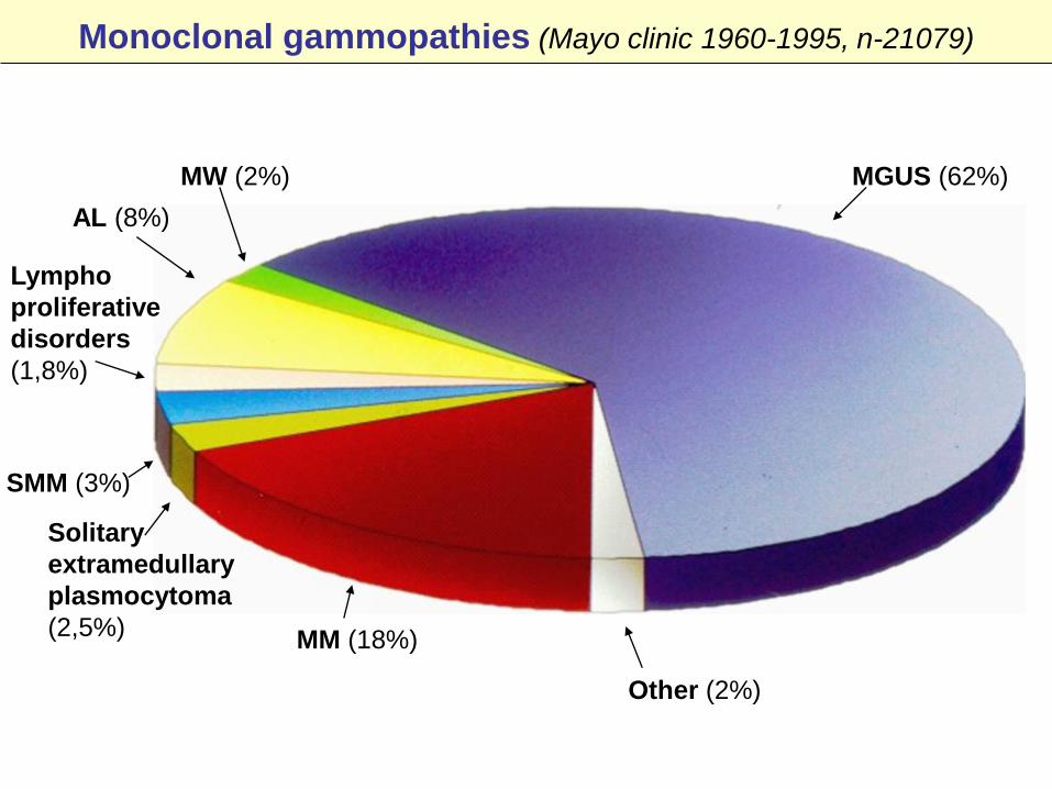
Monoclonal gammopathies (Mayo clinic 1960-1995, n-21079)
MGUS (62%)
Other (2%)
MM (18%)
Solitary
extramedullary
plasmocytoma
(2,5%)
SMM (3%)
MW (2%)
AL (8%)
Lympho
proliferative
disorders
(1,8%)
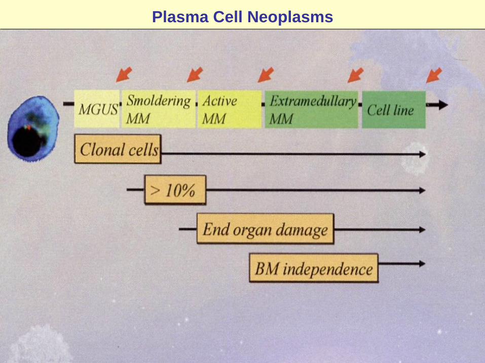
Plasma Cell Neoplasms
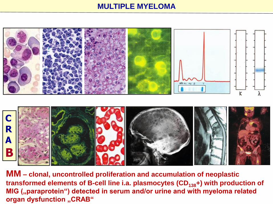
MULTIPLE MYELOMA
CRA
B
MM – clonal, uncontrolled proliferation and accumulation of neoplastic
transformed elements of B-cell line i.a. plasmocytes (CD138+) with production of
MIG („paraprotein“) detected in serum and/or urine and with myeloma related
organ dysfunction „CRAB“
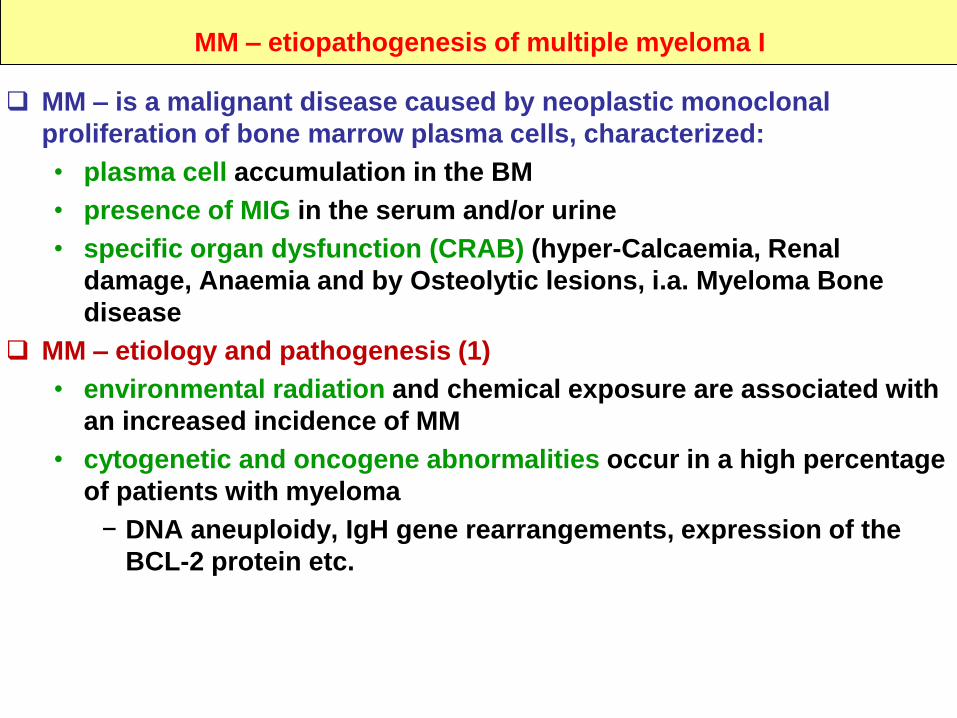
MM – etiopathogenesis of multiple myeloma I
MM – is a malignant disease caused by neoplastic monoclonal
proliferation of bone marrow plasma cells, characterized:
• plasma cell accumulation in the BM
• presence of MIG in the serum and/or urine
• specific organ dysfunction (CRAB) (hyper-Calcaemia, Renal
damage, Anaemia and by Osteolytic lesions, i.a. Myeloma Bone
disease
MM – etiology and pathogenesis (1)
• environmental radiation and chemical exposure are associated with
an increased incidence of MM
• cytogenetic and oncogene abnormalities occur in a high percentage
of patients with myeloma
− DNA aneuploidy, IgH gene rearrangements, expression of the
BCL-2 protein etc.
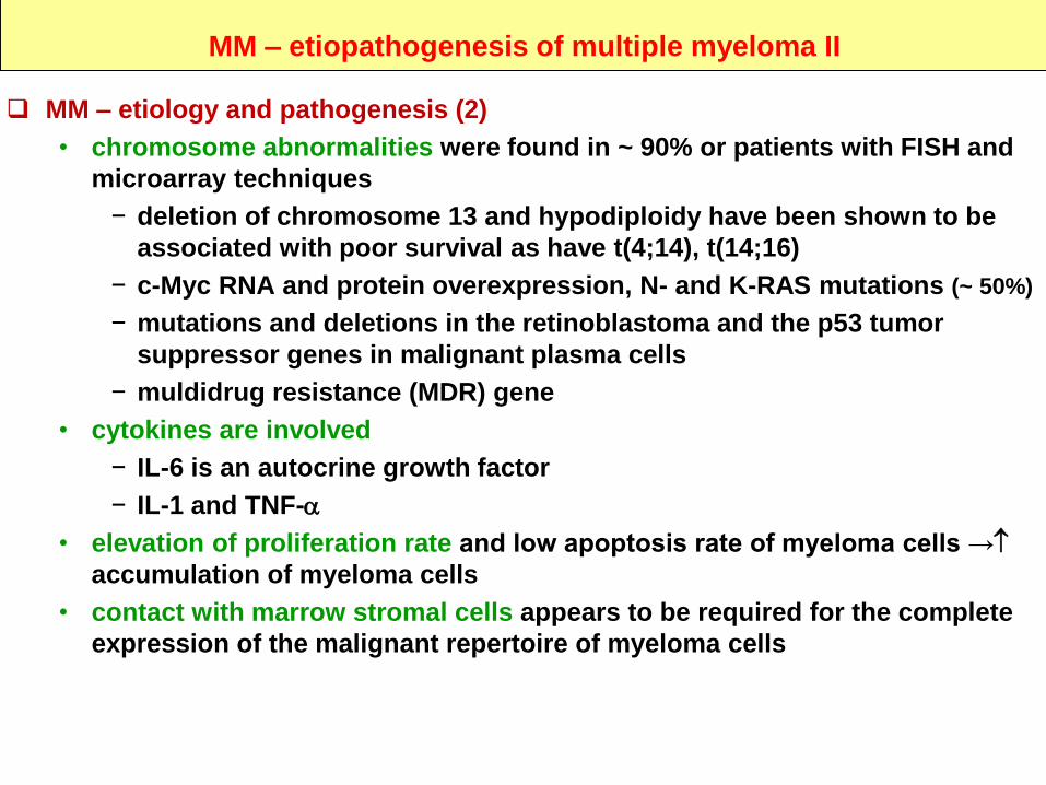
MM – etiopathogenesis of multiple myeloma II
MM – etiology and pathogenesis (2)
• chromosome abnormalities were found in ~ 90% or patients with FISH and
microarray techniques
− deletion of chromosome 13 and hypodiploidy have been shown to be
associated with poor survival as have t(4;14), t(14;16)
− c-Myc RNA and protein overexpression, N- and K-RAS mutations (~ 50%)
− mutations and deletions in the retinoblastoma and the p53 tumor
suppressor genes in malignant plasma cells
− muldidrug resistance (MDR) gene
• cytokines are involved
− IL-6 is an autocrine growth factor
− IL-1 and TNF-
• elevation of proliferation rate and low apoptosis rate of myeloma cells →
accumulation of myeloma cells
• contact with marrow stromal cells appears to be required for the complete
expression of the malignant repertoire of myeloma cells
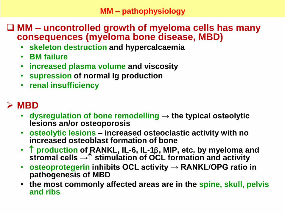
MM – pathophysiology
MM – uncontrolled growth of myeloma cells has many consequences (myeloma bone disease, MBD) • skeleton destruction and hypercalcaemia
• BM failure
• increased plasma volume and viscosity
• supression of normal Ig production
• renal insufficiency
MBD• dysregulation of bone remodelling → the typical osteolytic
lesions an/or osteoporosis
• osteolytic lesions – increased osteoclastic activity with no increased osteoblast formation of bone
• production of RANKL, IL-6, IL-1, MIP, etc. by myeloma and stromal cells → stimulation of OCL formation and activity
• osteoprotegerin inhibits OCL activity → RANKL/OPG ratio in pathogenesis of MBD
• the most commonly affected areas are in the spine, skull, pelvis and ribs
MM – pathophysiology
BMPC
bone marrow infiltration with plasma cells resulting in –anaemia, neutropenia, thrombocytopenia
overproduction of MIG – hyperviscosity syndrome• MIG – IgG (~ 50-60%), IgA (~ 20%), Bence-Jones ( or ) (~ 15%),
IgD, IgM, biclonal and nonsecretory type à ~ 1-2%
reduction in the normal Ig levels („immune paresis“)• tendency to recurrent infections (particularly of respiratory tract)
renal impairment – combination of• deposition of light chains in the renal tubules (cast formation)
• hypercalcaemia, hyperuricaemia, use of NSAID
• rarely deposition of amyloid
MM – DIAGNOSTIC CRITERIA (International Myeloma Working Group, 2003)
• All three diagnostic criteria required
1. Monoclonal BMPC ≥ 10%, and/or presence of biopsy – proven
plasmocytoma
2. MIG present in the serum and/or urine
3. Myeloma – releated organ dysfunction ( ≥1)
• Calcium elevation 2.8 mmol/l
• Renal insufficiency (S-creatinine 177 µmol/l)
• Anaemia Hb < 100 g/l
• Bone Osteolytic lesions or osteoporosis
- solitary plasmocytoma
- osteoporosis Pb 30%
• This criteria identify stage I-B and II-III – A/B myeloma by
Durie-Salmon stage
• Stage I-A becomes „smoldering“ or indolent MM
C
R
A
B
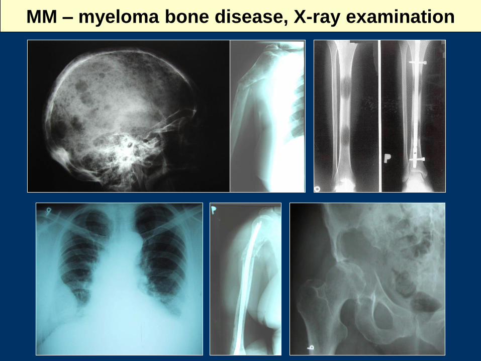
MM – myeloma bone disease, X-ray examination
MM – clinical manifestation
MM – incidence
• 3-4/100 000/year, variability from country to country
(1 in China, 4/100 000 in West Europe)
• is more common in blacks than white
• M/F ratio is 2-3:2
• age median 61 (63 years), the incidence rises with age
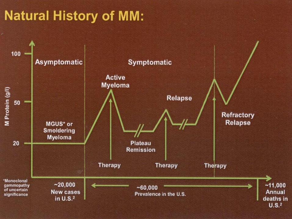
MM – clinical manifestation
nonetheless, the disease can remain „asymptomatic“ for many
years
disease phases
• asymptomatic (indolent, „smoldering“ MM, stage I-A according
D-S)
• symptomatic/“active“ MM
− remission, eventually „plateau“/stable phase
− relaps eventually progression
• refractory/terminal phase
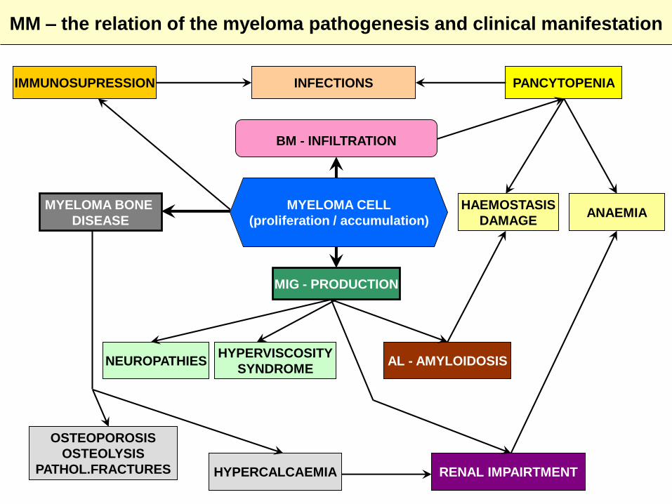
MM – the relation of the myeloma pathogenesis and clinical manifestation
IMMUNOSUPRESSION INFECTIONS PANCYTOPENIA
MYELOMA BONE
DISEASE
MIG - PRODUCTION
HAEMOSTASIS
DAMAGEANAEMIA
NEUROPATHIES AL - AMYLOIDOSISHYPERVISCOSITY
SYNDROME
OSTEOPOROSIS
OSTEOLYSIS
PATHOL.FRACTURES HYPERCALCAEMIA RENAL IMPAIRTMENT
BM - INFILTRATION
MYELOMA CELL
(proliferation / accumulation)
MM – clinical features
CLINICAL SYMPTOMS
Bone pain – most commonly lower back (60%)
• vertebral involvement (compression/pathological fractures of vertebral bodies)
• osteolytic bone lesions
• tumour growth on nerve roots or spinal cord compression
Features of anaemia
• lethargy, weakness, dyspnoe, pallor, etc.
Recurrent infections
• related to deficient normal immunoglobulins/antibody production and/or cell-mediated immunity
• bacterial, viral, etc. (frequently pneumonia)
Symptoms of renal failure (20-30%)
• nephrotic syndrome – in associated with AL amyloidosis and with B-J-proteinuria
Hypercalcaemia (20-30%)
• polydipsia, polyuria, anorexia, vomiting, constipation, mental disturbance
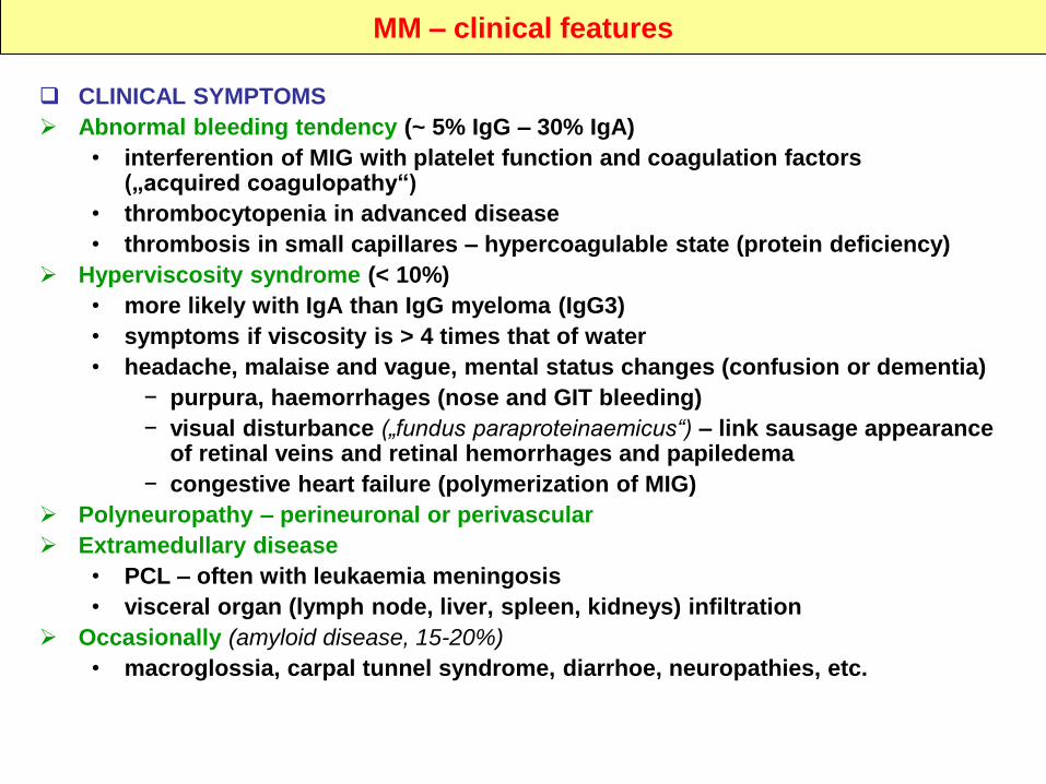
MM – clinical features
CLINICAL SYMPTOMS
Abnormal bleeding tendency (~ 5% IgG – 30% IgA)
• interferention of MIG with platelet function and coagulation factors („acquired coagulopathy“)
• thrombocytopenia in advanced disease
• thrombosis in small capillares – hypercoagulable state (protein deficiency)
Hyperviscosity syndrome (< 10%)
• more likely with IgA than IgG myeloma (IgG3)
• symptoms if viscosity is > 4 times that of water
• headache, malaise and vague, mental status changes (confusion or dementia)
− purpura, haemorrhages (nose and GIT bleeding)
− visual disturbance („fundus paraproteinaemicus“) – link sausage appearance of retinal veins and retinal hemorrhages and papiledema
− congestive heart failure (polymerization of MIG)
Polyneuropathy – perineuronal or perivascular
Extramedullary disease
• PCL – often with leukaemia meningosis
• visceral organ (lymph node, liver, spleen, kidneys) infiltration
Occasionally (amyloid disease, 15-20%)
• macroglossia, carpal tunnel syndrome, diarrhoe, neuropathies, etc.
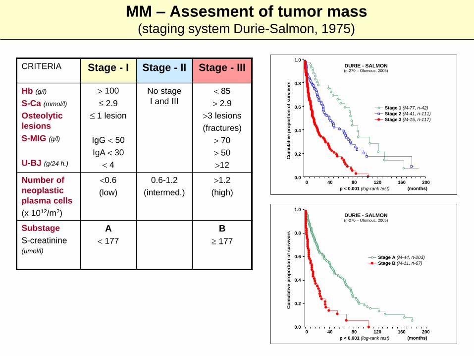
MM – Assesment of tumor mass (staging system Durie-Salmon, 1975)
CRITERIA Stage - I Stage - II Stage - III
Hb (g/l)
S-Ca (mmol/l)
Osteolytic
lesions
S-MIG (g/l)
U-BJ (g/24 h.)
100
2.9
1 lesion
IgG 50
IgA 30
4
No stage
I and III
85
2.9
3 lesions
(fractures)
70
50
12
Number of
neoplastic
plasma cells
(x 1012/m2)
0.6
(low)
0.6-1.2
(intermed.)
1.2
(high)
Substage
S-creatinine (µmol/l)
A
177
B
177
0.0
0.2
0.4
0.6
0.8
1.0
0 40 80 120 160 200
Cu
mu
lati
ve
pro
po
rtio
no
fs
urv
ivo
rs
p < 0.001 (log-rank test) (months)
DURIE - SALMON
Stage 1 (M-77, n-42)
Stage 2 (M-41, n-111)
Stage 3 (M-15, n-117)
(n-270)
0.0
0.2
0.4
0.6
0.8
1.0
0 40 80 120 160 200
Cu
mu
lati
ve
pro
po
rtio
no
fs
urv
ivo
rs
p < 0.001 (log-rank test) (months)
DURIE - SALMON
Stage 1 (M-77, n-42)
Stage 2 (M-41, n-111)
Stage 3 (M-15, n-117)
(n-270)
0.0
0.2
0.4
0.6
0.8
1.0
Cu
mu
lati
ve
pro
po
rtio
no
fs
urv
ivo
rs
0 40 80 120 160 200
p < 0.001 (log-rank test) (months)
DURIE - SALMON(n-270)
Stage A (M-44, n-203)
Stage B (M-11, n-67)
0.0
0.2
0.4
0.6
0.8
1.0
Cu
mu
lati
ve
pro
po
rtio
no
fs
urv
ivo
rs
0 40 80 120 160 200
p < 0.001 (log-rank test) (months)
DURIE - SALMON(n-270)
Stage A (M-44, n-203)
Stage B (M-11, n-67)
(n-270 – Olomouc, 2005)
(n-270 – Olomouc, 2005)
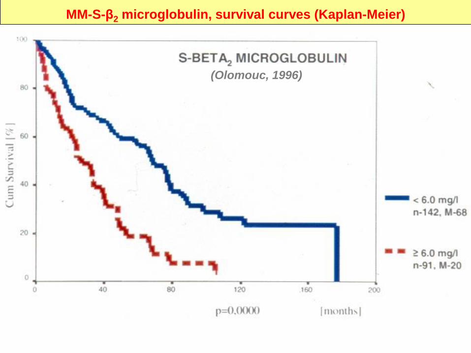
MM-S-β2 microglobulin, survival curves (Kaplan-Meier)
(Olomouc, 1996)
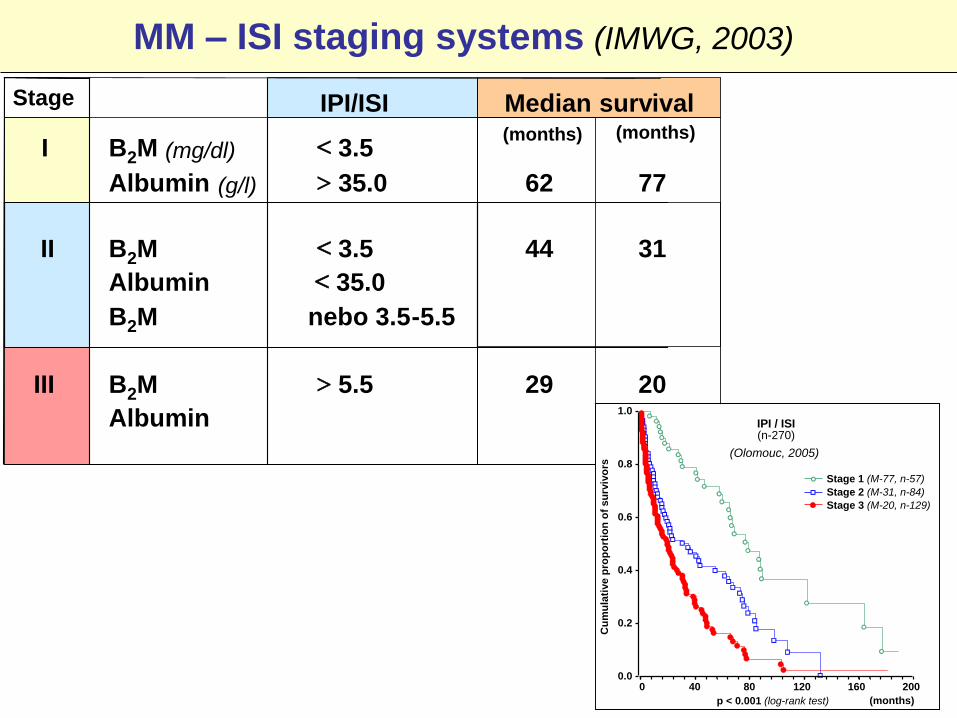
MM – ISI staging systems (IMWG, 2003)
IPI/ISI Median survival
I B2M (mg/dl) 3.5
Albumin (g/l) 35.0 62 77
II B2M 3.5 44 31
Albumin 35.0
B2M nebo 3.5-5.5
III B2M 5.5 29 20
Albumin
Stage
(months)(months)
0.0
0.2
0.4
0.6
0.8
1.0
Cu
mu
lati
ve
pro
po
rtio
no
fsu
rviv
ors
0 40 80 120 160 200
p < 0.001 (log-rank test) (months)
Stage 1 (M-77, n-57)
Stage 2 (M-31, n-84)
Stage 3 (M-20, n-129)
IPI / ISI(n-270)
(Olomouc, 2005)
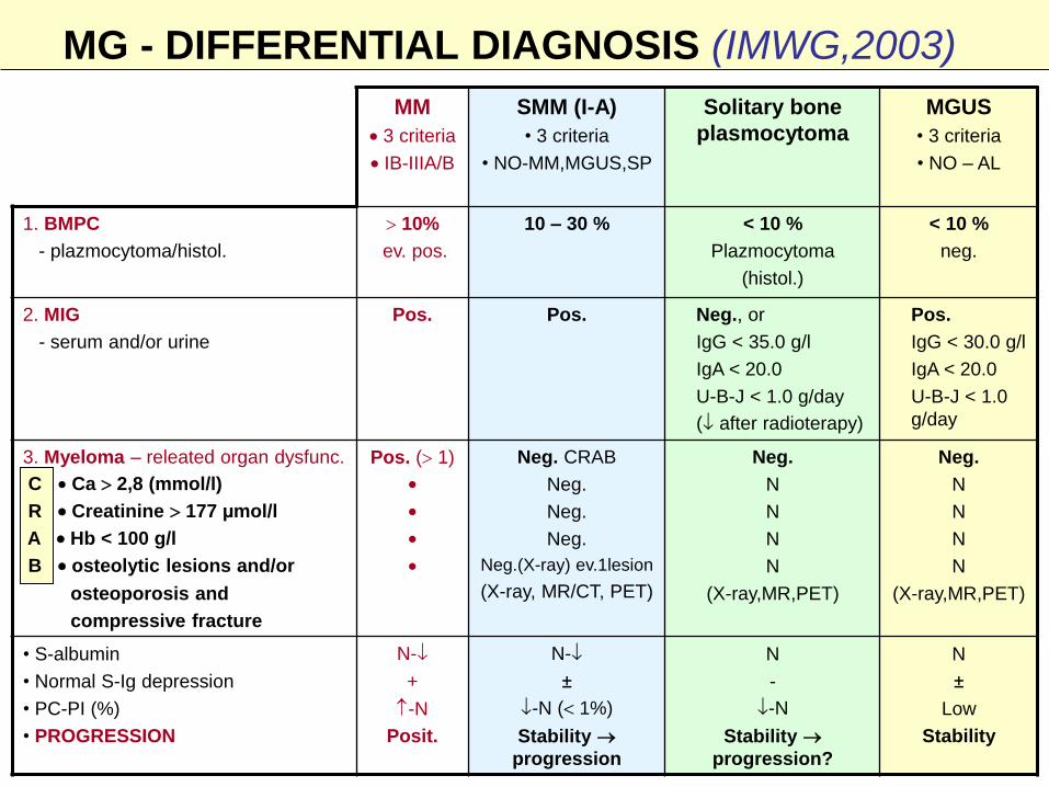
MG - DIFFERENTIAL DIAGNOSIS (IMWG,2003)
MM
3 criteria
IB-IIIA/B
SMM (I-A)
• 3 criteria
• NO-MM,MGUS,SP
Solitary bone
plasmocytoma
MGUS
• 3 criteria
• NO – AL
1. BMPC
- plazmocytoma/histol.
10%
ev. pos.
10 – 30 % < 10 %
Plazmocytoma
(histol.)
< 10 %
neg.
2. MIG
- serum and/or urine
Pos. Pos. Neg., or
IgG < 35.0 g/l
IgA < 20.0
U-B-J < 1.0 g/day
( after radioterapy)
Pos.
IgG < 30.0 g/l
IgA < 20.0
U-B-J < 1.0
g/day
3. Myeloma – releated organ dysfunc.
C Ca 2,8 (mmol/l)
R Creatinine 177 µmol/l
A Hb < 100 g/l
B osteolytic lesions and/or
osteoporosis and
compressive fracture
Pos. ( 1)
Neg. CRAB
Neg.
Neg.
Neg.
Neg.(X-ray) ev.1lesion
(X-ray, MR/CT, PET)
Neg.
N
N
N
N
(X-ray,MR,PET)
Neg.
N
N
N
N
(X-ray,MR,PET)
• S-albumin
• Normal S-Ig depression
• PC-PI (%)
• PROGRESSION
N-
+
-N
Posit.
N-
±
-N ( 1%)
Stability
progression
N
-
-N
Stability
progression?
N
±
Low
Stability
MM – investigations
MIG – in the serum, urine or both (98%)
• Bence-Jones in urine in two/thirds of cases
• dimished levels of an involved Ig classes is common
• total protein in the serum
• FLC serum levels (Freelite test)
BM - shows increased plasma cells (> 10%, usually 30-50% in BM aspirate or trephing)
- „myeloma cells“ – atypical PC, monoclonal cell expressing the same Ig as the S-MIG
• flow cytometric analysis
− DNA aneuploidy in 80% patients
− myeloma cells express positivity of CD138+, CD56+, CD38+, CD45-
• a high proliferative index (PC-propidium iodide) and low apoptotic index (e.g. annexin V) are an important negative prognostic features
• chromosomal abnormalities – FISH
Hematologic abnormalities
• normochromic/normocytic anaemia due to marrow replacement by plasmocytes, „rouleaux“ formation
• trombocytopenia and neutropenia – in the advanced phases of MM
• coagulopathy – interference with fibrin formation by M-protein
• abnormal plasma cells in the blood film (15%)
MM – investigations
Skeletal survey (x-ray, MRI, CT, FDG-PET/CT) of the axial skeleton
• osteolytic areas without evidence of surrounding osteoblastic reaction or sclerosis (60%)
• generalized bone rarefaction (20%)
• pathological fractures are common
• no bone lesions are found in 20% patients
• MIBI – is valuable detection and follow-up of treatment
• DEXA – evaluation of a grade of sceletal mineralisation
Other laboratory findings
• high ESR (this is almost always very high, > 100 mm/hour)
• serum elevations of: Ca, urea and creatinine (20%), uric acid, ALP (in fractures)
• 2-microglobulin, ↓ S-albumin, CRP, LDH, thymidinekinase → negative PF
• IL-6, VEGF, HGF, etc.
Bence-Jones
0,1
1
10
100
1000
0,1 1 10 100 1000FLC - KAPPA
FL
C -
LA
MB
DA
Active n-8
Stable n-5
MGUSn-54
0,1
1
10
100
1000
0,1 1 10 100 1000FLC - Kappa (mg/l)
FL
C -
La
mb
da
(m
g/l
)
MM n-116
0,1
1
10
100
1000
10000
0,1 1 10 100 1000 10000
FLC - KAPPA
FL
C -
LA
MB
DA
Stable n-56
Active n-60
0,1
1
10
100
1000
0,1 1 10 100 1000
FLC - KAPPA (mg/l)
FL
C -
LA
MB
DA
(m
g/l
)
AL solit. (n-4)
AL gen. (n-5)
MW (n-8)
Sol.plasmocytom(n-8)
Ostatní MG
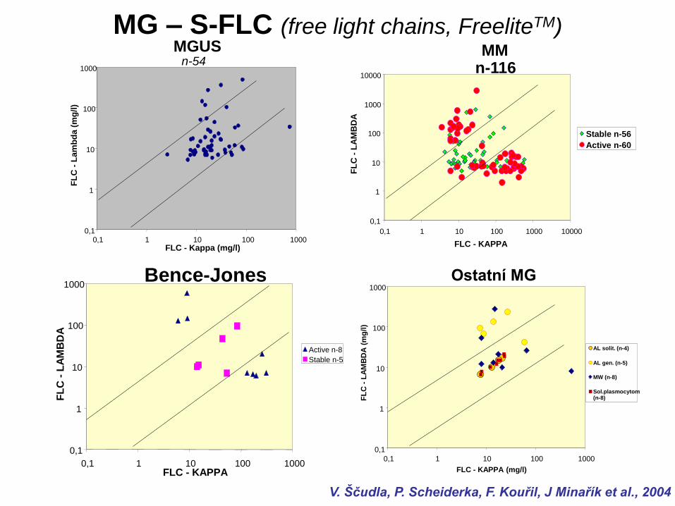
MG – S-FLC (free light chains, FreeliteTM)
V. Ščudla, P. Scheiderka, F. Kouřil, J Minařík et al., 2004
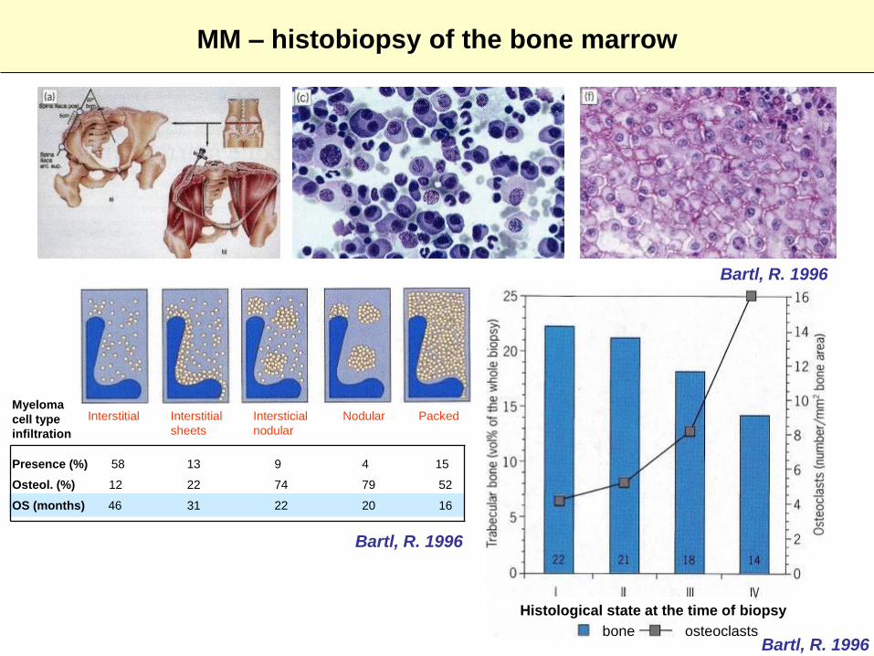
MM – histobiopsy of the bone marrow
Bartl, R. 1996
bone osteoclasts
Histological state at the time of biopsy
PackedNodularIntersticial
nodular
Interstitial
sheets
Interstitial
Presence (%) 58 13 9 4 15
Osteol. (%) 12 22 74 79 52
OS (months) 46 31 22 20 16
Bartl, R. 1996
Myeloma
cell type
infiltration
Bartl, R. 1996
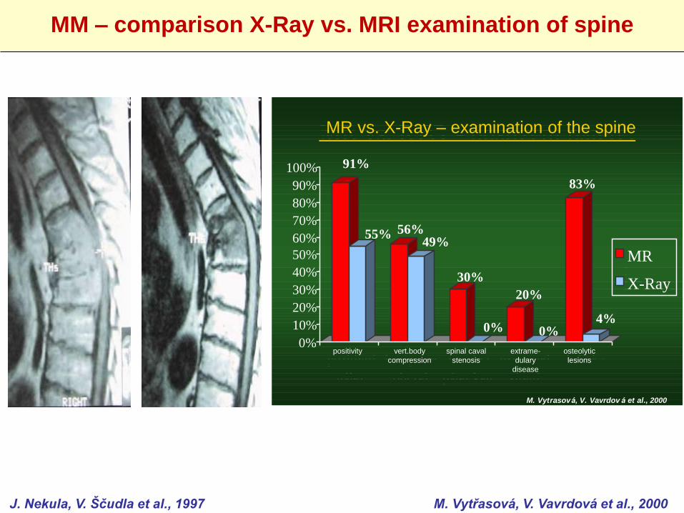
MM – comparison X-Ray vs. MRI examination of spine
J. Nekula, V. Ščudla et al., 1997 M. Vytřasová, V. Vavrdová et al., 2000
MR vs. RTG - postižení pátere:91%
55% 56%49%
30%
0%
20%
0%
83%
4%
0%
10%
20%
30%
40%
50%
60%
70%
80%
90%
100%
pozitivní
nález
komprese
obr.tel
stenóza
páter.kan.
extramed.
šírení
ložiska
MR
RTG
M. Vytrasová, V. Vavrdov á et al., 2000
MR vs. X-Ray – examination of the spine
91%
55% 56%49%
30%
0%
20%
0%
83%
4%
0%
10%
20%
30%
40%
50%
60%
70%
80%
90%
100%
MR
X-Ray
M. Vytrasová, V. Vavrdov á et al., 2000
positivity vert.body
compression
spinal caval
stenosis
extrame-
dulary
disease
osteolytic
lesions
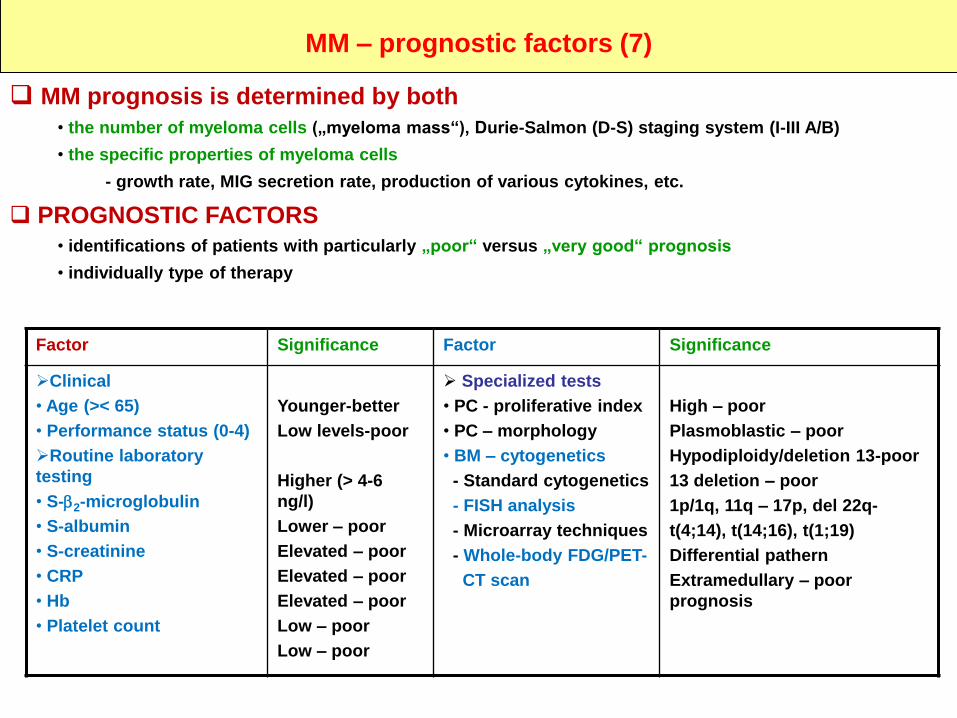
MM – prognostic factors (7)
MM prognosis is determined by both
• the number of myeloma cells („myeloma mass“), Durie-Salmon (D-S) staging system (I-III A/B)
• the specific properties of myeloma cells
- growth rate, MIG secretion rate, production of various cytokines, etc.
PROGNOSTIC FACTORS
• identifications of patients with particularly „poor“ versus „very good“ prognosis
• individually type of therapy
Factor Significance Factor Significance
Clinical
• Age (>< 65)
• Performance status (0-4)
Routine laboratory
testing
• S-2-microglobulin
• S-albumin
• S-creatinine
• CRP
• Hb
• Platelet count
Younger-better
Low levels-poor
Higher (> 4-6
ng/l)
Lower – poor
Elevated – poor
Elevated – poor
Elevated – poor
Low – poor
Low – poor
Specialized tests
• PC - proliferative index
• PC – morphology
• BM – cytogenetics
- Standard cytogenetics
- FISH analysis
- Microarray techniques
- Whole-body FDG/PET-
CT scan
High – poor
Plasmoblastic – poor
Hypodiploidy/deletion 13-poor
13 deletion – poor
1p/1q, 11q – 17p, del 22q-
t(4;14), t(14;16), t(1;19)
Differential pathern
Extramedullary – poor
prognosis
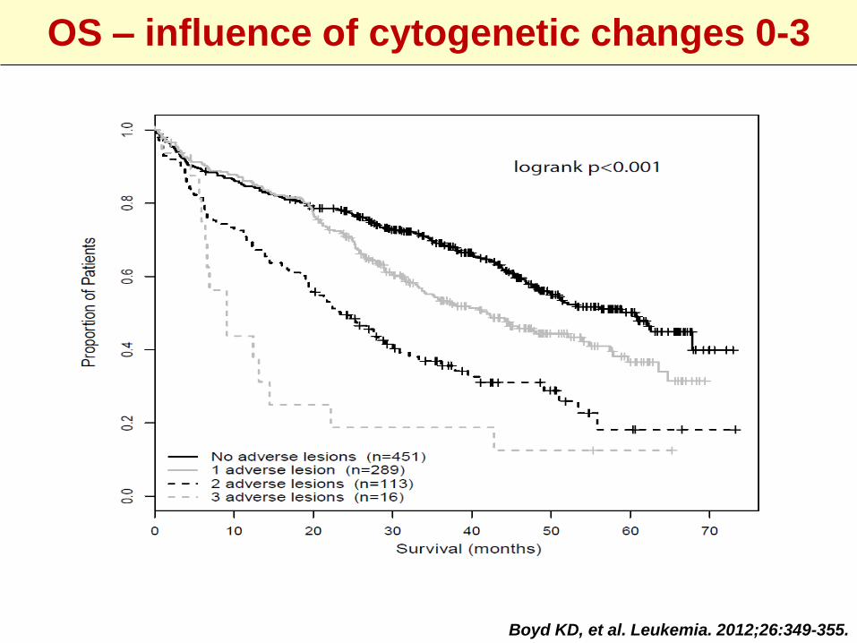
Boyd KD, et al. Leukemia. 2012;26:349-355.
OS – influence of cytogenetic changes 0-3
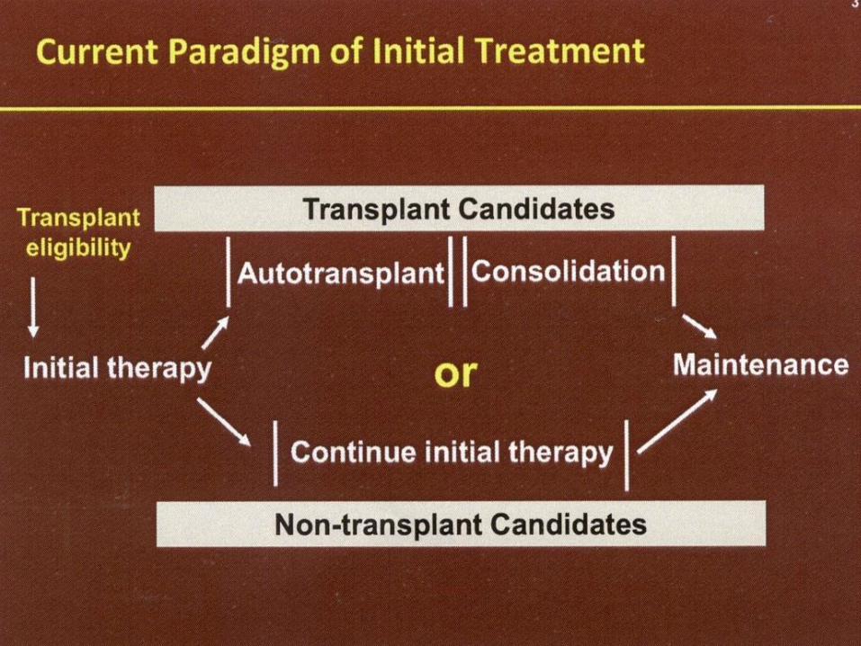
MM – frontline therapy
Exclusion of patient with MGUS and smoldering/indolent myeloma
There is yet no optimal consensus as to the best way to manage MM
• MM is not curable!
• patient with asymptomatic/indolent e.g. „smoldering-stable“ MM
− should be observed closely → „wait/watch and see“ and/or
supportive therapy only
Treatment is reccommended in active/symptomatic forms of MM
• advanced clinical stage (I-B-II/III-A/B according Durie Salmon)
• when the M-component/other laboratory features are increasing
• clinical problems or complications have emerged or are imminent
• problems sufficient to require treatment include
− bone lesions (X-Ray, MRI, CT, FDG-PET/CT)
− renal insufficiency
− reduced blood count (e.g. anaemia, neutropenia,
thrombocytopenia)
− elevated S-calcium
− other significant organ or tissue damage caused by MM
MM – overall summary of treatment options
BASIC THERAPY• Chemotherapy – standard/conventional treatment
• High dose therapy with autologous transplantation
• New „biologic“ therapy – „combination with conventional substances“
• Radiation
SUPPORTIVE CARE ASPECT• emergency care, e.g. hemodialysis, plasmapheresis, surgery, etc.
• pain medication
• rHuEPO (e.g. Eprex or Recormon, Darbepoietin)
• bisphosphonates (e.g. Aredia, Bonefos, Lodronat, Zometa, etc.)
• antibiotics
• growth factors (e.g. Neupogen)
• brace/corset
• diet
PERSPECTIVE TREATMENT OPTIONS (?)• new vaccines (e.g. antiidiotype)
• new chemotherapy and biological components
MM – conventional chemotherapy
INDUCTION CHEMOTHERAPY
MP – melphalan/prednison
• remains a valid option only for elderly patients
(> 80-85 years), pure PS and high commorbidity
(now it is not „gold standard“, but historical treatment)
• indication: „low risk“/> 80-85 years
• 40-60% of patients have an „OR“ (objective
response), good tolerance, OS – 19-40 months
• melphalan is not reccommended if stem cell
harvesting is planned
• cyclophosphamide – although less popular, is a
valid option (C or CP)
- similar anti-myeloma activity
MM – conventional chemotherapy
INDUCTION CHEMOTHERAPY More complex combination schedules
• higher and earlier response rate than MP but marginal overall better
outcome? (survival time)
• more toxic inconvenience and expense: VBMCP, VAD („historical treatment“)
Pulse dexamethasone alone – is widely used as frontline therapy
(40 mg 1.-4. day) → in renal insufficiency, pancytopenia (response
without injuring the BM stem cells!)
The current trend in „untrasplanted patients“ is to use chemo-
immunotherapy, eg.: MPT, MPV, R-Dex, ev. BAD, RAD as a first choice
• more complex („combination“) therapy is also reserved as a
back up approach for patients who fail to have a satisfactory
response or with severe complications (e.g. spinal cor
compression, etc.)
The major factor which determines outcome is the
intrinsic drug sensitivity or resistance of the myeloma
cells
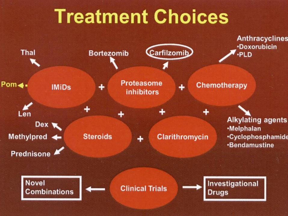
MM – novel therapies and new technologies
THE MOST PROMISING NOVEL BIOLOGIC THERAPIES
THALIDOMIDE
• Thal/Dex – it was active
− as a frontline approach 50-100 mg/day, minimal myelosupression
− Thal/Dex, event. MPT or CTD
Thal/Dex - ~ 50% OR (↓ to < 50% MIG)
• adverse reaction (~ 25-50%): severe neuropathy, constipation, lethargy, sleepines, skin reaction, etc.)
LEDALIDOMID (REVLIMID - thalidomide analogs)
• Revlimid/Dex
- very good efect, no neurologic side efect, but neutropenia
- R-Dex, RAD, RVD, RCD, etc.
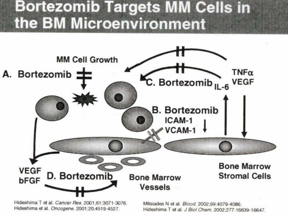
BORTEZOMIB (VELCADE)
• very good efect in patients with induction therapy and relapsed/refractory myeloma
- OR ~ 35%, median response ~ 12 months, OS ~ 16 months
• combination Velcade plus Thal, Doxil, Dex, etc.
New compunds: carfilzomib, pomalidomide, panobinostat, etc.
Assessment of all three new drug in the frontline therapy
– very good resultes before ASCT
− primary induction, consolidation with HDT/ASCT, post induction/consolidation and maintenance therapy
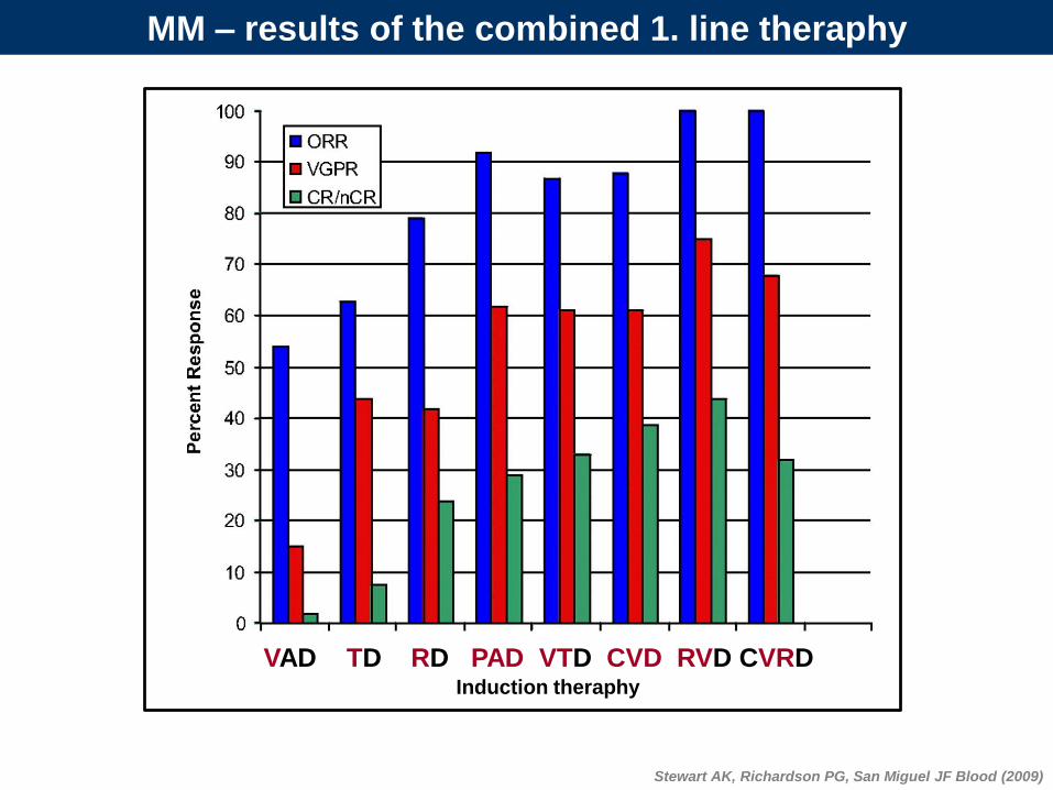
Stewart AK, Richardson PG, San Miguel JF Blood (2009)
Induction theraphy
VAD TD RD PAD VTD CVD RVD CVRD
MM – results of the combined 1. line theraphy
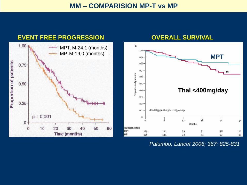
Thal <400mg/day
MM – COMPARISION MP-T vs MP
OVERALL SURVIVALEVENT FREE PROGRESSION
Palumbo, Lancet 2006; 367: 825-831
MPT
MPT, M-24,1 (months)
MP, M-19,0 (months)
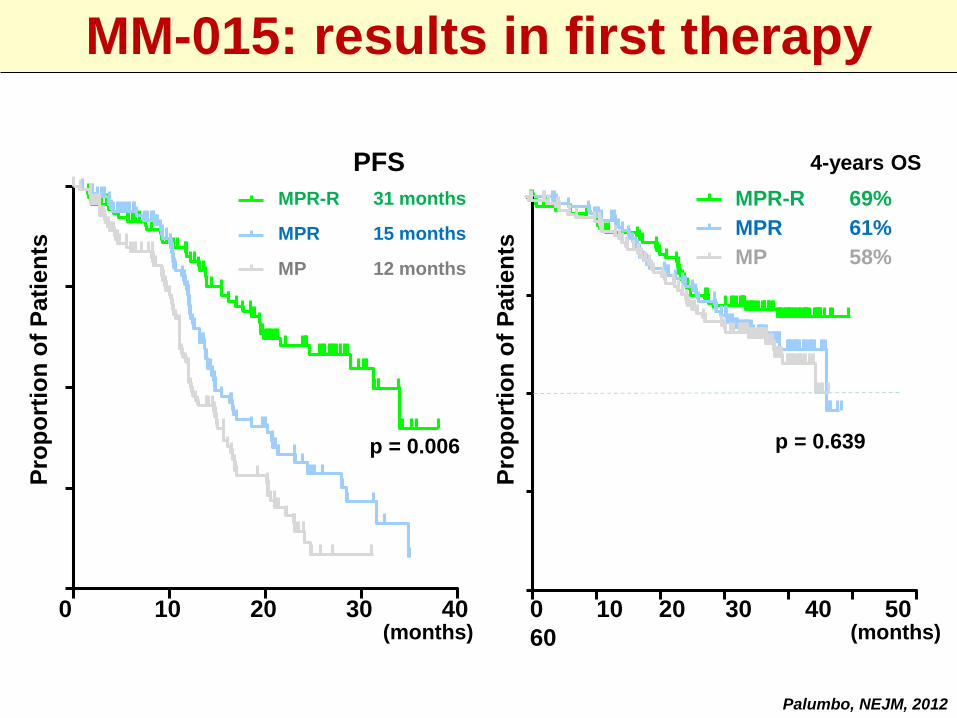
PFS
MPR-R 31 months
MPR 15 months
MP 12 months
Time (months)0 10 20 30 40
0
p = 0.006
Palumbo A, et al. Blood. 2011;118 [abstract 475; oral presentation at ASH 2011].
4-years OS
MPR-R 69%
MPR 61%
MP 58%
0 10 20 30 40 50 600
25
50
75
100
Time (months)
Pati
en
ts (
%)
p = 0.639
(months)0 10 20 30 40 0 10 20 30 40 50
60
Pro
po
rtio
n o
f P
ati
en
ts
Palumbo, NEJM, 2012
(months)
Pro
po
rtio
n o
f P
ati
en
ts
MM-015: results in first therapy
MM – high dose therapy with autotransplantation
HD – THERAPY with AUTOLOGOUS STEM CELL
TRANSPLANTATION (ASCT)
• HD-Th+ASCT → is the frontline „therapy of choice“ for newly
diagnosed patients with symptomatic MM in this time and
< 65 years
• complete remission (CR) rate range from 30-75%, PR from
75-90%
• This approach is not curative → 90% of patients relapse
− median overall survival with HDT/ASCT
is in 5-6 year range
− procedure related mortality with HDT/ASCT
is very low ~ 1%
• HD-Th – a more dramatic myeloma cell kill in comparison of
conventional therapy – „eradication of myeloma cells“?
MM – high dose therapy with autotransplantation
HD-therapy with ASCT
• induction therapy, e.g. CTD (Thalidomid), CTV (Velcade),
R-Dex (Revlimid), (↓ „myeloma mass“), R-Dex (Revlimide)
• „mobilisation“ and „collection“ of peripheral blood stem cells
(PBSC)
- cyclophosphamide (2-5 g) + G-CSF stimulation (Neupogen)
• „conditioning“ regimen – standard regimen is melphalan 200
mg/m2 i.v./1.day
• „transplantation“ of autologous PBSC (intravenously infusion)
− engrafment of haematopoietic stem cells (~ 2 weeks)
− supportive therapy (G-CSF, rHuEPO/antibiotics, etc.)
• maintenance therapy (<VGPR – Thalidomide,
Bortezomib/Velcade or Lenalidomide/Revlimid)
Double or tandem transplantation
• limited contribution
• useful and viable option is relapse after 1. ASCT
MM – HD-therapy/ASCT indications
Active/symptomatic/overt form of MM
Age - < 65 (70?) years
Performance status (ECOG/WHO) ≤ 2
Renal function
• normal function
• creatinine clearence 50 ml/min and/or S-creatinine < 3.-4.0 mg/dl can be
considered for autotransplantation
- but only at a center with special expertise in this setting
Patient without previous therapy: Melphalan, BCNU, extensive X-ray irradiation
Absence of advanced phases of „internal diseases“ (↓ low commorbidity)
ASCT is possible in ~ 50% of MM patients
Role of allogeneic transplantation
• despite medical improvements, allogeneic transplant is a high-risk procedure in the management
of MM
• initial treatment – related mortality is high (at least 20-30%)
• the GV-myeloma effect can be enhanced by using donor lymphocyte infusions (DLI)
• recent interest is in „non-myeloablative/or mini-allogeneic transplants“ in MM
− lesser toxicity (but still substantial acute (45%) and chronic (55%) GVH disease
indication in younger patients particularly with on HLA-matched, sibling donor of the same gender,
since the risk are lower
• „mini/allogeneic transplantation is a promising new approach“(?)
(months)
Cu
msu
rviv
al (%
)
1,0
0,8
0,6
0,4
0,2
0,0
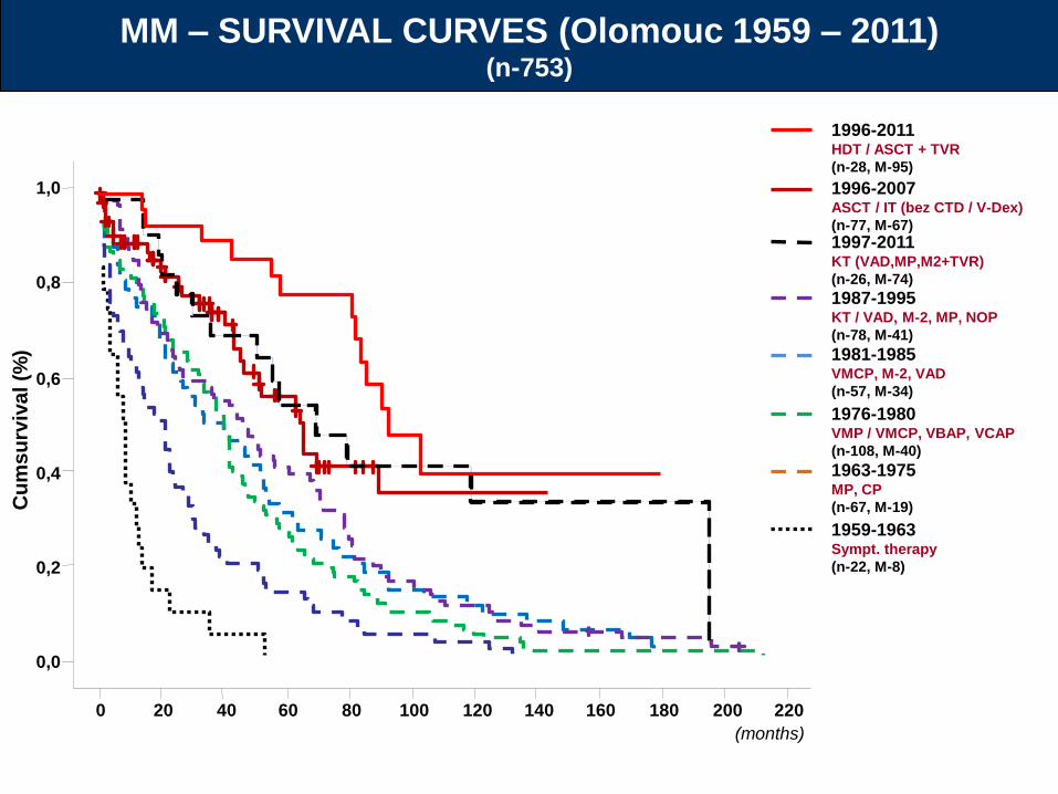
MM – SURVIVAL CURVES (Olomouc 1959 – 2011)(n-753)
1996-2007ASCT / IT (bez CTD / V-Dex)
(n-77, M-67)
1997-2011KT (VAD,MP,M2+TVR)
(n-26, M-74)
1987-1995KT / VAD, M-2, MP, NOP
(n-78, M-41)
1981-1985VMCP, M-2, VAD
(n-57, M-34)
1976-1980VMP / VMCP, VBAP, VCAP
(n-108, M-40)
1963-1975MP, CP
(n-67, M-19)
1959-1963Sympt. therapy
(n-22, M-8)
1996-2011HDT / ASCT + TVR
(n-28, M-95)
220200180160140120100806040200
MM – maintenance therapy
MAINTENANCE THERAPY The role of anti-myeloma maintenance therapy following frontline
therapy and ASCT is unclear
• continued alkylator therapy is not beneficial
• Several new agents are now beeing studied
- THALIDOMIDE
- REVLIMID (LEDALIDOMIDE) !
- VELCADE (BORTEZOMIB)- vaccine approaches
No strong reccommendation can be made for any particular maintenance strategy
− Lenalidomide (Revlimid) with or without steroids – is an option for maintenance, especially in high – risk settings
− Ledalidomid – is preffereded in this time
MM – treatment of emmergency situations
Renal failure („uraemia“)• rehydratation and treatment of underlying cause (e.g. hypercalcaemia,
hyperuricaemia)
• hemodialysis
Hypercalcaemia• rehydratation with isotonic saline
• corticosteroids intravenously
• bisphosphonates (pamidronate, zoledronic acid) intravenously
• active treatment of MM
Compression paraplegia• HD-steroids (dexamethasone, metylprednisolone), chemotherapy
• X-ray irradiation
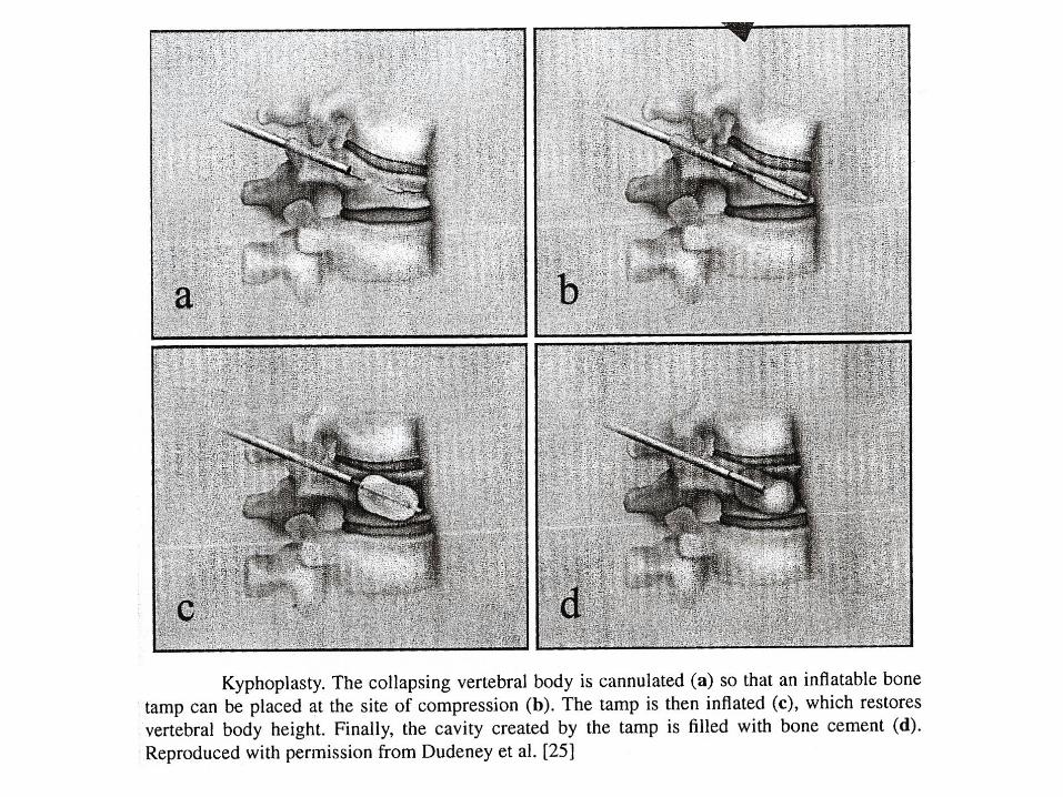
• decompression laminectomy
• vertebroplasty, kyphoplasty („vertebral body reconstruction“)
Single painful skeletal lesions• X-ray irradiation + chemotherapy
Severe anaemia• transfusion of packed red cells
Bleeding (MG interference with coagulation factors and HVS)• repeated plasmapheresis
• coagulation factors substitution, etc.
Severe reccurent infections• prophylactic infusions of immunoglobulin concentrates
• broad – spectrum antibiotics and antifugal agents
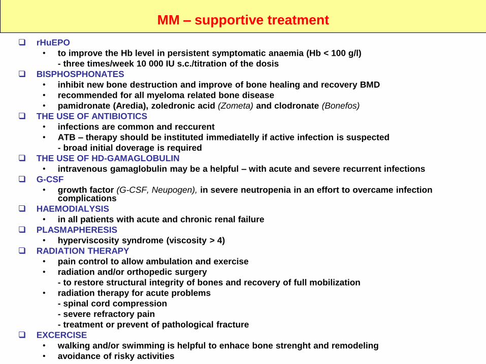
MM – supportive treatment
rHuEPO
• to improve the Hb level in persistent symptomatic anaemia (Hb < 100 g/l)
- three times/week 10 000 IU s.c./titration of the dosis
BISPHOSPHONATES
• inhibit new bone destruction and improve of bone healing and recovery BMD
• recommended for all myeloma related bone disease
• pamidronate (Aredia), zoledronic acid (Zometa) and clodronate (Bonefos)
THE USE OF ANTIBIOTICS
• infections are common and reccurent
• ATB – therapy should be instituted immediatelly if active infection is suspected
- broad initial doverage is required
THE USE OF HD-GAMAGLOBULIN
• intravenous gamaglobulin may be a helpful – with acute and severe recurrent infections
G-CSF
• growth factor (G-CSF, Neupogen), in severe neutropenia in an effort to overcame infection complications
HAEMODIALYSIS
• in all patients with acute and chronic renal failure
PLASMAPHERESIS
• hyperviscosity syndrome (viscosity > 4)
RADIATION THERAPY
• pain control to allow ambulation and exercise
• radiation and/or orthopedic surgery
- to restore structural integrity of bones and recovery of full mobilization
• radiation therapy for acute problems
- spinal cord compression
- severe refractory pain
- treatment or prevent of pathological fracture
EXCERCISE
• walking and/or swimming is helpful to enhace bone strenght and remodeling
• avoidance of risky activities