Embed Size (px)
Citation preview
MORPHOLOGICAL AND MOLECULAR CHARACTERIZATION OF POTENTIAL AMNESIC SHELLFISH POISONING
TOXIN-PRODUCING DIATOM Pseudo-nitzschia (Bacillariopbyeae) FROM MALAYSIA
Lim Hong Chang
Master of Science 2011
l'usat Khidm t M kJaU1\1VF.RSma UDUlt Akademik , MAUYSIA SARAWAK
P.KHIDMAT MAKLUMAT AKADI!MIK
11111 1lIllli'iTII 11111 III 1000246252
MORPHOLOGICAL AND MOLECULAR CHARACTERIZATION OF
POTENTIAL AMNESIC SHELLFISH POISONING TOXIN-PRODUCING
DIATOM Pseudo-nitzschia (BACILLARIOPHYCEAE) FROM MALAYSIA
Lim Hong Chang
A thesis submitted
In fulfillment of the requirements for the degree of
Master of Science
(Marine Science)
Faculty of Resource Science and Technology
UNIVERSITI MALAYSIA SARAWAK
2011
'.
I
DECLARATION
I hereby declare that the work in this thesis is my own except for quotations and
summaries which have been duly acknowledged.
LIM HONG CHANG
08021402
11
ACKNOWLEDGEMENTS
This dissertation would not have been possible without the supports of my supervisors,
Dr Lim Po Teen and Dr Leaw Chui Pin. I wish to express my indebtedness for their
patience and tireless efforts to help and guide me in data collections, data analyses
and paper preparations throughout these years. My special thanks also due to Zamalah
postgraduate scholarship UNIMAS, MOHE-FRGS grants to Dr Lim Po Teen and E
Science to Dr Leaw Chui Pin for financials support provided for the research. Special
thanks to Prof. Dr. Yuichi Kotaki, Kitasato University, Japan for the toxin analyses
and Prof. Dr. Gires Usup, National University Malaysia (UKM) for providing
financial support for DNA sequencing.
I would also like to thank members of Harmful Algal Blooms (HABs) research group:
Suriyanti Su Nyun Pau, Hartina Mohd Ali, Tan Toh Hii, Fareha Haji Hilaluddin, Siti
Zubaidah Kamarudin, Teng Sing Tung and Hii Kieng Soon whom assisted and
willingly in sharing their knowledge and experiences which contributed to this
dissertation in a vital way. I am eternally grateful to the faculty, staffs at laboratories
of Department of Aquatic Science, Institute of Biodiversity and Environmental
Conservation (IBEC) and also Science Officers (Madam Ting Woei, Mr Amin Mangi
and Mr Besar Ketol) for their assistances.
Special thanks to my friends Jiunn Liang, Yew Wei, Yong Lee, Kiaw Kiaw, Zi Ni,
Hardi Hakimi, Jasmina Majit and all, sincerely I thank you for your friendship and
being supportive in many occasion. Finally, I wish to express my gratitude to my Dad,
Mum, sisters Sin Hwa, Sin Min, brother Hong Hui and my life partner Chai Ying for
your encouragement and invaluable supports, it mean a lot to me.
III
I
ABSTRACT
( The genus Pseudo-nitzschia is one of the most well studied marine diatoms as fifteen
of the species are known to produce the neurotoxin, domoic acid (DA) that is
responsible for Amnesic Shellfish Poisoning (ASP). Thus far, little attention is given
to the distribution of Pseudo-nitzschia spp. in Malaysia. In this study, three species of
Pseudo-nitzschia were identified as P. pungens, P. brasiliana and P. dolorosa based
on detailed ultrastructure under light and electron microscopy. Species identification
was supported by LSU rDNA phylogenetic analyses. The application of sequence-
structure alignment based on the second internal transcribed spacer (ITS2) RNA
transcript was also inferred to improve the phylogeny which allows comparisons at
deeper taxonomic levels}The highly conserved ITS2 secondary structures of Pseudo
nitzschia spp. were predicted through free energy minimization and homologous
modeling. Results revealed two distinct types of ITS2 secondary structure models viz.
Type A and Type B. This region of ITS2, together with the structural information
obtained was further incorporated into our phylogenetic inference of Pseudo-nitzschia.
The study revealed that the phylogenetic position of Pseudo-nitzschia spp. in the ITS2
sequence-structure tree is better well resolved compared to the LSU rDNA tree
topology. On the other hand, mapping of 14 morphological characters on Pseudo
nitzschia tree showed that the character of central interspace (CIS) was of taxonomic
diagnostic value. The other 13 characters and toxicity were somehow homoplastic.
Intraspecific genetic variation of P. pungens and P. brasiliana from Borneo was also
investigated and results showed that Bornean popUlations of P. pungens were
distinctly isolated which could be due to the surface current pattern in the South China
1
IV
Sea. This study represents the first study of the Pseudo-nitzschia occurrence in the
region with extensive morphological and genetic information.
Key words: Pseudo-nitzschia, ITS2 transcript, LSU, population genetic
v
PENCIRIAN MORFOLOGI DAN MOLEKUL KERACUNAN KERANG
KERANGAN AMNESIK PENGHASIL TOKSIN DIATOM Pseudo-nitzschia
(BACILLARIOPHYCEAE) DI MALAYSIA
ABSTRAK
Genus Pseudo-nitzschia merupakan salah satu diatom laut yang dikaji secara meluas
dan 15 spesies telah dikenalpasti menghasilkan neurotoksin, asid domoic (DA) yang
bertanggungjawab ke atas keracunan kerang-kerangan amnesik (ASP). Setakat ini,
kajian ke atas taburan Pseudo-nitzschia di Malaysia adalah terhad. Dalam kajian ini,
tiga jenis Pseudo-nitzschia telah dikenal pasti sebagai P. pungens, P. brasiliana dan P.
dolorosa berdasarkan Giri-Giri ultrastruktur di bawah mikroskop cahaya dan elektron.
Pengecaman spesies telah disokong oleh analisis jilogenetik gen subunit besar (LSU)
rDNA. Aplikasi peyusunan jujukan-struktur berdasarkan struktur sekunder transkripsi
dalaman kedua (ITS2) juga digunakan membenarkan perbandingan-perbandingan di
peringkat taksonomi yang lebih terperinci. Struktur-struktur sekunder ITS2 Pseudo
nitzschia spp. telah dijanakan melalui kaedah pengurangan tenaga bebas dan kaedah
permodelan homologi. Keputusan kajian menunjukkan dua jenis struktur sekunder
ITS2 yang berbeza iaitu Jenis A dan Jenis B. Kawasan ITS2, bersama dengan
maklumat struktur yang diperolehi telah digabungkan ke dalam inferens jilogenetik
Pseudo-nitzschia selanjutnya. Kajian menunjukkan kedudukan jilogeni Pseudo
nitzschia dalam pokok jujukan-struktur ITS2 adalah lebih baik berbanding dengan
topologi pokok LSU rDNA. Selain itu, pemetaan 14 ciri-ciri morfologi pada pokok
Pseudo-nitzschia menunjukkan ruang antara pusat (CIS) mempunyai nilai diagnostik
taksonomi. Sebanyak 13 ciri-ciri yang lain dan toksisiti pula adalah homoplastik.
Variasi genetik intraspesies P. pungens dan P. brasiliana dari Borneo juga dikaji dan
VI
hasil kajian menunjukkan populasi P. pun gens dari Bornean amat berbeza yang
mungkin dipengaruhi oleh pengaliran arus permukaan Laut China Selatan. Kajian ini
merupakan kajian pertama kejadian Pseudo-nitzschia di rantau ini dengan maklumat
genetik dan mofologikal yang ekstensif
Kata kunci: Pseudo-nitzschia transkrip ITS2, LSU, genetik populasi
Vll
Pusat Khidmat Maklumat Akademik UNIVERSm MALAYSIA SARAWAK
TABLE OF CONTENTS
DECLARATION ACKNOWLEDGEMENT ABSTRACT ABSTRAK CONTENTS LIST OF TABLES LIST OF FIGURES
CHAPTER I
CHAPTER II
2.1
2.2
2.3
2.4
CHAPTER III
3.1
3.2
INTRODUCTION
Research questions and objectives
MORPHOLOGY AND TOXIN ASSESSMENT OF Pseudo-nitzschia (BACILLARIOPHYCEAE) SPECIES IN MALAYSIAN WATERS
INTRODUCTION
MATERIALS AND METHODS 2.2.1 Sampling and culture establishment 2.2.2 Morphological observation
2.2.2.1 Light microscopy 2.2.2.2 Sample preparation for electron microscopy 2.2.2.3 Electron microscopy (EM) 2.2.2.4 Morphometric measurement by EM
2.2.3 Toxin analysis
RESULTS 2.3.1
2.3.2 2.3.3 2.3.4 2.3.5
Pseudo-nitzschia brasiliana (Lundholm) Hasle et FryxeU 2002 Pseudo-nitzschia pungens (Cleve) Hasle 1993 Pseudo-nitzschia dolorosa Lundholm et Moestrup Identification key Toxicity
DISCUSSION
MOLECULAR PHYLOGENY AND CHARACTER EVOLUTION OF Pseudo-nitzschia (BACILLARIOPHYCEAE) REVEALED BY THE NUCLEAR RIBOSOMAL RNA GENES SEQUENCES
INTRODUCTION
MATERIALS AND METHODS 3.2.1 Algal cultures 3.2.2 Total genomic DNA extraction 3.2.3 Gene amplification and sequencing
viii
•
Page 11
III
VI
V11
Vlll
xi Xlll
1
6
8
8
11 11 14 14 14 16 16 17
18 19
22 27 30 32
34
40
40
43 43 43 44
3.3
3.4
3.5
CHAPTER IV
4.1
4.2
4.3
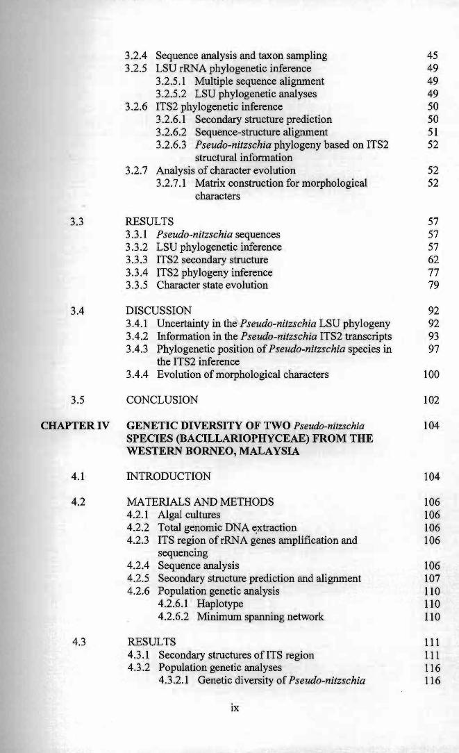
3.2.4 Sequence analysis and taxon sampling 45 3.2.5 LSU rRNA phylogenetic inference 49
3.2.5.1 Multiple sequence alignment 49 3.2.5.2 LSU phylogenetic analyses 49
3.2.6 ITS2 phylogenetic inference 50 3.2.6.1 Secondary structure prediction 50 3.2.6.2 Sequence-structure alignment 51 3.2.6.3 Pseudo-nitzschia phylogeny based on ITS2 52
structural information 3.2.7 Analysis of character evolution 52
3.2.7.1 Matrix construction for morphological 52 characters
RESULTS 57 3.3.1 Pseudo-nitzschia sequences 57 3.3.2 LSU phylogenetic inference 57 3.3.3 ITS2 secondary structure 62 3.3.4 ITS2 phylogeny inference 77 3.3.5 Character state evolution 79
DISCUSSION 92 3.4.1 Uncertainty in the Pseudo-nitzschia LSU phylogeny 92 3.4.2 Information in the Pseudo-nitzschia ITS2 transcripts 93 3.4.3 Phylogenetic position ofPseudo-nitzschia species in 97
the ITS2 inference 3.4.4 Evolution of morphological characters 100
CONCLUSION 102
GENETIC DIVERSITY OF TWO Pseudo-nitzschia 104 SPECIES (BACILLARIOPHYCEAE) FROM THE WESTERN BORNEO, MALAYSIA
INTRODUCTION 104
MATERIALS AND METHODS 106 4.2.1 Algal cultures 106 4.2.2 Total genomic DNA ~xtraction 106 4.2.3 ITS region ofrRNA genes amplification and 106
sequencing 4.2.4 Sequence analysis 106 4.2.5 Secondary structure prediction and alignment 107 4.2.6 Population genetic analysis 110
4.2.6.1 Haplotype 110 4.2.6.2 Minimum spanning network 110
RESULTS 111 4.3.1 Secondary structures of ITS region 111 4.3 .2 Population genetic analyses 116
4.3.2.1 Genetic diversity of Pseudo-nitzschia 116
IX
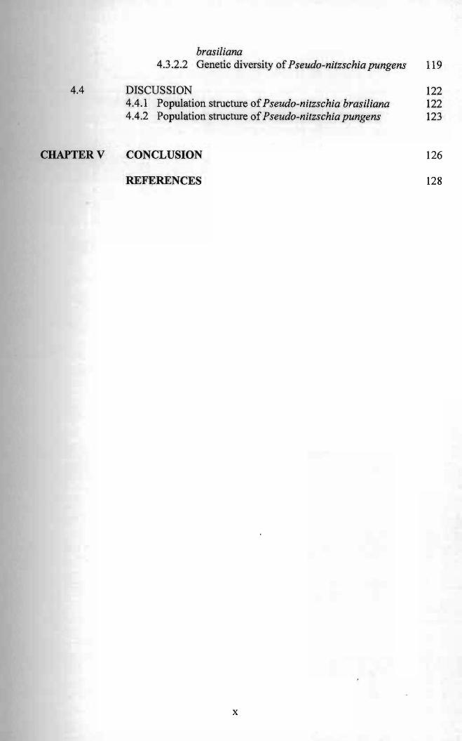
brasiliana 4.3.2.2 Genetic diversity of Pseudo-nitzschia pungens 119
4.4 DISCUSSION 122 4.4.1 Population structure ofPseudo-nitzschia brasiliana 122 4.4.2 Population structure ofPseudo-nitzschia pungens 123
CHAPTER V CONCLUSION 126
REFERENCES 128
x
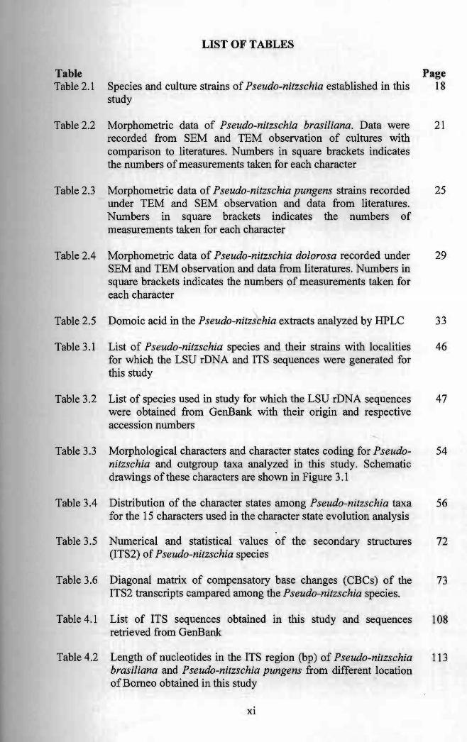
LIST OF TABLES
Table Table 2.1 Species and culture strains of Pseudo-nitzschia established i
study n this
Page 18
Table 2.2 Morphometric data of Pseudo-nitzschia brasiliana. Data recorded from SEM and TEM observation of cultures
were with
21
comparison to literatures. Numbers in square brackets indthe numbers of measurements taken for each character
icates
Table 2.3 Morphometric data of Pseudo-nitzschia pungens strains recorded 25 under TEM and SEM observation and data from literatures. Numbers in square brackets indicates the numbers of measurements taken for each character
Table 2.4 Morphometric data of Pseudo-nitzschia dolorosa recorded under 29 SEM and TEM observation and data from literatures. Numbers in square brackets indicates the numbers of measurements taken for each character
Table 2.5 Domoic acid in the Pseudo-nitzschia extracts analyzed by HPLC 33
Table 3.1 List of Pseudo-nitzschia species and their strains with localities 46 for which the LSU rDNA and ITS sequences were generated for this study
Table 3.2 List of species used in study for which the LSU rDNA sequences 47 were obtained from GenBank with their origin and respective accession numbers
Table 3.3 Morphological characters and character states coding for Pseudo- 54 nitzschia and outgroup taxa analyzed in this study. Schematic drawings of these characters are shown in Figure 3.1
Table 3.4 Distribution of the character states among Pseudo-nitzschia taxa 56 for the 15 characters used in the character state evolution analysis
Table 3.5 Numerical and statistical values of the secondary structures 72 (ITS2) of Pseudo-nitzschia species
Table 3.6 Diagonal matrix of compensatory base changes (CBCs) of the 73 ITS2 transcripts campared among the Pseudo-nitzschia species.
Table 4.1 List of ITS sequences obtained in this study and sequences 108 retrieved from GenBank
Table 4.2 Length of nucleotides in the ITS region (bp) of Pseudo-nitzschia 113 brasiliana and Pseudo-nitzschia pungens from different location of Borneo obtained in this study
Xl
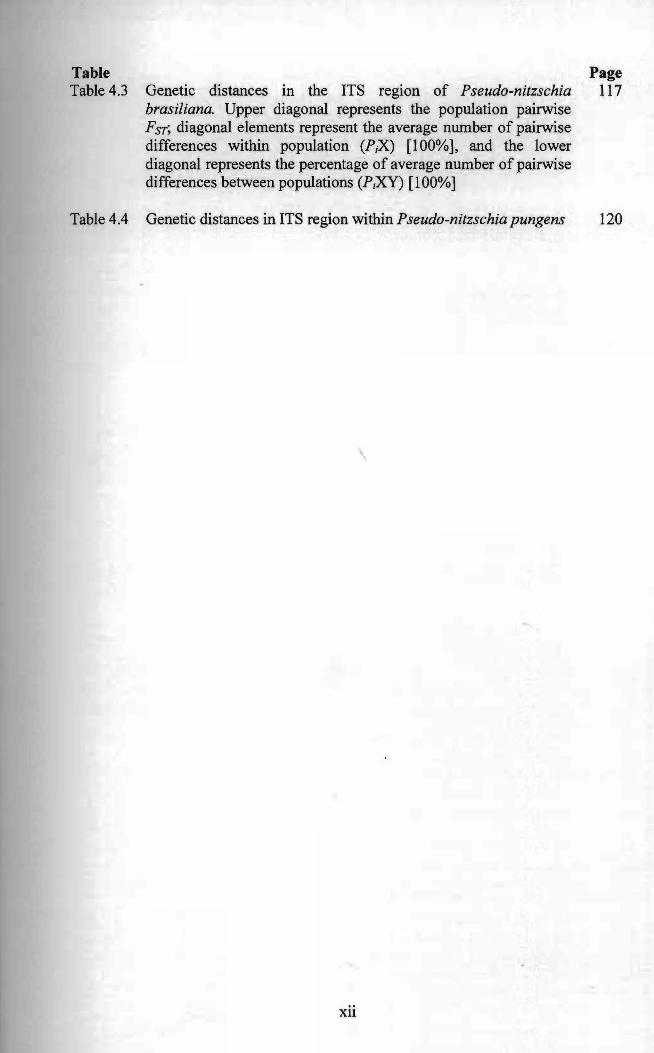
I Table Page Table 4.3 Genetic distances in the ITS region of Pseudo-nitzschia 117
brasiliana. Upper diagonal represents the population pairwise FST; diagonal elements represent the average number of pairwise differences within population (PX) [100%], and the lower diagonal represents the percentage of average number of pairwise differences between populations (PiXY) [100%]
Table 4.4 Genetic distances in ITS region within Pseudo-nitzschia pungens 120
xu
------~
LIST OF FIGURES
Figure Figure 2.1
Figure 2.2
Figure 2.3
Figure 2.4
Figure 2.5
Figure 2.6
Pseudo-nitzschia
Scale bar =
11m. (G) SEM.
Scale bar =
bar =
rows of poroi
Cell shape
collected from
Scale bar =
ds.
the
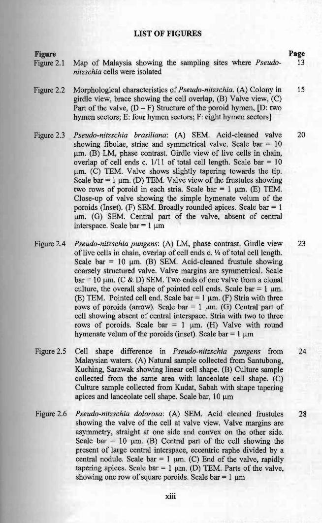
Page Map of Malaysia showing the sampling sites where Pseudo 13 nitzschia cells were isolated
Morphological characteristics of Pseudo-nitzschia. (A) Colony in 15 girdle view, brace showing the cell overlap, (B) Valve view, (C) Part of the valve, (D - F) Structure of the poroid hymen, [D: two hymen sectors; E: four hymen sectors; F: eight hymen sectors]
brasiliana: (A) SEM. Acid-cleaned valve 20 showing fibulae, striae and symmetrical valve. Scale bar = 10 11m. (B) LM, phase contrast. Girdle view of live cells in chain, overlap of cell ends c. 1/11 of total cell length. Scale bar = 10 11m. (C) TEM. Valve shows slightly tapering towards the tip.
111m. (D) TEM. Valve view of the frustules showing two rows of poroid in each stria. Scale bar = 111m. (E) TEM. Close-up of valve showing the simple hymen ate velum of the poroids (Inset). (F) SEM. Broadly rounded apices. Scale bar = 1
Central part of the valve, absent of central interspace. Scale bar = 1 11m
Pseudo-nitzschia pungens: (A) LM, phase contrast. Girdle view 23 of live cells in chain, overlap of cell ends c. Y4 of total cell length.
10 11m. (B) SEM. Acid-cleaned frustule showing coarsely structured valve. Valve margins are symmetrical. Scale
10 11m. (C & D) SEM. Two ends of one valve from a clonal culture, the overall shape of pointed cell ends. Scale bar = 111m. (E) TEM. Pointed cell end. Scale bar = 111m. (F) Stria with three rows of poroids (arrow). Scale bar = 111m. (G) Central part of cell showing absent of central interspace. Stria with two to three
Scale bar = 111m. (H) Valve with round hymenate velum of the poroids (inset). Scale bar = 1 11m
difference in Pseudo-nitzschia pungens from 24 Malaysian waters. (A) Natural sample collected from Santubong, Kuching, Sarawak showing linear cell shape. (B) Culture sample
same area with lanceolate cell shape. (C) Culture sample collected from Kudat, Sabah with shape tapering apices and lanceolate cell shape. Scale bar, 10 11m
Pseudo-nitzschia dolorosa: (A) SEM. Acid cleaned frustules 28 showing the valve of the cell at valve view. Valve margins are asymmetry, straight at one side and convex on the other side.
10 11m. (B) Central part of the cell showing the present of large central interspace, eccentric raphe divided by a central nodule. Scale bar = 111m. (C) End of the valve, rapidly tapering apices. Scale bar = 111m. (D) TEM. Parts of the valve, showing one row of square poroids. Scale bar = 1 11m
Xlll
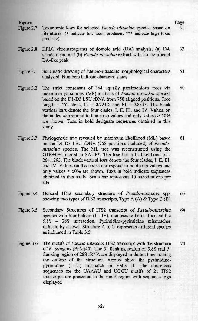
Figure Page Figure 2.7 Taxonomic keys for selected Pseudo-nitzschia species based on 31
literatures. (* indicate low toxin producer, *** indicate high toxin producer)
Figure 2.8 HPLC chromatograms of domoic acid (DA) analysis. (a) DA 32 standard run and (b) Pseudo-nitzschia extract with no significant DA-like peak
Figure 3.1 Schematic drawing of Pseudo-nitzschia morphological characters 53 analyzed. Numbers indicate character states
Figure 3.2 The strict consensus of 364 equally parsimonious trees via 60 maximum parsimony (MP) analysis of Pseudo-nitzschia species based on the DI-D3 LSU rDNA from 758 aligned positions. Tree length = 452 steps; CI = 0.7212; and Rl = 0.8313. The black vertical bars denote the four clades, I, II, III, and IV. Values on the nodes correspond to bootstrap values and only values> 50% are shown. Taxa in bold designate sequences obtained in this study
Figure 3.3 Phylogenetic tree revealed by maximum likelihood (ML) based 61 on the DI-D3 LSU rDNA (758 positions included) of Pseudonitzschia species. The ML tree was reconstructed using the GTR+G+I model in PAUP*. The tree has a In likelihood of 2641.293. The black vertical bars denote the four clades, I, II, III, and IV. Values on the nodes correspond to bootstrap values and only values> 50% are shown. Taxa in bold indicate sequences obtained in this study. Scale bar represents 10 substitutions per site
Figure 3.4 General ITS2 secondary structure of Pseudo-nitzschia spp. 63 showing two types ofITS2 transcripts, Type A (A) & Type B (B)
Figure 3.5 Secondary Structures of ITS2 transcript of Pseudo-nitzschia 64 species with four helices (I - IV), one pseudo-helix (IIa) and the 5.8S - 28S interaction. Pyrimidine-pyrimidine mismatches indicate by arrows. Structure A to U represents different species as indicated in Table 3.5
Figure 3.6 The motifs of Pseudo-nitzschia ITS2 transcript with the structure 74 of P. pungens (PnMt45). The 3' flanking region of 5.8S and 5' flanking region of 28S rRNA are displayed in dotted lines tracing the outline of the structure. Arrows show the pyrimidinepyrimidine (U-U) mismatch in Helix II. The consensus sequences for the UAAAU and UGGU motifs of 21 ITS2 transcripts are presented in the motif region with sequence logo displayed
xiv
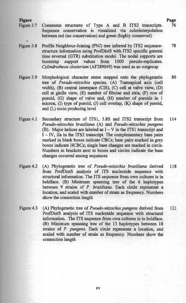
Figure Page Figure 3.7 Consensus structures of Type A and B ITS2 transcripts. 76
Sequence conservation IS visualized VIa colorinterpolation between red (no conservation) and green (highly conserved)
Figure 3.8 Profile Neighbour-Joining (PNJ) tree inferred by ITS2 sequence 78 structure information using ProfDistS with ITS2 specific general time reversal (GTR) substitution model. The nodal supports are bootstrap support values from 1000 pseudo-replicates. Cylindrotheca closterium (AF289049) was used as an outgroup
Figure 3.9 Morphological character states mapped onto the phylogenetic 80 tree of Pseudo-nitzschia species. (A) Transapical axis (cell width), (B) central interspace (CIS), (C) cell at valve view, (D) cell at girdle view, (E) number of fibulae and stria, (F) row of poroid, (G) shape of valve end, (H) number of poroids in 1 micron, (I) type of poroid, (J) cell overlap, (K) shape of poroid, and (L) toxin producing level
Figure 4.1 Secondary structure of ITSl , 5.8S and ITS2 transcript from 114 Pseudo-nitzschia brasiliana (A) and Pseudo-nitzschia pungens (B). Major helices are labeled as I - V in the ITS 1 transcript and I - IV, IIa in the ITS2 transcript. The complementary base pairs marked in black boxes indicate CBCs; base pairs marked in grey boxes indicate HCBCs; single base changes are marked in circle. Numbers in brackets next to boxes and circles indicate the base changes occurred among sequences
Figure 4.2 (A) Phylogenetic tree of Pseudo-nitzschia brasiliana derived U8 from ProfDistS analysis of ITS nucleotide sequence with structural information. The ITS sequence from own cultures is in boldface. (B) Minimum spanning tree of the 6 haplotypes between 9 strains of P. brasiliana. Each circle represents a location, and scaled with number of strain as frequency. Numbers show the connection length
Figure 4.3 (A) Phylogenetic tree of Pseudo-nitzschia pungens derived from 121 ProfDistS analysis -of ITS nucle<;>tide sequence with structural information. The ITS sequence from own cultures is in boldface. (B) Minimum spanning tree of the 13 haplotypes between 18 strains of P. pungens. Each circle represents a location, and scaled with number of strain as frequency. Numbers show the connection length
xv
CHAPTER I
INTRODUCTION
Diatom is an important marine phytoplankton characterized by having a siliceous cell
wall which is known as frustules. They contribute up to 45% of the total production in
the ocean (Werner 1977; Mann 1989). Diatoms form the base of the food web in
many marine ecosystems as they have highly efficient and adaptive survival
mechanisms and growth strategies. This may due to their use of silicate to form a
frustule which uses less energy to synthesis organic cell walls (Raven 1983). The
success of some diatoms species may be explained by their ability to form chains,
which deter some grazers. Diatoms require nutrient rich conditions for growth as well
as turbulence to keep them in suspension. They are therefore often found in coastal
regions, where their impacts on humans and marine food webs are often observed
(Bates & Trainer 2006).
Pseudo-nitzschia H. Peragallo was first identified as Nitzschia (Peragallo &
Peragallo 1900). In 1993, Hasle re-erected Pseudo-nitzschia as a separate genus from
section of Nitzschia Hassall after comparing the Iporphological characteristics of both
genera (Hasle 1993; 1994). Pseudo-nitzschia H. Peragallo is one of the most common
diatom genera among marine phytoplankton and it is cosmopolitan as it can be found
in polar, temperate, subtropical and tropical areas worldwide (Has Ie 2002). Pseudo
nitzschia received much more attention when the first occurrence of intoxication at
Prince Edward Island, Canada in 1987 which caused approximately 100 people
became ill while 4 patients died as a result of amnesic shellfish poisoning (ASP) after
1
consumption of contaminated mussels (Blue mussel, Mytilus edulis) (Perl et aI. 1987),
the first awareness of toxin producing diatoms (Bates et aI. 1989).
SeveraI Pseudo-nitzschia species had been reported to produce neurotoxin
domoic acid (DA). Blooms of Pseudo-nitzschia in natural water may result in
bioaccumulation of toxin in marine ecosystem, food web and thereby affecting marine
mammaIs, seabirds and potentially humans (Bates et aI. 1989; Work et aI. 1993;
Scholin et aI. 2000). These species include P. australis, P. calliantha, P. cuspidata, P.
delicatissima, P. fraudulenta, P. galaxiae, P, heimii, P. multiseries, P. multistriata, P.
pungens, P. pseudodelicatissima, P. seriata, P. subcurvata, P. subfraudulenta and P.
turgidula (Perl et aI. 1987; Whyte et al. 1995; Hasle et al. 1996; Parsons et aI. 1999;
Bargu et al. 2002; Lundholm et aI. 2003; Fehling et aI. 2004; Bill et al. 2005; Cerino
et aI. 2005; Fehling et aI. 2005; Schnetzer et aI. 2007; Besiktepe et al. 2008). There
were about thirty species in this genus but not all of the Pseudo-nitzschia is toxin
producer.
Amnesic shellfish poisoning (ASP) was first reported in Prince Edward Island,
Canada in 1987 with density of the pennate diatom Pseudo-nitzschia multiseries
(previously described as Nitzschia pungens) reaching up to 15 xl 06 cellslL (Bates et al.
1989). In subsequent years, bloom event of Pseudo-nitzschia australis (7 x lOs cells/L)
in Monterey Bay, California in 1991 was reported with mortality of 100 brown
pelicans and cormorants (Work et al. 1993). In the same year, dozens of cases of
human illness were reported along the Pacific coasts of Washington and Oregon,
where razor clams, Dungeness crabs, blue mussels and oyster were found to contain
up to 1541lg/g of domoic acid (Garrison et al. 1992). In year 2001, accumulation of
high concentration of 531lg DAig tissue were detected in shellfish samples (Wedge
shell, Donax trunculus) collected at Western Brittany, and the . diatom found
2
associated with this were P. pseudodelicatissima and P. multiseries (Amzil et al. 2001)
of which the toxin level had been exceeded the sanitary threshold i.e. 20Jlg DNg
tissue and might caused irreversible effect to casualty.
Domoic acid (DA) is the compound responsible for ASP. It is a water soluble
tricarboxylic amino acid with a molecular weight of 311.14, (ClsH21N06)' DA acts as
an analogue of the neurotransmitter glutamate and is a potent glutamate receptor
agonist (Frances 2000) that has high binding affinity towards both kainate and AMP A
(a-amino-3-hydroxy-5-methyl-4-isoxazol propionic acid) (Hampson et al. 1992). The
victims exhibited gastrointestinal disorders after digestion of any contaminated
shellfish. The symptoms include nausea, vomiting abdominal cramps, headache,
diarrhea, and memory loss. The loss of memory in patients intoxicated with ASP
appeared to be similar to patients with Alzheimer's disease (McKhann et al. 1984).
However, little is known about DA in tropical waters especially Malaysian
waters. Currently, only paralytic shellfish poisoning (PSP) and its related causative
organisms were monitored by the related authority in Malaysia focused mainly on the
west coast of Sabah and selected location in Peninsula Malaysia, but not for amnesic
shellfish poisoning (ASP). The raised of awareness regarding HABs in Malaysia were
mainly due to human intoxication reported in 1991, where three people were poisoned
after consuming mussels at Sebatu (Strait of Malacca) that were contaminated by a
type of dinofiagelates, Alexandrium tamiyavanichii (Usup et al. 2002a). In September
of year 2001, another case of shellfish intoxication was reported whereby six people
were hospitalized and one fatal after consuming benthic clam (Polymesoda similis) in
Tumpat, Kelantan. Investigation was carried out and Alexandrium minutum was
associated with the shellfish poisoning (Usup et al. 2002b). The status of ASP in
3
Malaysia is still remained unknown although Pseudo-nitzschia is commonly found in
coastal waters.
Contamination of domoic acid in shellfish molluscs had been reported in
several countries in South East Asian. In 2005, domoic acid was found accumulated
in bivalve Spondylus cruentus in Nha Trang Bay, Khanh Hoa Province of Vietnam
which ranged from 0.6 to 2.7~glg and the rate of depuration of toxin by the bivalve
species is extremely low in which it retained DA up to 45 days rearing under plankton
free conditions (Dao Viet et al. 2006). In addition, a high level ofDA up to 146.8~glg
was detected in tissues of bivalve (Spondylus versicolor) end of September 2005 in
Nha Phu Bay, Vietnam and it was associated with Pseudo-nitzschia abundances (Dao
Viet et al. 2009).
The ASP events in tropical countries might not as frequently reported as those
subtropical countries and temperate countries though Pseudo-nitzschia spp. was
detected almost through the year (Dao Viet et al. 2006, Huyen et al. 2006, Suriyanti
2011) most probably due to the non-toxic producing Pseudo-nitzschia are mopre
common in the region. Domoic acid was detected in shellfish at Haiphong area,
Vietnam but no significant toxicity was detected in net sample even the cell density of
Pseudo-nitzschia spp. was high suggesting that toxin contents was extremely low in
Pseudo-nitzschia spp. (Huyen et al. 2006). Besides, P. pungens was also found at
Manila Bay Philippines but no DA was detected from the cell extracts (Bajarias et al.
2006).
Pseudo-nitzschia taxonomy is rather complicated and confusing. General
features of the genus include the presence or absence of central interspace, valve
width and valve length, number of fibulae and stria in 10 ~m, number of poroids in
4
'Pusat Khidmat MakJumat Akademik UNlVERSm MALAYSIA SARAWAK
IJ,lm, row of poroid, overlapping/stepped cell forming a chain, shape and type of
poroids, valve view and girdle view of the cell, and shape of apices (See CHAPTER II).
The variation of types of poroids is sometimes used at the species level (Lundholm et
al. 2003). Current identification had been incorporated with molecular data, ITS-2
transcript analysis and sexual reproduction compatibility in describing new species
(Cerino et al. 2005; Lundholm et al. 2006; Amato & Montresor 2008; Quijano-
Scheggia et al. 2009). Molecular data are often included to strengthen the evidence
and to support the new species designation. The resolution of the species concept in
Pseudo-nitzschia will continue to be a subject of active discussion.
5
RESEARCH QUESTIONS AND OBJECTIVES
Malaysia is situated at the southern part of South China Sea which inhabits diverse
population of human and wildlife and with various fisheries activities especially
shellfish cultivations. Many types of harmful algal either diatoms or dinoflagelates
can be found in Malaysia waters including Pseudo-nitzschia species. Very little
research has been carried out due to lack of expertise in monitoring the potentially
toxin producing algal especially genus Pseudo-nitzschia species because it required
knowledge in detailed ultrastructure observations for morphological identification.
Besides, tedious preparations and certain skills in instrumentations like electron
microscopy are needed as normal stereo light microscopy is impossible to discern
them into species level. Thus the first objective of this study was to carry out a
regional survey of Pseudo-nitzschia species and to clarify the status of DA in
Malaysia waters in order to fill a gap in knowledge and engender a taxonomic key to
ease the identification and hence improve harmful algae monitoring in the region
(CHAPTER II).
Other than morphological characterization, molecular approaches had become
popular and widely used in delineating Pseudo-nitzschia into species level and at the
same time support the morphological identification. Large subunit (LSU) rDNA were
always the region being studied but the phylogeny resolution was rather low in
defining species and remained unresolved especially when dealing with large number
of species. Recently internal transcribed spacer (ITS) region had been used in
describing new species with the inference of secondary structure of the second
internal transcribed spacer (lTS-2) as this region possessed a highly conserved
secondary structure and useful in demarcating closely related species especially when
encounter with cryptic/pseudo-cryptic species. Thus the second objective was to .test
6
two hypotheses, that the phylogeny relationship of Pseudo-nitzschia species using
ITS-2 approaches with synchronous aligrunent together with the secondary structure
information would be useful in solving the phylogenetic resolution. Second, the
diagnostic features through character state evolution analysis might explain more
about the taxonomy nomenclature (CHAPTER III).
Pseudo-nitzschia is found globally but not all of the species in the genus
producing toxin and those potentially toxic producing Pseudo-nitzschia might not be
toxic due to different culture conditions and their diverse origins. Population genetics
is another approach that is useful to investigate organisms that originated from diverse
population and geographical variation in detail. The last objective of this thesis was to
investigate and assess potential intraspecific variation and biogeographic distribution
patterns of P. brasiliana and P. pungens found in Sarawak hence comparing the
population dynamics in Borneo Malaysia (CHAPTER IV).
7
-CHAPTER II
MORPHOLOGY AND TOXIN ASSESSMENT OF Pseudo-nitzschia
(BACILLARIOPHYCEAE) SPECIES IN MALAYSIAN WATERS
2.1 INTRODUCTION
Species in the genus of Pseudo-nitzschia Peragallo are marine diatoms that can be
found in estuarine to oceanic ecosystems. The genus was separated from Nitzschia
primarily due to the difficulty in differentiating the fibulae in the light microscope
(Hasle 1993). The common feature used to distinguish the genus from other closely
related genera of diatoms is the characteristic of the cells to form colonies/chain.
Pseudo-nitzschia cells might be solitary or in colonies and are capable of moving,
whole chain slides in one direction for a few seconds and followed by movement in
opposite direction (Skov et al. 1999), but individual cell in colonies is immotile
(Larsen & Nguyen 2004).
The taxonomy of Pseudo-nitzschia is rather complicated and species can only
be discernible with detailed ultrastructural observations. Fine morphological
observation by advanced scanning and transmission electron microscopy is usually
necessary for species identification. Hitherto, more than thirty species of Pseudo
nitzschia had been identified worldwide. Historically, the first Pseudo-nitzschia
species that being described was Pseudo-nitzschia delicatissima in 1928 followed by
Pseudo-nitzschia australis and Pseudo-nitzschia heimii in 1939 and 1957 respectively
(Hasle et al. 1996; Hasle & Syvertsen 1997). Subsequently, few more species were
8