Embed Size (px)
Citation preview
In this patient, retinal images of theleft eye were taken with the Imagine-Eyes (Imagine Eyes, Orsay, France)flood-illumination AO ophthalmo-scope from 3� to 8� temporal to the fix-ation point. Normally, retinal conesare seen as the bright spots on AOimages. Individual cones (bright spots)within the mosaic (regions containing aclose-packed arrangement of brightspots) were identified manually and theaverage nearest-neighbour spacing wasdetermined; thus, in turn, cone densitywas chosen over cone spacing as ameasure of photoreceptor distribution.
This quantitative analysis was com-pared with that from one age-matchedand refraction-matched control sub-ject. As the axial length of subjectswas not measured, we used the stan-dard eye [schematic (275 lm ⁄degree)]as previously reported (Drasdo &Fowler 1974). Overall, AO revealed apreservation of perifoveal cones overthe large colloid drusen (Fig. 1) at4–6� temporal to the fixation point(cone density was 18 267 ± 1187cones ⁄mm2), compared with anage-matched and refraction-matchedcontrol subject (cone density was 20628 ± 1392 cones ⁄mm2 at 4� tempo-ral to the fixation point).
Adaptive optics is a noninvasivetechnique that allows observing retinalpathology directly at a cellular leveland provides a measure of photore-ceptor loss in retinal diseases. Using
AO, we previously reported that inAMD drusen, the cone layout andphotoreceptor mosaic are disrupted,with residual cone photoreceptorsappearing sometimes isolated, some-times grouped into tight aggregates(Massamba et al. Invest OphthalmolVis Sci. E-abstract 2009). Generally,cone photoreceptors are visible inareas between drusen, and the mosaicimage sharpness is significantly lessuniform across the field than in youn-ger, healthy retinas (Massamba et al.Invest Ophthalmol Vis Sci. E-abstract2009). Interestingly, here, using AO,we demonstrated the preservation ofcones over drusen in a patient withearly onset large colloid drusen [a newtype of young-onset not age-relateddrusen recently described by ourgroup (Guigui et al. 2011)], as well asan overall preservation of perifovealcones over the large colloid drusen at4–6� temporal to the fixation point.
Similar findings have been recentlyreported (Godara et al. 2010) in a45-year-old asymptomatic woman withbasal laminar drusen. Also, in this casepresenting early onset drusen, theauthors found that the mosaic was visi-ble across the entire surface of the dru-sen, and, in turn, that the cones werestill present despite morphological dis-ruption on OCT (IS ⁄OS thinning).
The different AO features may sug-gest a different pathology and possibleevolution between AMD drusen and
this peculiar type of early onset dru-sen. Further analyses are needed toconfirm our preliminary findings onlarge colloid drusen.
ReferencesDrasdo N & Fowler CW (1974): Non-linear
projection of the retinal image in a wide-
angle schematic eye. Br J Ophthalmol 58:
709–714.
Gass JD (1973): Drusen and disciform macu-
lar detachment and degeneration. Arch
Ophthalmol 90: 206–217.
Godara P, Siebe C, Rha J, Michaelides M &
Carroll J (2010): Assessing the photorecep-
tor mosaic over drusen using adaptive
optics and spectral-domain optical coher-
ence tomography. Ophthalmic Surg Lasers
Imaging 41: S104–S108.
GuiguiB,LevezielN,MartinetV,MassambaN,
Sterkers M, Coscas G & Souied EH
(2011): Angiography features of early onset
drusen. Br J Ophthalmol 95: 238–244.
Liang J, Williams DR & Miller DT (1997):
Supernormal vision and high-resolution
retinal imaging through adaptive optics.
J Opt Soc Am A 14: 2884–2892.
Massamba N, Basurto A, Lamory B, Parier V
& Soubrane G (2009): In vivo microscopy
of macular soft drusen using adaptive
optics. Invest Ophthalmol Vis Sci 50: E-
Abstract 3300.
Pauleikhoff D, Barondes MJ, Minassian D,
Chrisholm J & Bird AC (1990): Drusen as
risk factors in age-related macular disease.
Am J Ophthalmol 109: 38–43.
Correspondence:
Giuseppe Querques
Department of Ophthalmology
Centre Hospitalier Intercommunal de Creteil
40 Avenue de Verdun
94000 Creteil
France
Tel: +33 1 45175908
Fax: +33 1 45175227
Email: [email protected]
Morphological versus
functional photoreceptor
viability of retinal pigment
epithelium tears
Albert Caramoy, Bernd Kirchhof andSascha Fauser
University of Cologne, Center of Oph-thalmology, Department of Vitreoretinal
Surgery, Cologne, Germany
doi: 10.1111/j.1755-3768.2011.02284.x
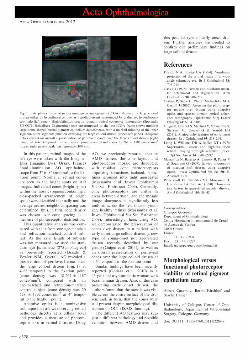
Fig. 1. Late phases frame of indocyanine green angiography (ICGA), showing the large colloid
drusen either as hyperfluorescent or as hypofluorescent surrounded by a discreet hyperfluores-
cent halo (left panel). High-definition spectral domain optical coherence tomography (Spectralis
SD-OCT; Heidelberg Engineering) scan superimposed to the late ICGA frame shows multiple
large dome-shaped retinal pigment epithelium detachments, with a marked thinning of the inner
segment ⁄ outer segments junction overlying the large colloid drusen (upper left panel). Adaptive
optics reveals an overall a preservation of perifoveal cones over the large colloid drusen (right
panel) at 4–6� temporal to the fixation point [cone density was 18 267 ± 1187 cones ⁄mm2
(upper right panel); scale bar represents 100 lm].
Acta Ophthalmologica 2012
e328
Editor,
R etinal pigment epithelium tears(RPE tears) are a serious com-
plication in patients with age-relatedmacular degeneration (AMD) leadingto a loss of photoreceptors. Previ-ously, we have shown that the photo-receptors can be rescued and functionrestored by autologous transplanta-tion of RPE and choroid in thesepatients (Caramoy et al. 2011a). Wehave also described previously thatthe morphological signs of viable pho-toreceptors can be seen in the area ofpigment epithelium tears up to325 days (Caramoy et al. 2011b),however little is known about thefunction of these photoreceptors inthe area without retinal pigmentepithelium.
In a retrospective study, weexamined the morphology of RPEtears using spectral-domain opticalcoherence tomography (SD-OCT)(Spectralis HRA + OCT; HeidelbergEngineering, Heidelberg, Germany)in relation to its function in microperi-metry (MP1; Nidek Technologies,Padova, Italy).
Mean age was 76 ± 7.9 years (range63–85 years) and mean best correctedvisual acuity was 0.54 ± 0.37 Log-MAR. Among the seven patients, threewere men and four were women. Fourpatients showed unstable fixation. Rela-tively unstable and stable fixations wereseen in two patients and in one patient,respectively. In the patient with stablefixation, the fovea was still supported bypigment epithelium. The fixationstability (percentage of fixation withinthe central 2� and 4�) was 42 ±29.65%and 75 ± 19.61%, respectively.
Among these patients, morphologi-cal signs of viable photoreceptors
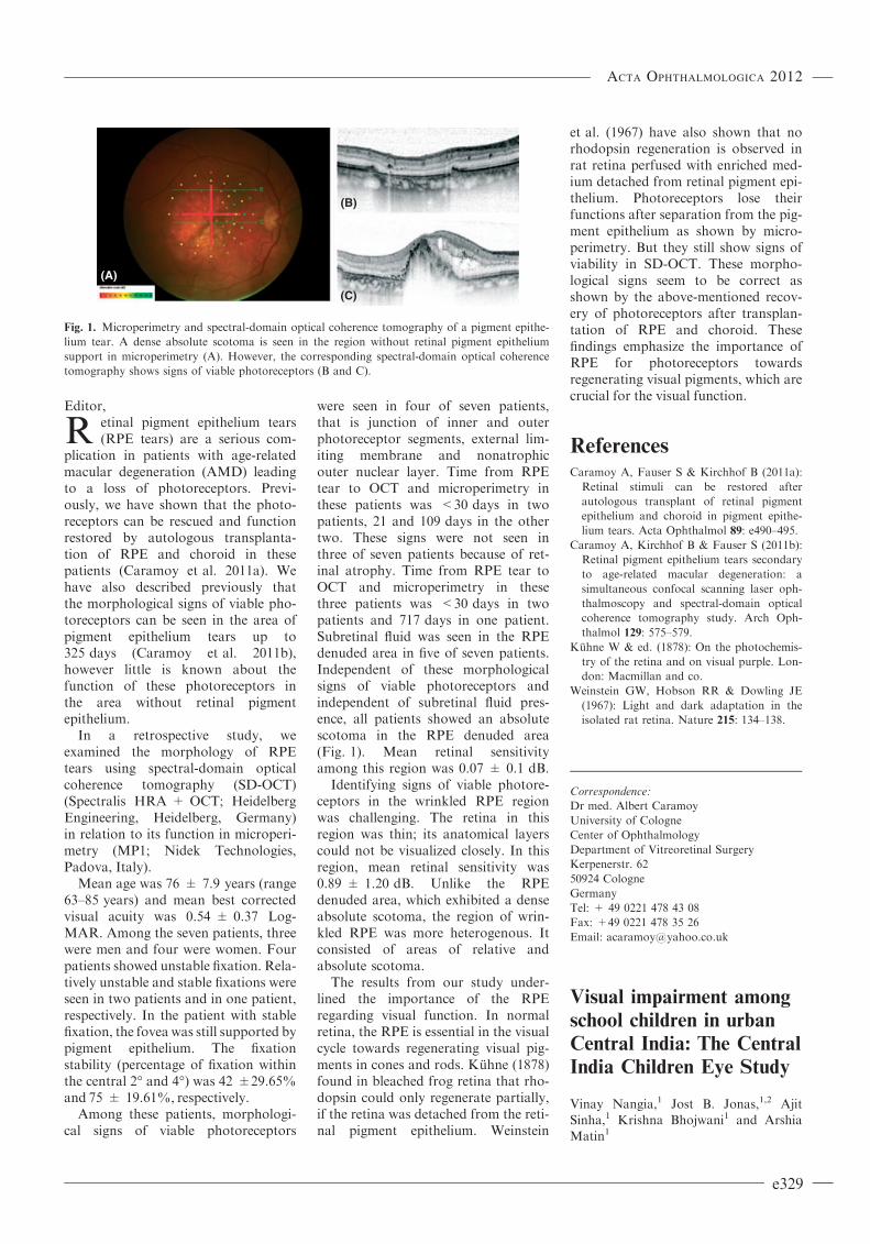
were seen in four of seven patients,that is junction of inner and outerphotoreceptor segments, external lim-iting membrane and nonatrophicouter nuclear layer. Time from RPEtear to OCT and microperimetry inthese patients was <30 days in twopatients, 21 and 109 days in the othertwo. These signs were not seen inthree of seven patients because of ret-inal atrophy. Time from RPE tear toOCT and microperimetry in thesethree patients was <30 days in twopatients and 717 days in one patient.Subretinal fluid was seen in the RPEdenuded area in five of seven patients.Independent of these morphologicalsigns of viable photoreceptors andindependent of subretinal fluid pres-ence, all patients showed an absolutescotoma in the RPE denuded area(Fig. 1). Mean retinal sensitivityamong this region was 0.07 ± 0.1 dB.
Identifying signs of viable photore-ceptors in the wrinkled RPE regionwas challenging. The retina in thisregion was thin; its anatomical layerscould not be visualized closely. In thisregion, mean retinal sensitivity was0.89 ± 1.20 dB. Unlike the RPEdenuded area, which exhibited a denseabsolute scotoma, the region of wrin-kled RPE was more heterogenous. Itconsisted of areas of relative andabsolute scotoma.
The results from our study under-lined the importance of the RPEregarding visual function. In normalretina, the RPE is essential in the visualcycle towards regenerating visual pig-ments in cones and rods. Kuhne (1878)found in bleached frog retina that rho-dopsin could only regenerate partially,if the retina was detached from the reti-nal pigment epithelium. Weinstein
et al. (1967) have also shown that norhodopsin regeneration is observed inrat retina perfused with enriched med-ium detached from retinal pigment epi-thelium. Photoreceptors lose theirfunctions after separation from the pig-ment epithelium as shown by micro-perimetry. But they still show signs ofviability in SD-OCT. These morpho-logical signs seem to be correct asshown by the above-mentioned recov-ery of photoreceptors after transplan-tation of RPE and choroid. Thesefindings emphasize the importance ofRPE for photoreceptors towardsregenerating visual pigments, which arecrucial for the visual function.
ReferencesCaramoy A, Fauser S & Kirchhof B (2011a):
Retinal stimuli can be restored after
autologous transplant of retinal pigment
epithelium and choroid in pigment epithe-
lium tears. Acta Ophthalmol 89: e490–495.
Caramoy A, Kirchhof B & Fauser S (2011b):
Retinal pigment epithelium tears secondary
to age-related macular degeneration: a
simultaneous confocal scanning laser oph-
thalmoscopy and spectral-domain optical
coherence tomography study. Arch Oph-
thalmol 129: 575–579.
Kuhne W & ed. (1878): On the photochemis-
try of the retina and on visual purple. Lon-
don: Macmillan and co.
Weinstein GW, Hobson RR & Dowling JE
(1967): Light and dark adaptation in the
isolated rat retina. Nature 215: 134–138.
Correspondence:
Dr med. Albert Caramoy
University of Cologne
Center of Ophthalmology
Department of Vitreoretinal Surgery
Kerpenerstr. 62
50924 Cologne
Germany
Tel: + 49 0221 478 43 08
Fax: +49 0221 478 35 26
Email: [email protected]
Visual impairment among
school children in urban
Central India: The Central
India Children Eye Study
Vinay Nangia,1 Jost B. Jonas,1,2 AjitSinha,1 Krishna Bhojwani1 and Arshia
Matin1
(A)
(B)
(C)
Fig. 1. Microperimetry and spectral-domain optical coherence tomography of a pigment epithe-
lium tear. A dense absolute scotoma is seen in the region without retinal pigment epithelium
support in microperimetry (A). However, the corresponding spectral-domain optical coherence
tomography shows signs of viable photoreceptors (B and C).
Acta Ophthalmologica 2012
e329