Embed Size (px)
Citation preview
Linda B Haramati MD, MS
Departments of Radiology and Medicine
Bronx, New York
MRI (AND CT) FOR REPAIRED
TETRALOGY OF FALLOT
OUTLINE
• Pathogenesis
• Variants
• Initial surgical treatments
• Basic MR protocols
• MR and CT for post-repair treatment planning (PVR)
• Ventricular size and function, pulmonary
insufficiency, pulmonary artery anatomy & flow,
coronary anomalies, lungs
INTRODUCTION
• Tetralogy of Fallot (TOF): 6.8% of all congenital heart disease
• Described in 1888 by Fallot- autopsy series cyanotic heart disease
• Malalignment of the conal septum in relation to the ventricular endocardial cushion lack of stimulus for membranous septum development
• Malaligned VSD, overriding aorta & RVOT obstruction
• Disease severity and age of presentation depends on degree of RVOT obstruction
• TOF represents a spectrum of disease with three major
variants:
• TOF with pulmonary valve or RVOT stenosis
• TOF with pulmonary atresia
• TOF with absent pulmonary valve
• TOF with pulmonary atresia is the most severe form,
present in 20%
• 1950s: initial surgical repair
• VSD closure and relief of RVOT obstruction
• Since then increasing population lives with TOF
• Unique problems related to longstanding postoperative physiology & delayed consequences of disease
• Most common problem & most frequent indication for imaging is prolonged post-op pulmonary insufficiency with consequent RV dilation
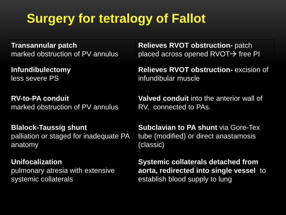
Transannular patch
marked obstruction of PV annulus
Relieves RVOT obstruction- patch
placed across opened RVOT free PI
Infundibulectomy
less severe PS
Relieves RVOT obstruction- excision of
infundibular muscle
RV-to-PA conduit
marked obstruction of PV annulus
Valved conduit into the anterior wall of
RV, connected to PAs.
Blalock-Taussig shunt
palliation or staged for inadequate PA
anatomy
Unifocalization
pulmonary atresia with extensive
systemic collaterals
Subclavian to PA shunt via Gore-Tex
tube (modified) or direct anastamosis
(classic)
Systemic collaterals detached from
aorta, redirected into single vessel to
establish blood supply to lung
Surgery for tetralogy of Fallot
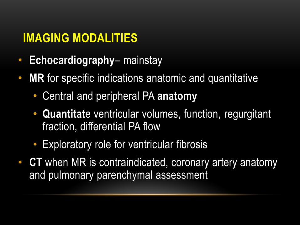
IMAGING MODALITIES
• Echocardiography– mainstay
• MR for specific indications anatomic and quantitative
• Central and peripheral PA anatomy
• Quantitate ventricular volumes, function, regurgitant fraction, differential PA flow
• Exploratory role for ventricular fibrosis
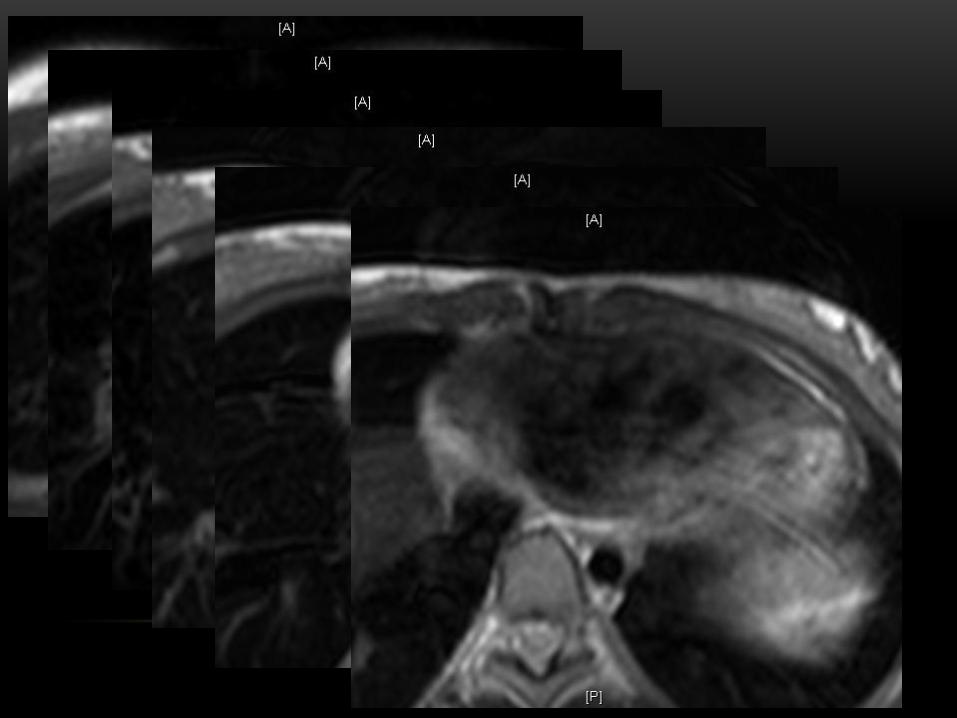
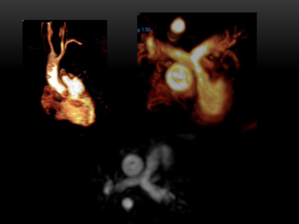
• CT when MR is contraindicated, coronary artery anatomy and pulmonary parenchymal assessment
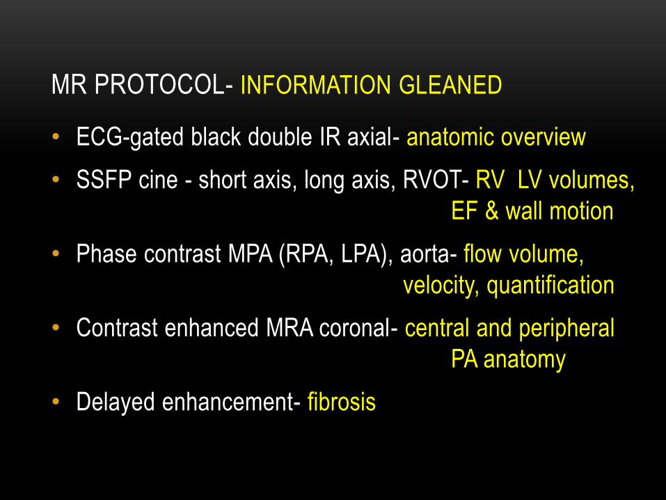
MR PROTOCOL- INFORMATION GLEANED
• ECG-gated black double IR axial- anatomic overview
• SSFP cine - short axis, long axis, RVOT- RV LV volumes,
EF & wall motion
• Phase contrast MPA (RPA, LPA), aorta- flow volume,
velocity, quantification
• Contrast enhanced MRA coronal- central and peripheral
PA anatomy
• Delayed enhancement- fibrosis
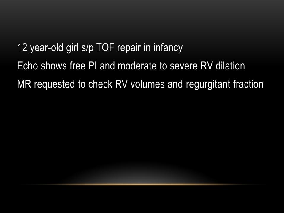
12 year-old girl s/p TOF repair in infancy
Echo shows free PI and moderate to severe RV dilation
MR requested to check RV volumes and regurgitant fraction
10/29/2010
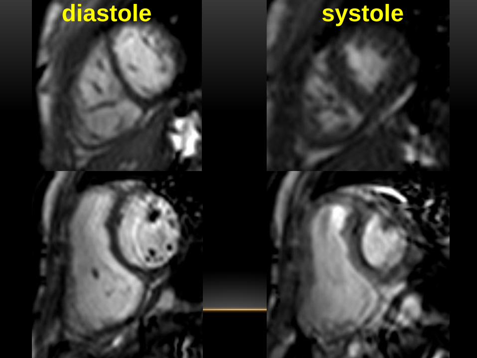
systole diastole
10/29/2010
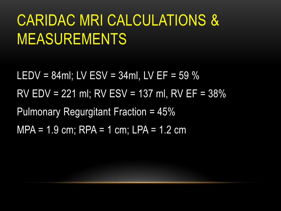
CARIDAC MRI CALCULATIONS &
MEASUREMENTS
LEDV = 84ml; LV ESV = 34ml, LV EF = 59 %
RV EDV = 221 ml; RV ESV = 137 ml, RV EF = 38%
Pulmonary Regurgitant Fraction = 45%
MPA = 1.9 cm; RPA = 1 cm; LPA = 1.2 cm
WHAT DOES THIS MEAN?
• Pulmonary insufficiency initiall well-tolerated post repair
• Over decades, morbidity & mortality from chronic PI RV
dilation, biventricular dysfunction, heart failure & arrhythmia
• PVR standard practice in symptomatic pts, improved PI &
sx, but not RVEF or ? mortality
• Optimal timing of PVR is still being explored, should
precede development of sxs
• RV ESV indexed to BSA <90 ml/m2, QRS <140 ms
associated optimal postoperative outcome (nl RV size, fxn)
Geva et al Circulation. 2010;122:S201–S208 Randomized Trial of Pulmonary
Valve Replacement Withand Without Right Ventricular Remodeling Surgery
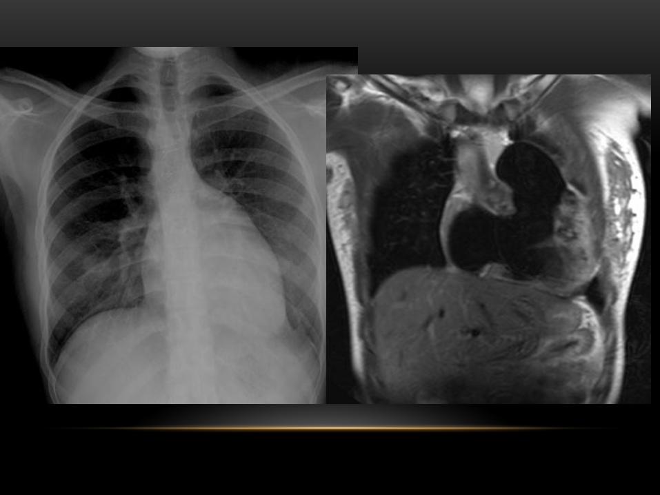
CASE 2 21-year-old woman s/p TOF repair in infancy who
developed shortness of breath on exertion
Echo showed TR and free PI with RV dilation
Underwent PVR and tricuspid valve annuloplasty
MR performed preoperatively & 1 and 2 years postop
4
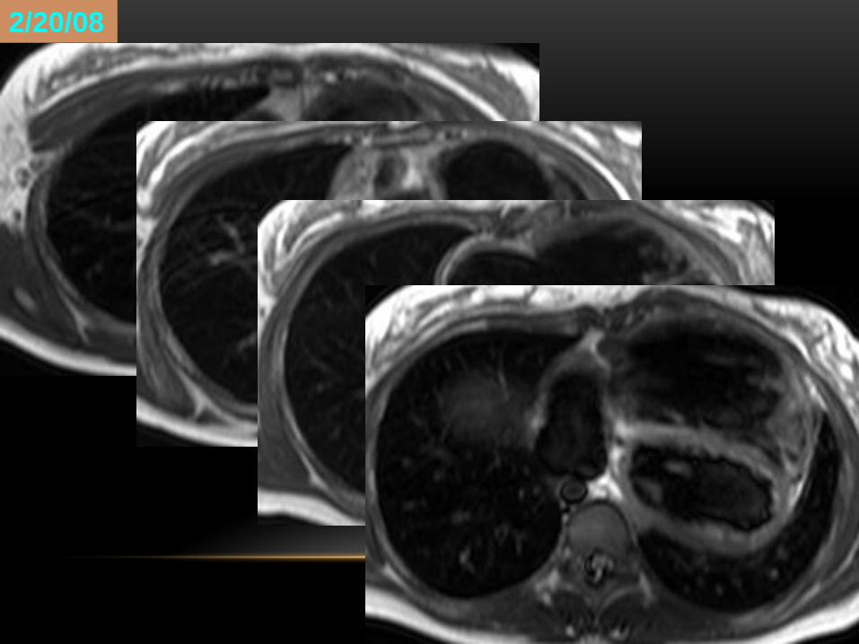
2/20/08
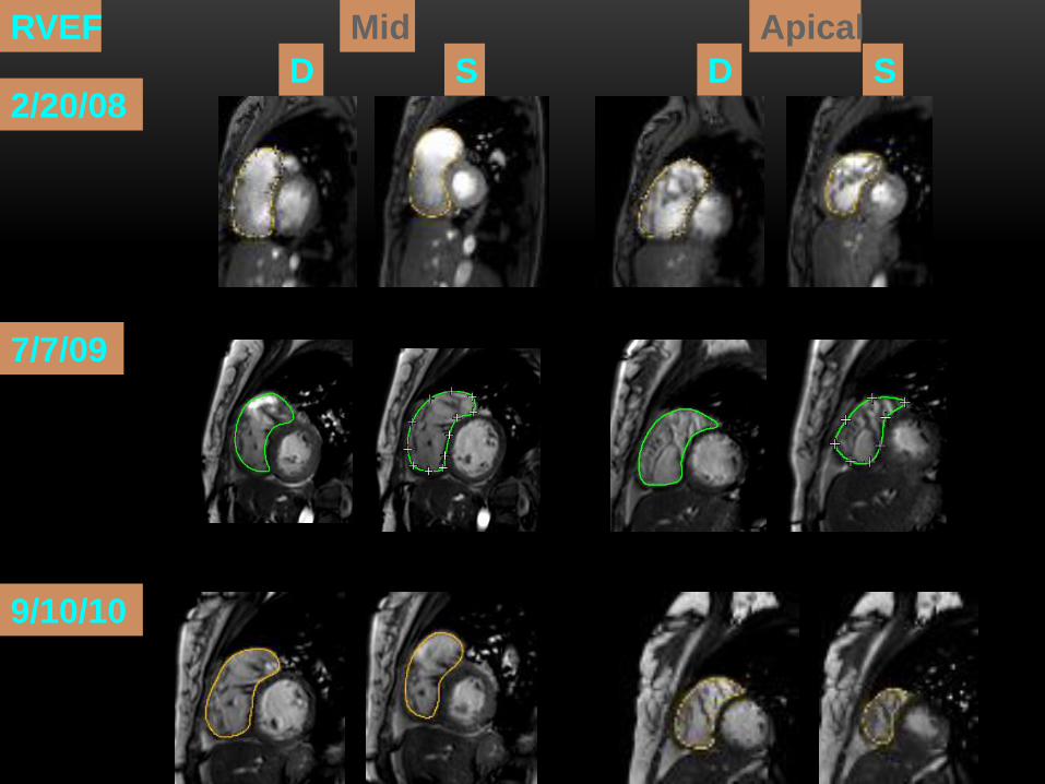
RVEF
2/20/08
9/10/10
7/7/09
D S D S
Mid Apical
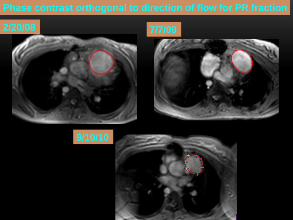
Phase contrast orthogonal to direction of flow for PR fraction
2/20/08
9/10/10
7/7/09
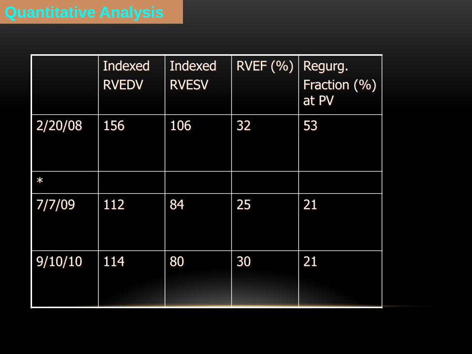
Quantitative Analysis
Indexed
RVEDV
Indexed
RVESV
RVEF (%) Regurg.
Fraction (%) at PV
2/20/08 156 106 32 53
*
7/7/09 112 84 25 21
9/10/10 114 80 30 21
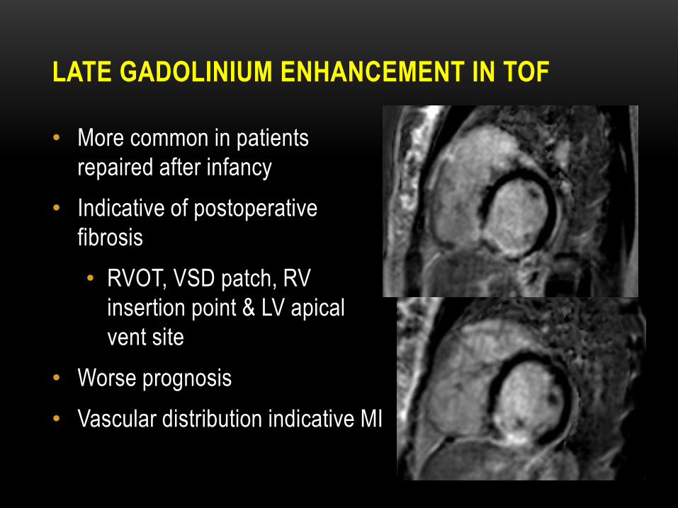
LATE GADOLINIUM ENHANCEMENT IN TOF
• More common in patients
repaired after infancy
• Indicative of postoperative
fibrosis
• RVOT, VSD patch, RV
insertion point & LV apical
vent site
• Worse prognosis
• Vascular distribution indicative MI
CT IN TOF
• Ventricular volumes, wall motion and ejection fractions-
retrospective ECG-gating
• Inferentially calculate regurgitant volume if isolated PI
(no TI or VSD) based on equal right and left heart
cardiac outputs
• RV SV-LV SV = pulmonary regurgitant volume
• Imaging modality of choice for coronary anomalies,
pulmonary parenchyma
20 YEAR OLD MALE S/P TOF REPAIR WITH
RV-PA CONDUIT AND SINGLE CORONARY
ARTERY S/P PACEMAKER
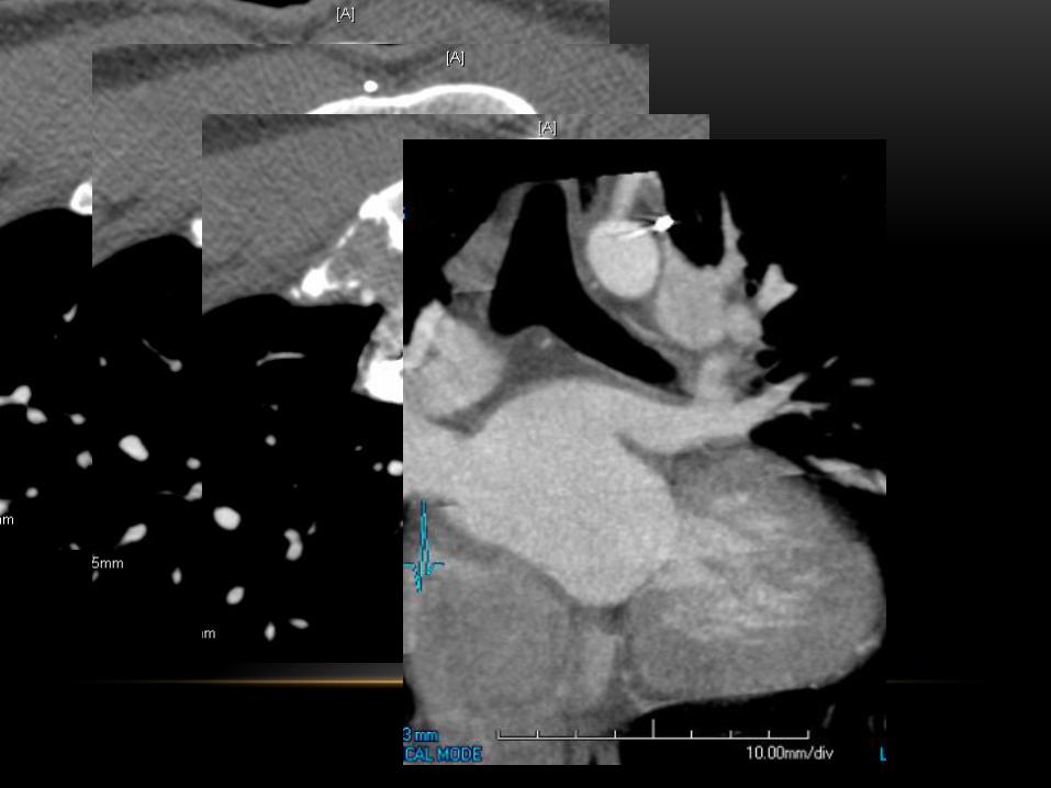
4/22/11
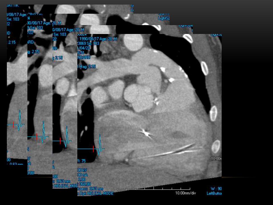
4/22/11
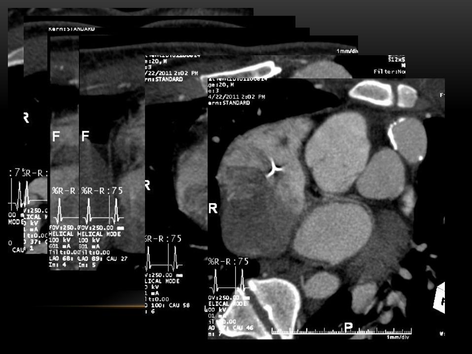
4/22/11
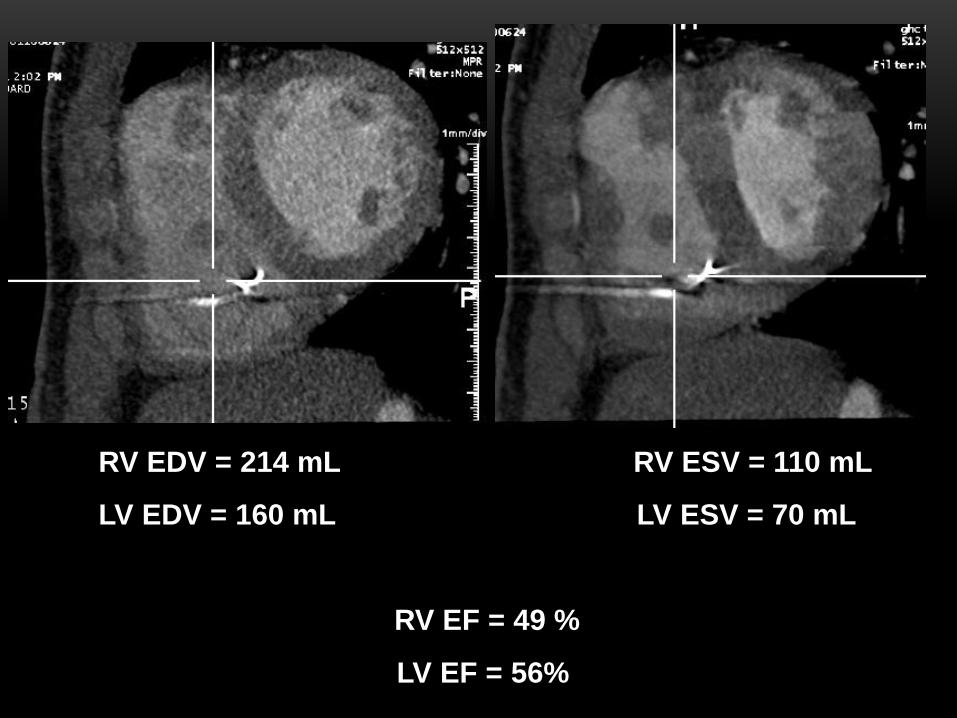
RV EDV = 214 mL RV ESV = 110 mL
LV EDV = 160 mL LV ESV = 70 mL
RV EF = 49 %
LV EF = 56% 4/22/11
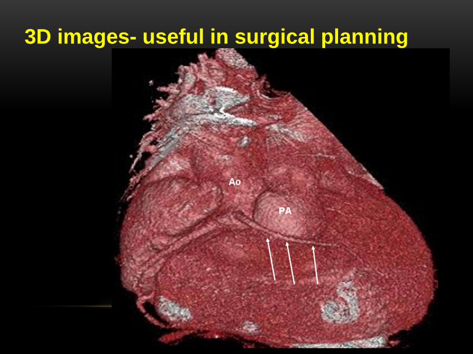
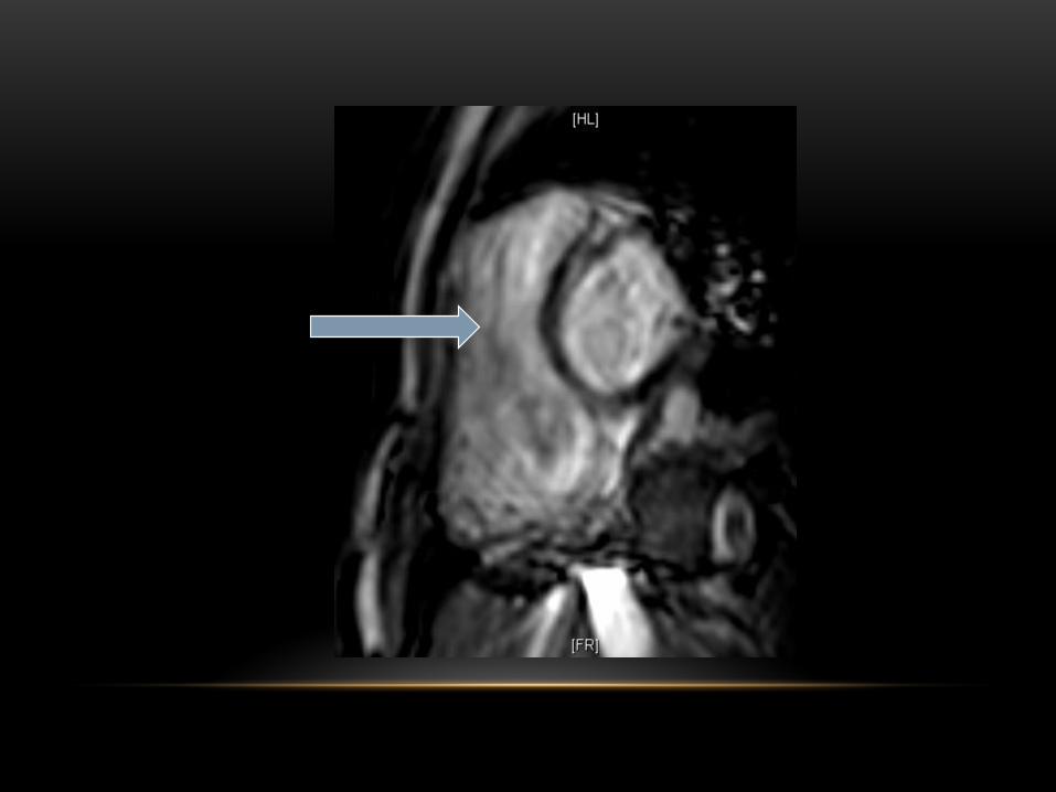
Fig 5c
3D images- useful in surgical planning
TOF WITH PULMONARY ATRESIA-SPECTRUM
• Large confluent central PAs discontinuous but near RVOT
• Absent central PAs with lungs supplied solely by systemic collaterals (pseudotruncus)
• Numerous intermediate forms
• Primary repair for milder forms
• Absent central PA’s difficult to repair (unifocalization)
• MR and CT add value to echo in surgical planning, depict central Pas, systemic collaterals
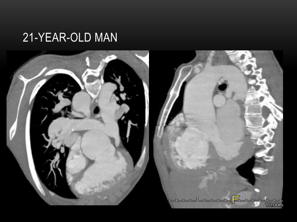
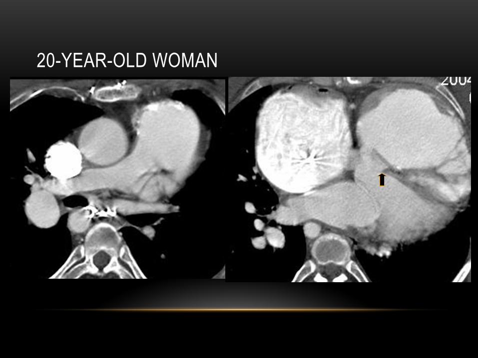
21-YEAR-OLD MAN
PULMONARY ARTERY ANEURYSM
• Aneurysmal central PAs in a subset of TOF- usually
absence of the pulmonary valve
• Cause unknown
• May be at Blalock- Taussig shunt insertion
• Collaterals
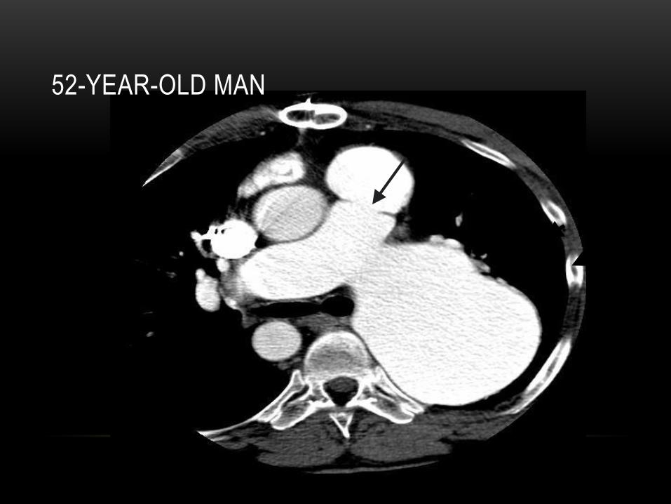
Fig 5a
52-YEAR-OLD MAN
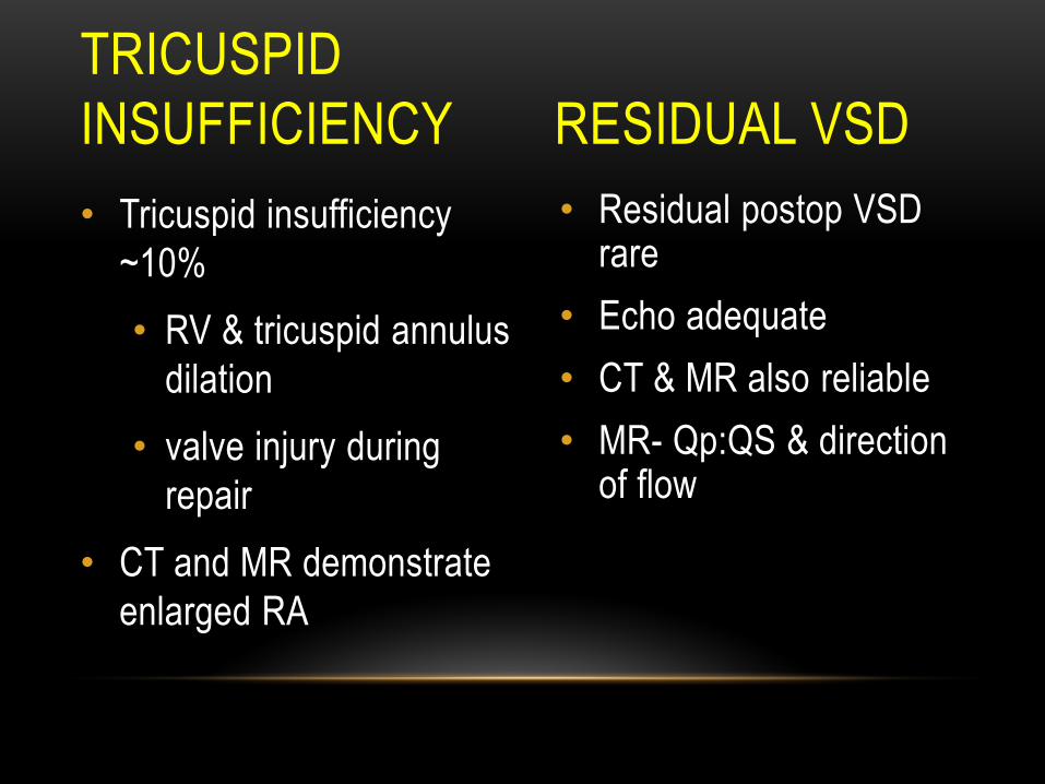
• Tricuspid insufficiency
~10%
• RV & tricuspid annulus
dilation
• valve injury during
repair
• CT and MR demonstrate
enlarged RA
• Residual postop VSD rare
• Echo adequate
• CT & MR also reliable
• MR- Qp:QS & direction of flow
TRICUSPID
INSUFFICIENCY RESIDUAL VSD
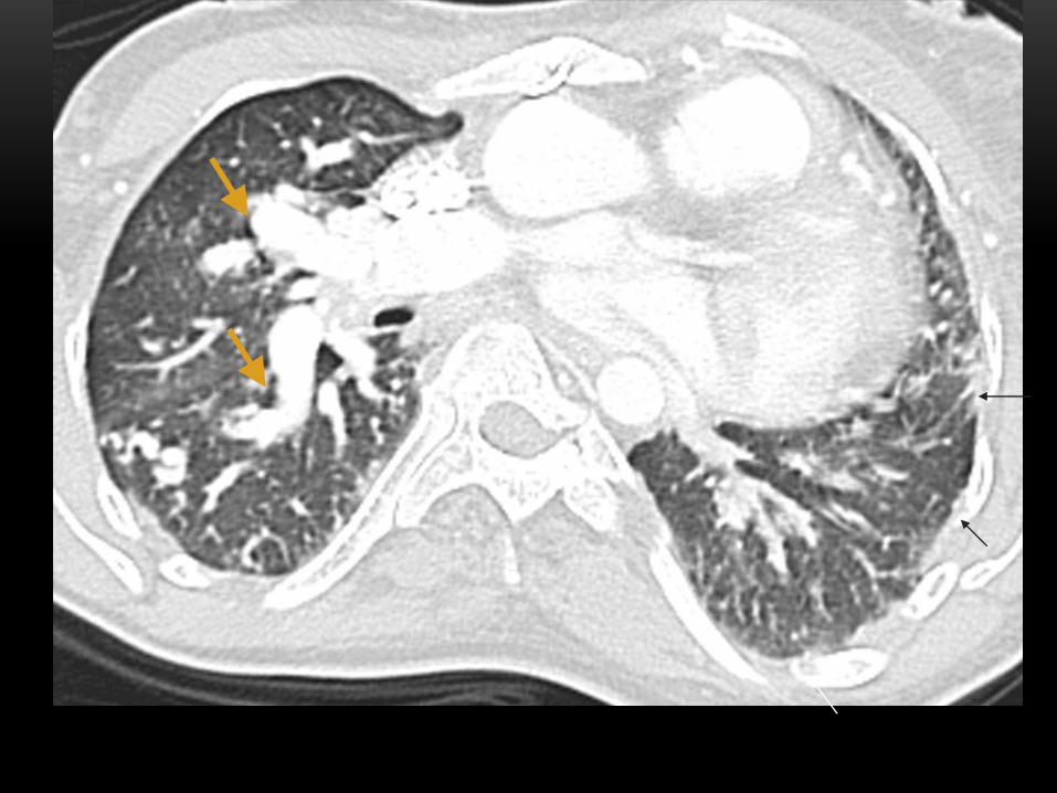
20-YEAR-OLD WOMAN
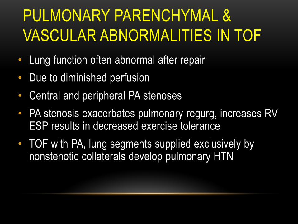
PULMONARY PARENCHYMAL &
VASCULAR ABNORMALITIES IN TOF
• Lung function often abnormal after repair
• Due to diminished perfusion
• Central and peripheral PA stenoses
• PA stenosis exacerbates pulmonary regurg, increases RV ESP results in decreased exercise tolerance
• TOF with PA, lung segments supplied exclusively by nonstenotic collaterals develop pulmonary HTN
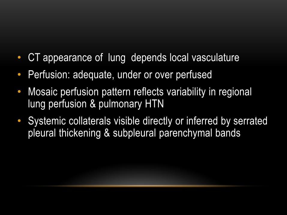
• CT appearance of lung depends local vasculature
• Perfusion: adequate, under or over perfused
• Mosaic perfusion pattern reflects variability in regional lung perfusion & pulmonary HTN
• Systemic collaterals visible directly or inferred by serrated pleural thickening & subpleural parenchymal bands
CONCLUSION
• TOF repair invariably leads to pulmonary insufficiency
• Well-tolerated early, but associated with morbidity & mortality over time
• MR primary tool for evaluating ventricular volumes & function, pulmonary insufficiency- guide to PVR
• MR useful for PA anatomy, differential flow thru PAs, fibrosis
• CT when MR contraindicated & primary tool for coronary anomalies & pulmonary parenchymal imaging

THANK YOU