Embed Size (px)
Citation preview
Muscles of facial
expressions
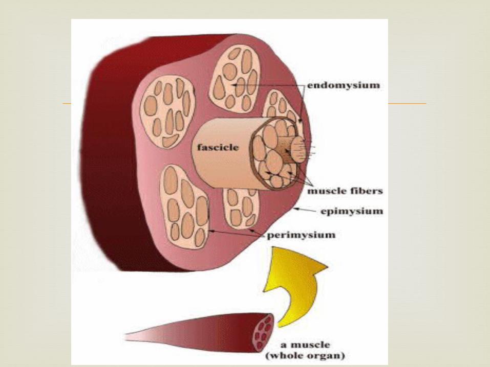
Muscle. Muscle classification. Structure of a muscle. Face. Skin fascia. various expressions of the face. Muscles involved in facial expression. Nerve supply. Lymphatic drainage. Blood supply. Applied anatomy. Conclusion
Contents
- Muscle- latin word musculus. Muscle tissue has two important properties: - contractility. - conductivity.
Muscle
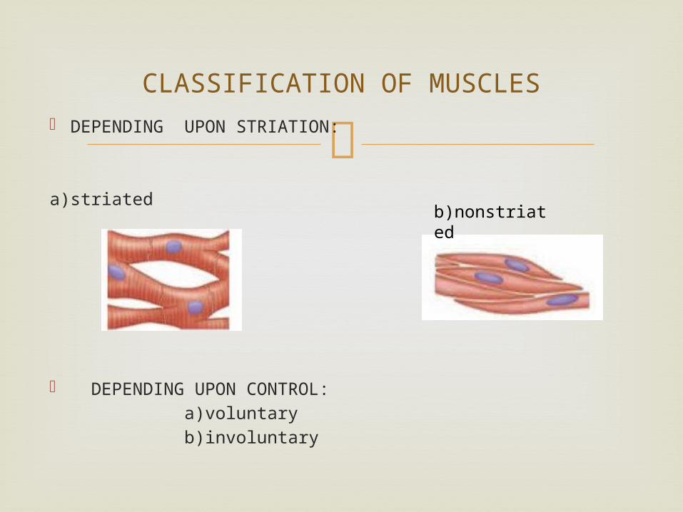
CLASSIFICATION OF MUSCLES
DEPENDING UPON STRIATION:
a)striated
DEPENDING UPON CONTROL: a)voluntary b)involuntary
b)nonstriated
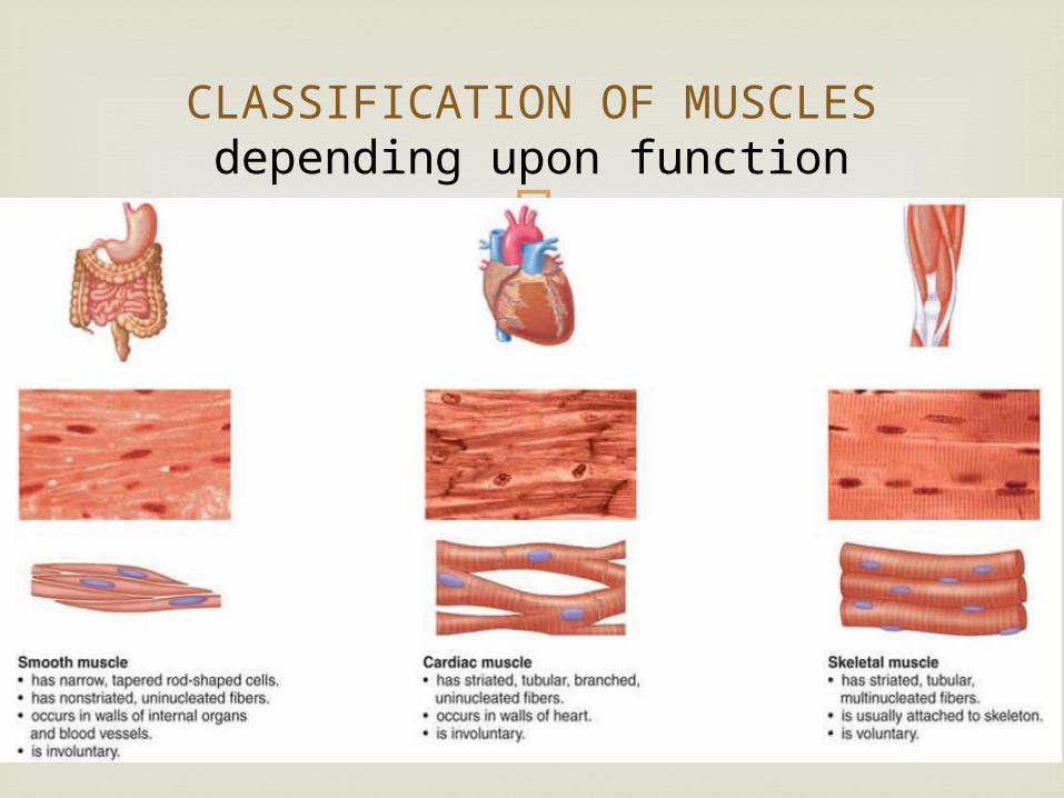
CLASSIFICATION OF MUSCLESdepending upon function
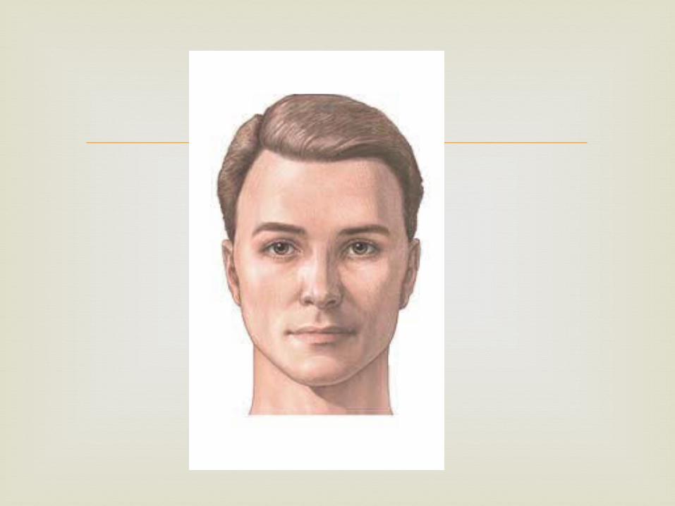
The face or countenance, extents superiorly
from the adolescent position of hairline, inferiorly to the chin and base of the mandible, and on each side to the auricle
The forehead is therefore common to scalp and face.
FACE
The face is covered by - Skin. - Fascia. - Muscle.
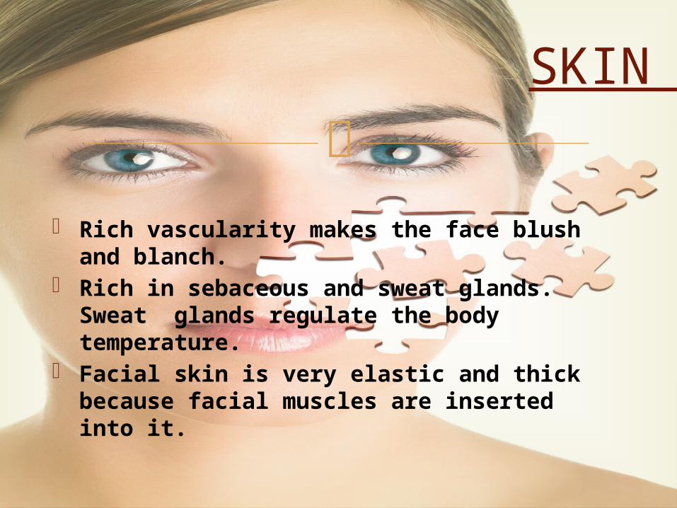
Rich vascularity makes the face blush and blanch.
Rich in sebaceous and sweat glands. Sweat glands regulate the body temperature.
Facial skin is very elastic and thick because facial muscles are inserted into it.
SKIN
Superficial fascia: - facial muscles. - vessels and nerves. - variable amount of fat.Deep fascia: - parotid fascia. - buccopharyngeal fascia.
FASCIA
FACIAL MUSCLES
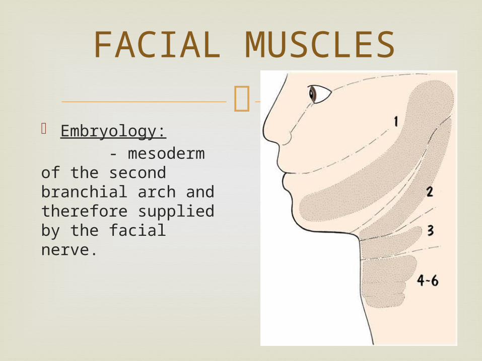
Embryology: - mesoderm of the second branchial arch and therefore supplied by the facial nerve.
CLASSIFICATION
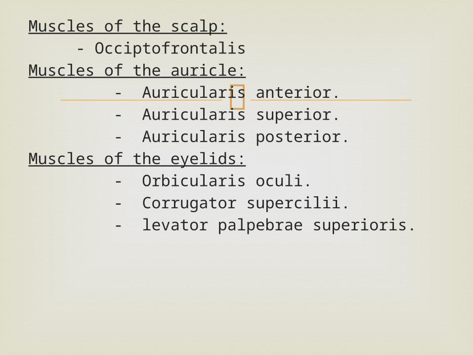
Muscles of the scalp: - Occiptofrontalis Muscles of the auricle: - Auricularis anterior. - Auricularis superior. - Auricularis posterior.Muscles of the eyelids: - Orbicularis oculi. - Corrugator supercilii. - levator palpebrae superioris.
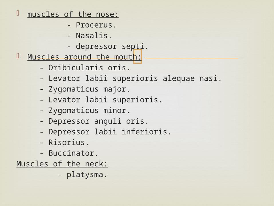
muscles of the nose: - Procerus. - Nasalis. - depressor septi. Muscles around the mouth: - Oribicularis oris. - Levator labii superioris alequae nasi. - Zygomaticus major. - Levator labii superioris. - Zygomaticus minor. - Depressor anguli oris. - Depressor labii inferioris. - Risorius. - Buccinator.Muscles of the neck: - platysma.
Most of these muscles may be regarded
primarily as regulators of the three openings situated on the face, namely
- palpebral fissure. - nostrils. - oral fissure. Each opening has a single sphinter and
variable number of dilators.
FUNCTIONS
Sphincters are naturally circular. Dilators are radial in their arrangments. The better developed around the eyes and
mouth than around nose.
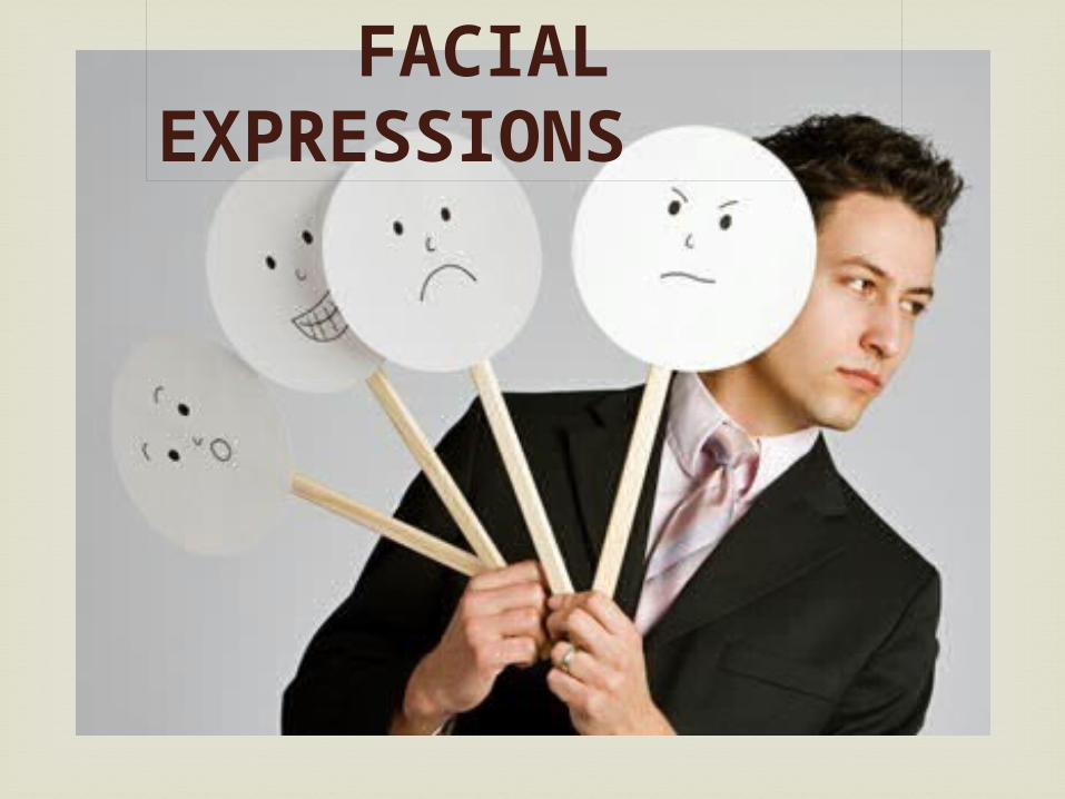
FACIAL EXPRESSIONS
Smiling Laughing Sadness Grief Anger Frowning Horror, terror and fright Doubt Grinning Contempt Closing the mouth whistling
MUSCLES OF SCALP
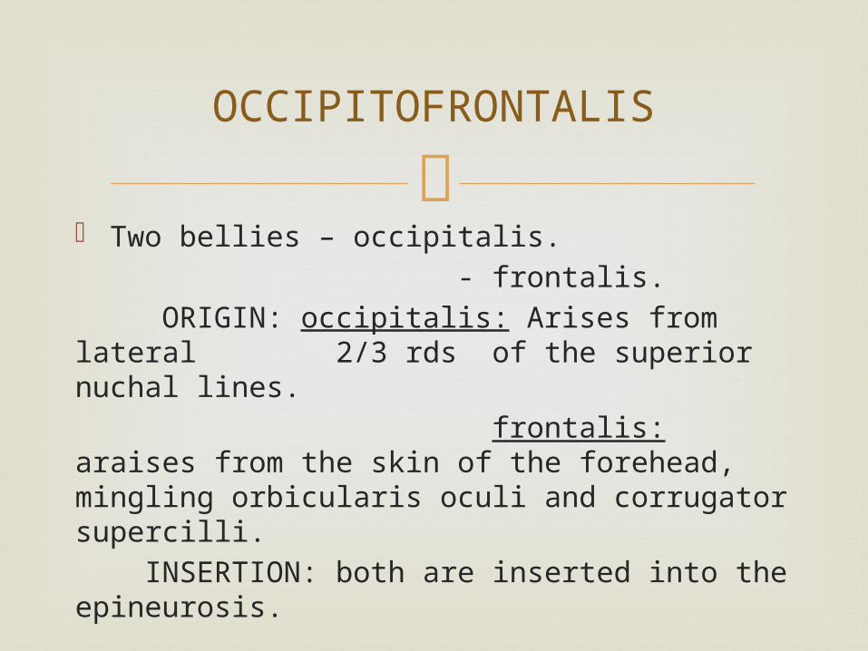
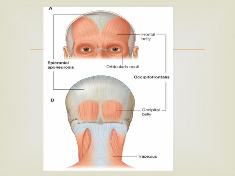
Two bellies – occipitalis. - frontalis. ORIGIN: occipitalis: Arises from lateral 2/3 rds of the superior nuchal lines. frontalis: araises from the skin of the forehead, mingling orbicularis oculi and corrugator supercilli. INSERTION: both are inserted into the epineurosis.
OCCIPITOFRONTALIS
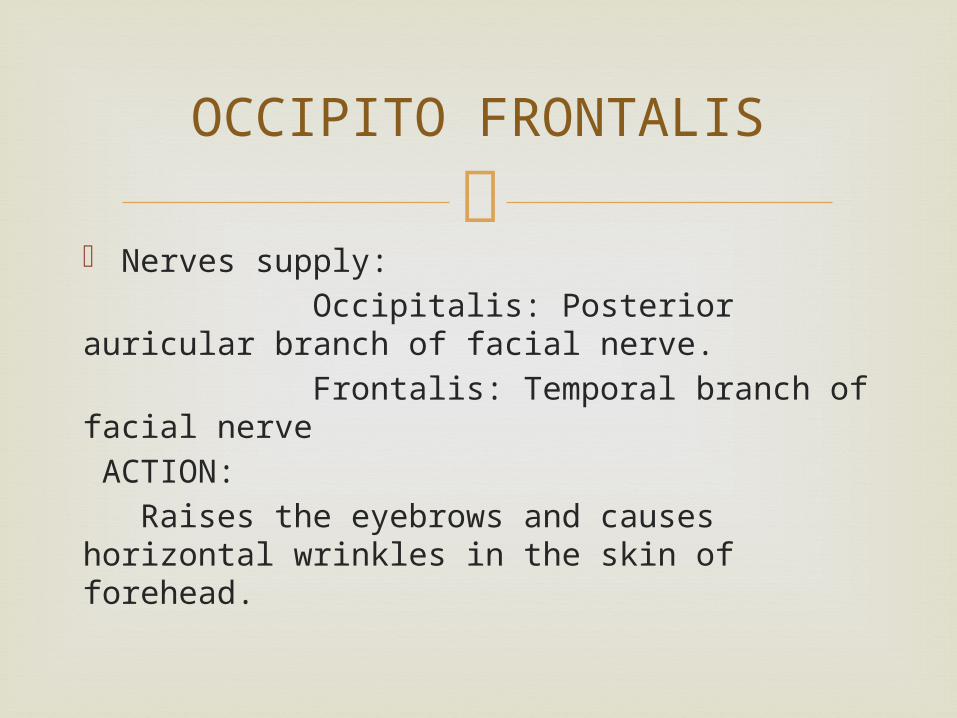
Nerves supply: Occipitalis: Posterior auricular branch of facial nerve. Frontalis: Temporal branch of facial nerve ACTION: Raises the eyebrows and causes horizontal wrinkles in the skin of forehead.
OCCIPITO FRONTALIS
Muscles of the auricle
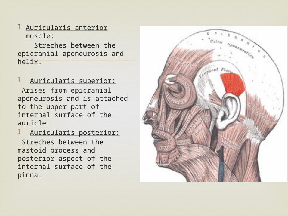
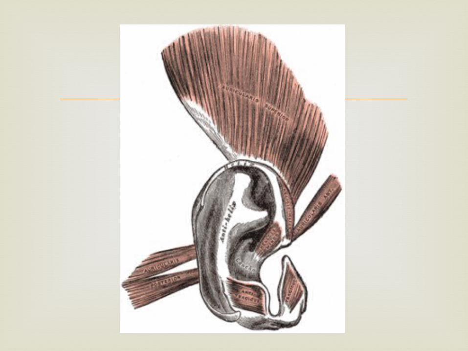
Auricularis anterior muscle:
Streches between the epicranial aponeurosis and helix. Auricularis superior: Arises from epicranial aponeurosis and is attached to the upper part of internal surface of the auricle. Auricularis posterior: Streches between the mastoid process and posterior aspect of the internal surface of the pinna.
Muscles around the eyelids
Orbicularis oculi
Orbicularis oculi: It is a broad, flat elliptical muscle which surrounds circumference of orbit & spreads in to eye lids, anterior temporal region, infraorbital, cheek & superciliary region.
It has orbital, lacrimal & palpebral part.
Orbicularis oculi
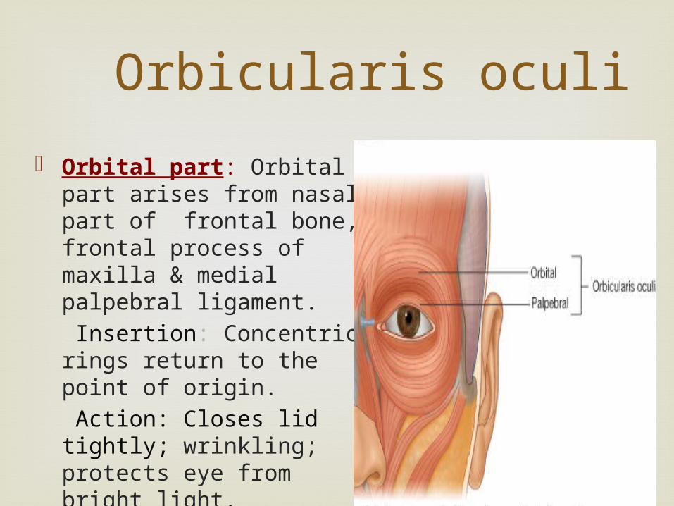
Orbital part: Orbital part arises from nasal part of frontal bone, frontal process of maxilla & medial palpebral ligament.
Insertion: Concentric rings return to the point of origin.
Action: Closes lid tightly; wrinkling; protects eye from bright light.
Orbicularis oculi
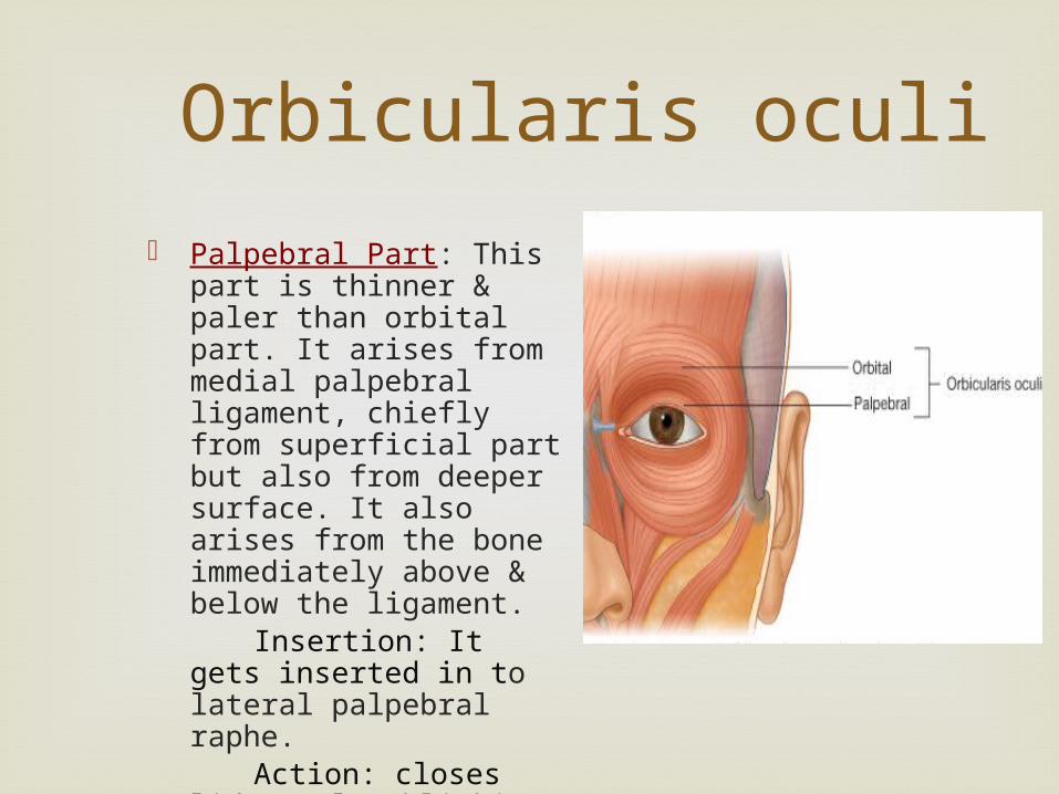
Palpebral Part: This part is thinner & paler than orbital part. It arises from medial palpebral ligament, chiefly from superficial part but also from deeper surface. It also arises from the bone immediately above & below the ligament.
Insertion: It gets inserted in to lateral palpebral raphe.
Action: closes lid gently, blinking.
Orbicularis oculi
Lacrimal Part: It is present lateral & deep to lacrimal sac.
Origin: Arises from lacrimal fascia & lacrimal bone.
Insertion: Upper & lower eye lids. Action: Dilates lacrimal sac; directs lacrimal
puncta in to lacus lacrimalis; supports the lower lid.
Orbicularis occuli
Nerve Supply: Orbicularis occuli is supplied by temporal & zygomatic branches of facial nerve.
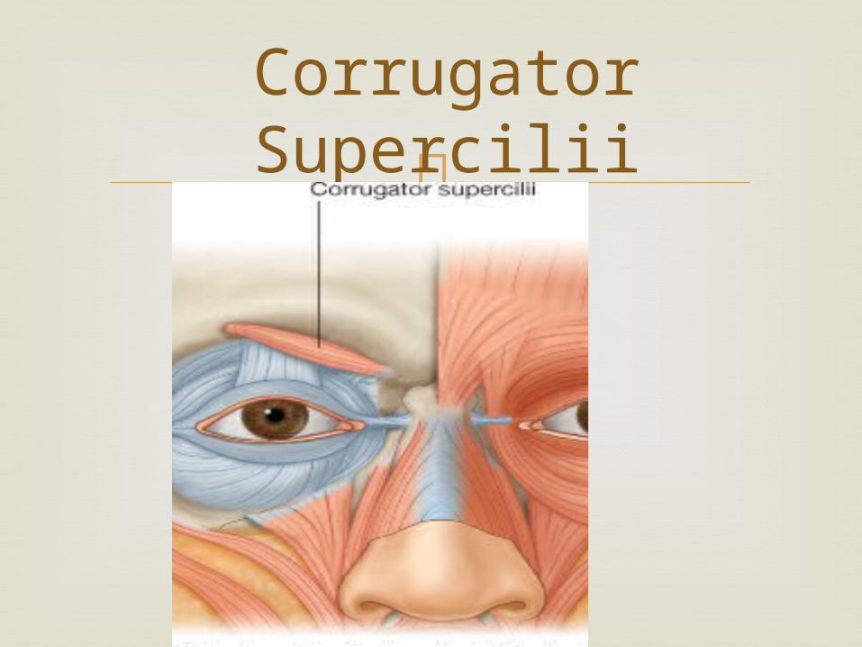
Corrugator Supercilii
It is a small pyramidal muscle located at medial end of each eyebrow, deep to frontal part of Occipitofrontalis & orbicularis occuli with which is partly blended.
Origin: Medial end of superciliary arch. Insertion: Skin of mid-eyebrow
Corrugator Supercilii
Corrugator Supercilii
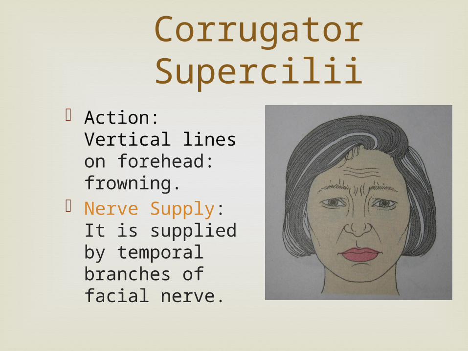
Action: Vertical lines on forehead: frowning.
Nerve Supply: It is supplied by temporal branches of facial nerve.
Levator Palpebral Superioris
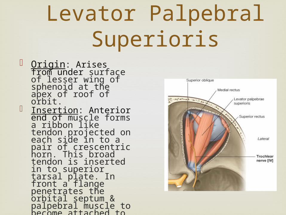
Origin: Arises from under surface of lesser wing of sphenoid at the apex of roof of orbit.
Insertion: Anterior end of muscle forms a ribbon like tendon projected on each side in to a pair of crescentric horn. This broad tendon is inserted in to superior tarsal plate. In front a flange penetrates the orbital septum & palpebral muscle to become attached to skin of upper lid, while behind a weaker flange is attached to the superior fornix of the conjunctiva.
Levator Palpebral Superioris
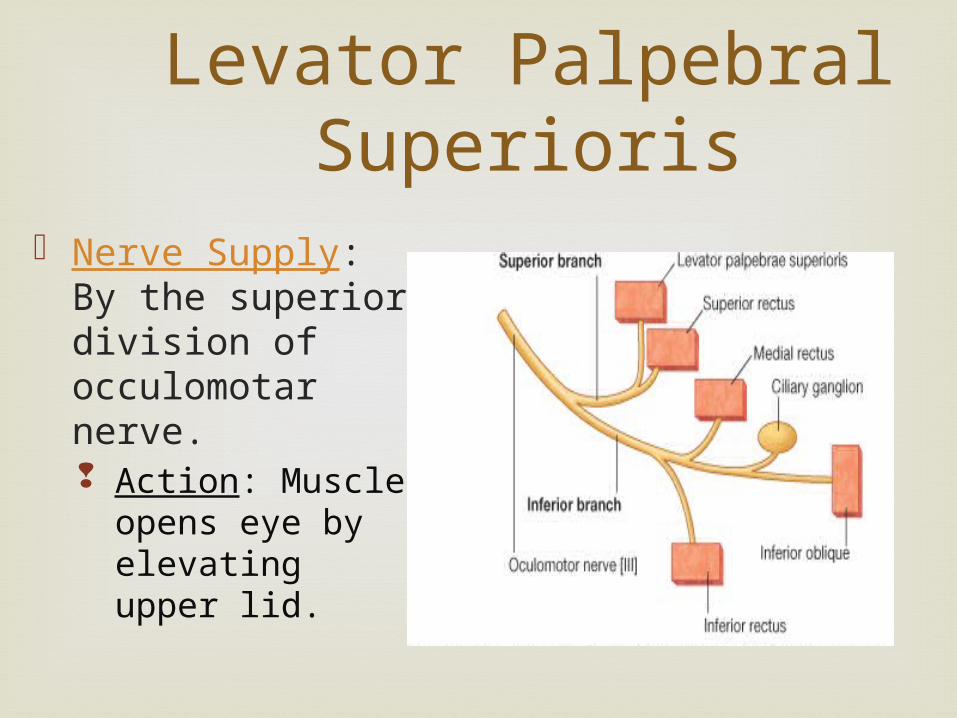
Nerve Supply: By the superior division of occulomotar nerve. Action: Muscle
opens eye by elevating upper lid.
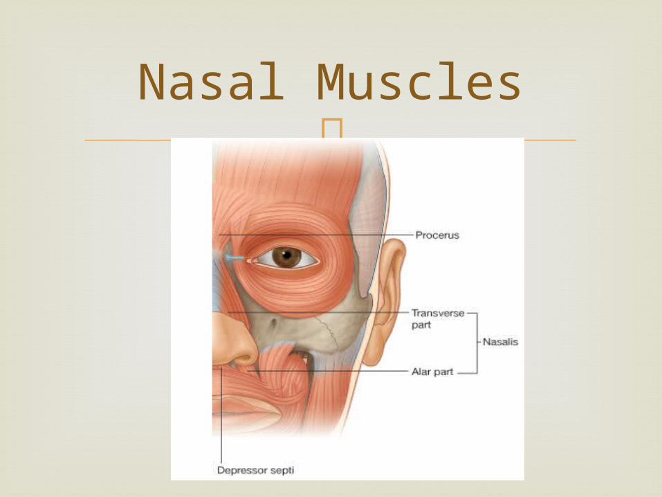
Muscles of nose
Nasalis
Consists of transverse & alar part. Transverse part arises from maxilla just lateral to nasal notch. Alar part arises from maxilla below & medial to transverse part with which it partly merges.
Action: Transverse part of Nasalis compresses the nasal aperture at the junction of vestibule with the nasal cavity, alar part assists in widening of anterior nasal aperture.

Nasal Muscles
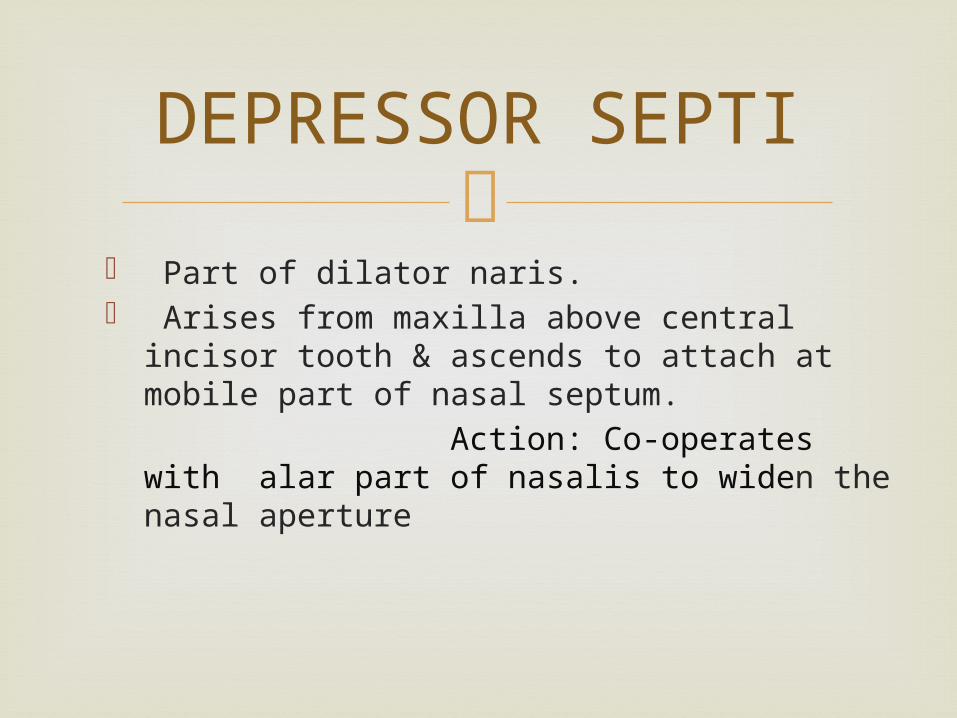
DEPRESSOR SEPTI
Part of dilator naris. Arises from maxilla above central incisor
tooth & ascends to attach at mobile part of nasal septum.
Action: Co-operates with alar part of nasalis to widen the nasal aperture

Nasal Muscles
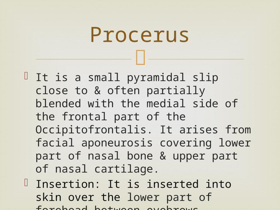
Procerus
It is a small pyramidal slip close to & often partially blended with the medial side of the frontal part of the Occipitofrontalis. It arises from facial aponeurosis covering lower part of nasal bone & upper part of nasal cartilage.
Insertion: It is inserted into skin over the lower part of forehead between eyebrows.
Nasal Muscles
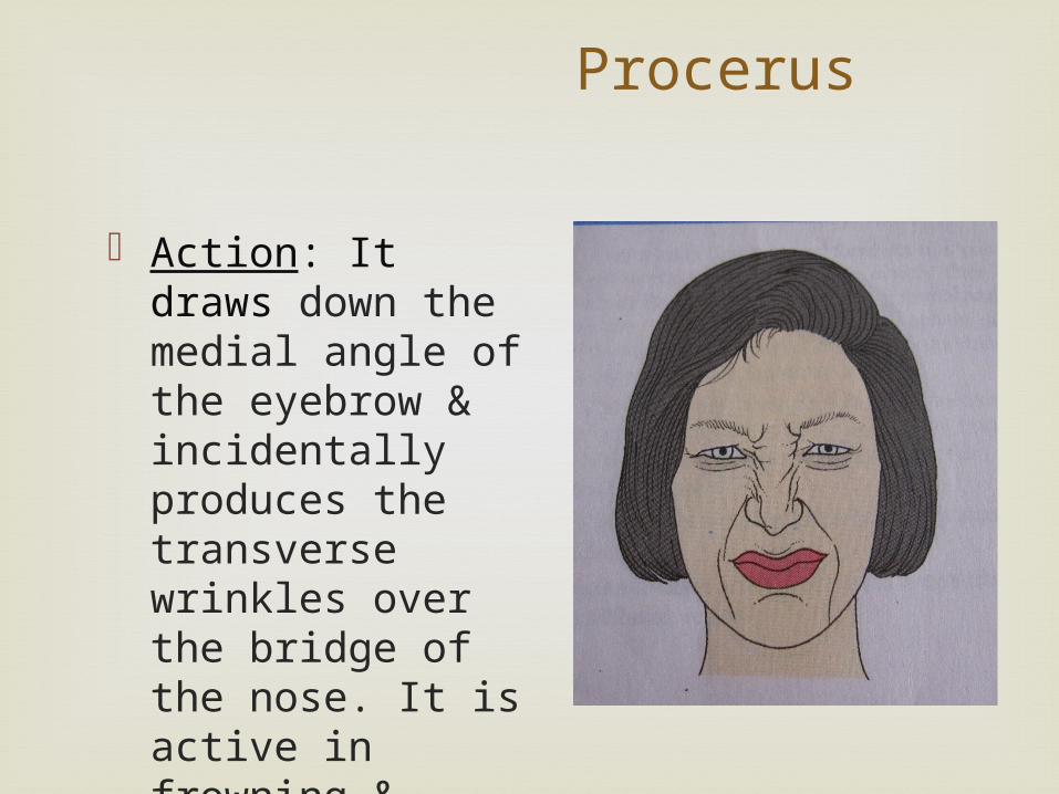
Procerus
Action: It draws down the medial angle of the eyebrow & incidentally produces the transverse wrinkles over the bridge of the nose. It is active in frowning & concentration.
Nerve Supply Of Nasal Muscles
All the muscles of this group are supplied by the superior buccal branch of the facial nerve.
Muscles around the mouth
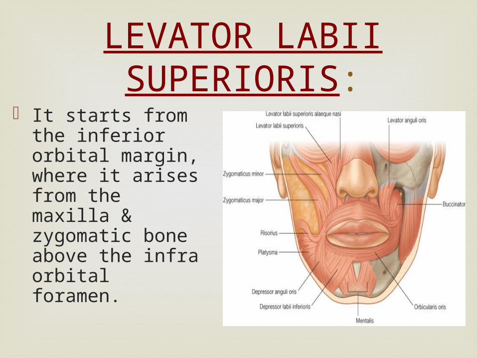
LEVATOR LABII SUPERIORIS:
It starts from the inferior orbital margin, where it arises from the maxilla & zygomatic bone above the infra orbital foramen.
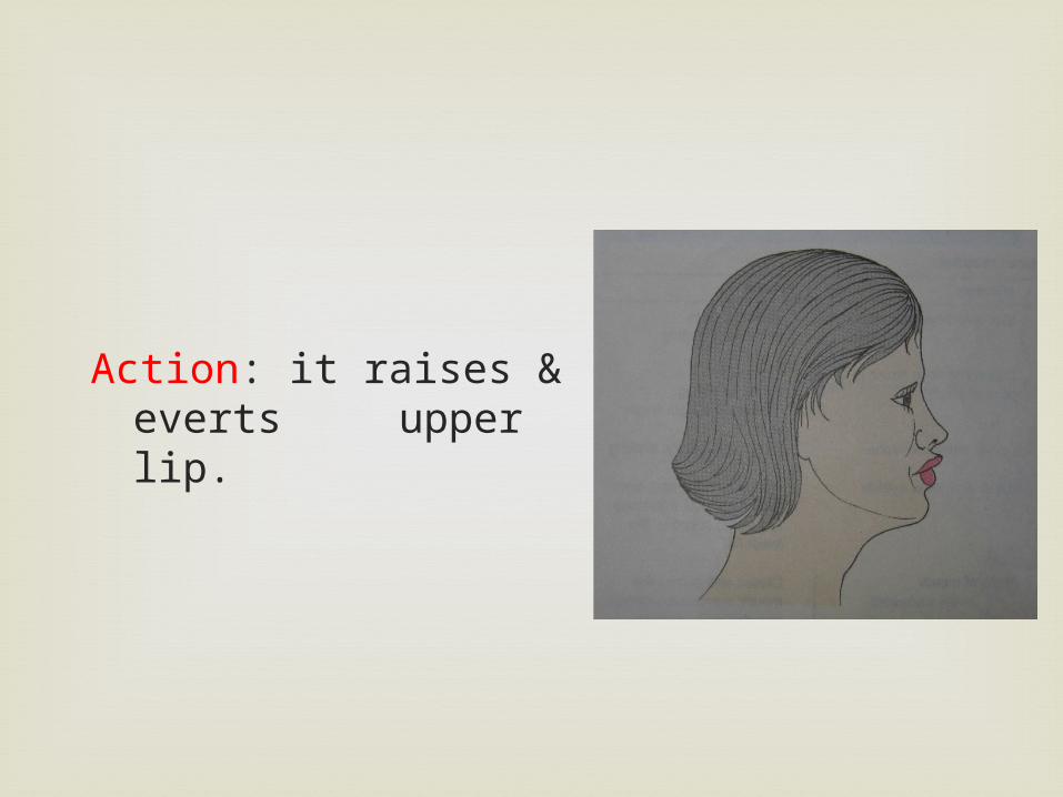
Action: it raises & everts upper lip.
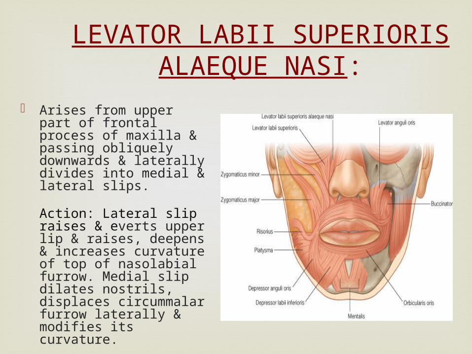
LEVATOR LABII SUPERIORIS ALAEQUE NASI:
Arises from upper part of frontal process of maxilla & passing obliquely downwards & laterally divides into medial & lateral slips.
Action: Lateral slip raises & everts upper lip & raises, deepens & increases curvature of top of nasolabial furrow. Medial slip dilates nostrils, displaces circummalar furrow laterally & modifies its curvature.
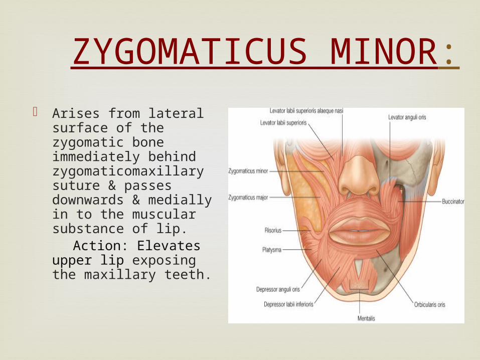
ZYGOMATICUS MINOR:
Arises from lateral surface of the zygomatic bone immediately behind zygomaticomaxillary suture & passes downwards & medially in to the muscular substance of lip.
Action: Elevates upper lip exposing the maxillary teeth.
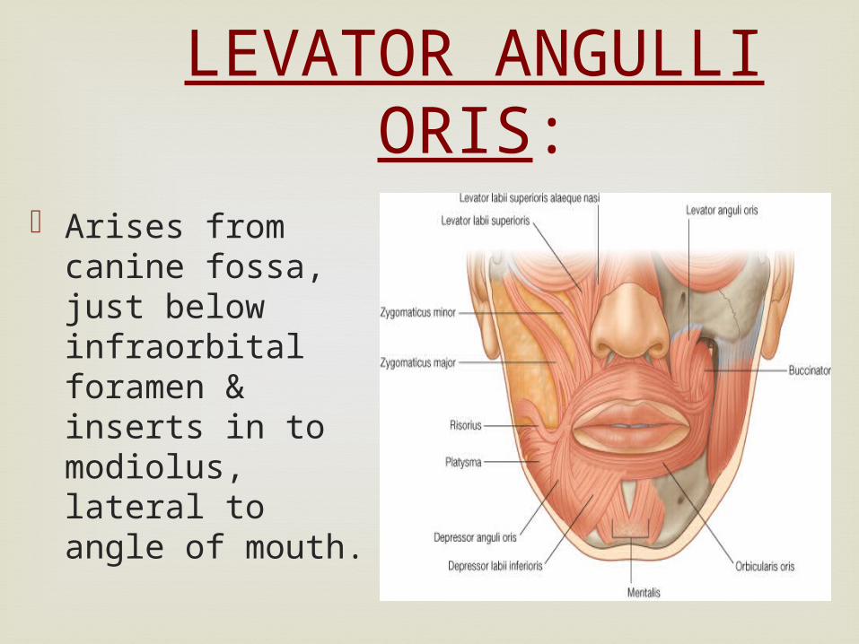
LEVATOR ANGULLI ORIS:
Arises from canine fossa, just below infraorbital foramen & inserts in to modiolus, lateral to angle of mouth.
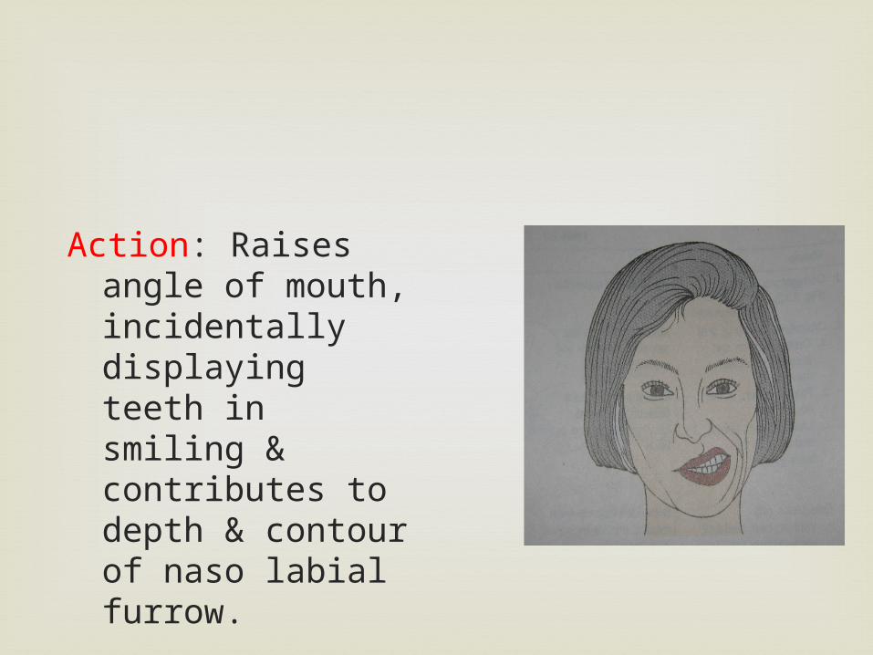
Action: Raises angle of mouth, incidentally displaying teeth in smiling & contributes to depth & contour of naso labial furrow.
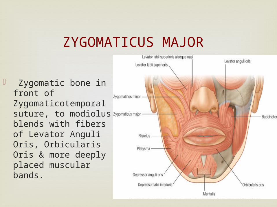
ZYGOMATICUS MAJOR
Zygomatic bone in front of Zygomaticotemporal suture, to modiolus, blends with fibers of Levator Anguli Oris, Orbicularis Oris & more deeply placed muscular bands.
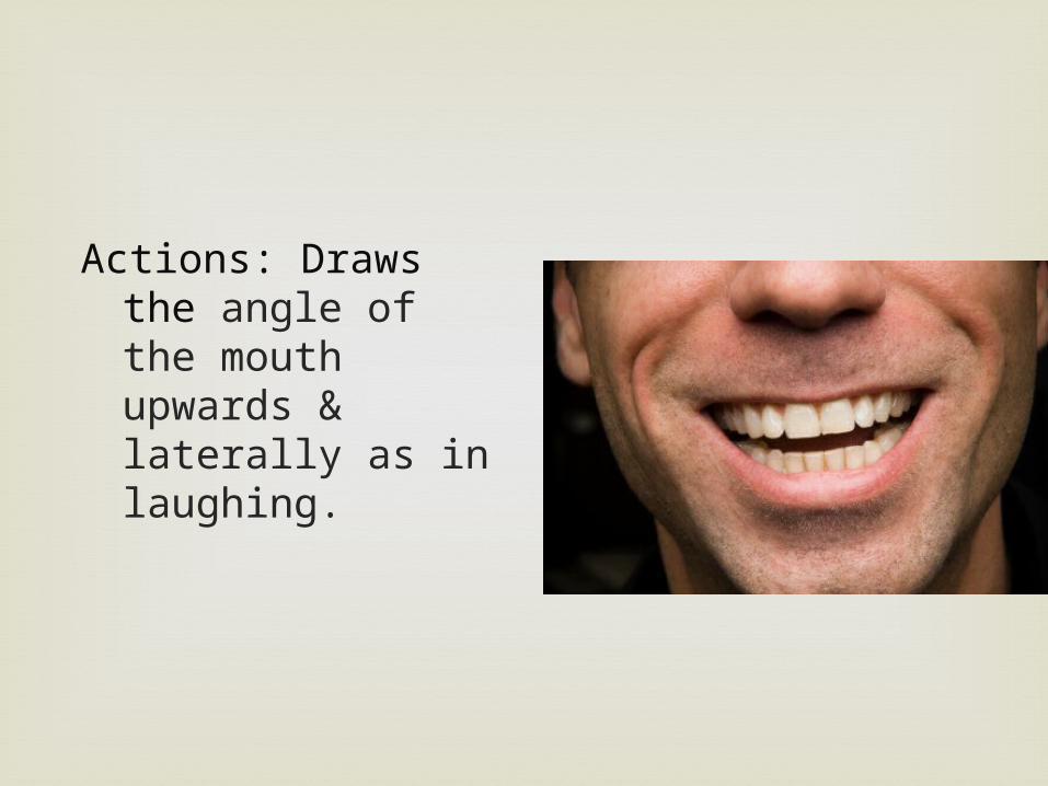
Actions: Draws the angle of the mouth upwards & laterally as in laughing.
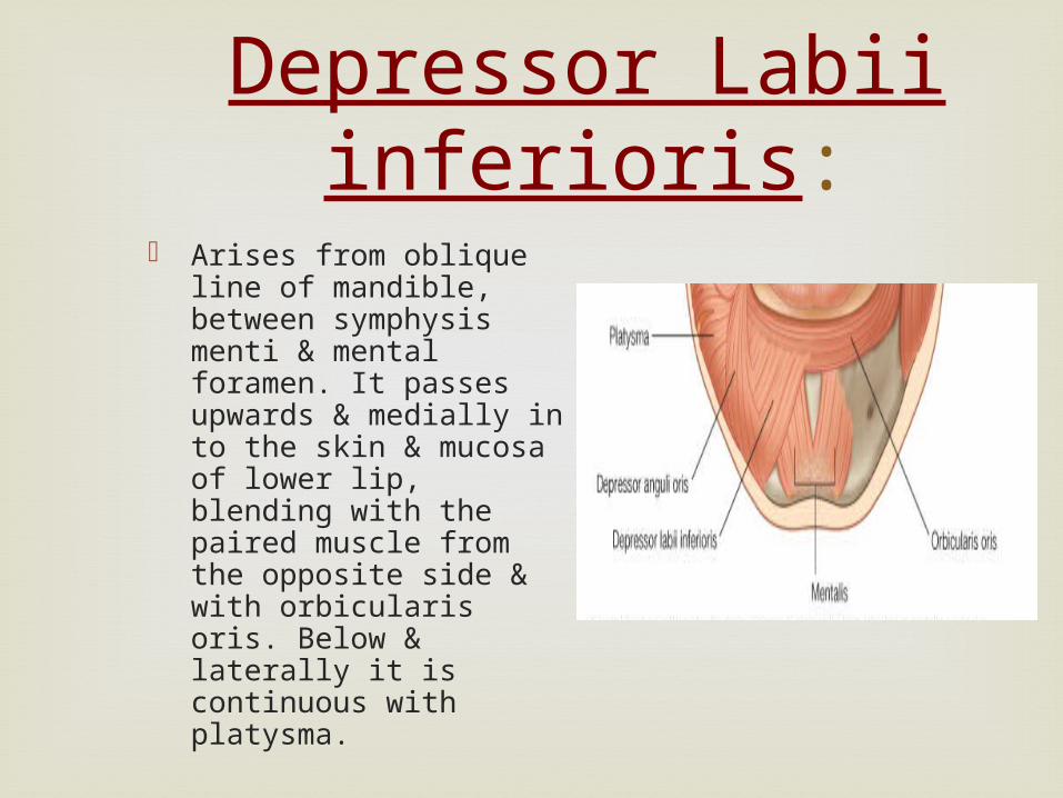
Depressor Labii inferioris:
Arises from oblique line of mandible, between symphysis menti & mental foramen. It passes upwards & medially in to the skin & mucosa of lower lip, blending with the paired muscle from the opposite side & with orbicularis oris. Below & laterally it is continuous with platysma.

Depressor Labii inferioris:
Action: It draws the lower lip downwards & a little laterally in masticatory activity, & may assist in its eversion. It contributes to the expression of Irony, sorrow, melancholy & doubt.
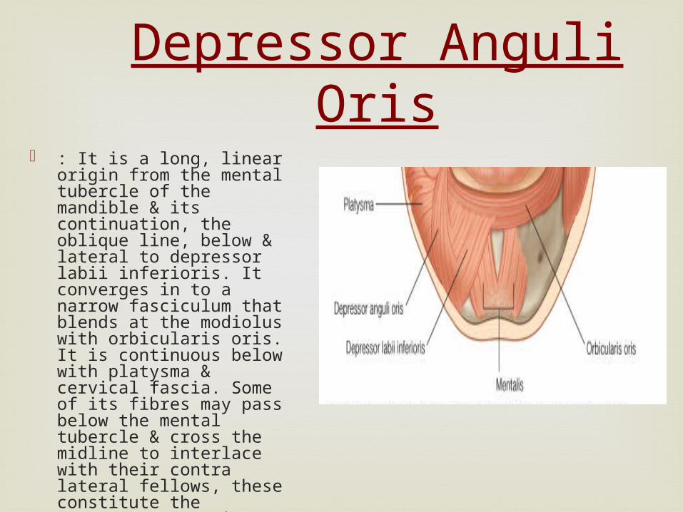
Depressor Anguli Oris : It is a long, linear origin
from the mental tubercle of the mandible & its continuation, the oblique line, below & lateral to depressor labii inferioris. It converges in to a narrow fasciculum that blends at the modiolus with orbicularis oris. It is continuous below with platysma & cervical fascia. Some of its fibres may pass below the mental tubercle & cross the midline to interlace with their contra lateral fellows, these constitute the transversus menti muscle.
Depressor Anguli Oris
Action: It draws the angle of mouth downwards & laterally in opening the mouth & in expressing sadness.
Mentalis:
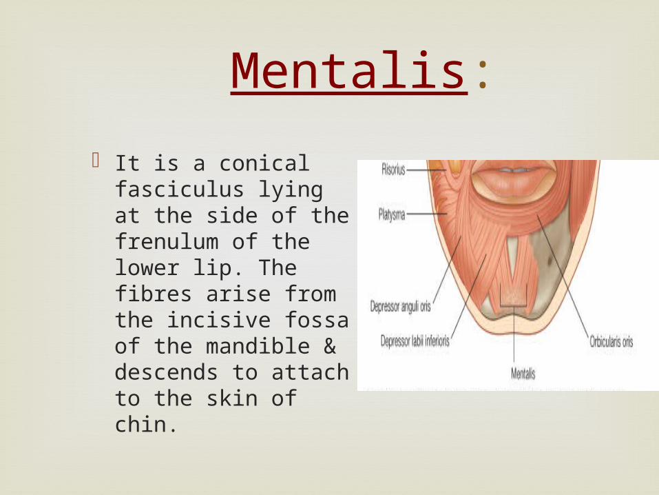
It is a conical fasciculus lying at the side of the frenulum of the lower lip. The fibres arise from the incisive fossa of the mandible & descends to attach to the skin of chin.
Mentalis:
Action: It raises the lower lip, mental tissues & mentolabial sulcus, wrinkling of the skin of chin, since it raises the base of the lower lip, it helps in protruding & everting it in drinking & also in expressing doubt or disdain.
Orbicularis oris:
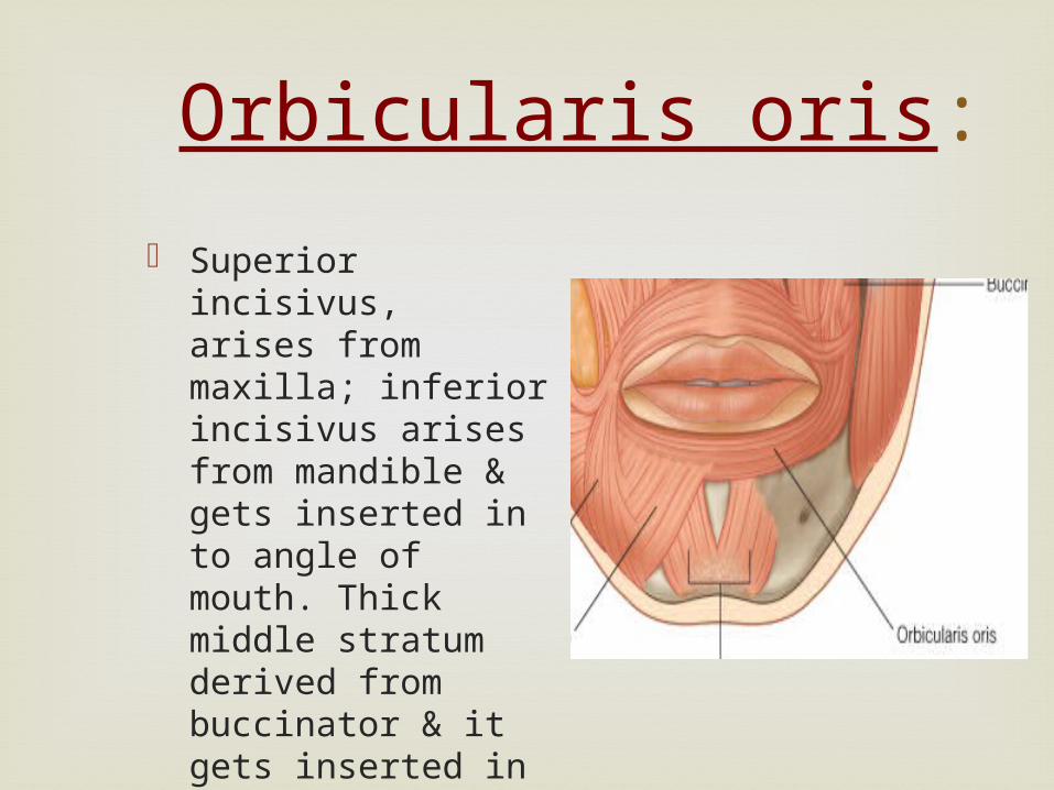
Superior incisivus, arises from maxilla; inferior incisivus arises from mandible & gets inserted in to angle of mouth. Thick middle stratum derived from buccinator & it gets inserted in to lips & angle of mouth.
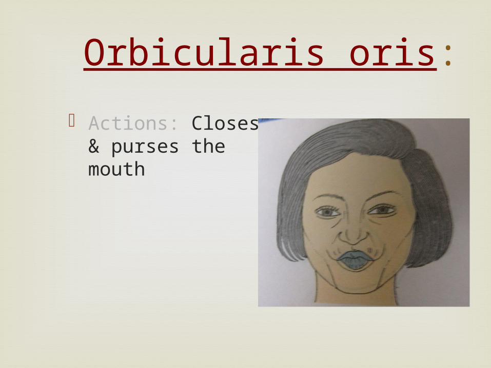
Orbicularis oris:
Actions: Closes & purses the mouth
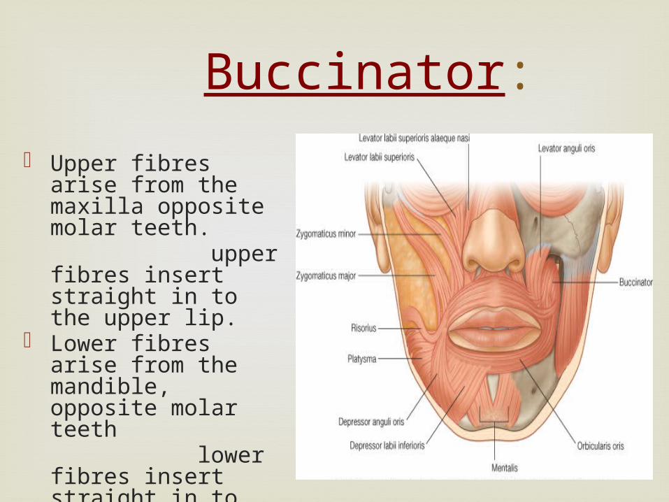
Buccinator:
Upper fibres arise from the maxilla opposite molar teeth.
upper fibres insert straight in to the upper lip.
Lower fibres arise from the mandible, opposite molar teeth
lower fibres insert straight in to the lower lip.
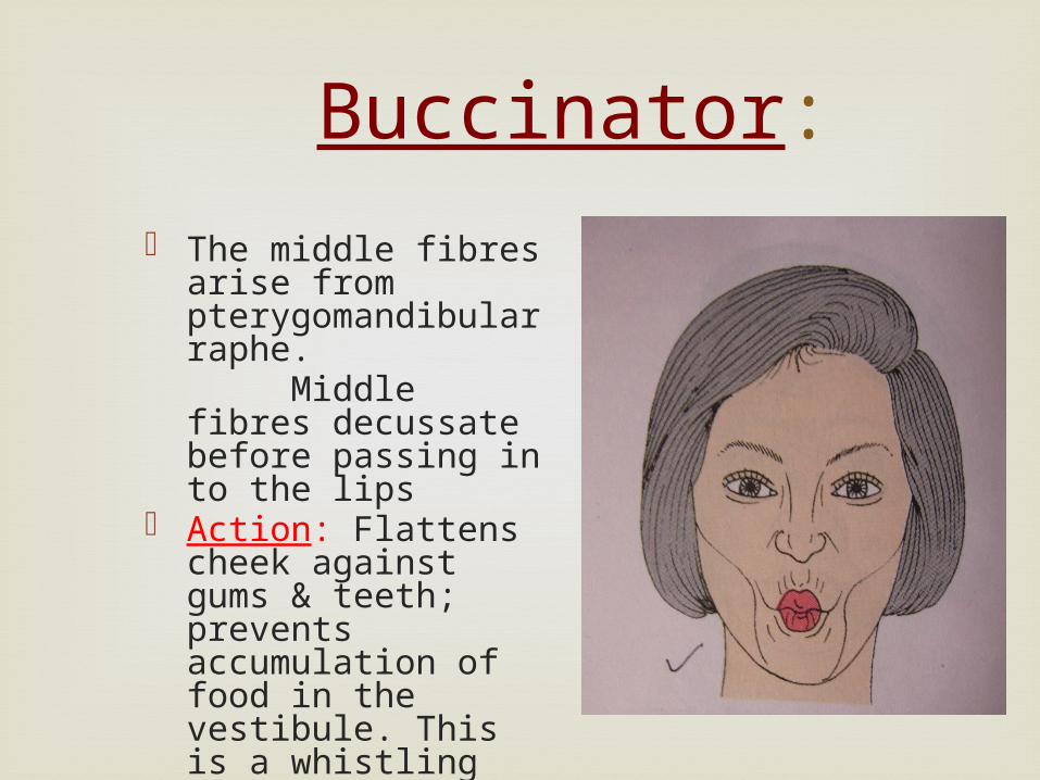
Buccinator:
The middle fibres arise from pterygomandibular raphe.
Middle fibres decussate before passing in to the lips
Action: Flattens cheek against gums & teeth; prevents accumulation of food in the vestibule. This is a whistling muscle.

Muscles of the neck
Platysma:
Broad sheet of muscle originates from the upper parts of pectoralis major & deltoid. Anterior fibre interlace across the midline with fibres of contralateral muscle, below & behind the symphysis menti.
Platysma:
Intermediate fibres attach to lower border of mandibular body or pass upwards & medially, deep to depressor anguli oris, to attachments in the lateral half of lower lip.
Posterior fibres cross the mandible & anteriolateral part of masseter to attach to skin & subcutaneous tissue of lower face.
Platysma:
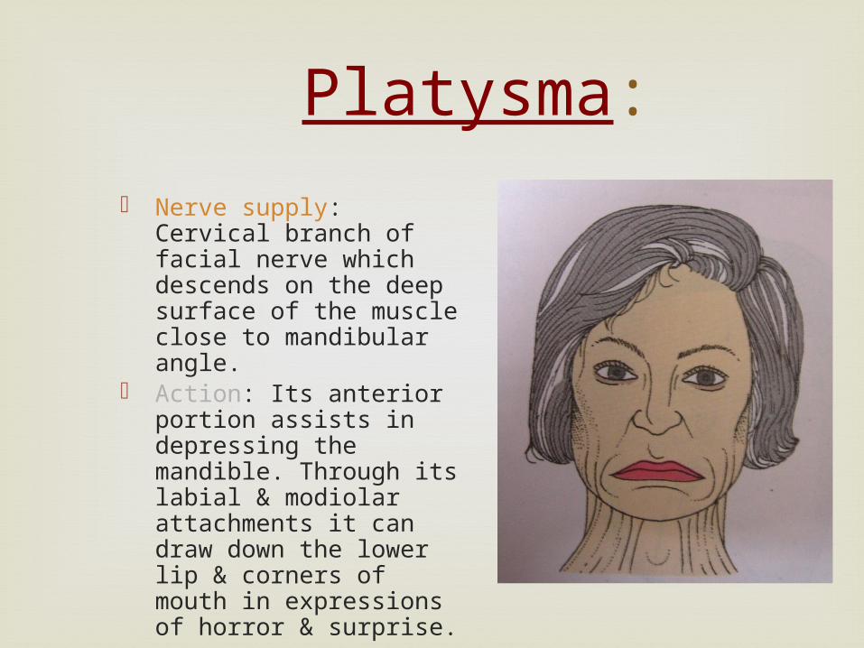
Nerve supply: Cervical branch of facial nerve which descends on the deep surface of the muscle close to mandibular angle.
Action: Its anterior portion assists in depressing the mandible. Through its labial & modiolar attachments it can draw down the lower lip & corners of mouth in expressions of horror & surprise.


Arterial Supply Of Face
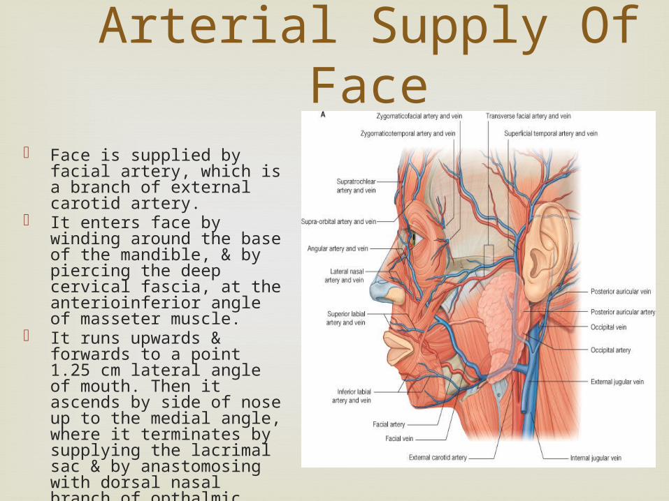
Face is supplied by facial artery, which is a branch of external carotid artery.
It enters face by winding around the base of the mandible, & by piercing the deep cervical fascia, at the anterioinferior angle of masseter muscle.
It runs upwards & forwards to a point 1.25 cm lateral angle of mouth. Then it ascends by side of nose up to the medial angle, where it terminates by supplying the lacrimal sac & by anastomosing with dorsal nasal branch of opthalmic artery.
Venous Drainage
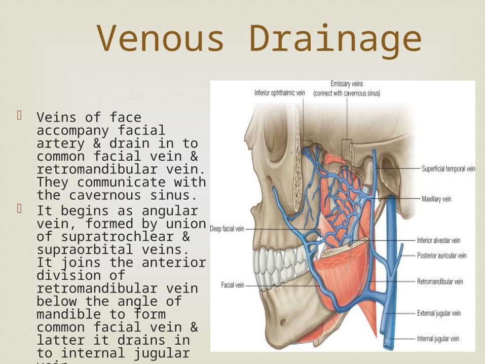
Veins of face accompany facial artery & drain in to common facial vein & retromandibular vein. They communicate with the cavernous sinus.
It begins as angular vein, formed by union of supratrochlear & supraorbital veins. It joins the anterior division of retromandibular vein below the angle of mandible to form common facial vein & latter it drains in to internal jugular vein
Lymphatic Drainage
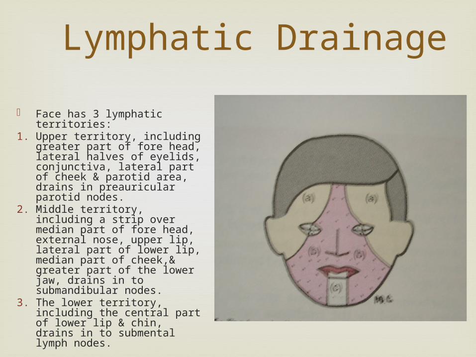
Face has 3 lymphatic territories:
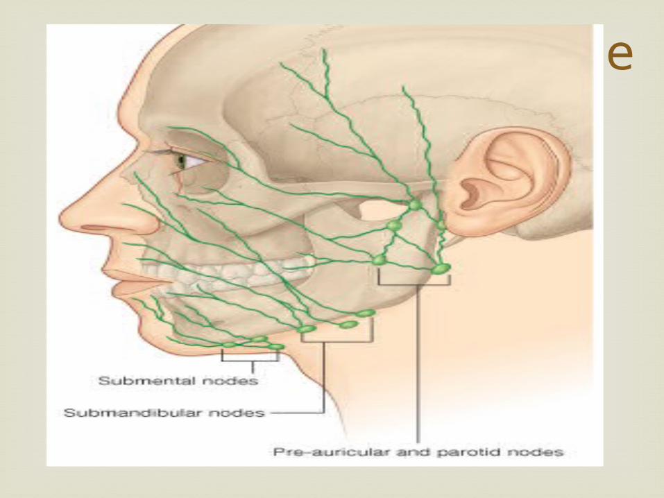
1. Upper territory, including greater part of fore head, lateral halves of eyelids, conjunctiva, lateral part of cheek & parotid area, drains in preauricular parotid nodes.
2. Middle territory, including a strip over median part of fore head, external nose, upper lip, lateral part of lower lip, median part of cheek,& greater part of the lower jaw, drains in to submandibular nodes.
3. The lower territory, including the central part of lower lip & chin, drains in to submental lymph nodes.
Lymphatic Drainage
Nerve Supply Of Face: Facial Nerve
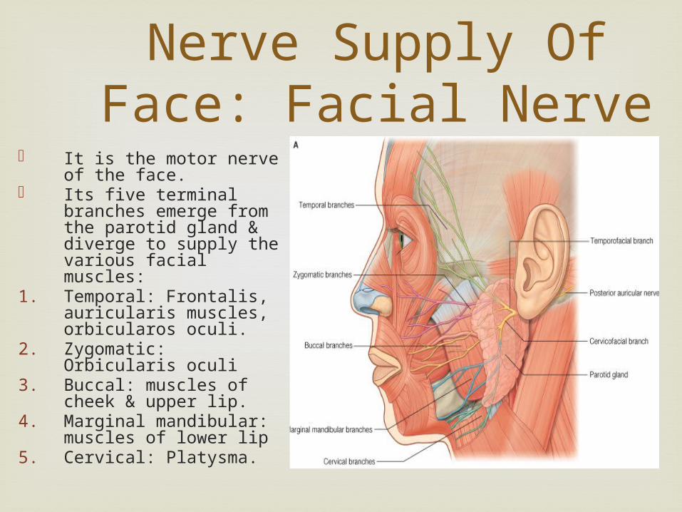
It is the motor nerve of the face.
Its five terminal branches emerge from the parotid gland & diverge to supply the various facial muscles:
1. Temporal: Frontalis, auricularis muscles, orbicularos oculi.
2. Zygomatic: Orbicularis oculi
3. Buccal: muscles of cheek & upper lip.
4. Marginal mandibular: muscles of lower lip
5. Cervical: Platysma.
Nerve Supply Of Face
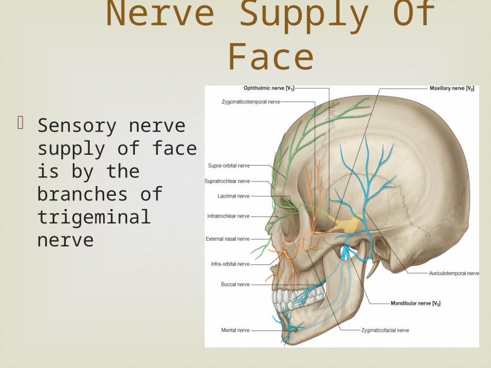
Sensory nerve supply of face is by the branches of trigeminal nerve
APPLIED ANATOMY
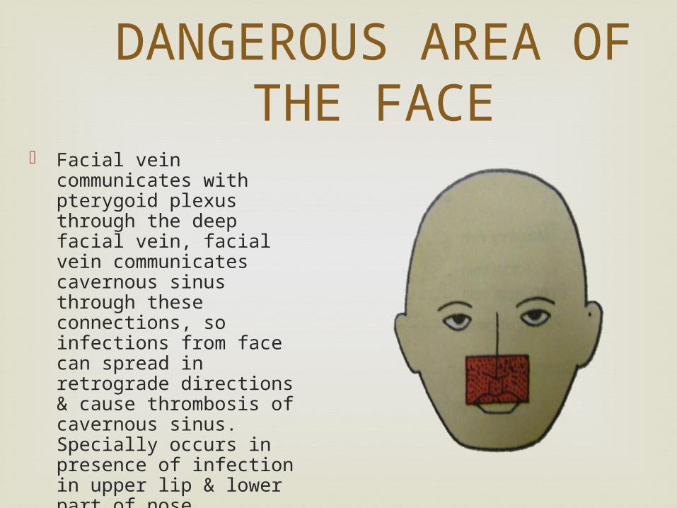
Facial vein communicates with pterygoid plexus through the deep facial vein, facial vein communicates cavernous sinus through these connections, so infections from face can spread in retrograde directions & cause thrombosis of cavernous sinus. Specially occurs in presence of infection in upper lip & lower part of nose.
DANGEROUS AREA OF THE FACE
Wounds of the face bleed profusely but heal
rapidly. The results of the plastic surgery on the face are excellent for the same reason.
Laxity greater part of the skin facilitates rapid spread of spread of oedema. Renal oedema appears first in the eyelids and face before spreading to the other parts of the body.
Boils in the nose and ear are acutely painfull
due to the fixity of the skin to the underlying cartilage.

INFRANUCLEAR LESIONS: Infranuclear lesion of the facial nerve known as bell’s palsy, the whole of the face of the same side get paralysed. - asymmentrical - drawn up to the normal side. - affected side is motionless. - wrinkles in the forehead disappears. - eye cannot be closed - smile draws the corners of the normal side.
Bell’s palsy
Supranuclear lesions of the facial nerve only
the lower part of the opposite side of the face is paralysed. The upper part of which is the frontalis and orbicularis oculi escapes due to the bilateral representation in the cerebral cortex.
Supranuclear lesion
The sensory distribution of the trigeminal
nerve explains why headache is a uniformly common symptoms in involvements of the nose, the paranasal air sinuses, refractive error in the eyes, glucoma and infections of the meningitis as in meningitis.
They may involve one or more of the divisions of the
trigeminal nerve. It causes very severe burning and scalding pain
along the distribution of the affected nerve. Pain is relieved by a) injection 90% alcohol in the affected division of the trigeminal ganglion. b) by sectioning the affected nerve, the main sensory root, or the spinal trait of the trigeminal nerve which is situated superfically in the medulla. This procedure is called medullary tractotomy.
Trigeminal neuralgia
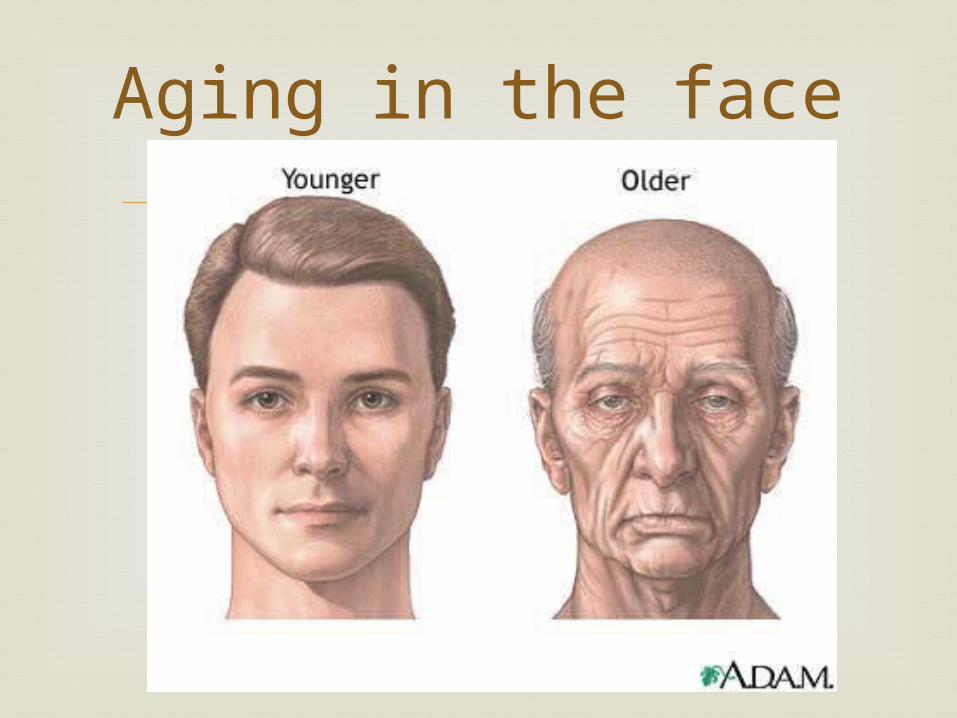
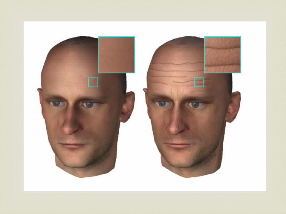
Aging in the face
The study of facial expressions is not only fascinating, but it has
practical uses as well. A good understanding of the information could allow people to
understand the feelings of others better, and help them in communicating their own.
A more detailed knowledge of the actual expressions themselves, and the signs of facial deceit could be useful for therapists who need to know how a patient is really feel.
Actors could use the information to help them match the emotions of their character.
In conclusion, facial expressions remain windows to the emotions that are universal across cultures, and have a great deal more relevance in every day life than most people give them credit.
Conclusion
Henry Gray, Susan Standring. Gray's Anatomy:
The Anatomical Basis of Clinical Practice. 2005.
Chaurasia. Textbook of Human Anatomy. 2008. Guyton & Hall. Textbook of Medical
Physiology . 11th edn. Ekman, P. & Friesen, W.V. (1975) Unmasking
the Face. Englewood Cliffs: Prentice-Hall.
references

Thank you