Embed Size (px)
Citation preview
Musculoskeletal Diagnostic Techniques
ATHT 305 Chapter 5
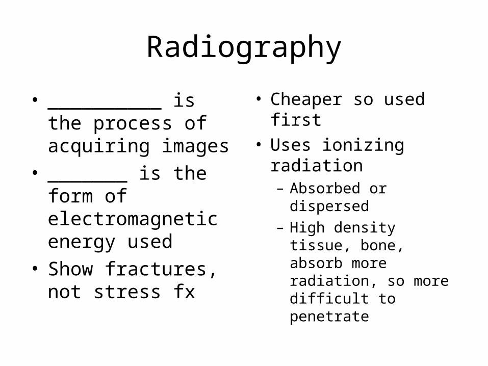
Radiography
• __________ is the process of acquiring images
• _______ is the form of electromagnetic energy used
• Show fractures, not stress fx
• Cheaper so used first• Uses ionizing
radiation– Absorbed or dispersed– High density tissue,
bone, absorb more radiation, so more difficult to penetrate
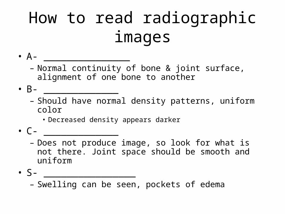
How to read radiographic images
• A- _______________– Normal continuity of bone & joint surface, alignment of
one bone to another
• B- _____________– Should have normal density patterns, uniform color
• Decreased density appears darker
• C- _____________– Does not produce image, so look for what is not there.
Joint space should be smooth and uniform
• S- ________________– Swelling can be seen, pockets of edema

Techniques
• Stress radiograph- stretch a ligament to determine amount of excessive movement
• Contrast imaging-injection of dye
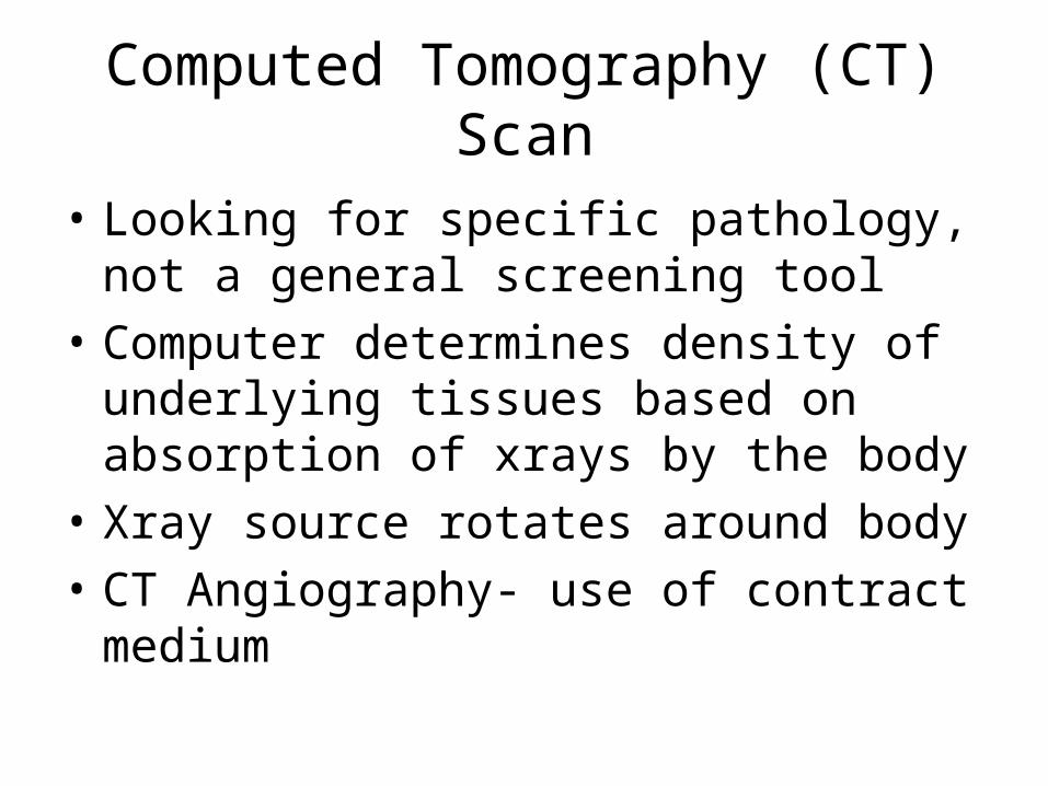
Computed Tomography (CT) Scan
• Looking for specific pathology, not a general screening tool
• Computer determines density of underlying tissues based on absorption of xrays by the body
• Xray source rotates around body
• CT Angiography- use of contract medium


Magnetic Resonance Imaging (MRI)
• For _________________• Machine creates a magnetic field, causing the
body’s hydrogen nuclei to align with the magnetic axis
• No potential harmful side effects like xray, unless you’re claustrophobic
• _________________ detect metabolic changes in brain
• Magnetic Resonance Angiography (MRA)- used to study blood vessels

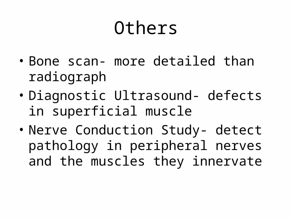
Others
• Bone scan- more detailed than radiograph
• Diagnostic Ultrasound- defects in superficial muscle
• Nerve Conduction Study- detect pathology in peripheral nerves and the muscles they innervate


![SYN ©EU ATHt^RUM - DiVA portal1145201/FULLTEXT01.pdf•*Hro) w FamiliarilTimum Graecis, in defignanda Ytligionc y S-py]](https://img.pdfslide.net/doc/110x75/5f7024ecd3ad193ed546c9af/syn-eu-athtrum-diva-1145201fulltext01pdf-ahro-w-familiariltimum-graecis.jpg)