Embed Size (px)
Citation preview
Musculoskeletal Radiology
Part one
• Imaging Techniques in Orthopaedics – Conventional Radiography– Fluoroscopy– Computed Tomography– Arthrography– Angiography– Ultrasound– Scintigraphy– Magnetic Resonance Imaging
Part two
• Upper limb MSK anatomy
• Lower limb MSK anatomy
Imaging Techniques in Orthopaedics.
• Use of Radiological Techniques methods in evaluating the presence, type, and extents of various bone, joints and soft tissue abnormality.
• Indications
• Limitations
• Appropriate imaging approach
The question
• “What modalities should I use for this particular problem” is frequently asked by Radiologists and Orthopaedic Surgeons alike.
• Conventional Radiograph
• The choice of imaging technique is dictated by the type of suspected abnormality
CONVENTIONAL RADIOGRAPHY:
• The most frequently used modality for evaluation of bone and joint disorder
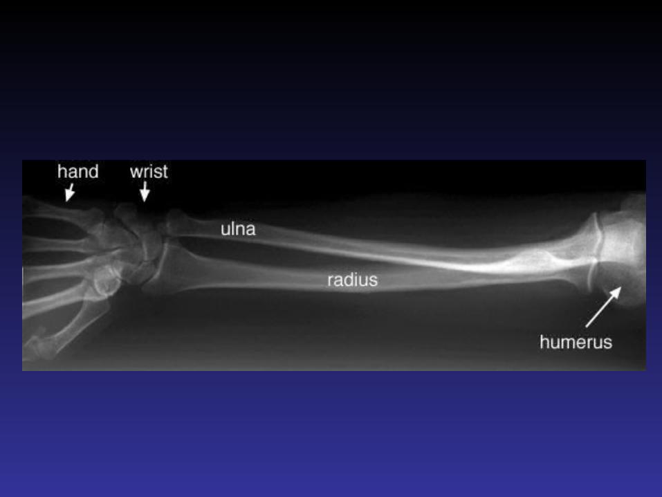
• The radiologist should obtain at least two (2) views of the bone involved at 90° angles to each other
• with each view including two adjacent joints
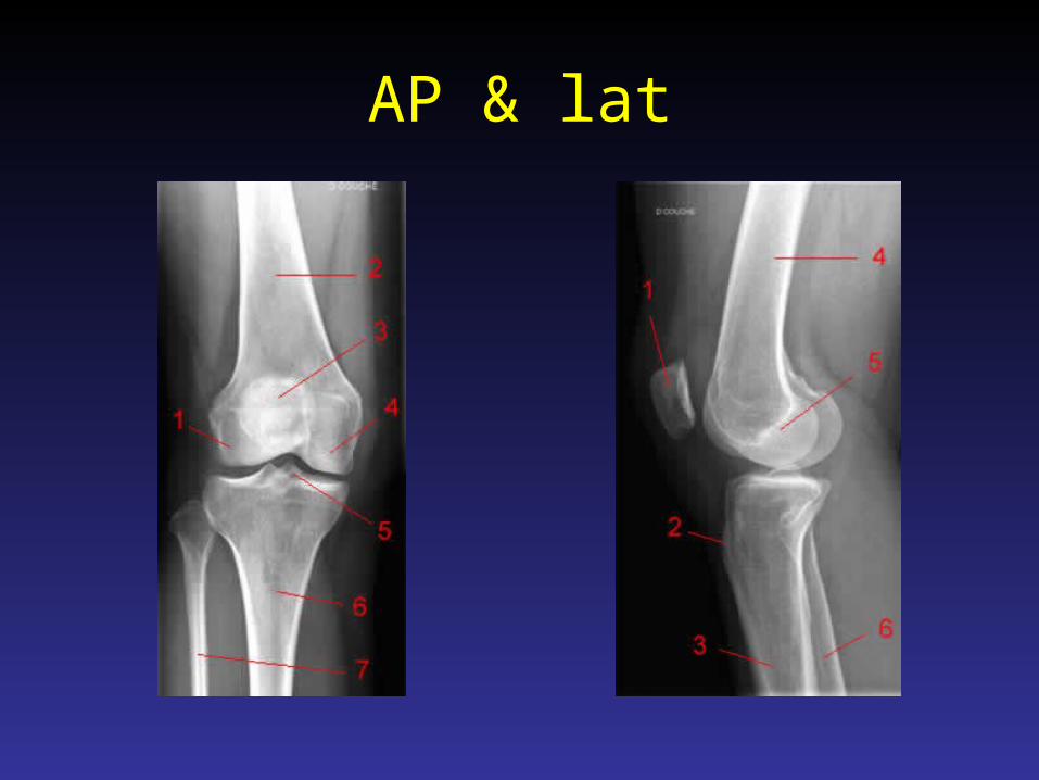
AP & lat
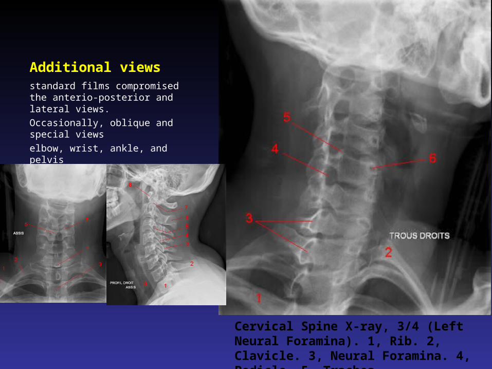
Additional viewsstandard films compromised the anterio-posterior and lateral views.
Occasionally, oblique and special views
elbow, wrist, ankle, and pelvis
Cervical Spine X-ray, 3/4 (Left Neural Foramina). 1, Rib. 2, Clavicle. 3, Neural Foramina. 4, Pedicle. 5, Trachea.
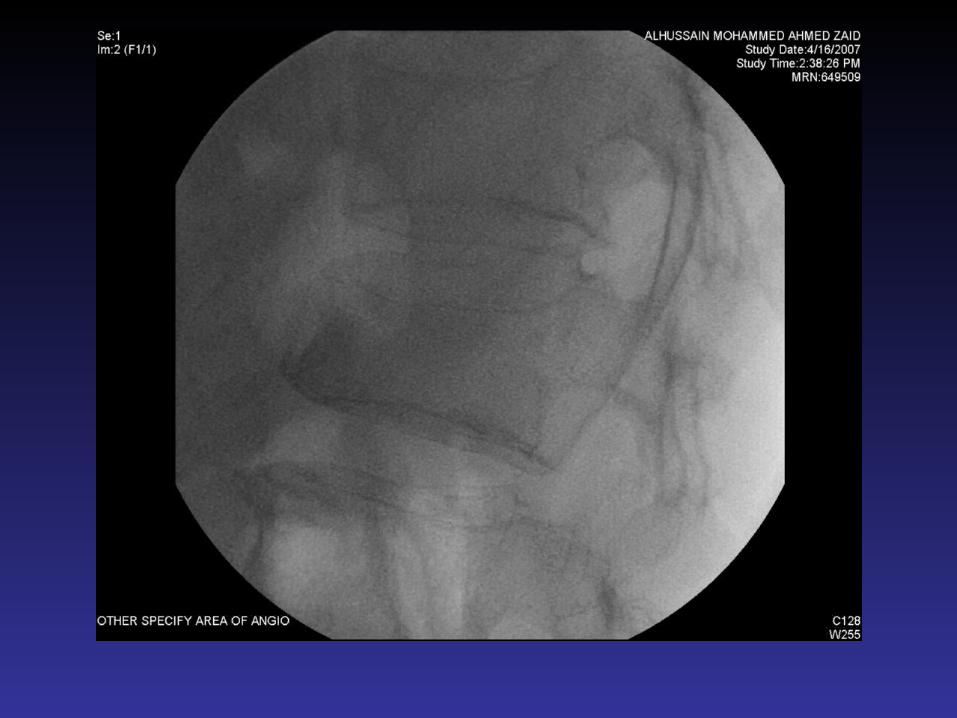
FLUOROSCOPY:
• Many radiologic procedures – Arthrography– Tenography– Versography– Arteriography – Percutaneous Bone or Soft Tissue Biopsy.
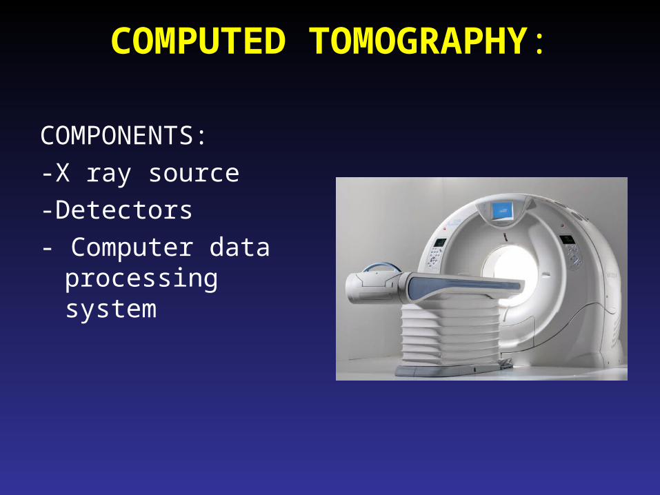
COMPUTED TOMOGRAPHY:
COMPONENTS:
-X ray source
-Detectors
- Computer data processing system
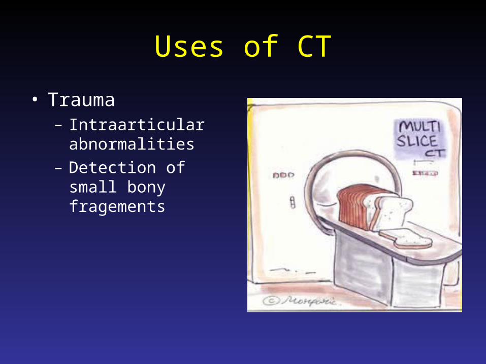
Uses of CT
• Trauma– Intraarticular
abnormalities– Detection of small
bony fragements
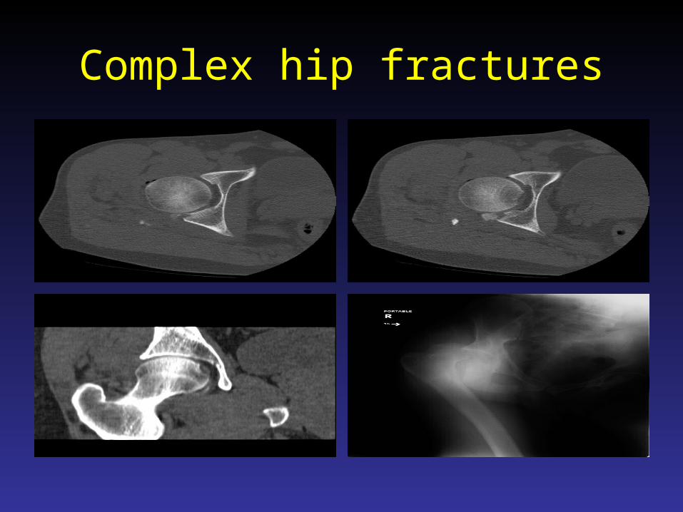
Complex hip fractures
CT Vs. Xray
• Advantages:– Excellent contrast
resolution.– Measures the tissue
attenuation coefficient– Obtain transaxial
images– Reformation
• Disadvantages:– Radiation– Inability to make a
specific diagnosis
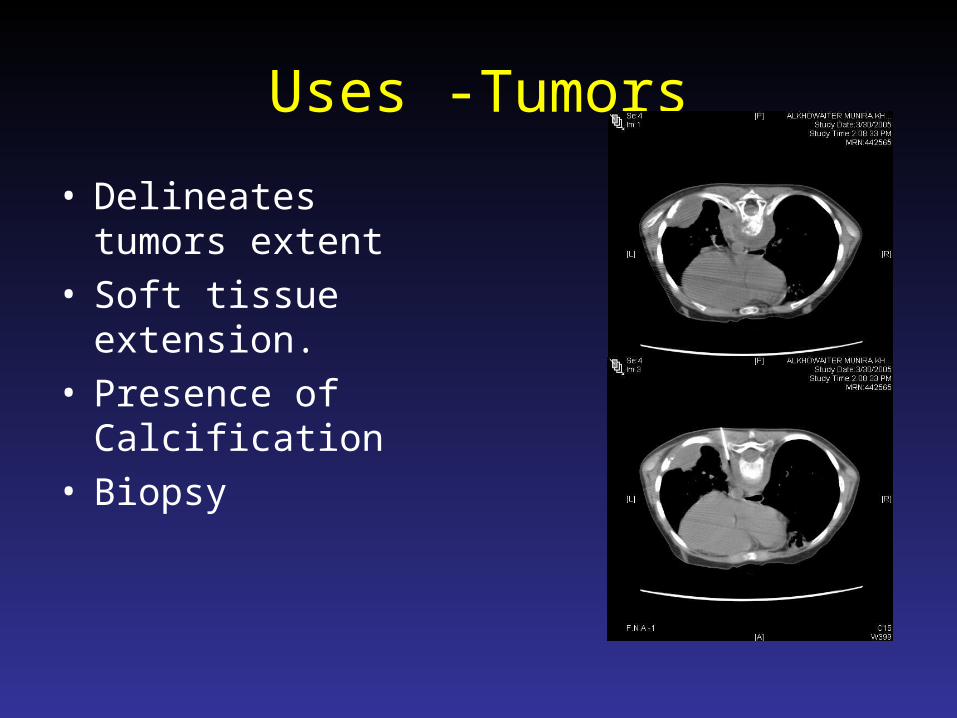
Uses -Tumors
• Delineates tumors extent
• Soft tissue extension.• Presence of
Calcification• Biopsy

Arthrography
• Arthrography is introduction of contrast agent positive contrast iodine iodide solution negative contrast, air or combination of both into the joint space.
• Advantages:– Simple– Effective
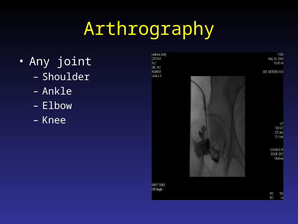
Arthrography
• Any joint– Shoulder– Ankle– Elbow– Knee
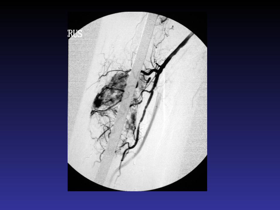
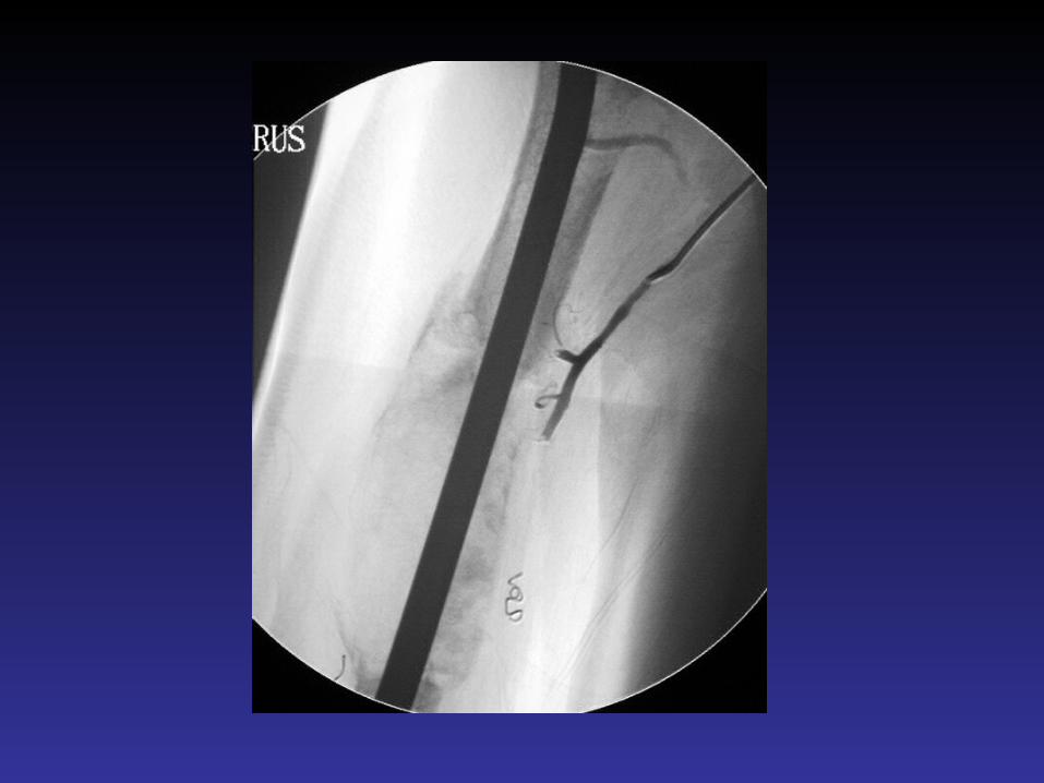
Angiography
• Advantages:1. Map-out bone lesions 2. Demonstrate the vascularity of the lesion. 3. Demonstrate the vascular supply of a
tumor4. Locate vessels suitable for pre operative
intraarterial chemotherapy. 5. Demonstrating the area suitable for open
biopsy.
ULTRASOUND:
• Rarely used • Advantages:
– inexpensive– allows comparison with
the opposite side, normal side
– uses no ionizing radiation,
– performed at bed side or in the operating room.
– It is a non invasive modality
Applications
• Evaluation of the rotator cuff
• Injuries to various tendons, e.g. the achilles tendons.
• Evaluation of the infant hip for which ultrasound has become the imaging modality of choice
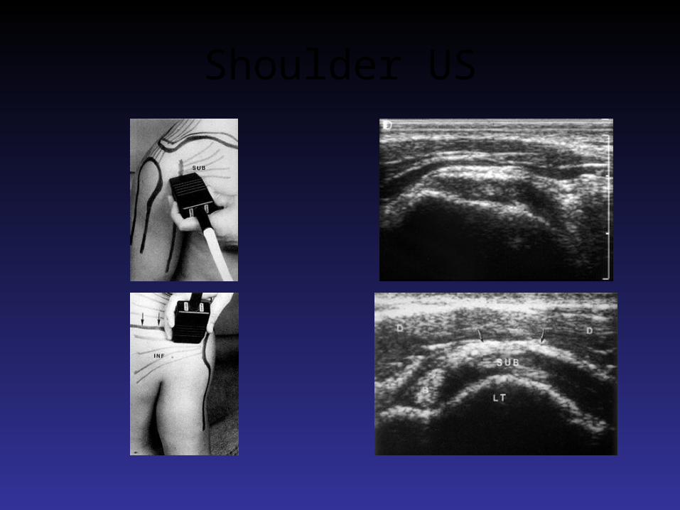
Shoulder US
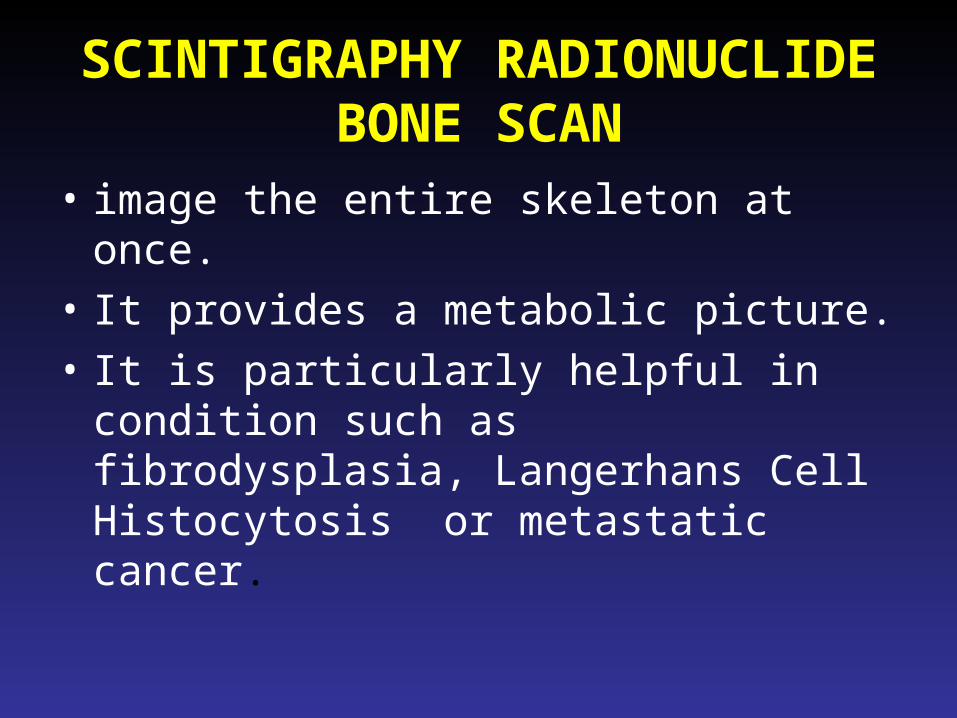
SCINTIGRAPHY RADIONUCLIDE BONE SCAN
• image the entire skeleton at once.
• It provides a metabolic picture.
• It is particularly helpful in condition such as fibrodysplasia, Langerhans Cell Histocytosis or metastatic cancer.
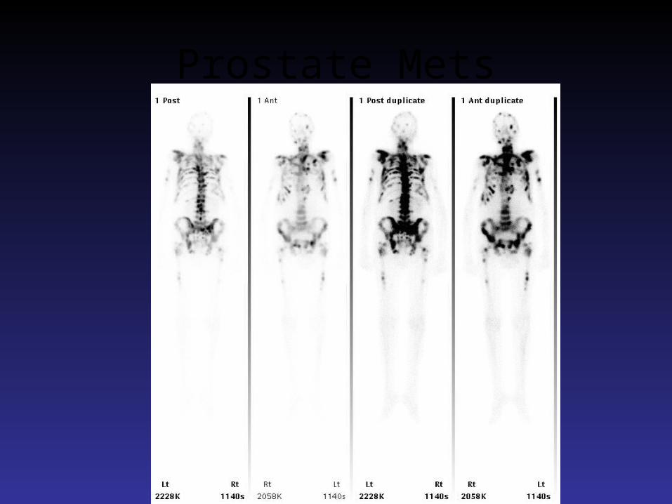
Prostate Mets
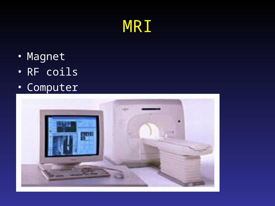
MRI
• Magnet• RF coils• Computer
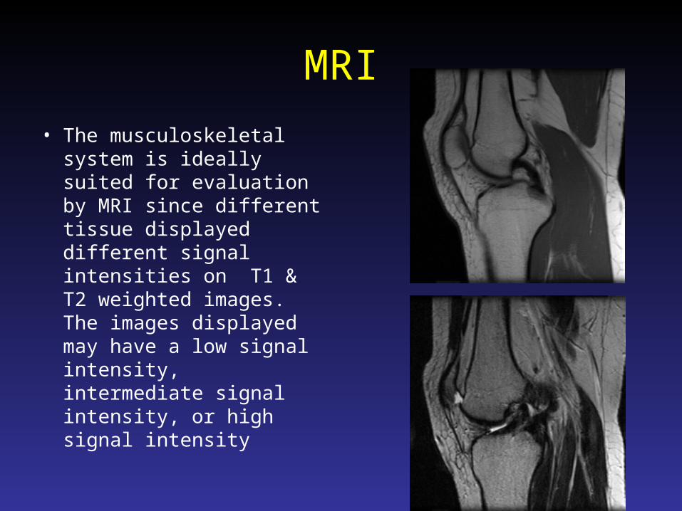
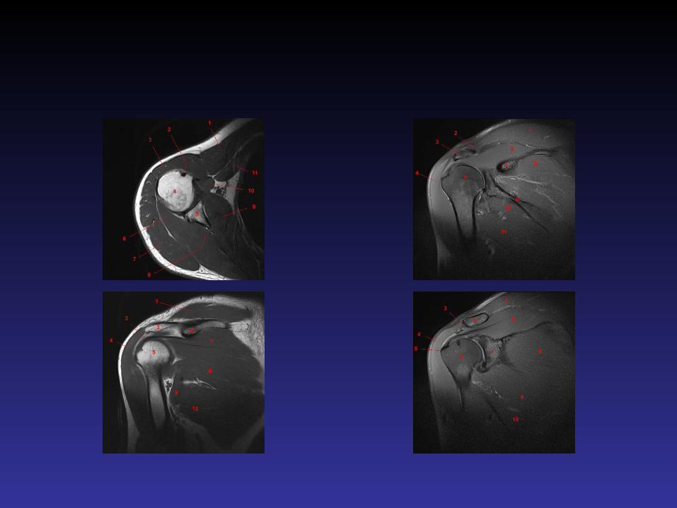
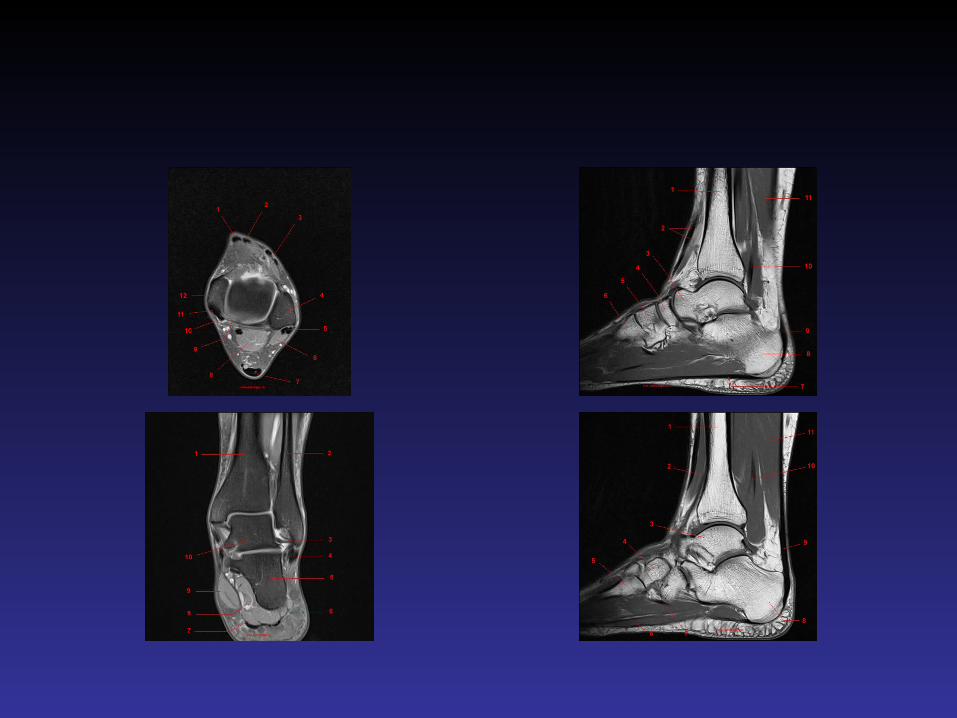
MRI
• The musculoskeletal system is ideally suited for evaluation by MRI since different tissue displayed different signal intensities on T1 & T2 weighted images. The images displayed may have a low signal intensity, intermediate signal intensity, or high signal intensity
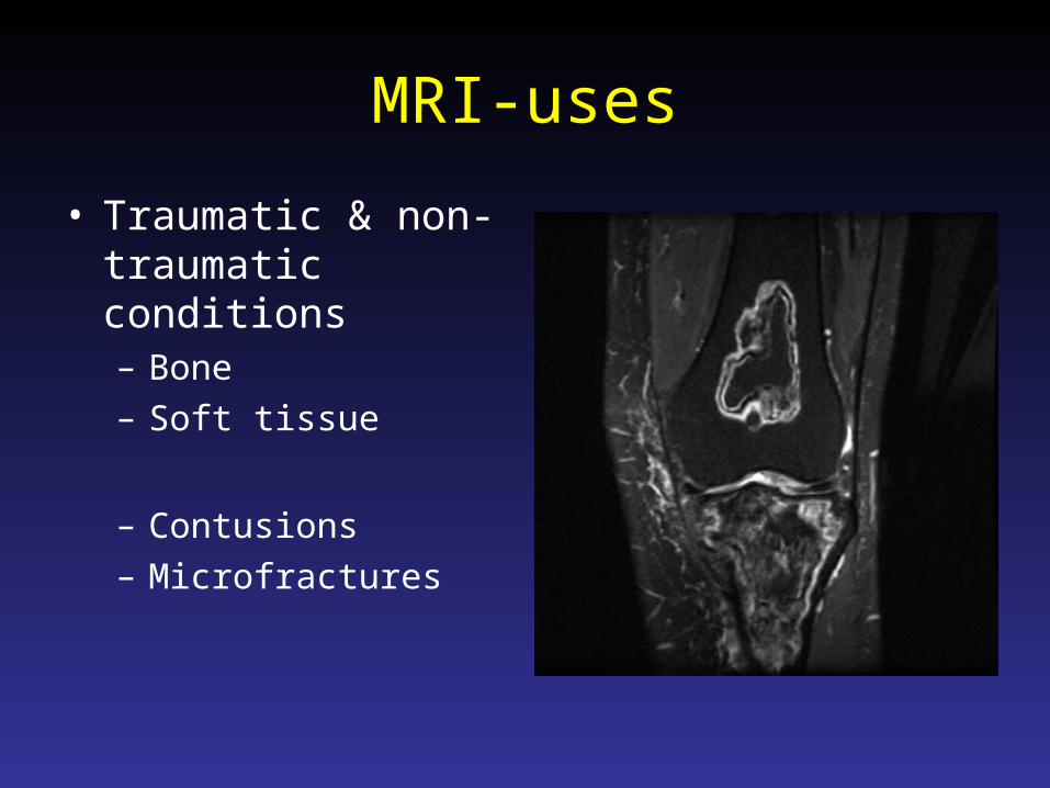
MRI-uses
• Traumatic & non-traumatic conditions– Bone– Soft tissue
– Contusions– Microfractures
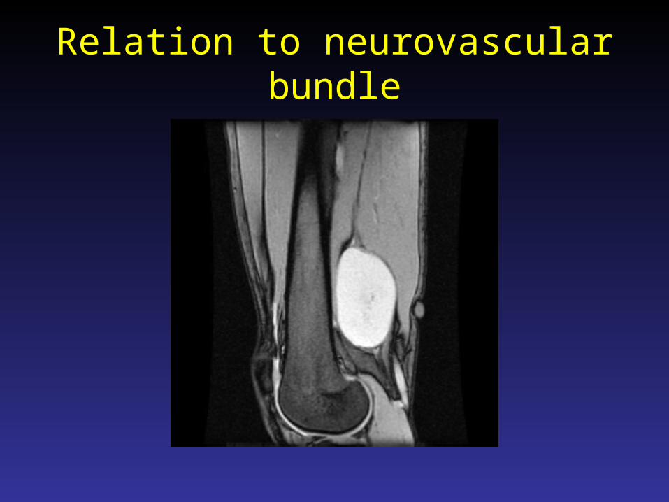
Relation to neurovascular bundle
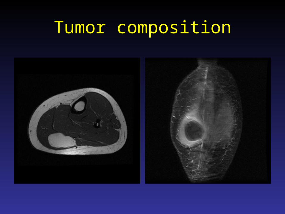
Tumor composition
MRI Contraindications
• ABSOLOUTE– Patients with cardiac pacemakers– Cerebral aneurysm clips
• RELATIVE:– Claustrophobia.
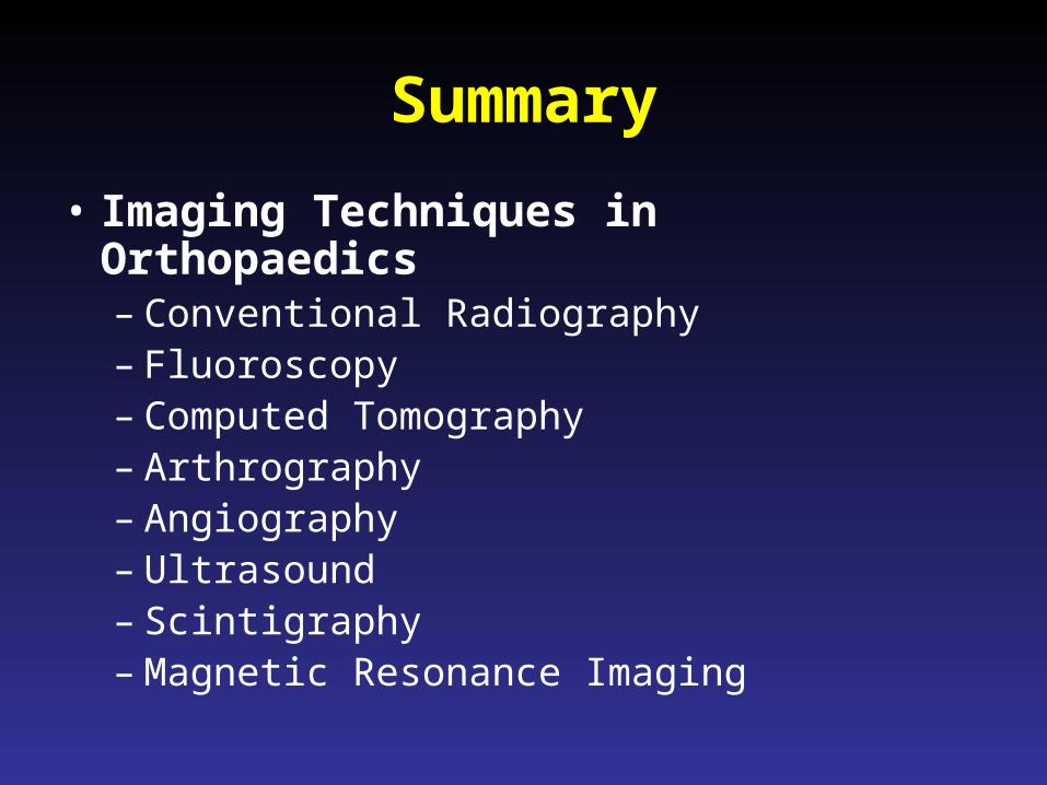
Summary
• Imaging Techniques in Orthopaedics– Conventional Radiography– Fluoroscopy– Computed Tomography– Arthrography– Angiography– Ultrasound– Scintigraphy– Magnetic Resonance Imaging
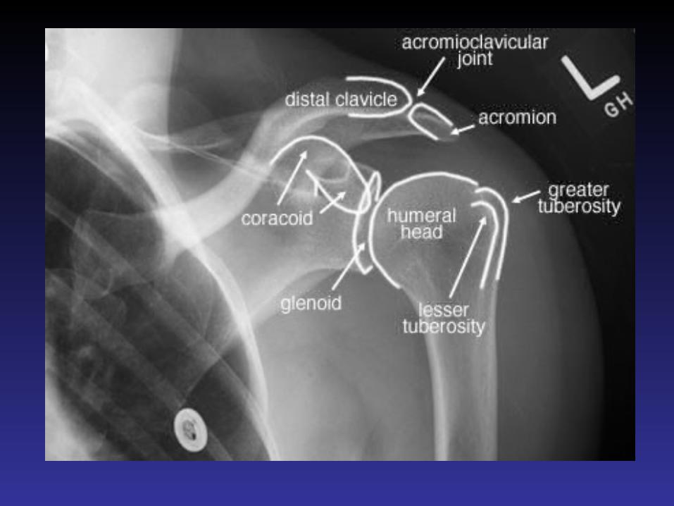
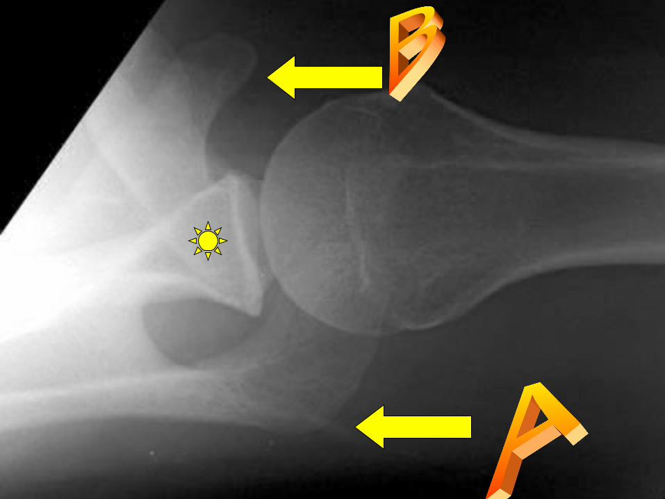
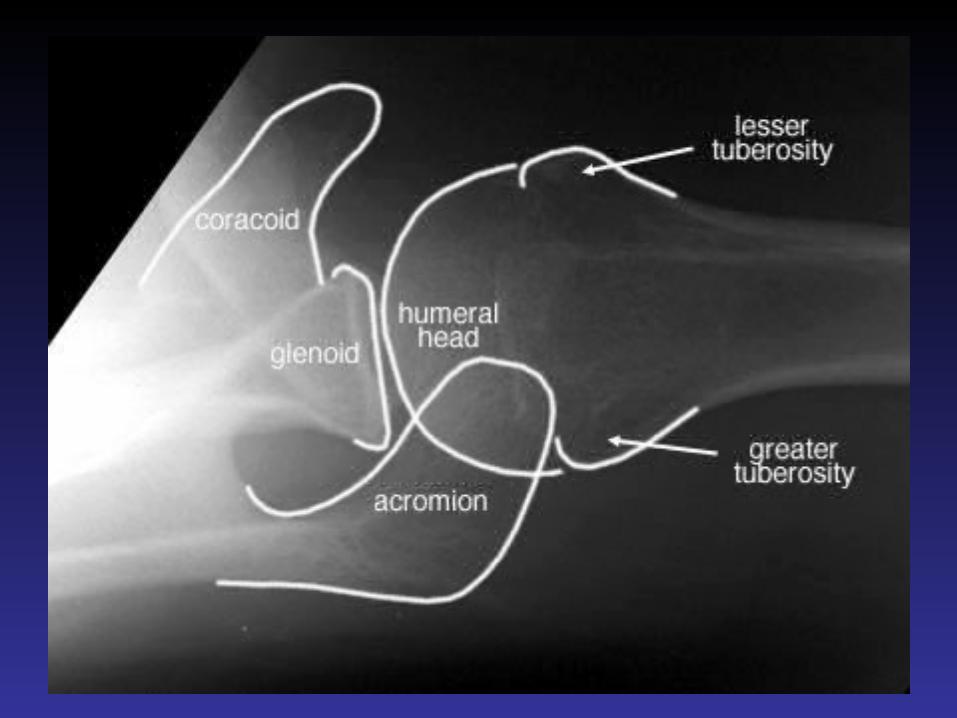
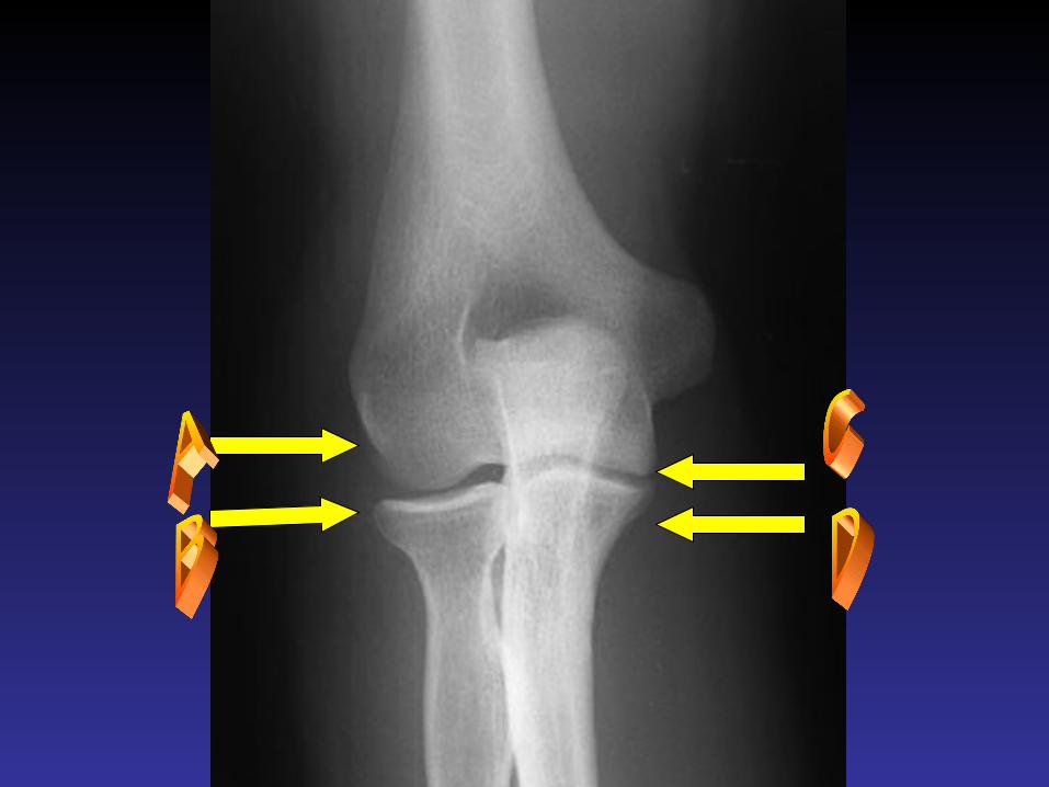
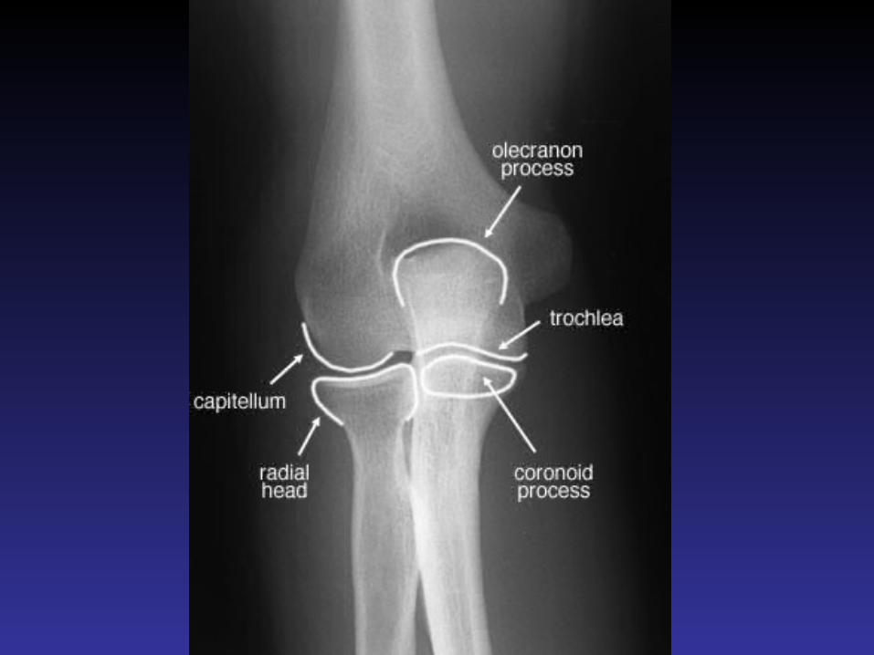
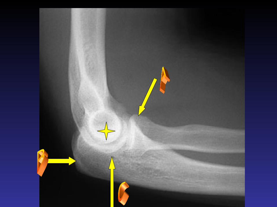
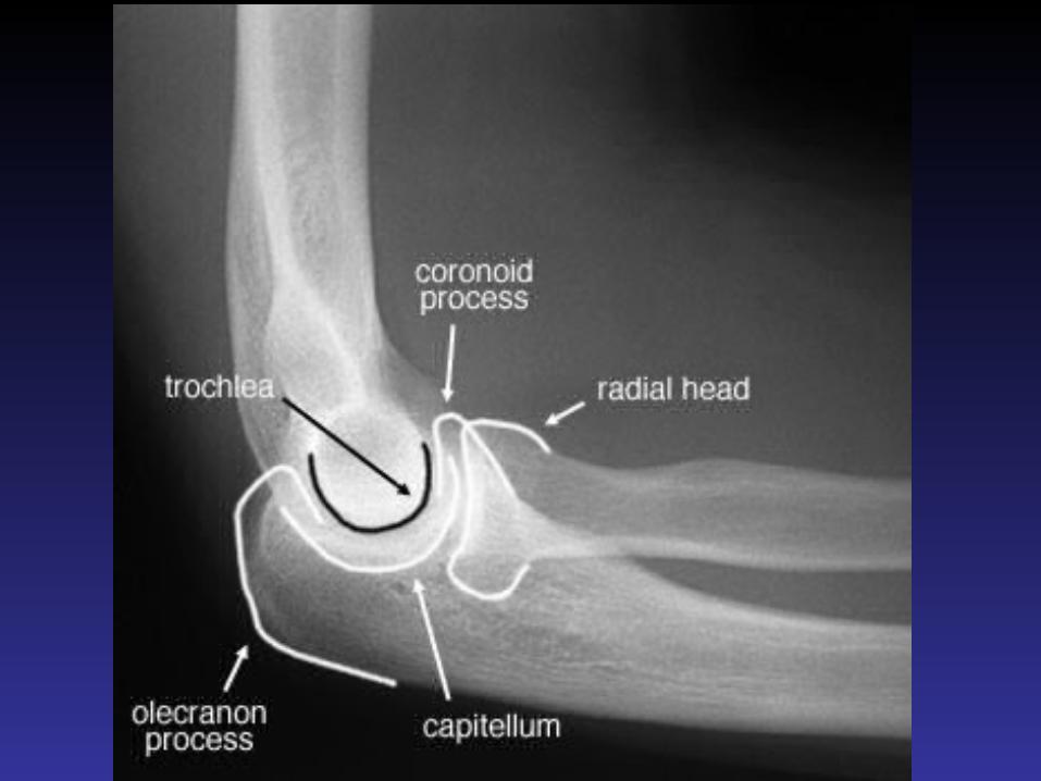
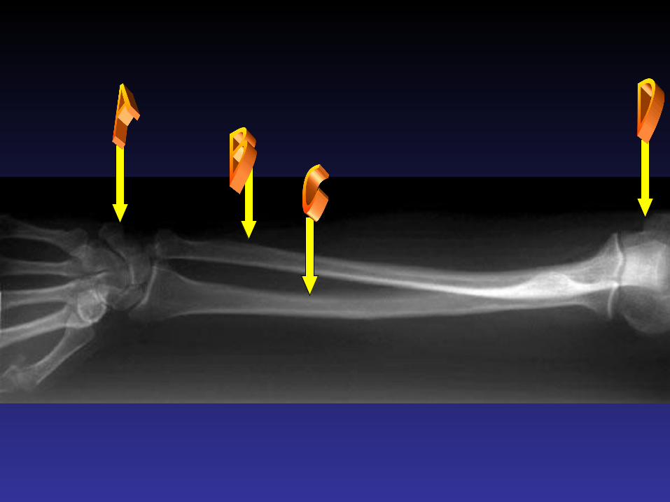
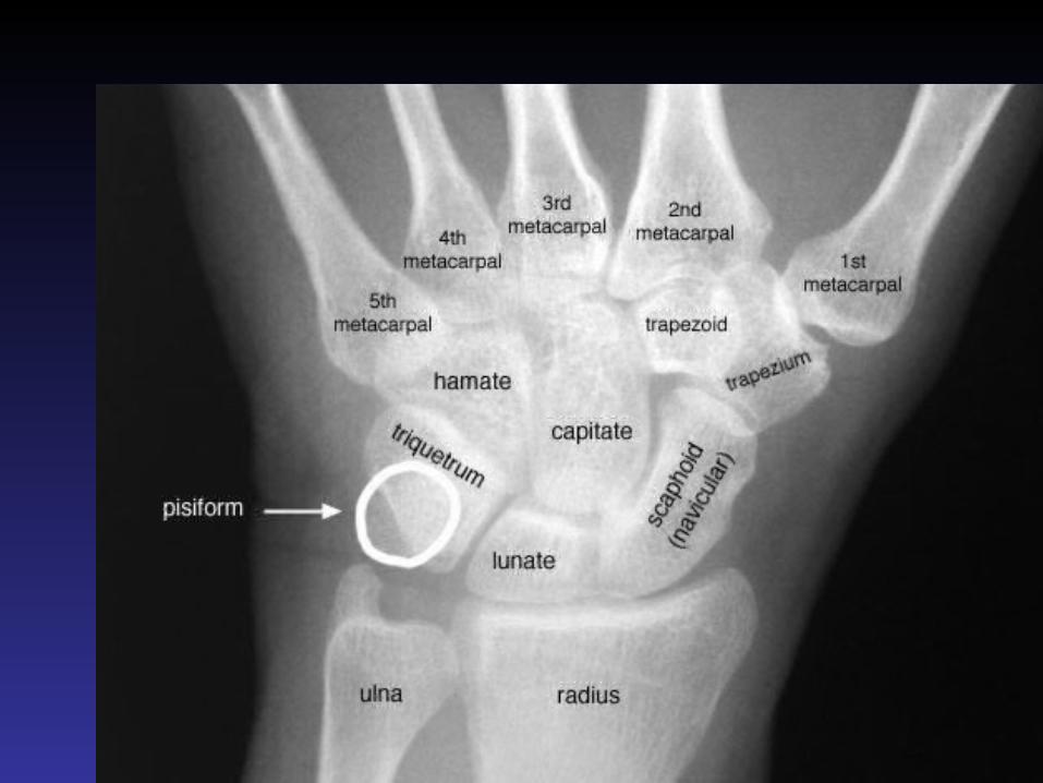
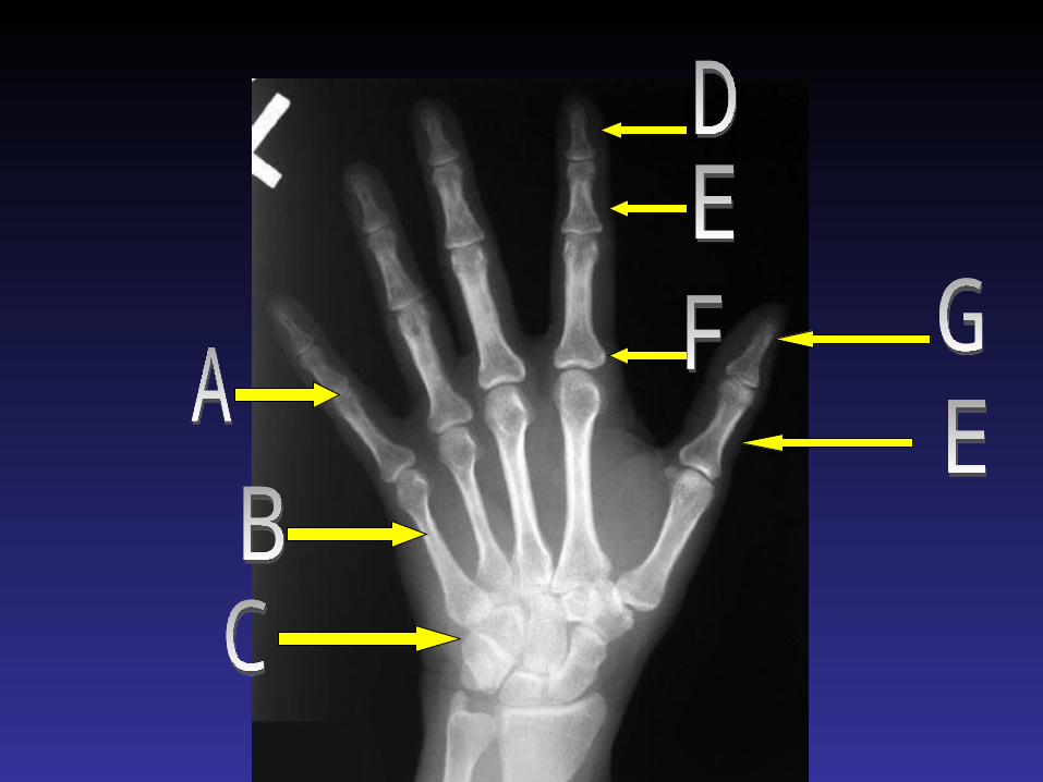
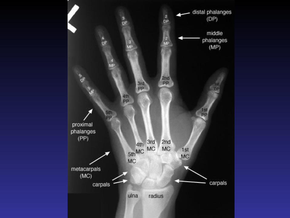
Radiologic Anatomy of the Musculoskeletal System
Upper limb

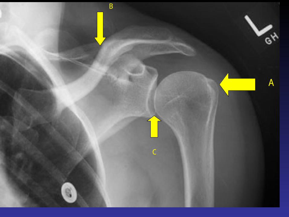
A
B
C


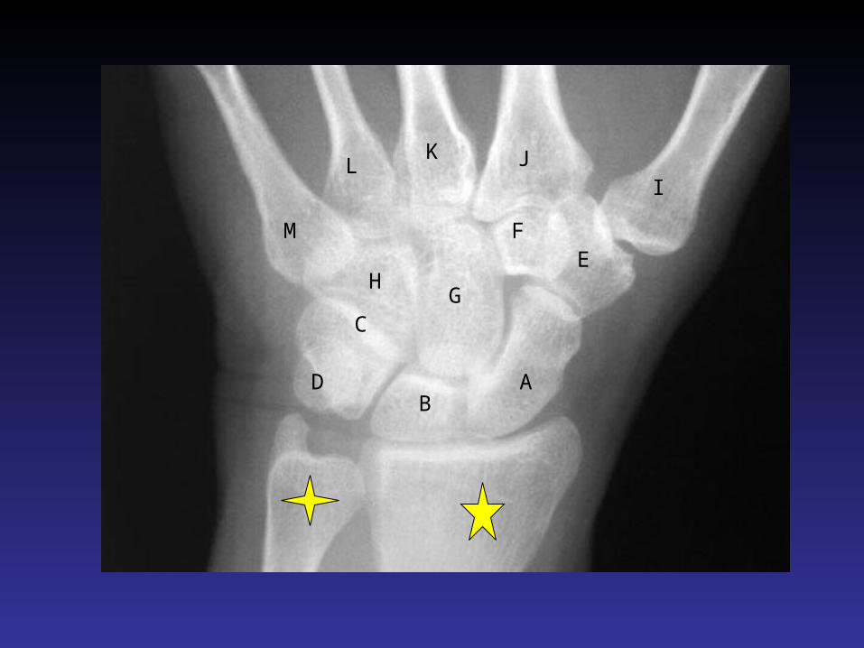
AB
C
D
EF
GH
IJK
L
M

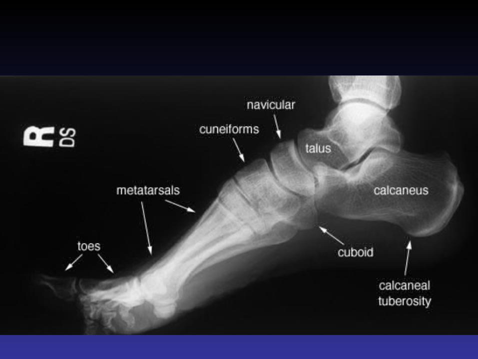
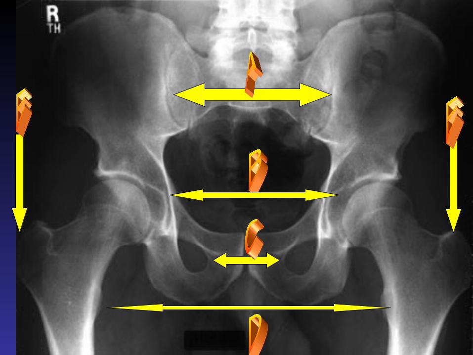
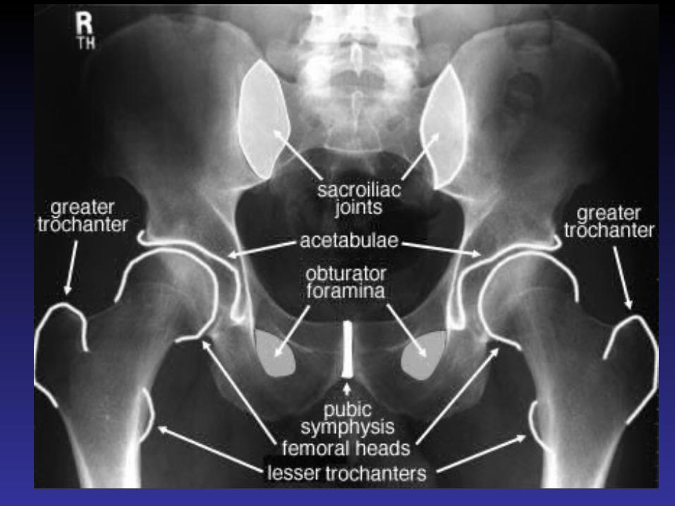
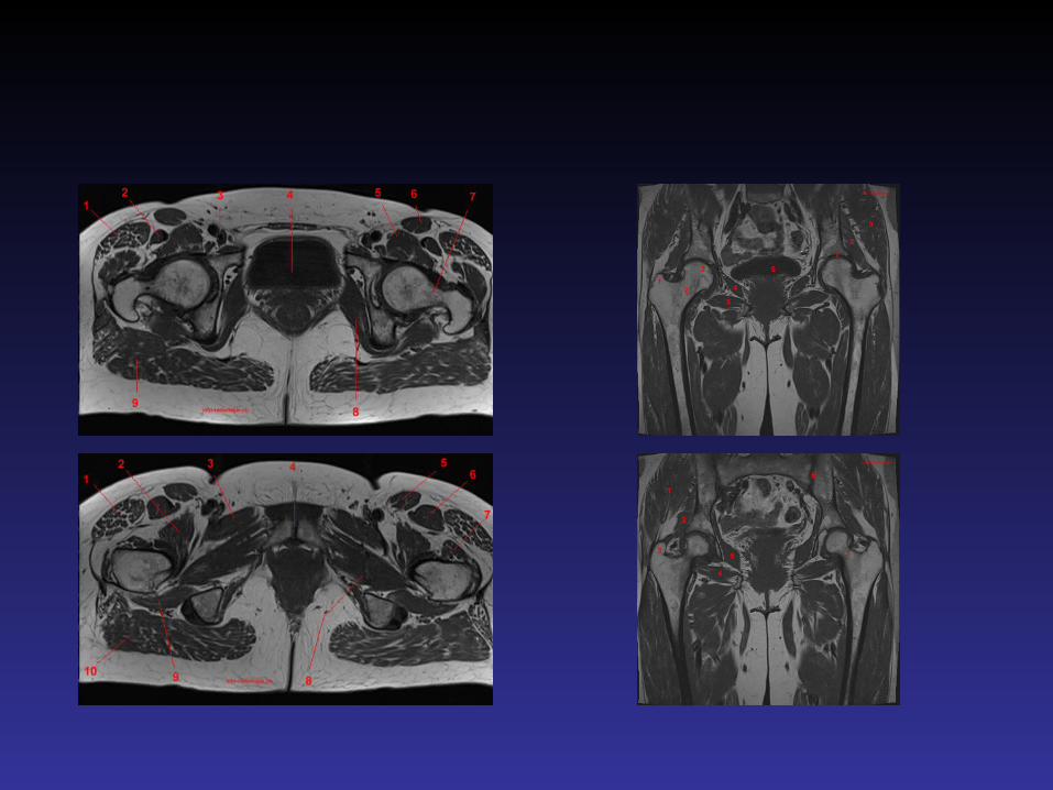
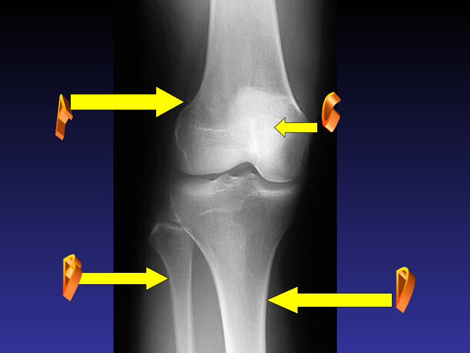
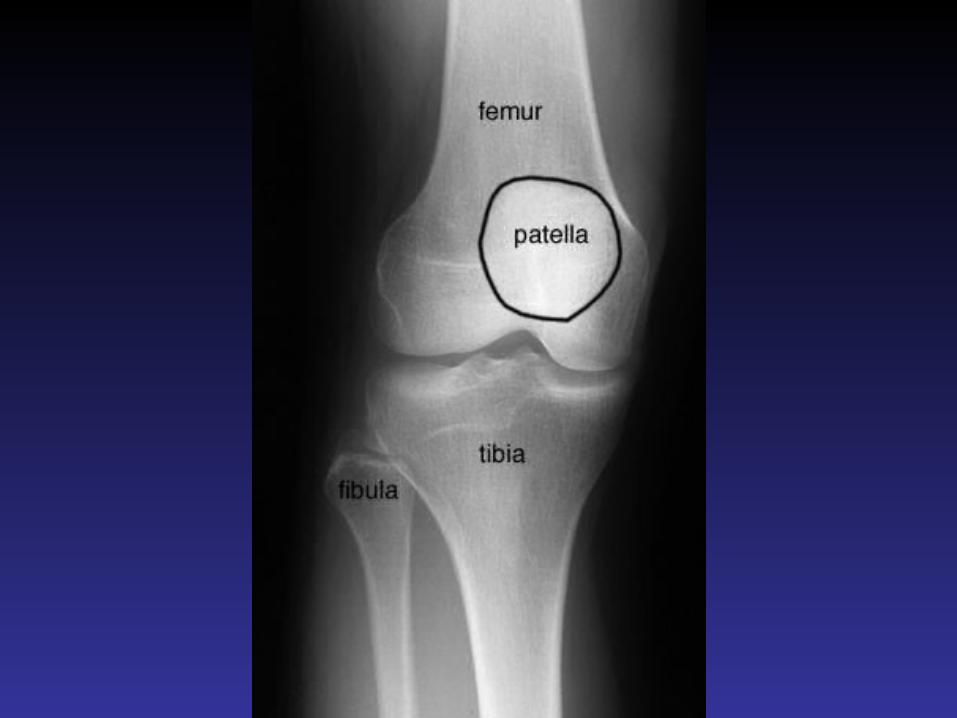
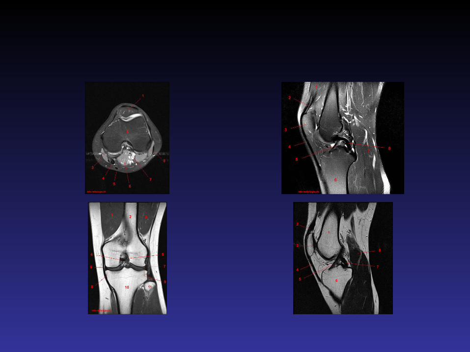
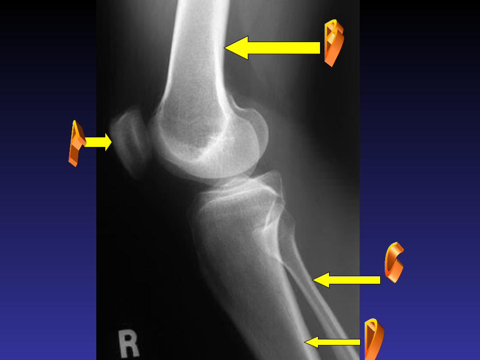
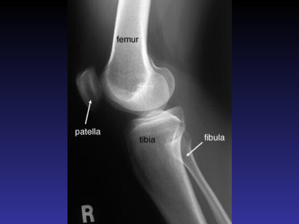
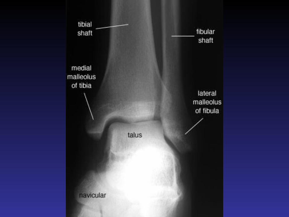
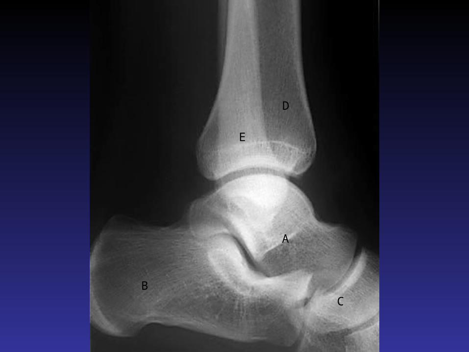
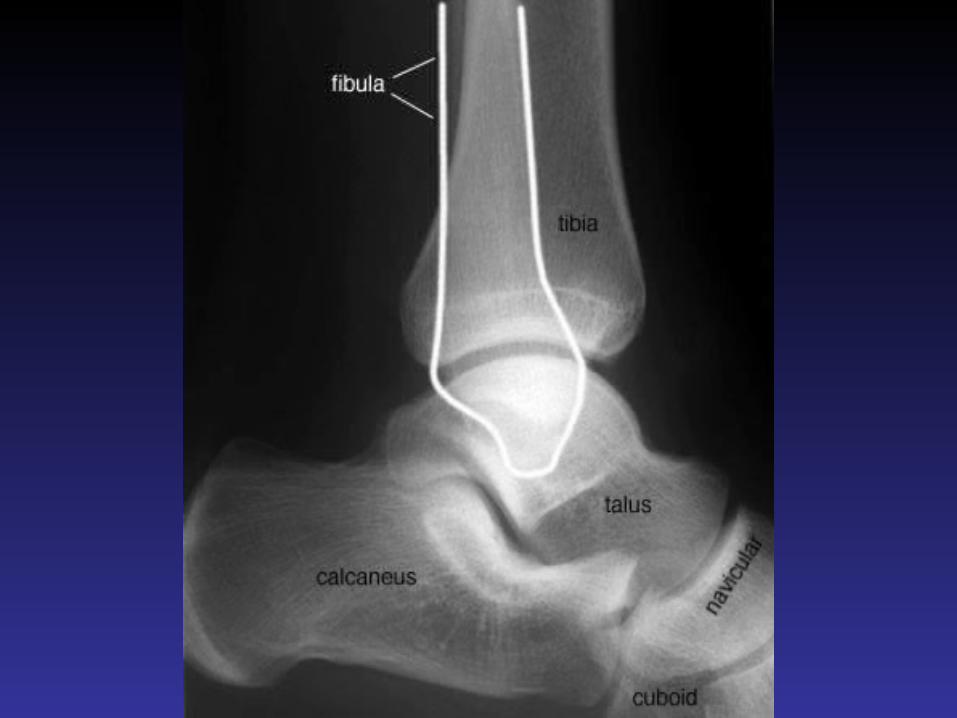
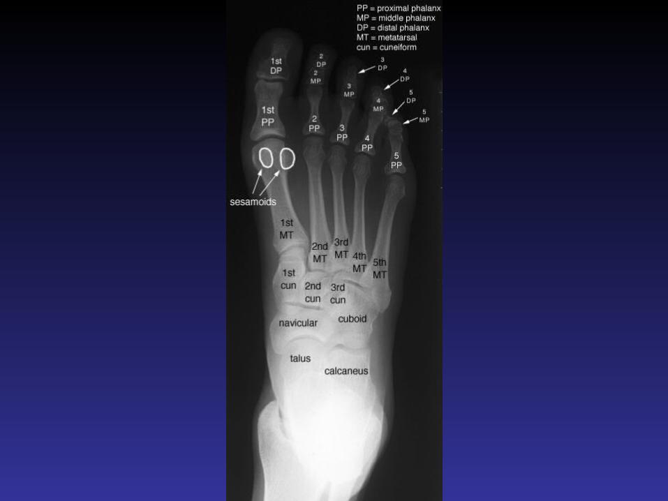
Radiologic Anatomy of the Musculoskeletal System
Lower limb


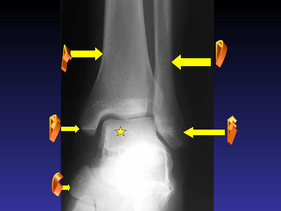

A
BC
D
E

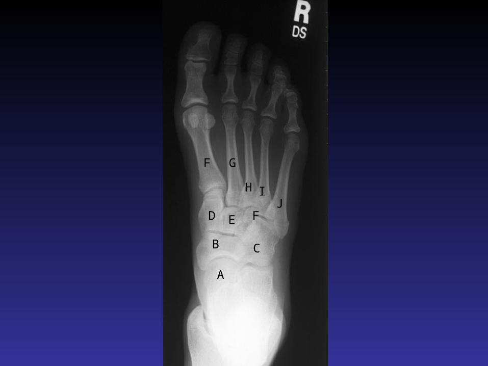
A
B C
D E F
F G
H IJ

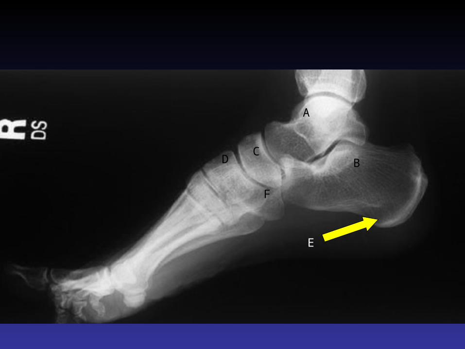
A
BC
D
E
F