Embed Size (px)
Citation preview
Cianca Atlas_00624_PTR_00_i-xviii_FM_09-14-17.indd Page PB 15/09/17 12:56 AM Cianca Atlas_00624_PTR_00_i-xviii_FM_09-14-17.indd Page i 15/09/17 12:56 AM
Mu s c u l o s k e l e ta l ult r a s o u n d cr o s s-Se c t i o n a l an at o M y
Cianca Atlas_00624_PTR_01_1-44_09-04-17.indd Page 6 13/09/17 7:03 PM Cianca Atlas_00624_PTR_01_1-44_09-04-17.indd Page 7 13/09/17 7:03 PMCianca Atlas_00624_PTR_00_i-xviii_FM_09-14-17.indd Page vi 15/09/17 12:56 AM Cianca Atlas_00624_PTR_00_i-xviii_FM_09-14-17.indd Page PB 15/09/17 12:56 AMCianca Atlas_00624_PTR_01_1-44_09-04-17.indd Page 6 13/09/17 7:03 PM Cianca Atlas_00624_PTR_01_1-44_09-04-17.indd Page 7 13/09/17 7:03 PM
This is a sample from MUSCULOSKELETAL ULTRASOUND CROSS-SECTIONAL ANATOMYVISIT THIS BOOK’S WEB PAGE BUY NOW
© Springer Publishing Company

Cianca Atlas_00624_PTR_00_i-xviii_FM_09-14-17.indd Page ii 15/09/17 12:56 AM Cianca Atlas_00624_PTR_00_i-xviii_FM_09-14-17.indd Page iii 15/09/17 12:56 AMCianca Atlas_00624_PTR_00_i-xviii_FM_09-14-17.indd Page ii 15/09/17 12:56 AM Cianca Atlas_00624_PTR_00_i-xviii_FM_09-14-17.indd Page iii 15/09/17 12:56 AMCianca Atlas_00624_PTR_01_1-44_09-04-17.indd Page 6 13/09/17 7:03 PM Cianca Atlas_00624_PTR_01_1-44_09-04-17.indd Page 7 13/09/17 7:03 PMCianca Atlas_00624_PTR_00_i-xviii_FM_09-14-17.indd Page vi 15/09/17 12:56 AM Cianca Atlas_00624_PTR_00_i-xviii_FM_09-14-17.indd Page PB 15/09/17 12:56 AMCianca Atlas_00624_PTR_01_1-44_09-04-17.indd Page 6 13/09/17 7:03 PM Cianca Atlas_00624_PTR_01_1-44_09-04-17.indd Page 7 13/09/17 7:03 PM
This is a sample from MUSCULOSKELETAL ULTRASOUND CROSS-SECTIONAL ANATOMYVISIT THIS BOOK’S WEB PAGE BUY NOW
© Springer Publishing Company
Cianca Atlas_00624_PTR_00_i-xviii_FM_09-14-17.indd Page ii 15/09/17 12:56 AM Cianca Atlas_00624_PTR_00_i-xviii_FM_09-14-17.indd Page iii 15/09/17 12:56 AMCianca Atlas_00624_PTR_00_i-xviii_FM_09-14-17.indd Page ii 15/09/17 12:56 AM Cianca Atlas_00624_PTR_00_i-xviii_FM_09-14-17.indd Page iii 15/09/17 12:56 AM
Mu s c u l o s k e l e ta l ult r a s o u n d cr o s s-se c t i o n a l an at o M y
John C. CianCa, MDAdjunct Associate Professor of Physical Medicine and RehabilitationBaylor College of MedicineUniversity of Texas Health Science CenterHuman Performance CenterHouston, Texas
ShounuCk i. Patel, Do, MMSInterventional Regenerative Orthopedic MedicineRegenexx-Health Link Medical CenterLos Angeles, California
An Imprint of Springer Publishing
Cianca Atlas_00624_PTR_01_1-44_09-04-17.indd Page 6 13/09/17 7:03 PM Cianca Atlas_00624_PTR_01_1-44_09-04-17.indd Page 7 13/09/17 7:03 PMCianca Atlas_00624_PTR_00_i-xviii_FM_09-14-17.indd Page vi 15/09/17 12:56 AM Cianca Atlas_00624_PTR_00_i-xviii_FM_09-14-17.indd Page PB 15/09/17 12:56 AMCianca Atlas_00624_PTR_01_1-44_09-04-17.indd Page 6 13/09/17 7:03 PM Cianca Atlas_00624_PTR_01_1-44_09-04-17.indd Page 7 13/09/17 7:03 PM
This is a sample from MUSCULOSKELETAL ULTRASOUND CROSS-SECTIONAL ANATOMYVISIT THIS BOOK’S WEB PAGE BUY NOW
© Springer Publishing Company
Cianca Atlas_00624_PTR_00_i-xviii_FM_09-14-17.indd Page iv 15/09/17 12:56 AM Cianca Atlas_00624_PTR_00_i-xviii_FM_09-14-17.indd Page v 15/09/17 12:56 AM
Visit our website at www.demosmedical.com
ISBN: 9781620700624ebook ISBN: 9781617052279
Acquisitions Editor: Beth BarryCompositor: diacriTech
Copyright © 2018 Springer Publishing Company. Demos Medical Publishing is an imprint of Springer Publishing Company, LLC.
All rights reserved. This book is protected by copyright. No part of it may be reproduced, stored in a retrieval system, or transmitted in any form or by any means, electronic, mechanical, photocopying, recording, or otherwise, without the prior written permission of the publisher.
Medicine is an ever-changing science. Research and clinical experience are continually expanding our knowledge, in particular our understanding of proper treatment and drug therapy. The authors, editors, and publisher have made every effort to ensure that all information in this book is in accordance with the state of knowledge at the time of production of the book. Nevertheless, the authors, editors, and publisher are not responsible for errors or omissions or for any consequences from application of the information in this book and make no warranty, expressed or implied, with respect to the contents of the publication. Every reader should examine carefully the package inserts accompanying each drug and should carefully check whether the dosage schedules mentioned therein or the contraindications stated by the manufacturer differ from the statements made in this book. Such examination is particularly important with drugs that are either rarely used or have been newly released on the market.
Library of Congress Cataloging-in-Publication DataNames: Cianca, John C., author. | Patel, Shounuck I., author.Title: Musculoskeletal ultrasound cross-sectional anatomy / John C. Cianca, Shounuck I. Patel.Description: New York: Demos Medical Publishing, [2017] | Includes bibliographical references and index.Identifiers: LCCN 2017011023| ISBN 9781620700624 | ISBN 9781617052279 (ebook)Subjects: | MESH: Musculoskeletal System—anatomy & histology | Anatomy, Cross-Sectional | Ultrasonography—methods | AtlasesClassification: LCC QM100 | NLM WE 17 | DDC 612.7—dc23 LC record available at https://lccn.loc.gov/2017011023
Contact us to receive discount rates on bulk purchases.
We can also customize our books to meet your needs.
For more information please contact: [email protected]
Printed in the United States of America by Bang Printing.
17 18 19 20 21 / 5 4 3 2 1
Cianca Atlas_00624_PTR_01_1-44_09-04-17.indd Page 6 13/09/17 7:03 PM Cianca Atlas_00624_PTR_01_1-44_09-04-17.indd Page 7 13/09/17 7:03 PMCianca Atlas_00624_PTR_00_i-xviii_FM_09-14-17.indd Page vi 15/09/17 12:56 AM Cianca Atlas_00624_PTR_00_i-xviii_FM_09-14-17.indd Page PB 15/09/17 12:56 AMCianca Atlas_00624_PTR_01_1-44_09-04-17.indd Page 6 13/09/17 7:03 PM Cianca Atlas_00624_PTR_01_1-44_09-04-17.indd Page 7 13/09/17 7:03 PM
This is a sample from MUSCULOSKELETAL ULTRASOUND CROSS-SECTIONAL ANATOMYVISIT THIS BOOK’S WEB PAGE BUY NOW
© Springer Publishing Company

Cianca Atlas_00624_PTR_00_i-xviii_FM_09-14-17.indd Page iv 15/09/17 12:56 AM Cianca Atlas_00624_PTR_00_i-xviii_FM_09-14-17.indd Page v 15/09/17 12:56 AM
To my parents, Louis and Antoinette Cianca, who gave me their unwavering support and confidence. Their selfless dedication to my growth and
development enabled me to achieve my goals. My mother’s gentle and compassionate thoughtfulness and my father’s devotion to a job well done
have inspired me and shaped me.
—John Cianca
To my mom, thank you for teaching me how to draw and for cultivating my love of art.
To Chloe, thank you for making me chase you, your energy never ceases to motivate me.
And most of all to my wife, Payal, thank you for your unequivocal love and support. You are my rock. Thank you for putting up with me and
for encouraging me to pursue my passions.
—Shounuck Patel
Cianca Atlas_00624_PTR_01_1-44_09-04-17.indd Page 6 13/09/17 7:03 PM Cianca Atlas_00624_PTR_01_1-44_09-04-17.indd Page 7 13/09/17 7:03 PMCianca Atlas_00624_PTR_00_i-xviii_FM_09-14-17.indd Page vi 15/09/17 12:56 AM Cianca Atlas_00624_PTR_00_i-xviii_FM_09-14-17.indd Page PB 15/09/17 12:56 AMCianca Atlas_00624_PTR_01_1-44_09-04-17.indd Page 6 13/09/17 7:03 PM Cianca Atlas_00624_PTR_01_1-44_09-04-17.indd Page 7 13/09/17 7:03 PM
This is a sample from MUSCULOSKELETAL ULTRASOUND CROSS-SECTIONAL ANATOMYVISIT THIS BOOK’S WEB PAGE BUY NOW
© Springer Publishing Company

Cianca Atlas_00624_PTR_00_i-xviii_FM_09-14-17.indd Page vi 15/09/17 12:56 AM Cianca Atlas_00624_PTR_00_i-xviii_FM_09-14-17.indd Page PB 15/09/17 12:56 AMCianca Atlas_00624_PTR_01_1-44_09-04-17.indd Page 6 13/09/17 7:03 PM Cianca Atlas_00624_PTR_01_1-44_09-04-17.indd Page 7 13/09/17 7:03 PMCianca Atlas_00624_PTR_00_i-xviii_FM_09-14-17.indd Page vi 15/09/17 12:56 AM Cianca Atlas_00624_PTR_00_i-xviii_FM_09-14-17.indd Page PB 15/09/17 12:56 AMCianca Atlas_00624_PTR_01_1-44_09-04-17.indd Page 6 13/09/17 7:03 PM Cianca Atlas_00624_PTR_01_1-44_09-04-17.indd Page 7 13/09/17 7:03 PM
This is a sample from MUSCULOSKELETAL ULTRASOUND CROSS-SECTIONAL ANATOMYVISIT THIS BOOK’S WEB PAGE BUY NOW
© Springer Publishing Company
Cianca Atlas_00624_PTR_00_i-xviii_FM_09-14-17.indd Page PB 15/09/17 12:56 AM Cianca Atlas_00624_PTR_00_i-xviii_FM_09-14-17.indd Page vii 15/09/17 12:56 AM
co n t e n t s
Foreword Jay Smith, MD xiii
Preface xv
Acknowledgments xvii
I . UPPER EXTREMITY
1. Shoulder 3
Superior and Anterior Shoulder1.1 Superior Shoulder 41.2 Coracoacromial Arch 61.3 Superior Rotator Interval 81.4 Rotator Interval Middle 101.5 Rotator Interval Distal 121.6 Rotator Interval at the Intertubercular Sulcus 141.7 Biceps Musculotendinous Junction 161.8 Biceps Musculotendinous Junction 18
Posterior Shoulder1.9 Medial Scapula Sagittal Plane 201.10 Lateral Scapula Sagittal Plane 221.11 Far Lateral Scapula Sagittal Plane 241.12 Posterior Rotator Cuff Sagittal Plane 26
Rotator Cuff1.13 Rotator Cuff Tendons 281.14 Supraspinatus/Infraspinatus Tendons 301.15 Greater Tuberosity Facets 321.16 Infraspinatus 341.17 Supraspinatus 361.18 Infraspinatus Insertion 381.19 Subscapularis 401.20 Long Head of Biceps 42
Share Musculoskeletal Ultrasound Cross-Sectional Anatomy
Cianca Atlas_00624_PTR_01_1-44_09-04-17.indd Page 6 13/09/17 7:03 PM Cianca Atlas_00624_PTR_01_1-44_09-04-17.indd Page 7 13/09/17 7:03 PMCianca Atlas_00624_PTR_00_i-xviii_FM_09-14-17.indd Page vi 15/09/17 12:56 AM Cianca Atlas_00624_PTR_00_i-xviii_FM_09-14-17.indd Page PB 15/09/17 12:56 AMCianca Atlas_00624_PTR_01_1-44_09-04-17.indd Page 6 13/09/17 7:03 PM Cianca Atlas_00624_PTR_01_1-44_09-04-17.indd Page 7 13/09/17 7:03 PM
This is a sample from MUSCULOSKELETAL ULTRASOUND CROSS-SECTIONAL ANATOMYVISIT THIS BOOK’S WEB PAGE BUY NOW
© Springer Publishing Company
2. Arm 45
Anterior Compartment2.1 Biceps Tendons 462.2 Biceps Brachii 482.3 Brachialis Origin 502.4 Mid Arm 522.5 Biceps and Brachialis 542.6 Distal Arm 562.7 Neurovascular Bundle Proximal 582.8 Neurovascular Bundle Mid Humerus 602.9 Neurovascular Bundle Distal 62
Posterior Compartment2.10 Triceps Origin 642.11 Spiral Groove 662.12 Triceps Common Aponeurosis 682.13 Radial Nerve and Deep Brachial Artery 702.14 Supracondylar Ridge 722.15 Distal Triceps 742.16 Olecranon Fossa 76
3. Elbow 79
Anterior Elbow3.1 Distal Arm 803.2 Elbow Articular Surface 823.3 Proximal Forearm 843.4 Supinator 86
Posterior Elbow3.5 Distal Triceps and Fat Pad 883.6 Posterior Articular Surface 903.7 Radial Head 92
4. Forearm 95
Volar Forearm4.1 Mobile Wad and Flexors 964.2 Superficial and Deep Flexors 984.3 Mid Forearm 1004.4 Transverse Flexor Septum 102
Cianca Atlas_00624_PTR_01_1-44_09-04-17.indd Page 6 13/09/17 7:03 PM Cianca Atlas_00624_PTR_01_1-44_09-04-17.indd Page 7 13/09/17 7:03 PMCianca Atlas_00624_PTR_00_i-xviii_FM_09-14-17.indd Page vi 15/09/17 12:56 AM Cianca Atlas_00624_PTR_00_i-xviii_FM_09-14-17.indd Page PB 15/09/17 12:56 AMCianca Atlas_00624_PTR_01_1-44_09-04-17.indd Page 6 13/09/17 7:03 PM Cianca Atlas_00624_PTR_01_1-44_09-04-17.indd Page 7 13/09/17 7:03 PM
This is a sample from MUSCULOSKELETAL ULTRASOUND CROSS-SECTIONAL ANATOMYVISIT THIS BOOK’S WEB PAGE BUY NOW
© Springer Publishing Company
Dorsal Forearm4.5 Extensor Compartments 1044.6 Interosseous Membrane 1064.7 Extensor Muscles 1084.8 Mobile Wad 1104.9 Supinator and Extensors 1124.10 Biceps Insertion 114
5. Wrist 117
Volar Wrist5.1 Pronator Quadratus 1185.2 Radio Ulnar Joint 1205.3 Scapholunate 1225.4 Proximal Carpal Tunnel 1245.5 Transverse Carpal Ligament 1265.6 Distal Carpal Tunnel 128
Dorsal Wrist5.7 Extensor Compartments 1305.8 3rd Compartment Variant 1325.9 1st Carpal Row 1345.10 2nd Carpal Row 1365.11 Radial Wrist Sagittal Plane 1385.12 Ulnar Wrist Sagittal Plane 1405.13 Volar Digit 142
I I . LOWER EXTREMITY
6. Hip 147
Anterior Hip6.1 Anterior Hip 1506.2 Anterior Inferior Iliac Spine 1526.3 Rectus Femoris Origin 1546.4 Femoral Head 1566.5 Femoral Neck 1586.6 Tensor Fascia Lata 1606.7 Rectus Femoris 1626.8 Psoas Sagittal and Rectus Femoris Sagittal 1646.9 Rectus Femoris Sagittal 166
Cianca Atlas_00624_PTR_01_1-44_09-04-17.indd Page 6 13/09/17 7:03 PM Cianca Atlas_00624_PTR_01_1-44_09-04-17.indd Page 7 13/09/17 7:03 PMCianca Atlas_00624_PTR_00_i-xviii_FM_09-14-17.indd Page vi 15/09/17 12:56 AM Cianca Atlas_00624_PTR_00_i-xviii_FM_09-14-17.indd Page PB 15/09/17 12:56 AMCianca Atlas_00624_PTR_01_1-44_09-04-17.indd Page 6 13/09/17 7:03 PM Cianca Atlas_00624_PTR_01_1-44_09-04-17.indd Page 7 13/09/17 7:03 PM
This is a sample from MUSCULOSKELETAL ULTRASOUND CROSS-SECTIONAL ANATOMYVISIT THIS BOOK’S WEB PAGE BUY NOW
© Springer Publishing Company
Lateral Hip6.10 Iliotibial Band 1686.11 Anterior Facet 1706.12 Gluteus Maximus 1726.13 Gluteus Medius 1746.14 Middle Ileum 1766.15 Proximal Ileum 1786.16 Gluteal Muscles Sagittal Plane 1806.17 Gluteus Medius and Minimus Sagittal Plane 1826.18 Gluteus Medius Sagittal Plane 184
Posterior Hip6.19 Gluteal Muscles Long Axis 1866.20 Piriformis and Sciatic Foramen 1886.21 Piriformis and Superior Gemellus 1906.22 Superior Gemellus 1926.23 Obturator Internus 1946.24 Inferior Gemellus 1966.25 Quadratus Femoris 1986.26 Hamstring Origin Sagittal Plane and Sciatic Nerve Sagittal Plane 2006.27 External Rotators Medial and External Rotators Lateral 202
7. Thigh 205
Anterior Thigh7.1 Anterior and Medial Thigh 2067.2 Femoral and Saphenous Nerves 2087.3 Quadriceps Proximal 2107.4 Quadriceps Middle 2127.5 Quadriceps Distal 2147.6 Rectus Femoris Tendon 216
Medial Thigh7.7 Pectineus and Obturator Nerve 2187.8 Adductors 2207.9 Neurovascular Bundle 2227.10 Adductor Longus 2247.11 Adductor Longus and Adductor Magnus 2267.12 Adductor Magnus 2287.13 Adductor Hiatus 230
Posterior Thigh7.14 Ischial Tuberosity 2327.15 Hyperechoic Triangle 234
Cianca Atlas_00624_PTR_01_1-44_09-04-17.indd Page 6 13/09/17 7:03 PM Cianca Atlas_00624_PTR_01_1-44_09-04-17.indd Page 7 13/09/17 7:03 PMCianca Atlas_00624_PTR_00_i-xviii_FM_09-14-17.indd Page vi 15/09/17 12:56 AM Cianca Atlas_00624_PTR_00_i-xviii_FM_09-14-17.indd Page PB 15/09/17 12:56 AMCianca Atlas_00624_PTR_01_1-44_09-04-17.indd Page 6 13/09/17 7:03 PM Cianca Atlas_00624_PTR_01_1-44_09-04-17.indd Page 7 13/09/17 7:03 PM
This is a sample from MUSCULOSKELETAL ULTRASOUND CROSS-SECTIONAL ANATOMYVISIT THIS BOOK’S WEB PAGE BUY NOW
© Springer Publishing Company
7.16 Conjoint Tendon and Sciatic Nerve 2367.17 Mid Posterior Thigh 2387.18 Hamstring Group 2407.19 Sciatic Nerve 2427.20 Femoral Artery and Adductor Hiatus 244
8. Knee 247
Anterior Knee8.1: Extensor Mechanism Sagittal Plane and
Extensor Mechanism With Effusion 2488.2: Superior Knee Sagittal Plane 2508.3: Superior Knee Cross Section 2528.4: Dry Knee Cross Section 2548.5: Effusion Cross Section 2568.6: Very Large Effusion 2588.7: Anterior Medial Knee 260
Posterior Knee8.8: Superior Popliteal Fossa 2628.9: Mid Popliteal Fossa 2648.10: Posterior Femoral Condyles 2668.11: Tibial Plateaus 2688.12: Popliteus 270
9. Leg 273
Anterior Leg9.1: Common Fibular Nerve 2749.2: Common Fibular Nerve Bifurcation 2769.3: Extensor Hallucis Longus Origin 2789.4: Superficial Fibular Nerve 2809.5: Distal Anterior Leg 282
Posterior Leg9.6: Proximal Posterior Leg 2849.7: Flexor Hallucis Longus Origin 2869.8: Mid Posterior Leg 2889.9: Intermuscular Membrane 2909.10: Distal Posterior Leg 2929.11: Posterior Leg Sagittal Plane 2949.12: Plantaris Tendon 2969.13: Ruptured Popliteal Cyst 298
Cianca Atlas_00624_PTR_01_1-44_09-04-17.indd Page 6 13/09/17 7:03 PM Cianca Atlas_00624_PTR_01_1-44_09-04-17.indd Page 7 13/09/17 7:03 PMCianca Atlas_00624_PTR_00_i-xviii_FM_09-14-17.indd Page vi 15/09/17 12:56 AM Cianca Atlas_00624_PTR_00_i-xviii_FM_09-14-17.indd Page PB 15/09/17 12:56 AMCianca Atlas_00624_PTR_01_1-44_09-04-17.indd Page 6 13/09/17 7:03 PM Cianca Atlas_00624_PTR_01_1-44_09-04-17.indd Page 7 13/09/17 7:03 PM
This is a sample from MUSCULOSKELETAL ULTRASOUND CROSS-SECTIONAL ANATOMYVISIT THIS BOOK’S WEB PAGE BUY NOW
© Springer Publishing Company
9.14: Partial Gastrocnemius Tear 3009.15: Dissecting Hematoma 302
10. Ankle 305
Anterior Lateral Ankle10.1: Anterior Lateral Muscles 30610.2: Extensor Tendons 30810.3: Dorsalis Pedis Artery 31010.4: Talar Dome 31210.5: Extensor Hallucis Longus 31410.6: Tibialis Anterior 31610.7: Extensor Digitorum Longus 318
Medial Posterior Ankle10.8: Medial Posterior Ankle 32010.9: Tarsal Tunnel 32210.10: Flexor Retinaculum 32410.11: Sustentaculum Tali 326
Posterior Ankle10.12: Posterior Ankle Sagittal Plane 328
Index 331
Cianca Atlas_00624_PTR_01_1-44_09-04-17.indd Page 6 13/09/17 7:03 PM Cianca Atlas_00624_PTR_01_1-44_09-04-17.indd Page 7 13/09/17 7:03 PMCianca Atlas_00624_PTR_00_i-xviii_FM_09-14-17.indd Page vi 15/09/17 12:56 AM Cianca Atlas_00624_PTR_00_i-xviii_FM_09-14-17.indd Page PB 15/09/17 12:56 AMCianca Atlas_00624_PTR_01_1-44_09-04-17.indd Page 6 13/09/17 7:03 PM Cianca Atlas_00624_PTR_01_1-44_09-04-17.indd Page 7 13/09/17 7:03 PM
This is a sample from MUSCULOSKELETAL ULTRASOUND CROSS-SECTIONAL ANATOMYVISIT THIS BOOK’S WEB PAGE BUY NOW
© Springer Publishing Company
Cianca Atlas_00624_PTR_00_i-xviii_FM_09-14-17.indd Page PB 15/09/17 12:56 AM Cianca Atlas_00624_PTR_00_i-xviii_FM_09-14-17.indd Page xiii 15/09/17 12:56 AM
The field of musculoskeletal sonography has advanced rapidly during the past decade. Today, technological innovations, supportive research, and an accumulated collective clin-ical experience continue to inspire clinicians and sonographers to integrate this powerful modality into clinical practice. Many medical schools, residency programs, and fellowships now offer MSK sonography training as part of their standard curricula. The proliferation of MSK sonography has created a knowledge gap with respect to education. Although this gap has been narrowed by an increasing number of textbooks, online courses, and live educa-tional experiences, there remains a need for a contemporary, high-quality MSK sonographic anatomical atlas. This atlas fulfills that need.
When I started learning MSK sonography in 2003, I was continually reminded by my mentors that anatomy was the key to MSK sonography—ANATOMY—ANATOMY—ANATOMY. Despite reading articles and textbooks and attending courses, the foundation of my MSK sonography training was built using a standard anatomy textbook while I scanned myself, someone else, or worked in the anatomy lab. In fact, it was only after truly understanding the sonographic anatomy that I could optimally benefit from reading those articles and text-books, or attending those courses.
As an experienced sonologist, Dr. John Cianca also recognizes the importance of cross-sec-tional anatomy in the field of MSK sonography. When John informed me he was working on this project, I immediately acknowledged the need for a high- quality atlas demonstrat-ing MSK sonographic cross-sectional anatomy. This atlas takes the user on a comprehensive ultrasound guided tour of the MSK system. The atlas is well organized, intuitive, and user friendly. Although it can be read “cover to cover” it is certainly not necessary to do so. The user can easily find an anatomic region of interest and rapidly learn or review the relevant sonographic anatomy. The combination of figures and extensively labeled ultrasound images is a differentiating factor among ultrasound texts. The emphasis on pattern recognition—echotextural differences between various tissue types—is appreciated. A major strength of this atlas is its wide scope of influence, as its content is equally applicable to both the medical
Fo r e w o r d
Cianca Atlas_00624_PTR_01_1-44_09-04-17.indd Page 6 13/09/17 7:03 PM Cianca Atlas_00624_PTR_01_1-44_09-04-17.indd Page 7 13/09/17 7:03 PMCianca Atlas_00624_PTR_00_i-xviii_FM_09-14-17.indd Page vi 15/09/17 12:56 AM Cianca Atlas_00624_PTR_00_i-xviii_FM_09-14-17.indd Page PB 15/09/17 12:56 AMCianca Atlas_00624_PTR_01_1-44_09-04-17.indd Page 6 13/09/17 7:03 PM Cianca Atlas_00624_PTR_01_1-44_09-04-17.indd Page 7 13/09/17 7:03 PM
This is a sample from MUSCULOSKELETAL ULTRASOUND CROSS-SECTIONAL ANATOMYVISIT THIS BOOK’S WEB PAGE BUY NOW
© Springer Publishing Company
student initially learning MSK sonography and the experienced sonologist or sonographer needing to quickly review a specific region in the clinic.
I am privileged to contribute this foreword to Dr. Cianca’s Musculoskeletal Ultrasound Cross-Sectional Anatomy. This work represents a much-needed addition to the exciting and expand-ing field of MSK sonography. Although I regret that I did not have access to it during my initial MSK sonographic training, I am pleased that it is now available for everyone to learn from and use to improve the care of our patients. I will certainly have a copy on my shelf.
Jay Smith, MDRochester, Minnesota
Cianca Atlas_00624_PTR_01_1-44_09-04-17.indd Page 6 13/09/17 7:03 PM Cianca Atlas_00624_PTR_01_1-44_09-04-17.indd Page 7 13/09/17 7:03 PMCianca Atlas_00624_PTR_00_i-xviii_FM_09-14-17.indd Page vi 15/09/17 12:56 AM Cianca Atlas_00624_PTR_00_i-xviii_FM_09-14-17.indd Page PB 15/09/17 12:56 AMCianca Atlas_00624_PTR_01_1-44_09-04-17.indd Page 6 13/09/17 7:03 PM Cianca Atlas_00624_PTR_01_1-44_09-04-17.indd Page 7 13/09/17 7:03 PM
This is a sample from MUSCULOSKELETAL ULTRASOUND CROSS-SECTIONAL ANATOMYVISIT THIS BOOK’S WEB PAGE BUY NOW
© Springer Publishing Company
Cianca Atlas_00624_PTR_00_i-xviii_FM_09-14-17.indd Page PB 15/09/17 12:56 AM Cianca Atlas_00624_PTR_00_i-xviii_FM_09-14-17.indd Page xv 15/09/17 12:56 AM
Pr e Fa c e
Diagnostic and interventional sonography has become an important clinical tool for physi-cians treating musculoskeletal conditions. It is practical to use in the clinic setting and even in the field. It is cost-effective and provides point-of-care results. Furthermore, as a clinical imaging modality it is much more adaptable to a clinical question than other modalities. It has made my practice more informed and using sonography has brought anatomy to life in my office. Sonography is a very useful tool for those who desire to fill out and test the differ-ential diagnosis that is created with history and physical examination.
Having taught musculoskeletal ultrasound for more than 10 years, I have become aware that learners often lack understanding of spatial relationships within an anatomic region. They need to develop visual acuity to cross-sectional anatomy, which is not a focus of teach-ing anatomy in medical school. However, this visual awareness is essential to being able to understand sonographic images.
It is the aim of this atlas to use cross-sectional sonographic images to promote a better under-standing of clinical anatomy. Each image is accompanied by an illustration that serves as a road map to the image and highlights important structures. This will help the learner develop an eye for anatomic relationships in the body by comparing the sonograph to a more famil-iar format (the illustration). Furthermore, both image and illustration will make apparent the dynamic nature of these anatomic relationships.
One of the hurdles I have seen in mastering ultrasound as a diagnostic tool is learning to understand and recognize the spatial relationships of anatomic structures. One of the first things taught in training sonographers is the echogenic appearance of various tissues. This pattern recognition is critical to deciphering a sonographic image. This atlas will expand that pattern recognition to include all of the tissues in a cross-sectional image and each accompa-nying illustration provides a visual translation of the sonograph.
An ultrasound beam is remarkably thin and gives a very narrow field of view. When a struc-ture is viewed in long axis, it is a very limited field of view indeed. However, when viewed
Cianca Atlas_00624_PTR_01_1-44_09-04-17.indd Page 6 13/09/17 7:03 PM Cianca Atlas_00624_PTR_01_1-44_09-04-17.indd Page 7 13/09/17 7:03 PMCianca Atlas_00624_PTR_00_i-xviii_FM_09-14-17.indd Page vi 15/09/17 12:56 AM Cianca Atlas_00624_PTR_00_i-xviii_FM_09-14-17.indd Page PB 15/09/17 12:56 AMCianca Atlas_00624_PTR_01_1-44_09-04-17.indd Page 6 13/09/17 7:03 PM Cianca Atlas_00624_PTR_01_1-44_09-04-17.indd Page 7 13/09/17 7:03 PM
This is a sample from MUSCULOSKELETAL ULTRASOUND CROSS-SECTIONAL ANATOMYVISIT THIS BOOK’S WEB PAGE BUY NOW
© Springer Publishing Company
in short axis that field of view expands dramatically and becomes demonstrative of the regional anatomic relationships, albeit in a 1-mm slice. This is how a learner develops spatial awareness of a region. The cross-sectional images displayed in this atlas, many of which are extended field of view, were chosen to highlight important aspects of an anatomic region. There are also selected extended field of view images along the long axis of a structure to add another dimension to the area of study. Using an extended field of view image can make long axis imaging much more demonstrative of an entire structure. These images can be very striking in their illustrative nature. In some areas, this image can take on the visual magni-tude of an axial MRI image.
I have identified cross-sections of each segment of the upper extremity and lower extremity that are important to understand as visual touchstones for anyone performing diagnostic sonography. Each is placed alongside an illustration that documents the structures in the sonograph. I am indebted to Shounuck Patel, DO, my coauthor. His artistic talent coupled with his understanding of anatomy will allow this atlas to be a guide to sonographers. Each illustration is a key that unlocks the content of the sonographic image it accompanies by translating each sonograph into the more common visual language of anatomy. These trans-lations will enable the reader to assimilate the sonographs into their visual acumen and thus move forward in their use of musculoskeletal sonography.
There is also a legend for every image/illustration couplet that allows the user to identify individual structures in each. There is a body icon with the cursor in the location of each image/illustration couplet to help the reader localize the position of each sonograph. In addi-tion to the normal anatomy, there are also a few images of pathology that demonstrate how pathology is represented in a sonograph.
My journey into diagnostic sonography has been wonder-filled and fascinating. I hope that you find this atlas helpful in guiding you to understand the complex and dynamic musculoskeletal relationships of the human body in a more complete spatial context.
John Cianca, MD
Cianca Atlas_00624_PTR_01_1-44_09-04-17.indd Page 6 13/09/17 7:03 PM Cianca Atlas_00624_PTR_01_1-44_09-04-17.indd Page 7 13/09/17 7:03 PMCianca Atlas_00624_PTR_00_i-xviii_FM_09-14-17.indd Page vi 15/09/17 12:56 AM Cianca Atlas_00624_PTR_00_i-xviii_FM_09-14-17.indd Page PB 15/09/17 12:56 AMCianca Atlas_00624_PTR_01_1-44_09-04-17.indd Page 6 13/09/17 7:03 PM Cianca Atlas_00624_PTR_01_1-44_09-04-17.indd Page 7 13/09/17 7:03 PM
This is a sample from MUSCULOSKELETAL ULTRASOUND CROSS-SECTIONAL ANATOMYVISIT THIS BOOK’S WEB PAGE BUY NOW
© Springer Publishing Company

Cianca Atlas_00624_PTR_00_i-xviii_FM_09-14-17.indd Page PB 15/09/17 12:56 AM Cianca Atlas_00624_PTR_00_i-xviii_FM_09-14-17.indd Page xvii 15/09/17 12:56 AM
ac k n o w l e d g M e n t s
I would like to thank Christopher Visco, MD, and Jeffrey Strakowski, MD, for reviewing this work. Their thoughtful edits and insights have been invaluable.
I would also like to thank Sandra Shriner, MD; Kathy Travnicek, MD; Carolyn Kienstra, MD; Katie Cannizzaro, PT; Ugochi Azuike, MD; Barbara Trautner, MD; Cathy Thompson; Uzoh Ikpeama, MD; Phuong Nguyen; Alan Swearingen, MD; Angela Cortez, MD; Bao Van, MD; Joslyn John, MD; and Prathap Jayaram, MD, for their help in creating these images.
I would also like to thank Joe Stubenrauch and the staff at Demos Medical Publishing, LLC, for their technical support. Finally, I would like to thank Beth Barry, whose support gave me the courage to pursue this idea and whose patience with me allowed it to be completed.
John Cianca, MD
If I have seen further than others, it is by standing upon the shoulders of giants.
Isaac Newton
I would like to thank my influencers and mentors who have been instrumental in my learn-ing art, anatomy, and ultrasound: Ila Patel; Jim Lee; Frank Netter, MD; Dennis Dowling, DO; Todd Stitik, MD; Patrick Foye, MD; Gautam Malhotra, MD; Susan Garstang, MD; Rex Ma, MD; Gerard Malanga, MD; Mooyeon Oh-Park, MD; Gary Chimes, MD; Chris Visco, MD; Mike Furman, MD; Jim Gilhool, DO; Marco Bodor, MD; Scott Primack, DO; and John Cianca, MD.
I would also like to thank Joslyn and Prathap for connecting me with John for this collabora-tion. John, thank you for this amazing opportunity and for opening all our eyes to this new perspective on musculoskeletal ultrasound. Finally, thank you to Beth; this project would not have been possible without your guidance and perseverance.
Shounuck Patel, DO
Cianca Atlas_00624_PTR_01_1-44_09-04-17.indd Page 6 13/09/17 7:03 PM Cianca Atlas_00624_PTR_01_1-44_09-04-17.indd Page 7 13/09/17 7:03 PMCianca Atlas_00624_PTR_00_i-xviii_FM_09-14-17.indd Page vi 15/09/17 12:56 AM Cianca Atlas_00624_PTR_00_i-xviii_FM_09-14-17.indd Page PB 15/09/17 12:56 AMCianca Atlas_00624_PTR_01_1-44_09-04-17.indd Page 6 13/09/17 7:03 PM Cianca Atlas_00624_PTR_01_1-44_09-04-17.indd Page 7 13/09/17 7:03 PM
This is a sample from MUSCULOSKELETAL ULTRASOUND CROSS-SECTIONAL ANATOMYVISIT THIS BOOK’S WEB PAGE BUY NOW
© Springer Publishing Company

Cianca Atlas_00624_PTR_00_i-xviii_FM_09-14-17.indd Page xviii 15/09/17 12:56 AM Cianca Atlas_00624_PTR_00_i-xviii_FM_09-14-17.indd Page PB 15/09/17 12:56 AMCianca Atlas_00624_PTR_01_1-44_09-04-17.indd Page 6 13/09/17 7:03 PM Cianca Atlas_00624_PTR_01_1-44_09-04-17.indd Page 7 13/09/17 7:03 PMCianca Atlas_00624_PTR_00_i-xviii_FM_09-14-17.indd Page vi 15/09/17 12:56 AM Cianca Atlas_00624_PTR_00_i-xviii_FM_09-14-17.indd Page PB 15/09/17 12:56 AMCianca Atlas_00624_PTR_01_1-44_09-04-17.indd Page 6 13/09/17 7:03 PM Cianca Atlas_00624_PTR_01_1-44_09-04-17.indd Page 7 13/09/17 7:03 PM
This is a sample from MUSCULOSKELETAL ULTRASOUND CROSS-SECTIONAL ANATOMYVISIT THIS BOOK’S WEB PAGE BUY NOW
© Springer Publishing Company
Cianca Atlas_00624_PTR_00_i-xviii_FM_09-14-17.indd Page xviii 15/09/17 12:56 AM Cianca Atlas_00624_PTR_00_i-xviii_FM_09-14-17.indd Page PB 15/09/17 12:56 AM
ShareMusculoskeletal Ultrasound Cross-Sectional Anatomy
Cianca Atlas_00624_PTR_01_1-44_09-04-17.indd Page 6 13/09/17 7:03 PM Cianca Atlas_00624_PTR_01_1-44_09-04-17.indd Page 7 13/09/17 7:03 PMCianca Atlas_00624_PTR_00_i-xviii_FM_09-14-17.indd Page vi 15/09/17 12:56 AM Cianca Atlas_00624_PTR_00_i-xviii_FM_09-14-17.indd Page PB 15/09/17 12:56 AMCianca Atlas_00624_PTR_01_1-44_09-04-17.indd Page 6 13/09/17 7:03 PM Cianca Atlas_00624_PTR_01_1-44_09-04-17.indd Page 7 13/09/17 7:03 PM
This is a sample from MUSCULOSKELETAL ULTRASOUND CROSS-SECTIONAL ANATOMYVISIT THIS BOOK’S WEB PAGE BUY NOW
© Springer Publishing Company
Cianca Atlas_00624_PTR_01_1-44_09-04-17.indd Page PB 13/09/17 7:03 PM Cianca Atlas_00624_PTR_01_1-44_09-04-17.indd Page 1 13/09/17 7:03 PM
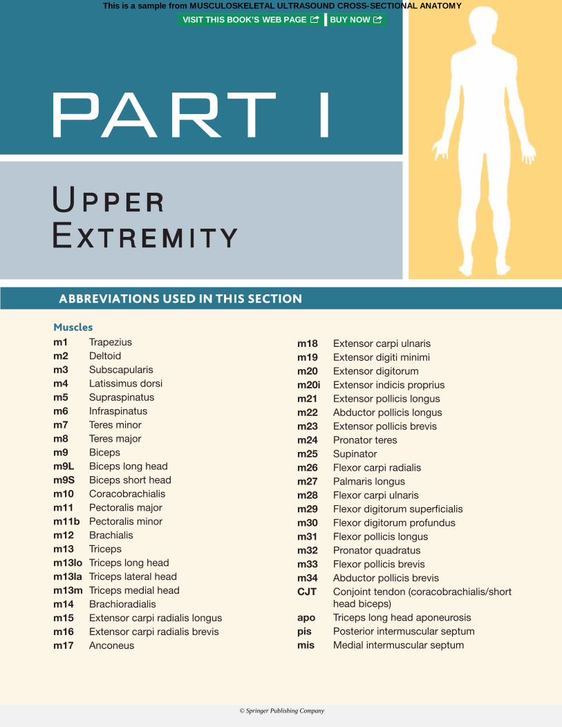
m18 Extensor carpi ulnaris m19 Extensor digiti minimi m20 Extensor digitorum m20i Extensor indicis proprius m21 Extensor pollicis longus m22 Abductor pollicis longus m23 Extensor pollicis brevis m24 Pronator teres m25 Supinator m26 Flexor carpi radialis m27 Palmaris longus m28 Flexor carpi ulnaris m29 Flexor digitorum superfi cialis m30 Flexor digitorum profundus m31 Flexor pollicis longus m32 Pronator quadratus m33 Flexor pollicis brevis m34 Abductor pollicis brevis CJT Conjoint tendon (coracobrachialis/short
head biceps) apo Triceps long head aponeurosis pis Posterior intermuscular septum mis Medial intermuscular septum
Muscles m1 Trapezius m2 Deltoid m3 Subscapularis m4 Latissimus dorsi m5 Supraspinatus m6 Infraspinatus m7 Teres minor m8 Teres major m9 Biceps m9L Biceps long head m9S Biceps short head m10 Coracobrachialis m11 Pectoralis major m11b Pectoralis minor m12 Brachialis m13 Triceps m13lo Triceps long head m13la Triceps lateral head m13m Triceps medial head m14 Brachioradialis m15 Extensor carpi radialis longus m16 Extensor carpi radialis brevis m17 Anconeus
PART IU P P E RE X T R E M I T Y
ABBREVIATIONS USED IN THIS SECTION
Cianca Atlas_00624_PTR_01_1-44_09-04-17.indd Page 6 13/09/17 7:03 PM Cianca Atlas_00624_PTR_01_1-44_09-04-17.indd Page 7 13/09/17 7:03 PMCianca Atlas_00624_PTR_00_i-xviii_FM_09-14-17.indd Page vi 15/09/17 12:56 AM Cianca Atlas_00624_PTR_00_i-xviii_FM_09-14-17.indd Page PB 15/09/17 12:56 AMCianca Atlas_00624_PTR_01_1-44_09-04-17.indd Page 6 13/09/17 7:03 PM Cianca Atlas_00624_PTR_01_1-44_09-04-17.indd Page 7 13/09/17 7:03 PM
This is a sample from MUSCULOSKELETAL ULTRASOUND CROSS-SECTIONAL ANATOMYVISIT THIS BOOK’S WEB PAGE BUY NOW
© Springer Publishing Company
Cianca Atlas_00624_PTR_01_1-44_09-04-17.indd Page 2 13/09/17 7:03 PM Cianca Atlas_00624_PTR_01_1-44_09-04-17.indd Page 3 13/09/17 7:03 PM
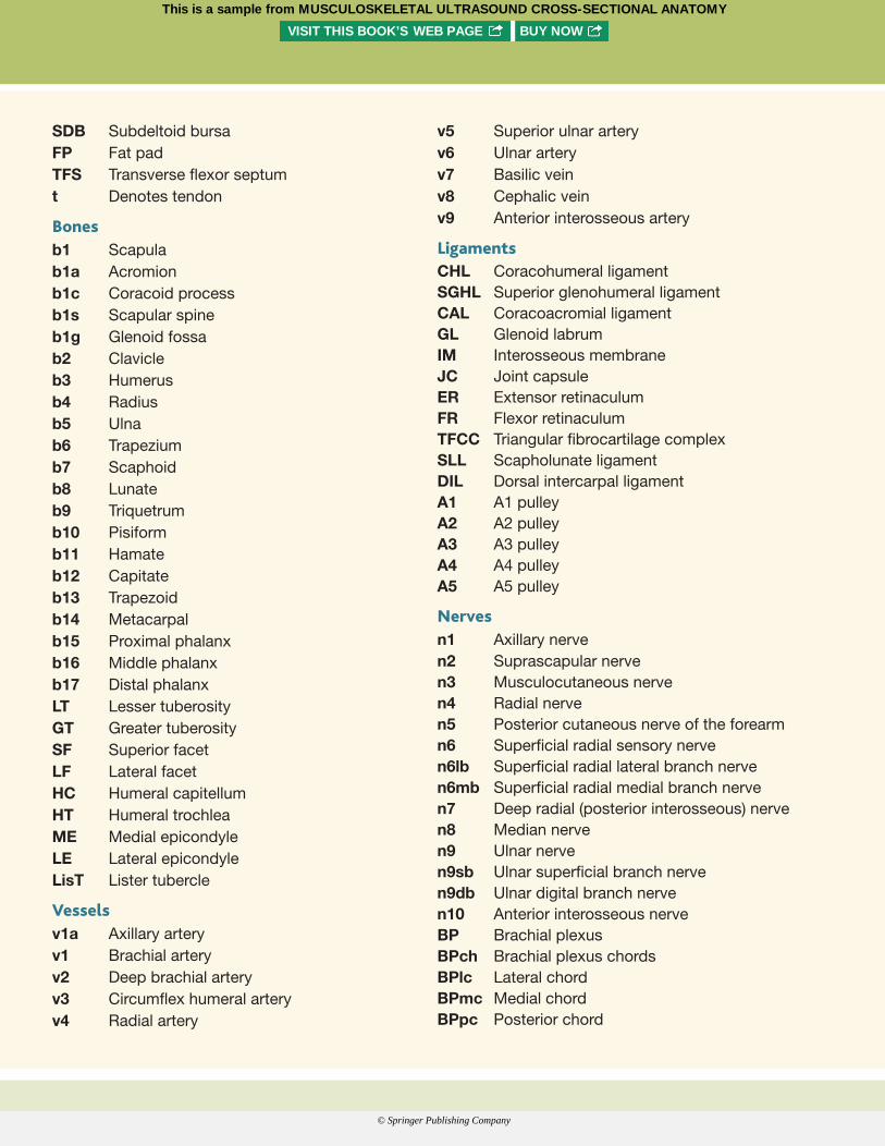
v5 Superior ulnar arteryv6 Ulnar arteryv7 Basilic veinv8 Cephalic veinv9 Anterior interosseous artery
LigamentsCHL Coracohumeral ligamentSGHL Superior glenohumeral ligamentCAL Coracoacromial ligamentGL Glenoid labrumIM Interosseous membraneJC Joint capsuleER Extensor retinaculumFR Flexor retinaculumTFCC Triangular fibrocartilage complexSLL Scapholunate ligamentDIL Dorsal intercarpal ligamentA1 A1 pulleyA2 A2 pulleyA3 A3 pulleyA4 A4 pulleyA5 A5 pulley
Nervesn1 Axillary nerven2 Suprascapular nerven3 Musculocutaneous nerven4 Radial nerven5 Posterior cutaneous nerve of the forearmn6 Superficial radial sensory nerven6lb Superficial radial lateral branch nerven6mb Superficial radial medial branch nerven7 Deep radial (posterior interosseous) nerven8 Median nerven9 Ulnar nerven9sb Ulnar superficial branch nerven9db Ulnar digital branch nerven10 Anterior interosseous nerveBP Brachial plexusBPch Brachial plexus chordsBPlc Lateral chordBPmc Medial chordBPpc Posterior chord
SDB Subdeltoid bursaFP Fat padTFS Transverse flexor septumt Denotes tendon
Bonesb1 Scapulab1a Acromionb1c Coracoid processb1s Scapular spineb1g Glenoid fossab2 Clavicleb3 Humerusb4 Radiusb5 Ulnab6 Trapeziumb7 Scaphoidb8 Lunateb9 Triquetrumb10 Pisiformb11 Hamateb12 Capitateb13 Trapezoidb14 Metacarpalb15 Proximal phalanxb16 Middle phalanxb17 Distal phalanxLT Lesser tuberosityGT Greater tuberositySF Superior facetLF Lateral facetHC Humeral capitellumHT Humeral trochleaME Medial epicondyleLE Lateral epicondyleLisT Lister tubercle
Vesselsv1a Axillary arteryv1 Brachial arteryv2 Deep brachial arteryv3 Circumflex humeral arteryv4 Radial artery
Cianca Atlas_00624_PTR_01_1-44_09-04-17.indd Page 6 13/09/17 7:03 PM Cianca Atlas_00624_PTR_01_1-44_09-04-17.indd Page 7 13/09/17 7:03 PMCianca Atlas_00624_PTR_00_i-xviii_FM_09-14-17.indd Page vi 15/09/17 12:56 AM Cianca Atlas_00624_PTR_00_i-xviii_FM_09-14-17.indd Page PB 15/09/17 12:56 AMCianca Atlas_00624_PTR_01_1-44_09-04-17.indd Page 6 13/09/17 7:03 PM Cianca Atlas_00624_PTR_01_1-44_09-04-17.indd Page 7 13/09/17 7:03 PM
This is a sample from MUSCULOSKELETAL ULTRASOUND CROSS-SECTIONAL ANATOMYVISIT THIS BOOK’S WEB PAGE BUY NOW
© Springer Publishing Company
Cianca Atlas_00624_PTR_01_1-44_09-04-17.indd Page 2 13/09/17 7:03 PM Cianca Atlas_00624_PTR_01_1-44_09-04-17.indd Page 3 13/09/17 7:03 PM
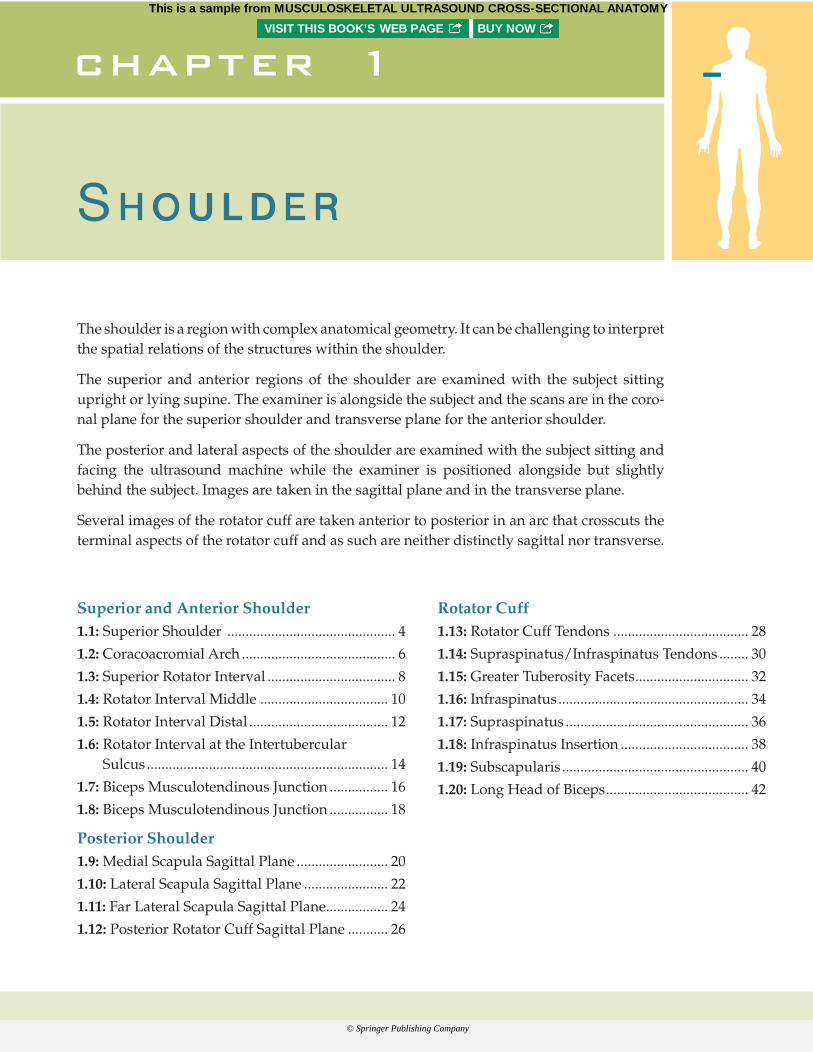
The shoulder is a region with complex anatomical geometry. It can be challenging t o interpret the spatial relations of the structures within the shoulder.
The superior and anterior regions of the shoulder are examined with the subject sitting upright or lying supine. The examiner is alongside the subject and the scans are in the coro-nal plane for the superior shoulder and transverse plane for the anterior shoulder.
The posterior and lateral aspects of the shoulder are examined with the subject sitting and facing the ultrasound machine while the examiner is positioned alongside but slightly behind the subject. Images are taken in the sagittal plane and in the transverse plane.
Several images of the rotator cuff are taken anterior to posterior in an arc that crosscuts the terminal aspects of the rotator cuff and as such are neither distinctly sagittal nor transverse.
S H O U L D E R
chapter 1
Superior and Anterior Shoulder1.1: Superior Shoulder .............................................. 4
1.2: Coracoacromial Arch .......................................... 6
1.3: Superior Rotator Interval ................................... 8
1.4: Rotator Interval Middle ................................... 10
1.5: Rotator Interval Distal ...................................... 12
1.6: Rotator Interval at the Intertubercular Sulcus .................................................................. 14
1.7: Biceps Musculotendinous Junction ................ 16
1.8: Biceps Musculotendinous Junction ................ 18
Posterior Shoulder1.9: Medial Scapula Sagittal Plane ......................... 20
1.10: Lateral Scapula Sagittal Plane ....................... 22
1.11: Far Lateral Scapula Sagittal Plane................. 24
1.12: Posterior Rotator Cuff Sagittal Plane ........... 26
Rotator Cuff1.13: Rotator Cuff Tendons ..................................... 28
1.14: Supraspinatus/Infraspinatus Tendons ........ 30
1.15: Greater Tuberosity Facets ............................... 32
1.16: Infraspinatus .................................................... 34
1.17: Supraspinatus .................................................. 36
1.18: Infraspinatus Insertion ................................... 38
1.19: Subscapularis ................................................... 40
1.20: Long Head of Biceps ....................................... 42
Cianca Atlas_00624_PTR_01_1-44_09-04-17.indd Page 6 13/09/17 7:03 PM Cianca Atlas_00624_PTR_01_1-44_09-04-17.indd Page 7 13/09/17 7:03 PMCianca Atlas_00624_PTR_00_i-xviii_FM_09-14-17.indd Page vi 15/09/17 12:56 AM Cianca Atlas_00624_PTR_00_i-xviii_FM_09-14-17.indd Page PB 15/09/17 12:56 AMCianca Atlas_00624_PTR_01_1-44_09-04-17.indd Page 6 13/09/17 7:03 PM Cianca Atlas_00624_PTR_01_1-44_09-04-17.indd Page 7 13/09/17 7:03 PM
This is a sample from MUSCULOSKELETAL ULTRASOUND CROSS-SECTIONAL ANATOMYVISIT THIS BOOK’S WEB PAGE BUY NOW
© Springer Publishing Company
Cianca Atlas_00624_PTR_01_1-44_09-04-17.indd Page 4 13/09/17 7:03 PM Cianca Atlas_00624_PTR_01_1-44_09-04-17.indd Page 5 13/09/17 7:03 PM
SUPERIOR AND ANTERIOR SHOULDER
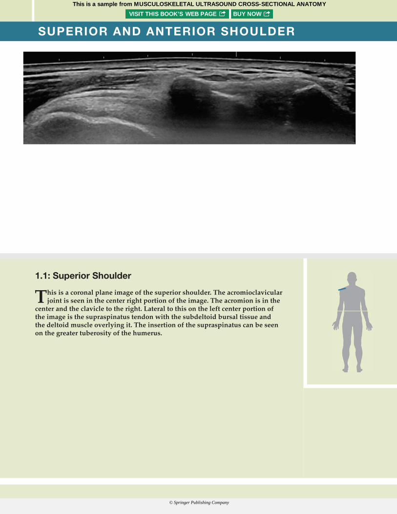
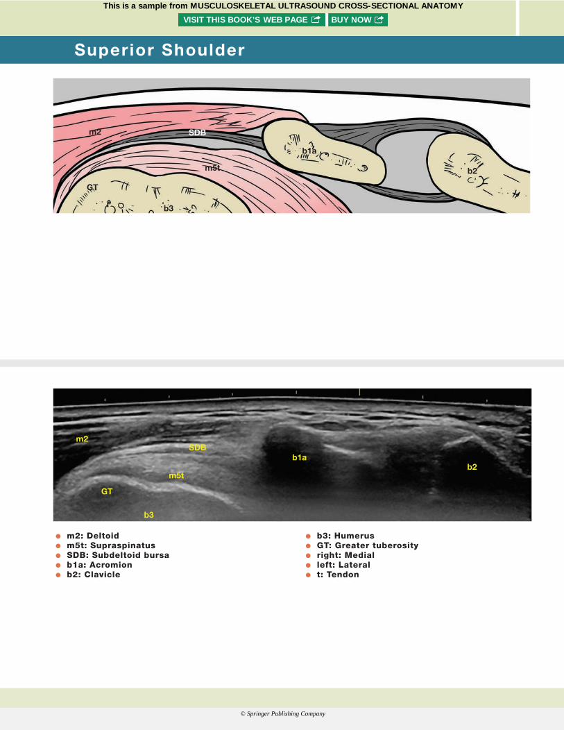
1.1: Superior Shoulder
This is a coronal plane image of the superior shoulder. The acromioclavicular joint is seen in the center right portion of the image. The acromion is in the
center and the clavicle to the right. Lateral to this on the left center portion of the image is the supraspinatus tendon with the subdeltoid bursal tissue and the deltoid muscle overlying it. The insertion of the supraspinatus can be seen on the greater tuberosity of the humerus.
Cianca Atlas_00624_PTR_01_1-44_09-04-17.indd Page 6 13/09/17 7:03 PM Cianca Atlas_00624_PTR_01_1-44_09-04-17.indd Page 7 13/09/17 7:03 PMCianca Atlas_00624_PTR_00_i-xviii_FM_09-14-17.indd Page vi 15/09/17 12:56 AM Cianca Atlas_00624_PTR_00_i-xviii_FM_09-14-17.indd Page PB 15/09/17 12:56 AMCianca Atlas_00624_PTR_01_1-44_09-04-17.indd Page 6 13/09/17 7:03 PM Cianca Atlas_00624_PTR_01_1-44_09-04-17.indd Page 7 13/09/17 7:03 PM
This is a sample from MUSCULOSKELETAL ULTRASOUND CROSS-SECTIONAL ANATOMYVISIT THIS BOOK’S WEB PAGE BUY NOW
© Springer Publishing Company
Cianca Atlas_00624_PTR_01_1-44_09-04-17.indd Page 4 13/09/17 7:03 PM Cianca Atlas_00624_PTR_01_1-44_09-04-17.indd Page 5 13/09/17 7:03 PM
O m2: Deltoid O m5t: Supraspinatus O SDB: Subdeltoid bursa O b1a: Acromion O b2: Clavicle
O b3: Humerus O GT: Greater tuberosity O right: Medial O left: Lateral O t: Tendon
m2
b3
GT
b1a
SDB
m5t b2
m2
b3
GT
b1aSDB
m5tb2
Superior Shoulder
Cianca Atlas_00624_PTR_01_1-44_09-04-17.indd Page 6 13/09/17 7:03 PM Cianca Atlas_00624_PTR_01_1-44_09-04-17.indd Page 7 13/09/17 7:03 PMCianca Atlas_00624_PTR_00_i-xviii_FM_09-14-17.indd Page vi 15/09/17 12:56 AM Cianca Atlas_00624_PTR_00_i-xviii_FM_09-14-17.indd Page PB 15/09/17 12:56 AMCianca Atlas_00624_PTR_01_1-44_09-04-17.indd Page 6 13/09/17 7:03 PM Cianca Atlas_00624_PTR_01_1-44_09-04-17.indd Page 7 13/09/17 7:03 PM
This is a sample from MUSCULOSKELETAL ULTRASOUND CROSS-SECTIONAL ANATOMYVISIT THIS BOOK’S WEB PAGE BUY NOW
© Springer Publishing Company
Cianca Atlas_00624_PTR_01_1-44_09-04-17.indd Page 6 13/09/17 7:03 PM Cianca Atlas_00624_PTR_01_1-44_09-04-17.indd Page 7 13/09/17 7:03 PM
SUPERIOR AND ANTERIOR SHOULDER
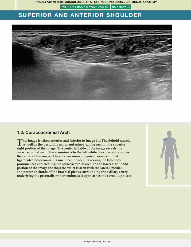
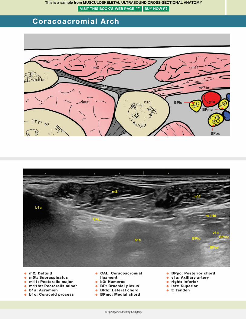
1.2: Coracoacromial Arch
This image is taken anterior and inferior to Image 1.1. The deltoid muscle, as well as the pectoralis major and minor, can be seen in the superior
right portion of the image. The center left side of the image reveals the coracoacromial arch. The acromion is to the left while the coracoid occupies the center of the image. The coracoacromial ligament coracoacromial ligament coracoacromial ligament can be seen traversing the two bony prominences and creating the coracoacromial arch. In the lower right-hand portion of the image the thoracic outlet is seen with the lateral, medial, and posterior chords of the brachial plexus surrounding the axillary artery underlying the pectoralis minor tendon as it approaches the coracoid process.
Cianca Atlas_00624_PTR_01_1-44_09-04-17.indd Page 6 13/09/17 7:03 PM Cianca Atlas_00624_PTR_01_1-44_09-04-17.indd Page 7 13/09/17 7:03 PMCianca Atlas_00624_PTR_00_i-xviii_FM_09-14-17.indd Page vi 15/09/17 12:56 AM Cianca Atlas_00624_PTR_00_i-xviii_FM_09-14-17.indd Page PB 15/09/17 12:56 AMCianca Atlas_00624_PTR_01_1-44_09-04-17.indd Page 6 13/09/17 7:03 PM Cianca Atlas_00624_PTR_01_1-44_09-04-17.indd Page 7 13/09/17 7:03 PM
This is a sample from MUSCULOSKELETAL ULTRASOUND CROSS-SECTIONAL ANATOMYVISIT THIS BOOK’S WEB PAGE BUY NOW
© Springer Publishing Company
Cianca Atlas_00624_PTR_01_1-44_09-04-17.indd Page 6 13/09/17 7:03 PM Cianca Atlas_00624_PTR_01_1-44_09-04-17.indd Page 7 13/09/17 7:03 PM
b1a
m2
m5t b1c
b3
BPlc
BPpc
v1a
BPmc
m11bt
m11
CAL
b1a
m2
CAL
b1c
m11
m11bt
v1aBPmc
BPpc
BPIc
O m2: Deltoid O m5t: Supraspinatus O m11: Pectoralis major O m11bt: Pectoralis minor O b1a: Acromion O b1c: Coracoid process
O CAL: Coracoacromial ligament
O b3: Humerus O BP: Brachial plexus O BPlc: Lateral chord O BPmc: Medial chord
O BPpc: Posterior chord O v1a: Axillary artery O right: Inferior O left: Superior O t: Tendon
Coracoacromial Arch
Cianca Atlas_00624_PTR_01_1-44_09-04-17.indd Page 6 13/09/17 7:03 PM Cianca Atlas_00624_PTR_01_1-44_09-04-17.indd Page 7 13/09/17 7:03 PMCianca Atlas_00624_PTR_00_i-xviii_FM_09-14-17.indd Page vi 15/09/17 12:56 AM Cianca Atlas_00624_PTR_00_i-xviii_FM_09-14-17.indd Page PB 15/09/17 12:56 AMCianca Atlas_00624_PTR_01_1-44_09-04-17.indd Page 6 13/09/17 7:03 PM Cianca Atlas_00624_PTR_01_1-44_09-04-17.indd Page 7 13/09/17 7:03 PM
This is a sample from MUSCULOSKELETAL ULTRASOUND CROSS-SECTIONAL ANATOMYVISIT THIS BOOK’S WEB PAGE BUY NOW
© Springer Publishing Company
Cianca Atlas_00624_PTR_01_1-44_09-04-17.indd Page 8 13/09/17 7:03 PM Cianca Atlas_00624_PTR_01_1-44_09-04-17.indd Page 9 13/09/17 7:03 PM
SUPERIOR AND ANTERIOR SHOULDER
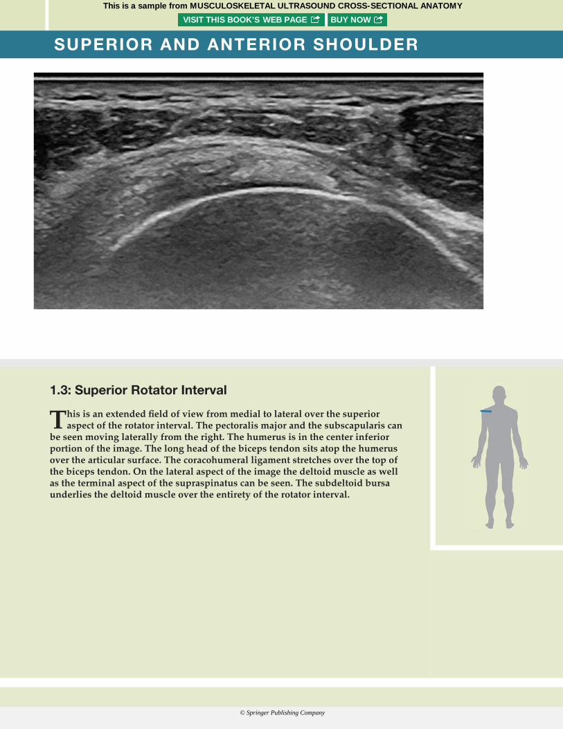
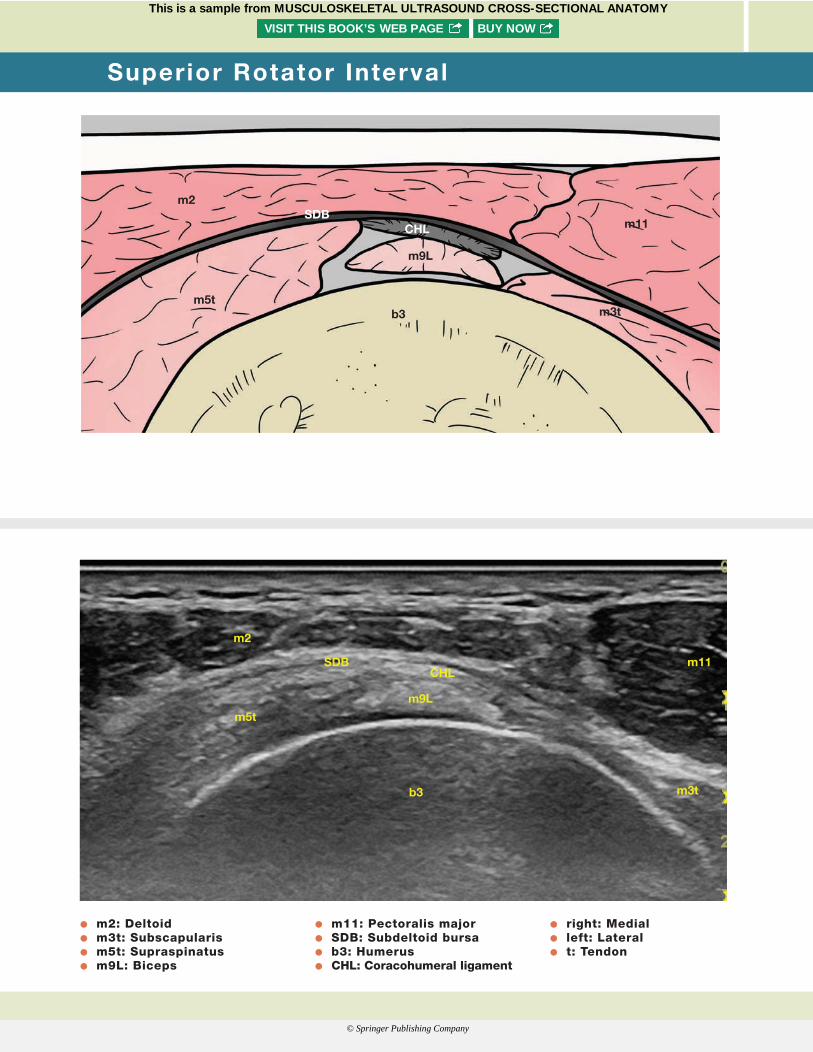
1.3: Superior Rotator Interval
This is an extended fi eld of view from medial to lateral over the superior aspect of the rotator interval. The pectoralis major and the subscapularis can
be seen moving laterally from the right. The humerus is in the center inferior portion of the image. The long head of the biceps tendon sits atop the humerus over the articular surface. The coracohumeral ligament stretches over the top of the biceps tendon. On the lateral aspect of the image the deltoid muscle as well as the terminal aspect of the supraspinatus can be seen. The subdeltoid bursa underlies the deltoid muscle over the entirety of the rotator interval.
Cianca Atlas_00624_PTR_01_1-44_09-04-17.indd Page 6 13/09/17 7:03 PM Cianca Atlas_00624_PTR_01_1-44_09-04-17.indd Page 7 13/09/17 7:03 PMCianca Atlas_00624_PTR_00_i-xviii_FM_09-14-17.indd Page vi 15/09/17 12:56 AM Cianca Atlas_00624_PTR_00_i-xviii_FM_09-14-17.indd Page PB 15/09/17 12:56 AMCianca Atlas_00624_PTR_01_1-44_09-04-17.indd Page 6 13/09/17 7:03 PM Cianca Atlas_00624_PTR_01_1-44_09-04-17.indd Page 7 13/09/17 7:03 PM
This is a sample from MUSCULOSKELETAL ULTRASOUND CROSS-SECTIONAL ANATOMYVISIT THIS BOOK’S WEB PAGE BUY NOW
© Springer Publishing Company
Cianca Atlas_00624_PTR_01_1-44_09-04-17.indd Page 8 13/09/17 7:03 PM Cianca Atlas_00624_PTR_01_1-44_09-04-17.indd Page 9 13/09/17 7:03 PM
m2
m5t
SDBCHL
m9L
b3 m3t
m11
m2
m5t
SDBCHL
m9L
b3 m3t
m11
O m2: Deltoid O m3t: Subscapularis O m5t: Supraspinatus O m9L: Biceps
O m11: Pectoralis major O SDB: Subdeltoid bursa O b3: Humerus O CHL: Coracohumeral ligament
O right: Medial O left: Lateral O t: Tendon
Superior Rotator Interval
Cianca Atlas_00624_PTR_01_1-44_09-04-17.indd Page 6 13/09/17 7:03 PM Cianca Atlas_00624_PTR_01_1-44_09-04-17.indd Page 7 13/09/17 7:03 PMCianca Atlas_00624_PTR_00_i-xviii_FM_09-14-17.indd Page vi 15/09/17 12:56 AM Cianca Atlas_00624_PTR_00_i-xviii_FM_09-14-17.indd Page PB 15/09/17 12:56 AMCianca Atlas_00624_PTR_01_1-44_09-04-17.indd Page 6 13/09/17 7:03 PM Cianca Atlas_00624_PTR_01_1-44_09-04-17.indd Page 7 13/09/17 7:03 PM
This is a sample from MUSCULOSKELETAL ULTRASOUND CROSS-SECTIONAL ANATOMYVISIT THIS BOOK’S WEB PAGE BUY NOW
© Springer Publishing Company

Cianca Atlas_00624_PTR_01_1-44_09-04-17.indd Page 10 13/09/17 7:03 PM Cianca Atlas_00624_PTR_01_1-44_09-04-17.indd Page 11 13/09/17 7:03 PM
SUPERIOR AND ANTERIOR SHOULDER
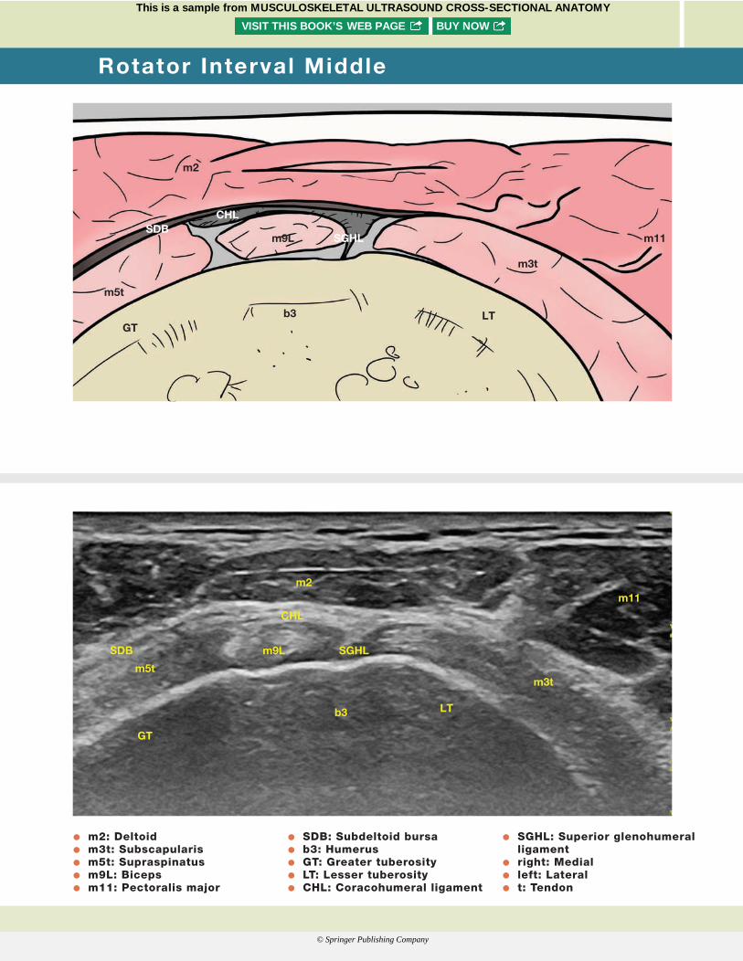
1.4: Rotator Interval Middle
This image spans the middle aspect of the rotator interval. The same structures are highlighted as in the previous image with the addition of
the superior glenohumeral ligament. This structure is seen interposed between the biceps tendon and the humerus and together with the coracohumeral ligament form the reflection pulley of the long head of the biceps tendon.
Cianca Atlas_00624_PTR_01_1-44_09-04-17.indd Page 6 13/09/17 7:03 PM Cianca Atlas_00624_PTR_01_1-44_09-04-17.indd Page 7 13/09/17 7:03 PMCianca Atlas_00624_PTR_00_i-xviii_FM_09-14-17.indd Page vi 15/09/17 12:56 AM Cianca Atlas_00624_PTR_00_i-xviii_FM_09-14-17.indd Page PB 15/09/17 12:56 AMCianca Atlas_00624_PTR_01_1-44_09-04-17.indd Page 6 13/09/17 7:03 PM Cianca Atlas_00624_PTR_01_1-44_09-04-17.indd Page 7 13/09/17 7:03 PM
This is a sample from MUSCULOSKELETAL ULTRASOUND CROSS-SECTIONAL ANATOMYVISIT THIS BOOK’S WEB PAGE BUY NOW
© Springer Publishing Company
Cianca Atlas_00624_PTR_01_1-44_09-04-17.indd Page 10 13/09/17 7:03 PM Cianca Atlas_00624_PTR_01_1-44_09-04-17.indd Page 11 13/09/17 7:03 PM
SDB
m5t
GTLT
m2
CHL
SGHLm9L
b3
m11
m3t
SDB
m5t
GT
m2
CHL
SGHLm9L
LTb3
m11
m3t
O m2: Deltoid O m3t: Subscapularis O m5t: Supraspinatus O m9L: Biceps O m11: Pectoralis major
O SDB: Subdeltoid bursa O b3: Humerus O GT: Greater tuberosity O LT: Lesser tuberosity O CHL: Coracohumeral ligament
O SGHL: Superior glenohumeral ligament
O right: Medial O left: Lateral O t: Tendon
Rotator Interval Middle
Cianca Atlas_00624_PTR_01_1-44_09-04-17.indd Page 6 13/09/17 7:03 PM Cianca Atlas_00624_PTR_01_1-44_09-04-17.indd Page 7 13/09/17 7:03 PMCianca Atlas_00624_PTR_00_i-xviii_FM_09-14-17.indd Page vi 15/09/17 12:56 AM Cianca Atlas_00624_PTR_00_i-xviii_FM_09-14-17.indd Page PB 15/09/17 12:56 AMCianca Atlas_00624_PTR_01_1-44_09-04-17.indd Page 6 13/09/17 7:03 PM Cianca Atlas_00624_PTR_01_1-44_09-04-17.indd Page 7 13/09/17 7:03 PM
This is a sample from MUSCULOSKELETAL ULTRASOUND CROSS-SECTIONAL ANATOMYVISIT THIS BOOK’S WEB PAGE BUY NOW
© Springer Publishing Company
Cianca Atlas_00624_PTR_01_1-44_09-04-17.indd Page 12 13/09/17 7:03 PM Cianca Atlas_00624_PTR_01_1-44_09-04-17.indd Page 13 13/09/17 7:03 PM
SUPERIOR AND ANTERIOR SHOULDER
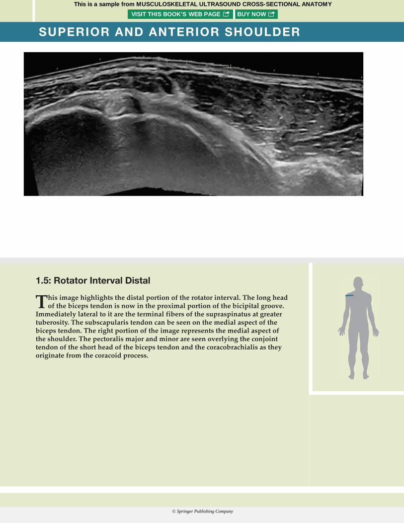
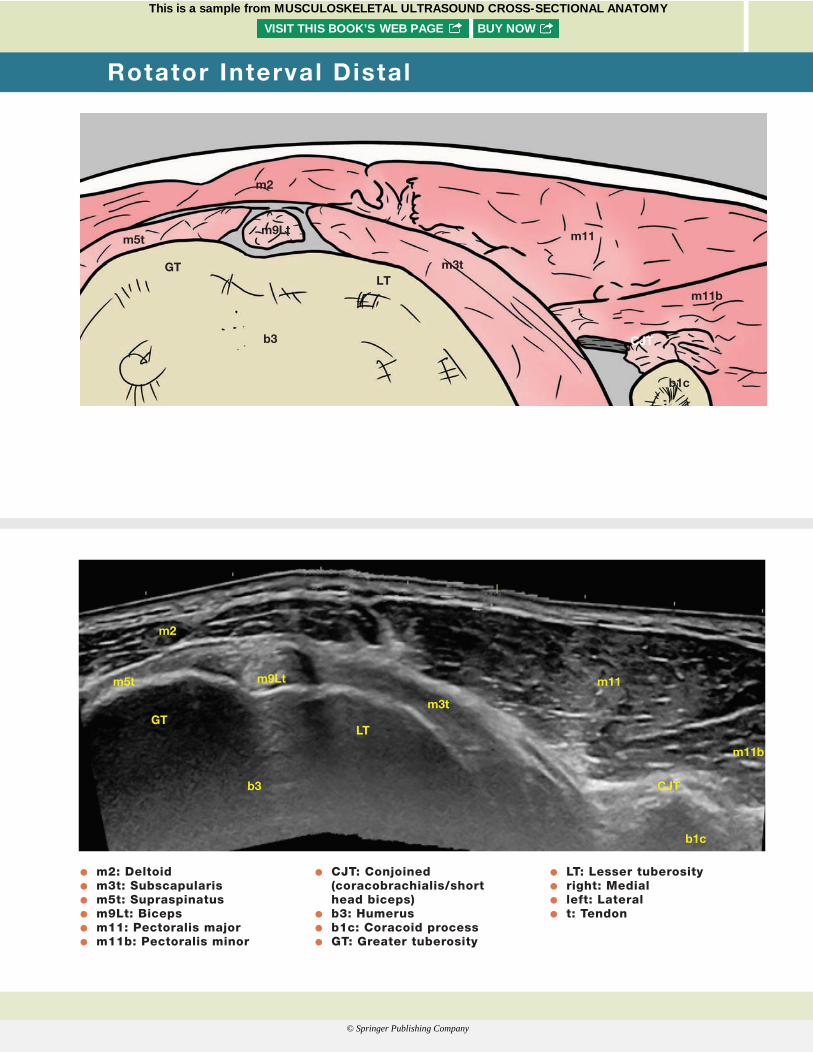
1.5: Rotator Interval Distal
This image highlights the distal portion of the rotator interval. The long head of the biceps tendon is now in the proximal portion of the bicipital groove.
Immediately lateral to it are the terminal fibers of the supraspinatus at greater tuberosity. The subscapularis tendon can be seen on the medial aspect of the biceps tendon. The right portion of the image represents the medial aspect of the shoulder. The pectoralis major and minor are seen overlying the conjoint tendon of the short head of the biceps tendon and the coracobrachialis as they originate from the coracoid process.
Cianca Atlas_00624_PTR_01_1-44_09-04-17.indd Page 6 13/09/17 7:03 PM Cianca Atlas_00624_PTR_01_1-44_09-04-17.indd Page 7 13/09/17 7:03 PMCianca Atlas_00624_PTR_00_i-xviii_FM_09-14-17.indd Page vi 15/09/17 12:56 AM Cianca Atlas_00624_PTR_00_i-xviii_FM_09-14-17.indd Page PB 15/09/17 12:56 AMCianca Atlas_00624_PTR_01_1-44_09-04-17.indd Page 6 13/09/17 7:03 PM Cianca Atlas_00624_PTR_01_1-44_09-04-17.indd Page 7 13/09/17 7:03 PM
This is a sample from MUSCULOSKELETAL ULTRASOUND CROSS-SECTIONAL ANATOMYVISIT THIS BOOK’S WEB PAGE BUY NOW
© Springer Publishing Company
Cianca Atlas_00624_PTR_01_1-44_09-04-17.indd Page 12 13/09/17 7:03 PM Cianca Atlas_00624_PTR_01_1-44_09-04-17.indd Page 13 13/09/17 7:03 PM
m2
b1c
b3
GTLT
m3t
m5t
CJT
m11
m11b
m9Lt
m2
b1c
b3
GTLT
m3t
m5t m9Lt
CJT
m11
m11b
O m2: Deltoid O m3t: Subscapularis O m5t: Supraspinatus O m9Lt: Biceps O m11: Pectoralis major O m11b: Pectoralis minor
O CJT: Conjoined (coracobrachialis/short head biceps)
O b3: Humerus O b1c: Coracoid process O GT: Greater tuberosity
O LT: Lesser tuberosity O right: Medial O left: Lateral O t: Tendon
Rotator Interval Distal
Cianca Atlas_00624_PTR_01_1-44_09-04-17.indd Page 6 13/09/17 7:03 PM Cianca Atlas_00624_PTR_01_1-44_09-04-17.indd Page 7 13/09/17 7:03 PMCianca Atlas_00624_PTR_00_i-xviii_FM_09-14-17.indd Page vi 15/09/17 12:56 AM Cianca Atlas_00624_PTR_00_i-xviii_FM_09-14-17.indd Page PB 15/09/17 12:56 AMCianca Atlas_00624_PTR_01_1-44_09-04-17.indd Page 6 13/09/17 7:03 PM Cianca Atlas_00624_PTR_01_1-44_09-04-17.indd Page 7 13/09/17 7:03 PM
This is a sample from MUSCULOSKELETAL ULTRASOUND CROSS-SECTIONAL ANATOMYVISIT THIS BOOK’S WEB PAGE BUY NOW
© Springer Publishing Company
Cianca Atlas_00624_PTR_01_1-44_09-04-17.indd Page 14 13/09/17 7:03 PM Cianca Atlas_00624_PTR_01_1-44_09-04-17.indd Page 15 13/09/17 7:03 PM
SUPERIOR AND ANTERIOR SHOULDER
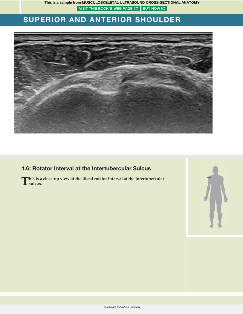
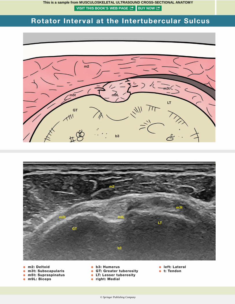
1.6: Rotator Interval at the Intertubercular Sulcus
This is a close-up view of the distal rotator interval at the intertubercular sulcus.
Cianca Atlas_00624_PTR_01_1-44_09-04-17.indd Page 6 13/09/17 7:03 PM Cianca Atlas_00624_PTR_01_1-44_09-04-17.indd Page 7 13/09/17 7:03 PMCianca Atlas_00624_PTR_00_i-xviii_FM_09-14-17.indd Page vi 15/09/17 12:56 AM Cianca Atlas_00624_PTR_00_i-xviii_FM_09-14-17.indd Page PB 15/09/17 12:56 AMCianca Atlas_00624_PTR_01_1-44_09-04-17.indd Page 6 13/09/17 7:03 PM Cianca Atlas_00624_PTR_01_1-44_09-04-17.indd Page 7 13/09/17 7:03 PM
This is a sample from MUSCULOSKELETAL ULTRASOUND CROSS-SECTIONAL ANATOMYVISIT THIS BOOK’S WEB PAGE BUY NOW
© Springer Publishing Company
Cianca Atlas_00624_PTR_01_1-44_09-04-17.indd Page 14 13/09/17 7:03 PM Cianca Atlas_00624_PTR_01_1-44_09-04-17.indd Page 15 13/09/17 7:03 PM
O m2: Deltoid O m3t: Subscapularis O m5t: Supraspinatus O m9L: Biceps
O b3: Humerus O GT: Greater tuberosity O LT: Lesser tuberosity O right: Medial
O left: Lateral O t: Tendon
GTLT
m3t
m5t
m2
m9L
b3
GT
LT
m3t
m5t
m2
m9L
b3
Rotator Interval at the Intertubercular Sulcus
Cianca Atlas_00624_PTR_01_1-44_09-04-17.indd Page 6 13/09/17 7:03 PM Cianca Atlas_00624_PTR_01_1-44_09-04-17.indd Page 7 13/09/17 7:03 PMCianca Atlas_00624_PTR_00_i-xviii_FM_09-14-17.indd Page vi 15/09/17 12:56 AM Cianca Atlas_00624_PTR_00_i-xviii_FM_09-14-17.indd Page PB 15/09/17 12:56 AMCianca Atlas_00624_PTR_01_1-44_09-04-17.indd Page 6 13/09/17 7:03 PM Cianca Atlas_00624_PTR_01_1-44_09-04-17.indd Page 7 13/09/17 7:03 PM
This is a sample from MUSCULOSKELETAL ULTRASOUND CROSS-SECTIONAL ANATOMYVISIT THIS BOOK’S WEB PAGE BUY NOW
© Springer Publishing Company
Cianca Atlas_00624_PTR_01_1-44_09-04-17.indd Page 16 13/09/17 7:03 PM Cianca Atlas_00624_PTR_01_1-44_09-04-17.indd Page 17 13/09/17 7:03 PM
SUPERIOR AND ANTERIOR SHOULDER
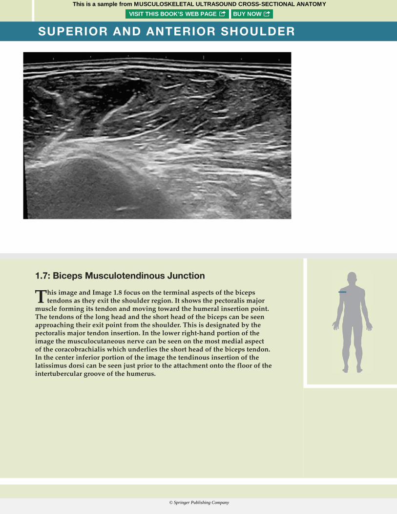
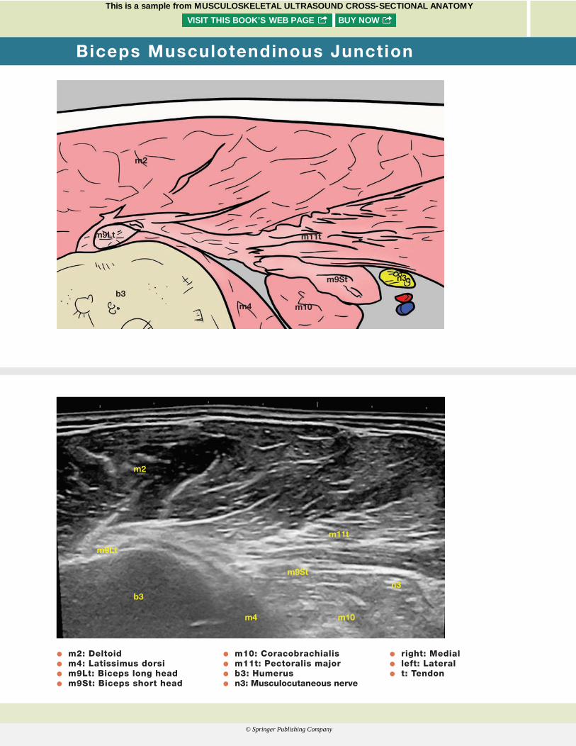
1.7: Biceps Musculotendinous Junction
This image and Image 1.8 focus on the terminal aspects of the biceps tendons as they exit the shoulder region. It shows the pectoralis major
muscle forming its tendon and moving toward the humeral insertion point. The tendons of the long head and the short head of the biceps can be seen approaching their exit point from the shoulder. This is designated by the pectoralis major tendon insertion. In the lower right-hand portion of the image the musculocutaneous nerve can be seen on the most medial aspect of the coracobrachialis which underlies the short head of the biceps tendon. In the center inferior portion of the image the tendinous insertion of the latissimus dorsi can be seen just prior to the attachment onto the floor of the intertubercular groove of the humerus.
Cianca Atlas_00624_PTR_01_1-44_09-04-17.indd Page 6 13/09/17 7:03 PM Cianca Atlas_00624_PTR_01_1-44_09-04-17.indd Page 7 13/09/17 7:03 PMCianca Atlas_00624_PTR_00_i-xviii_FM_09-14-17.indd Page vi 15/09/17 12:56 AM Cianca Atlas_00624_PTR_00_i-xviii_FM_09-14-17.indd Page PB 15/09/17 12:56 AMCianca Atlas_00624_PTR_01_1-44_09-04-17.indd Page 6 13/09/17 7:03 PM Cianca Atlas_00624_PTR_01_1-44_09-04-17.indd Page 7 13/09/17 7:03 PM
This is a sample from MUSCULOSKELETAL ULTRASOUND CROSS-SECTIONAL ANATOMYVISIT THIS BOOK’S WEB PAGE BUY NOW
© Springer Publishing Company
Cianca Atlas_00624_PTR_01_1-44_09-04-17.indd Page 16 13/09/17 7:03 PM Cianca Atlas_00624_PTR_01_1-44_09-04-17.indd Page 17 13/09/17 7:03 PM
b3
m4
m9Lt
m2
m11t
m10
m9St
n3
b3m4
m9Lt
m2
m11t
m10
m9St n3
O m2: Deltoid O m4: Latissimus dorsi O m9Lt: Biceps long head O m9St: Biceps short head
O m10: Coracobrachialis O m11t: Pectoralis major O b3: Humerus O n3: Musculocutaneous nerve
O right: Medial O left: Lateral O t: Tendon
Biceps Musculotendinous Junction
Cianca Atlas_00624_PTR_01_1-44_09-04-17.indd Page 6 13/09/17 7:03 PM Cianca Atlas_00624_PTR_01_1-44_09-04-17.indd Page 7 13/09/17 7:03 PMCianca Atlas_00624_PTR_00_i-xviii_FM_09-14-17.indd Page vi 15/09/17 12:56 AM Cianca Atlas_00624_PTR_00_i-xviii_FM_09-14-17.indd Page PB 15/09/17 12:56 AMCianca Atlas_00624_PTR_01_1-44_09-04-17.indd Page 6 13/09/17 7:03 PM Cianca Atlas_00624_PTR_01_1-44_09-04-17.indd Page 7 13/09/17 7:03 PM
This is a sample from MUSCULOSKELETAL ULTRASOUND CROSS-SECTIONAL ANATOMYVISIT THIS BOOK’S WEB PAGE BUY NOW
© Springer Publishing Company
Cianca Atlas_00624_PTR_01_1-44_09-04-17.indd Page 18 13/09/17 7:03 PM Cianca Atlas_00624_PTR_01_1-44_09-04-17.indd Page 19 13/09/17 7:03 PM
SUPERIOR AND ANTERIOR SHOULDER
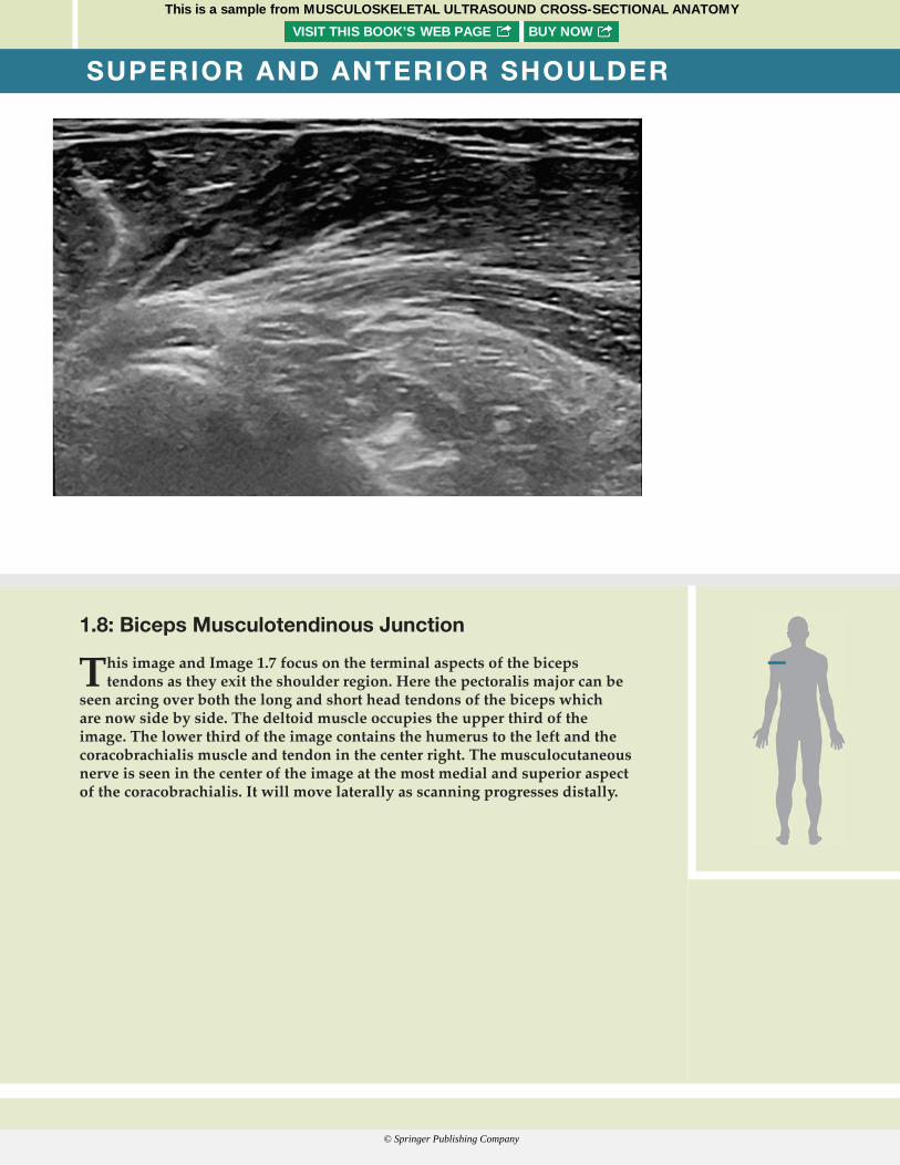
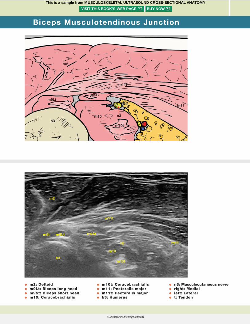
1.8: Biceps Musculotendinous Junction
This image and Image 1.7 focus on the terminal aspects of the biceps tendons as they exit the shoulder region. Here the pectoralis major can be
seen arcing over both the long and short head tendons of the biceps which are now side by side. The deltoid muscle occupies the upper third of the image. The lower third of the image contains the humerus to the left and the coracobrachialis muscle and tendon in the center right. The musculocutaneous nerve is seen in the center of the image at the most medial and superior aspect of the coracobrachialis. It will move laterally as scanning progresses distally.
Cianca Atlas_00624_PTR_01_1-44_09-04-17.indd Page 6 13/09/17 7:03 PM Cianca Atlas_00624_PTR_01_1-44_09-04-17.indd Page 7 13/09/17 7:03 PMCianca Atlas_00624_PTR_00_i-xviii_FM_09-14-17.indd Page vi 15/09/17 12:56 AM Cianca Atlas_00624_PTR_00_i-xviii_FM_09-14-17.indd Page PB 15/09/17 12:56 AMCianca Atlas_00624_PTR_01_1-44_09-04-17.indd Page 6 13/09/17 7:03 PM Cianca Atlas_00624_PTR_01_1-44_09-04-17.indd Page 7 13/09/17 7:03 PM
This is a sample from MUSCULOSKELETAL ULTRASOUND CROSS-SECTIONAL ANATOMYVISIT THIS BOOK’S WEB PAGE BUY NOW
© Springer Publishing Company
Cianca Atlas_00624_PTR_01_1-44_09-04-17.indd Page 18 13/09/17 7:03 PM Cianca Atlas_00624_PTR_01_1-44_09-04-17.indd Page 19 13/09/17 7:03 PM
b3
m2
m11t
m9Ltm9St
n3
m11
m10
m10t
b3
m2
m11t
m11
m9Ltm9t m9St
n3
m10
m10t
O m2: Deltoid O m9Lt: Biceps long head O m9St: Biceps short head O m10: Coracobrachialis
O m10t: Coracobrachialis O m11: Pectoralis major O m11t: Pectoralis major O b3: Humerus
O n3: Musculocutaneous nerve O right: Medial O left: Lateral O t: Tendon
Biceps Musculotendinous Junction
Cianca Atlas_00624_PTR_01_1-44_09-04-17.indd Page 6 13/09/17 7:03 PM Cianca Atlas_00624_PTR_01_1-44_09-04-17.indd Page 7 13/09/17 7:03 PMCianca Atlas_00624_PTR_00_i-xviii_FM_09-14-17.indd Page vi 15/09/17 12:56 AM Cianca Atlas_00624_PTR_00_i-xviii_FM_09-14-17.indd Page PB 15/09/17 12:56 AMCianca Atlas_00624_PTR_01_1-44_09-04-17.indd Page 6 13/09/17 7:03 PM Cianca Atlas_00624_PTR_01_1-44_09-04-17.indd Page 7 13/09/17 7:03 PM
This is a sample from MUSCULOSKELETAL ULTRASOUND CROSS-SECTIONAL ANATOMYVISIT THIS BOOK’S WEB PAGE BUY NOW
© Springer Publishing Company
Cianca Atlas_00624_PTR_01_1-44_09-04-17.indd Page 20 13/09/17 7:03 PM Cianca Atlas_00624_PTR_01_1-44_09-04-17.indd Page 21 13/09/17 7:03 PM
POSTERIOR SHOULDER
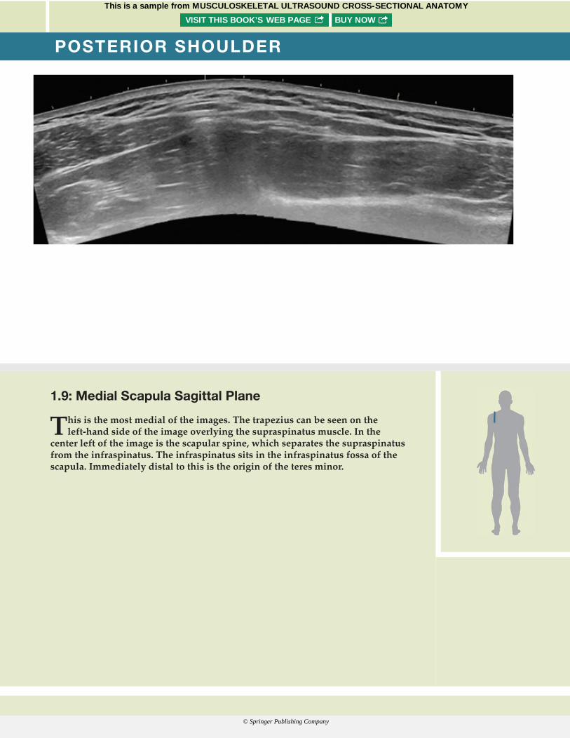
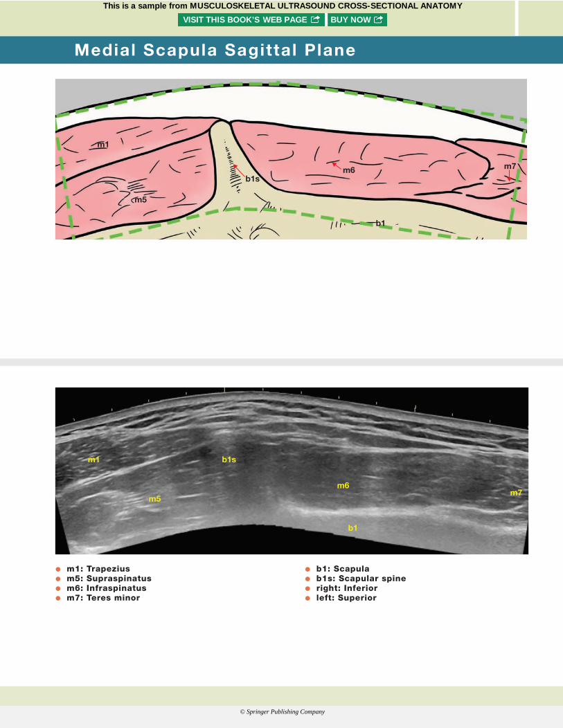
1.9: Medial Scapula Sagittal Plane
This is the most medial of the images. The trapezius can be seen on the left-hand side of the image overlying the supraspinatus muscle. In the
center left of the image is the scapular spine, which separates the supraspinatus from the infraspinatus. The infraspinatus sits in the infraspinatus fossa of the scapula. Immediately distal to this is the origin of the teres minor.
Cianca Atlas_00624_PTR_01_1-44_09-04-17.indd Page 6 13/09/17 7:03 PM Cianca Atlas_00624_PTR_01_1-44_09-04-17.indd Page 7 13/09/17 7:03 PMCianca Atlas_00624_PTR_00_i-xviii_FM_09-14-17.indd Page vi 15/09/17 12:56 AM Cianca Atlas_00624_PTR_00_i-xviii_FM_09-14-17.indd Page PB 15/09/17 12:56 AMCianca Atlas_00624_PTR_01_1-44_09-04-17.indd Page 6 13/09/17 7:03 PM Cianca Atlas_00624_PTR_01_1-44_09-04-17.indd Page 7 13/09/17 7:03 PM
This is a sample from MUSCULOSKELETAL ULTRASOUND CROSS-SECTIONAL ANATOMYVISIT THIS BOOK’S WEB PAGE BUY NOW
© Springer Publishing Company
Cianca Atlas_00624_PTR_01_1-44_09-04-17.indd Page 20 13/09/17 7:03 PM Cianca Atlas_00624_PTR_01_1-44_09-04-17.indd Page 21 13/09/17 7:03 PM
m1
m5
m6 m7
b1
b1s
m1
m5m6
m7
b1
b1s
O m1: Trapezius O m5: Supraspinatus O m6: Infraspinatus O m7: Teres minor
O b1: Scapula O b1s: Scapular spine O right: Inferior O left: Superior
Medial Scapula Sagittal Plane
Cianca Atlas_00624_PTR_01_1-44_09-04-17.indd Page 6 13/09/17 7:03 PM Cianca Atlas_00624_PTR_01_1-44_09-04-17.indd Page 7 13/09/17 7:03 PMCianca Atlas_00624_PTR_00_i-xviii_FM_09-14-17.indd Page vi 15/09/17 12:56 AM Cianca Atlas_00624_PTR_00_i-xviii_FM_09-14-17.indd Page PB 15/09/17 12:56 AMCianca Atlas_00624_PTR_01_1-44_09-04-17.indd Page 6 13/09/17 7:03 PM Cianca Atlas_00624_PTR_01_1-44_09-04-17.indd Page 7 13/09/17 7:03 PM
This is a sample from MUSCULOSKELETAL ULTRASOUND CROSS-SECTIONAL ANATOMYVISIT THIS BOOK’S WEB PAGE BUY NOW
© Springer Publishing Company